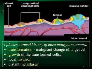
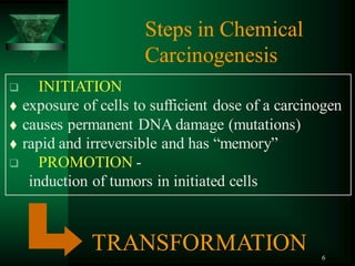
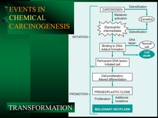
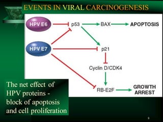
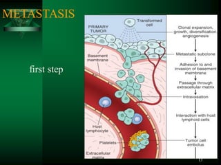
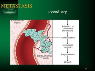
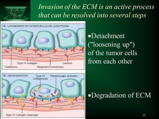
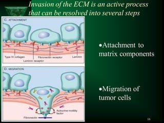
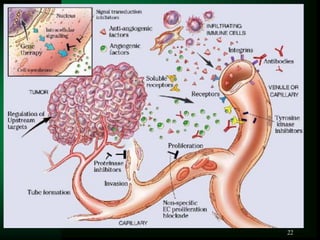
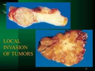
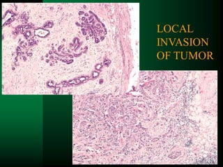
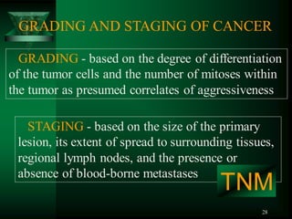
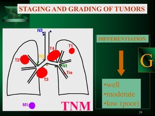
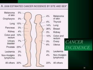
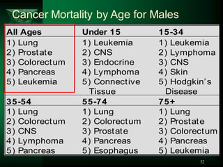
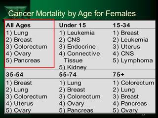
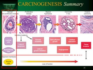
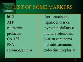
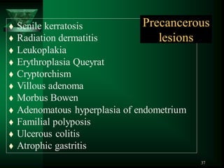
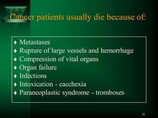
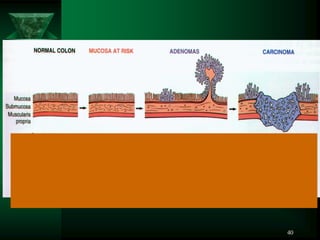
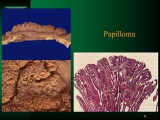
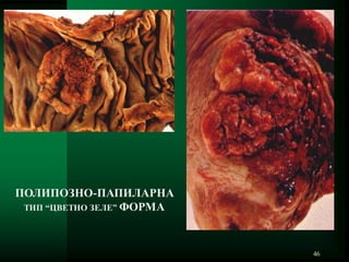
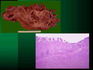
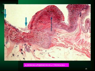
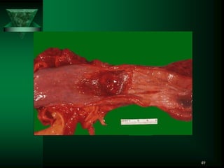
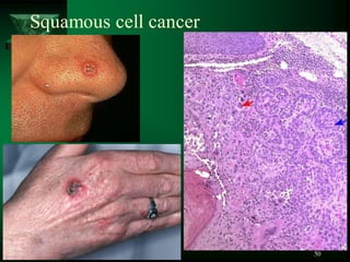
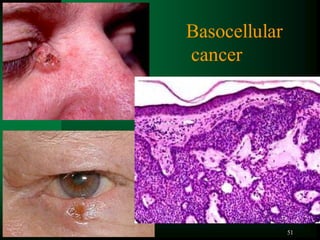
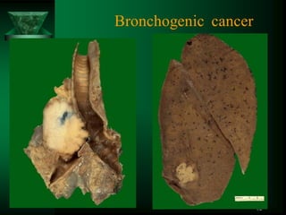
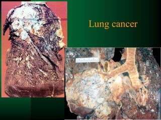
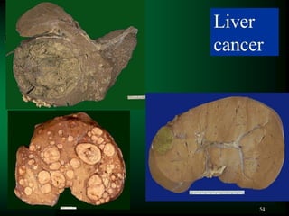
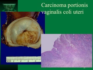
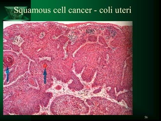
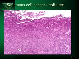
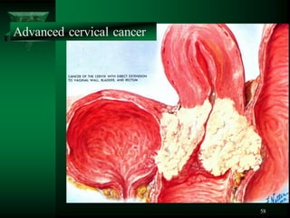
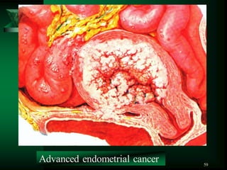
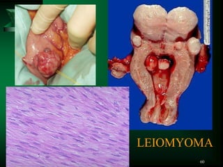
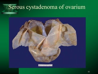
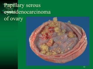
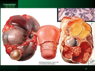
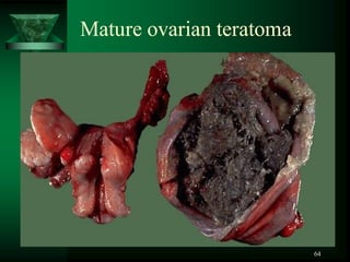
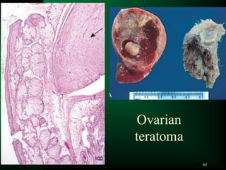
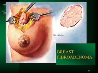
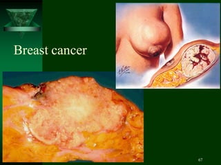
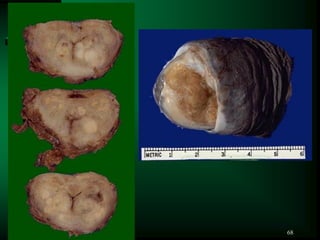
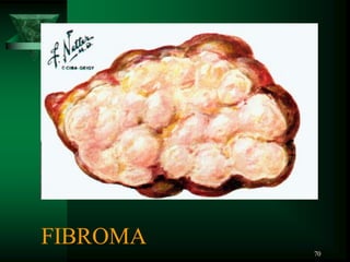
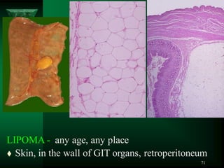
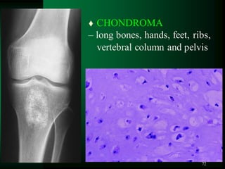
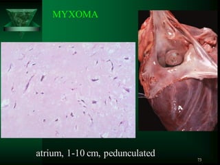
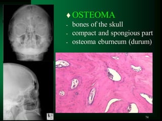
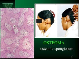
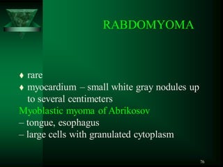
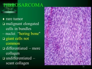
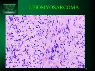
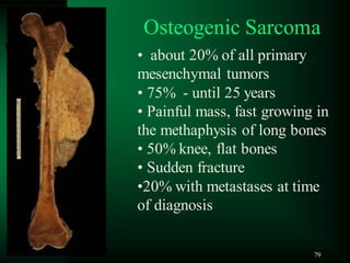
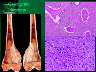
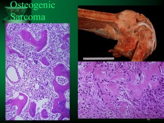
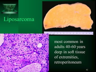
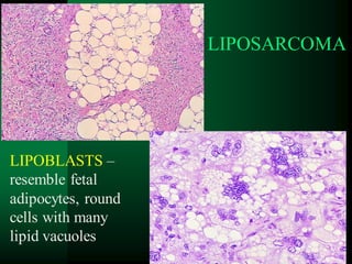
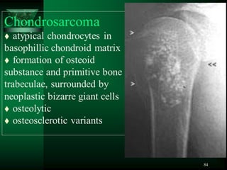
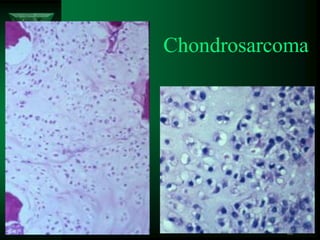
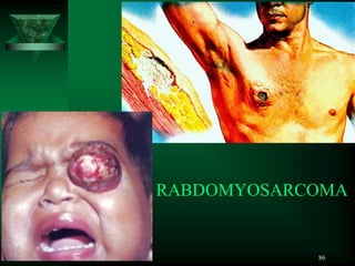
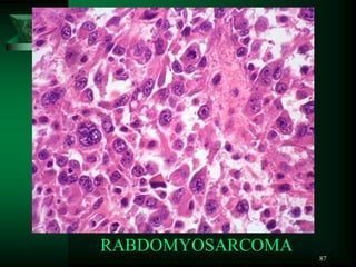
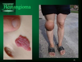
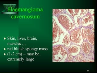
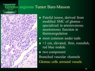
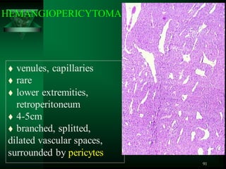
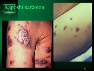
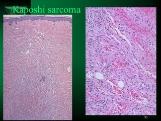
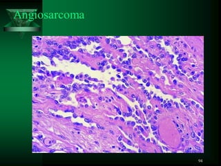
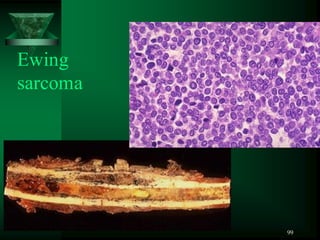
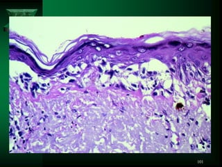
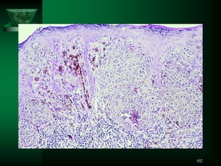
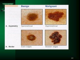
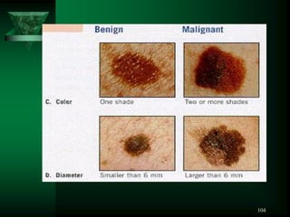
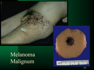
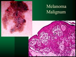
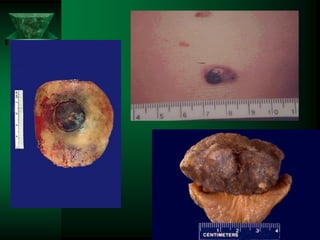
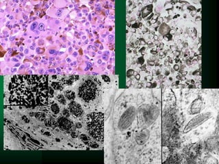
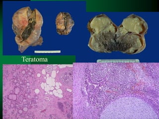
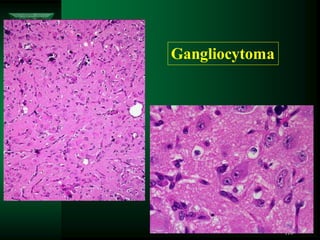
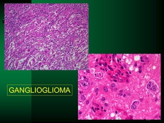
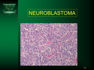
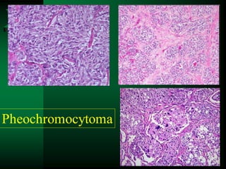
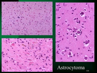
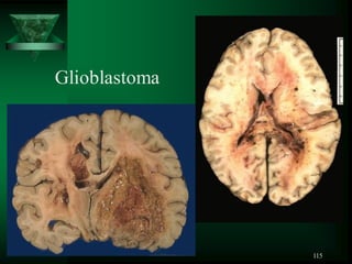
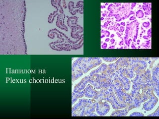
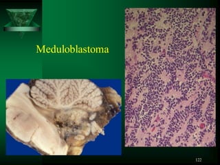
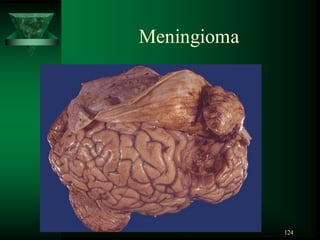
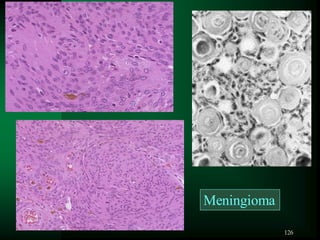
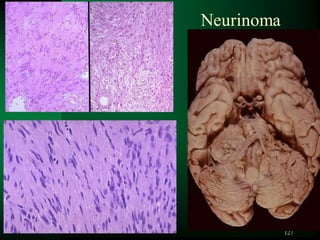
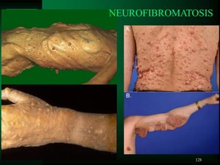
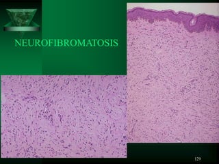
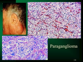
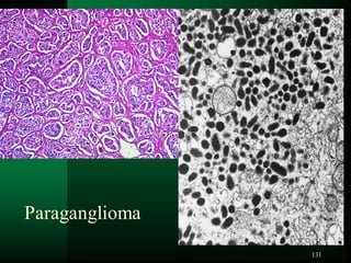
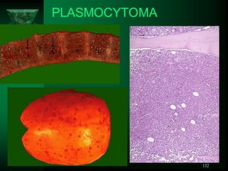
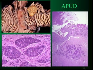
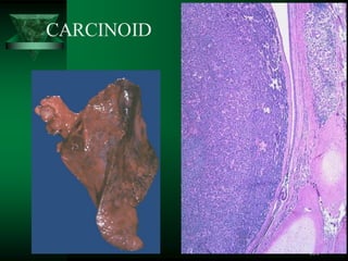
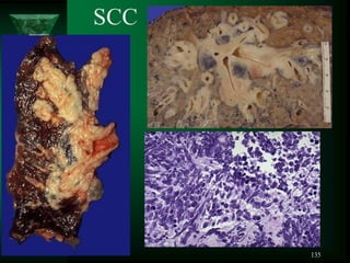
MODULE 5.
Neoplastic growth
Department of General and Clinical Pathology
Medical University - Varna
Peter Ghenev MD, PhD
2
ETIOLOGY OF TUMOR GROWTH
Theories
 Chemical (irritative) R.Virchow
 Viral (genetic)
 Dysonthogenetic (Conheim)
 Polietiological
3
 Sir Percival Pott, London surgeon -
more than 200 years ago correctly attributed
scrotal skin cancer in chimney sweeps to
chronic exposure to soot
Chemical Carcinogenesis
4
Carcinogenic agents from the environment
 Chemicals: insecticides, herbicides ...
 Drugs: anticancer, immunosuppressors
 Tobaco smoke
 Ethanol
 Heavy metals
 Microbial: Helicobacter pylori...
 VIRAL incl. Sexually transmitted: HIV,
Herpes simplex, HCV, Human papilloma virus
 Radiation: UVL - formation of pyrimidine
dimers in DNA
5
4 phases natural history of most malignant tumors:
 transformation - malignant change of target cell
 growth of the transformed cells;
 local invasion
 distant metastases
6
Steps in Chemical
Carcinogenesis
 INITIATION
 exposure of cells to sufficient dose of a carcinogen
 causes permanent DNA damage (mutations)
 rapid and irreversible and has “memory”
 PROMOTION -
induction of tumors in initiated cells
TRANSFORMATION
7
TRANSFORMATION
EVENTS IN
CHEMICAL
CARCINOGENESIS
8
The net effect of
HPV proteins -
block of apoptosis
and cell proliferation
EVENTS IN VIRAL CARCINOGENESIS
9
Dissemination of cancers may occur through
one of three pathways:
(1) direct seeding of body cavities or surfaces,
(2) lymphatic spread, and
(3) hematogenous spread
+Implantation during surgery
Pathways of Spread
10
DEFINITION: Tumor implants discontinuous
with the primary tumor
Metastasis unequivocally marks a tumor as malignant
because benign neoplasms do not metastasize
The invasiveness of cancers permits them to
penetrate into blood vessels, lymphatics, and
body cavities, providing the opportunity for spread
With few exceptions, all cancers can metastasize
Metastases
11
Millions of cells are released into
the circulation each day from
a primary tumor, but only a few
metastases are produced
A clear understanding of the origin of metastasis is
of major importance for the management of cancer
patients and the development of effective therapies
to prevent tumor spread
METASTASIS
12
involves two phases:
(1) invasion of the extracellular matrix and
(2) vascular dissemination and homing of tumor cells
The metastatic cascade
13
METASTASIS
first step
14
METASTASIS
second step
15
Detachment
("loosening up")
of the tumor cells
from each other
Degradation of ECM
Invasion of the ECM is an active process
that can be resolved into several steps
16
Attachment to
matrix components
Migration of
tumor cells
Invasion of the ECM is an active process
that can be resolved into several steps
17
Certain subclones possess the right combination of
gene products to complete all the steps for metastasis
Mechanisms of metastasis development
within a primary tumor
18
Metastases are due to gene expression pattern
called a "metastasis signature”
Mechanisms of metastasis development
within a primary tumor
19
Mechanisms of metastasis development
within a primary tumor
A combination of A and B, in which
metastatic variants appear in a tumor
with a metastatic gene signature
20
Metastasis development is influenced by the
tumor stroma, which regulates angiogenesis,
local invasiveness and resistance to immune
elimination
21
Metastasis signature represents a general
predisposition for metastasis, due to multiple
abnormalities that occur in many, perhaps in
most, cells of a primary tumor.
This signature may involve not only properties
intrinsic to the cancer cells, but also the
characteristics of the stroma, the presence of
infiltrating immune cells, and angiogenesis.
Nat Rev Cancer. 2005; 5 (5): 338.
22
23
LOCAL
INVASION
OF TUMORS
24
LOCAL
INVASION
OF TUMOR
25
Peritoneal
carcinosis by
ovarian cancer
26
Metastatic adenocarcinoma
from colon
27
Axillary lymph node with
metastatic breast carcinoma
28
GRADING AND STAGING OF CANCER
GRADING - based on the degree of differentiation
of the tumor cells and the number of mitoses within
the tumor as presumed correlates of aggressiveness
STAGING - based on the size of the primary
lesion, its extent of spread to surrounding tissues,
regional lymph nodes, and the presence or
absence of blood-borne metastases
TNM
29
STAGING AND GRADING OF TUMORS
TNM
DIFFERENTIATION
•well
•moderate
•low (poor)
G
30
CANCER
INCIDENCE
31
CANCER
INCIDENCE
32
Cancer Mortality by Age for Males
All Ages Under 15 15-34
1) Lung
2) Prostate
3) Colorectum
4) Pancreas
5) Leukemia
1) Leukemia
2) CNS
3) Endocrine
4) Lymphoma
5) Connective
Tissue
1) Leukemia
2) Lymphoma
3) CNS
4) Skin
5) Hodgkin’s
Disease
35-54 55-74 75+
1) Lung
2) Colorectum
3) CNS
4) Lymphoma
5) Pancreas
1) Lung
2) Colorectum
3) Prostate
4) Pancreas
5) Esophagus
1) Lung
2) Prostate
3) Colorectum
4) Pancreas
5) Leukemia
33
Cancer Mortality by Age for Females
All Ages Under 15 15-34
1) Lung
2) Breast
3) Colorectum
4) Ovary
5) Pancreas
1) Leukemia
2) CNS
3) Endocrine
4) Connective
Tissue
5) Kidney
1) Breast
2) Leukemia
3) Uterus
4) CNS
5) Lymphoma
35-54 55-74 75+
1) Breast
2) Lung
3) Colorectum
4) Uterus
5) Ovary
1) Lung
2) Breast
3) Colorectum
4) Ovary
5) Pancreas
1) Colorectum
2) Lung
3) Breast
4) Pancreas
5) Ovary
34
CARCINOGENESIS Summary
35
BIOCHEMICAL MARKERS
 tumor markers – diagnostic and prognostic
 Follow up - therapeutic effect and recurrent
disease
36
LIST OF SOME MARKERS
hCG choriocarcinoma
AFP hepatocellular ca
calcitonin thyroid medullary ca
prolactin pituitary adenomas
CA 125 ovarian carcinoma
PSA prostate carcinoma
chromogranin A endocrine neoplasias
37
 Senile kerratosis
 Radiation dermatitis
 Leukoplakia
 Erythroplasia Queyrat
 Cryptorchism
 Villous adenoma
 Morbus Bowen
 Adenomatous hyperplasia of endometrium
 Familial polyposis
 Ulcerous colitis
 Atrophic gastritis
Precancerous
lesions
38
Cancer patients usually die because of:
 Metastases
 Rupture of large vessels and hemorrhage
 Compression of vital organs
 Organ failure
 Infections
 Intoxication - cacchexia
 Paraneoplastic syndrome - tromboses
39
Histogenetic
Classification
of Tumors
40
41
Papilloma
42
Adenoma of adrenal gland
43
Breast papilloma
44
45
Cancer of colon
46
ПОЛИПОЗНО-ПАПИЛАРНА
ТИП “ЦВЕТНО ЗЕЛЕ” ФОРМА
47
48
АДЕНОКАРЦИНОМ НА СТОМАХА
49
50
Squamous cell cancer
51
Basocellular
cancer
52
Bronchogenic cancer
53
Lung cancer
54
Liver
cancer
55
Carcinoma portionis
vaginalis coli uteri
56
Squamous cell cancer - coli uteri
57
Squamous cell cancer - coli uteri
58
Advanced cervical cancer
59
Advanced endometrial cancer
60
LEIOMYOMA
61
Serous cystadenoma of ovarium
62
Papillary serous
cystadenocarcinoma
of ovary
63
64
Mature ovarian teratoma
65
Ovarian
teratoma
66
BREAST
FIBROADENOMA
67
Breast cancer
68
69
Seminoma
70
FIBROMA
71
LIPOMA - any age, any place
 Skin, in the wall of GIT organs, retroperitoneum
72
 CHONDROMA
– long bones, hands, feet, ribs,
vertebral column and pelvis
73
atrium, 1-10 cm, pedunculated
MYXOMA
74
 OSTEOMA
- bones of the skull
- compact and spongious part
- osteoma eburneum (durum)
75
OSTEOMA
osteoma spongiosum
76
 rare
 myocardium – small white gray nodules up
to several centimeters
Myoblastic myoma of Abrikosov
– tongue, esophagus
– large cells with granulated cytoplasm
RABDOMYOMA
77
 rare tumor
 malignant elongated
cells in bundles
– nuclei “hering bone"
 giant cells not
common
 differentiated – more
collagen
 undifferentiated –
scant collagen
FIBROSARCOMA
78
LEIOMYOSARCOMA
79
• about 20% of all primary
mesenchymal tumors
• 75% - until 25 years
• Painful mass, fast growing in
the methaphysis of long bones
• 50% knee, flat bones
• Sudden fracture
•20% with metastases at time
of diagnosis
Osteogenic Sarcoma
80
Osteogenic
Sarcoma
81
Osteogenic
Sarcoma
82
most common in
adults 40-60 years
deep in soft tissue
of extremities,
retroperitoneum
Liposarcoma
83
LIPOSARCOMA
LIPOBLASTS –
resemble fetal
adipocytes, round
cells with many
lipid vacuoles
84
Chondrosarcoma
 atypical chondrocytes in
basophillic chondroid matrix
 formation of osteoid
substance and primitive bone
trabeculae, surrounded by
neoplastic bizarre giant cells
 osteolytic
 osteosclerotic variants
85
Chondrosarcoma
86
RABDOMYOSARCOMA
87
RABDOMYOSARCOMA
88
Hemangioma
89
Haemangioma
cavernosum
 Skin, liver, brain,
muscles ...
 red bluish spongy mass
 (1-2 cm) – may be
extremely large
90
Glomus angioma Tumor Bare-Masson
 Painful tumor, derived from
modified SMC of glomus -
specialized in arteriovenous
anastomoses function in
thermoregulation
 most common under nails
 >1 cm, elevated, firm, roundish,
red blue nodule
 two component‫׃‬
Branched vascular channels
Glomus cells arround vessels
91
HEMANGIOPERICYTOMA
 venules, capillaries
 rare
 lower extremities,
retroperitoneum
 4-5cm
 branched, splitted,
dilated vascular spaces,
surrounded by pericytes
92
Kaposhi sarcoma
93
Kaposhi sarcoma
94
Angiosarcoma
95
Angiosarcoma
96
Synovioma
97
Ewing sarcoma
 Първични малигнени дребноклетъчни тумори на
костта /сарком на Юинг и PNET/
 Неврална диференциация
 PNET – примитивни невроектодермални тумори
 Различават се по диференциация
 Туморите с неврална диференциация на светлинна
микроскопия, имунохистохимично и ЕМ - PNET,
а тези, които не се позитивират - Юинг сарком
 Двата тумора – 6-10% от първичните малигнени
костни тумори
 При деца на 2-ро място след остеосаркома
 10-15 г- възраст
98
Сарком на Юинг и PNET
 Произлизат от медуларния канал, инвазират
кортекса и периоста и образува болезнена
мекотъканна маса – жълтеникавобелезникава
често с кръвоизливи и некрози
 В диафизата на дългите тръбести кости,
плоските кости на таза
 Сарком на Юинг - малки сини кръгли клетки -
ПАС + цитоплазма - гликоген.
 PNET - розетки на Homer-Wright розетки
/туморни клетки подредени около централно
фибриларно пространство/ е индикатор за
невронална диференциация
99
Ewing
sarcoma
100
NEVUS
101
102
103
104
105
Melanoma
Malignum
106
Melanoma
Malignum
107
108
109
Teratoma
110
Gangliocytoma
111
GANGLIOGLIOMA
112
NEUROBLASTOMA
113
Pheochromocytoma
114
Astrocytoma
115
Glioblastoma
116
Glioblastoma
117
Oligodendroglioma
118
Епендимом /Ependymoma/ и тумори от плексус
хориоидеус
- 5%-10% от туморите до 20 г. - в съседство на епендима на
вентрикулите /най-често IV/
- При възрастни – гръбначния мозък
- Солиден или папиларни маси
- Трудно се екстирпират – близо до понса и продълговатия
мозък
- образува розетки /наподобява ембрионален епендимален
канал/ и периваскуларни розетки /около съдове/ - добре
диференциран епендимом /II степен/
- Анапластичен/ епендимом /III степен/
119
Ependymoma
120
Епендимни тумори и тумори от плексус
хориоидеус
 Папилом на Plexus chorioideus
 при деца латералните вентрикули; при
възрастни - IV вентрикул
 папили /съединителнотъканна сърцевина
покрита с кубичен или цилиндричен
епител
 Хориоиден карцином !!!
121
Папилом на
Plexus chorioideus
122
Meduloblastoma
123
Meningioma
124
Meningioma
125
Meningioma
Meningiothelial type
126
Meningioma
127
Neurinoma
128
NEUROFIBROMATOSIS
129
NEUROFIBROMATOSIS
130
Paraganglioma
131
Paraganglioma
132
PLASMOCYTOMA
133
APUD
134
CARCINOID
135
SCC

ELGP 22-23_Neoplastic growth.pdf