This document discusses the use of electrosurgery in dentistry. It begins by describing the basic components and mechanism of electrosurgery, noting that it uses high frequency alternating current to generate heat and cut or coagulate tissue without significant bleeding. It then discusses various electrosurgery techniques and their indications, benefits including minimal trauma and hemostasis, and potential risks like odor and heat damage. It also compares electrosurgery to lasers, noting their similar applications but differences in costs, learning curves, and heat production. In summary, the document presents electrosurgery as a safe and effective soft tissue management tool when used properly.
Electrosurgery uses high frequency electrical current to cut, coagulate, and destroy soft tissue. It has several advantages over scalpels for dental procedures, allowing for precise sculpting of tissue without pressure and inherent concurrent hemostasis. Potential disadvantages include unpleasant odor, risk of damaging bone or teeth if contact is made. Proper technique involves using different electrode types and currents depending on the procedure, with rapid movements to prevent tissue burning. Healing occurs via clot formation, inflammation, and growth of new connective and epithelial tissue over several days.
This document discusses common white lesions of the oral cavity that can be seen during examination. It describes several conditions that present as white or pale lesions including leukoedema, white sponge nevus, frictional keratosis, morsicatio mucosae oris, smokeless tobacco keratosis, nicotine stomatitis, leukoplakia, hairy leukoplakia, candidiasis, oral submucous fibrosis, oral lichen planus, and chemical injuries of the oral mucosa. Differential diagnoses and clinical features of each condition are provided.
Lasers and its application in periodonticsShilpa Shiv
The document discusses different types of lasers used in periodontology, including their properties, mechanisms of interaction with tissue, safety classifications, and clinical applications. It provides details on lasers such as the argon, diode, Nd:YAG, Er:YAG, and CO2 lasers, covering their wavelengths, active mediums, delivery systems, absorption characteristics, and periodontal uses. The document also examines laser tissue interactions, safety considerations, and the theoretical zones of tissue change caused by laser exposure.
Valuable clinical guide for soft tissue diode laser users with pre & post operative pics , useful , fully informative with tips helps my beloved coleagues to enjoy & profession the use of soft tissue dental laser #clinical_dental_laser #dental_laser #soft_tissue_laser
This document discusses the use of electrosurgery in esthetic dental procedures. It begins by outlining some common esthetic considerations like color, spacing, and soft tissue arrangement. It then describes how electrosurgery can be used conservatively for soft tissue alterations without the need for surgery. The document provides details on how electrosurgery works, producing heat through radiofrequency waves to contour soft tissues. Various electrosurgery techniques are explained for incisions, coagulation, and tissue removal. The document outlines appropriate uses of electrosurgery and important safety considerations for patient care.
Peripheral giant cell granuloma (giant cell epulisKhin Soe
The document discusses two types of giant cell lesions of the jaw: peripheral giant cell granuloma (PGG) and central giant cell granuloma (CGG). PGG is a reactive lesion caused by local irritation or trauma that occurs on the gingiva. CGG is a benign process that occurs within the jaw bones and can be non-aggressive or aggressive depending on symptoms and growth rate. Both lesions contain multinucleated giant cells and are treated with surgical excision, with CGG having a slightly higher recurrence rate. Key distinguishing features and histological characteristics are provided.
This document provides an overview of lasers in dentistry. It discusses the history and development of lasers, how lasers are designed and how laser light interacts with tissues. It describes common dental lasers like CO2 and argon lasers, and their applications. CO2 lasers are well absorbed in oral tissues and useful for soft tissue procedures. Argon lasers are absorbed by hemoglobin and melanin, making them good for coagulation. The document outlines the benefits of lasers for various dental procedures.
This document discusses the use of lasers in periodontal treatment. It begins by introducing several types of lasers approved for soft tissue treatments in dentistry, including CO2, Nd:YAG, and diode lasers. The Er:YAG laser is also noted as being approved for hard tissue treatments. The document then lists advantages of laser surgery over conventional treatments. It provides examples of soft tissue applications like gingivectomy, gingivoplasty, and frenectomy. Hard tissue applications mentioned include scaling and root planing, bone procedures, whitening, and crown lengthening. Specific case examples are also included to illustrate laser procedures.
Radicular cysts originate from epithelial cell rests of Malassez and form through three phases: initiation, cyst formation, and growth/enlargement. They are usually painless unless infected and associated with nonvital teeth. Calcifying odontogenic cysts contain ghost cells, which represent abnormal keratinization and have an affinity for calcification. They may also induce dental tissue formation. Keratocystic odontogenic tumors initiate from dental lamina proliferation and enlarge through factors like osmolality, inflammatory exudate, glycosaminoglycans, and collagenolytic/bone resorbing molecules. Their thin fragile linings and intrinsic growth potential contribute to high recurrence rates.
Three sentences:
This document discusses oral pemphigus vulgaris, an autoimmune disease causing blistering of the mucosa. It defines the disease, describes its pathogenesis involving autoantibodies against desmoglein proteins, and outlines the clinical features, diagnosis using biopsy, immunofluorescence and Tzank smear, and treatment typically involving corticosteroids. Differential diagnoses include bullous pemphigoid and diagnosis is confirmed through histology demonstrating acantholysis and direct immunofluorescence showing intercellular IgG deposits.
This document discusses several laser dentistry systems and their applications. It provides information on the Philips ZoomWhiteSpeed light-activated whitening system, which can whiten teeth up to 8 shades in under an hour. It also discusses the Waterlase laser system which uses water and air to cut tissue without heat, vibration or pressure. Finally, it summarizes several other dental laser systems including models from Dentmat, CAO Group, Sirona, AMD, and Epic by Biolase.
This document provides an overview of chronic periodontitis. It discusses the introduction, history, prevalence, etiology, pathogenesis, clinical features, models of disease progression, microbiological and immunological considerations, and risk factors of chronic periodontitis. Chronic periodontitis is a complex polymicrobial infection that results from an imbalance between pathogenic bacteria in plaque and the host immune response. It begins as plaque-induced gingivitis and progresses to the destruction of connective tissue and alveolar bone through periods of activity and remission, leading to pocket formation and potential tooth loss over time if left untreated. Systemic and environmental risk factors like smoking can increase the risk and rate of disease progression.
The document discusses gingival curettage, which involves scraping diseased soft tissue from periodontal pockets. It describes different types of curettage including surgical, chemical, ultrasonic, and laser. Indications for curettage include shallow pockets and as maintenance treatment for recurrent inflammation. Contraindications include acute infections and pockets extending beyond the mucogingival junction. The procedure involves scraping the pocket wall with a curette. Excisional new attachment procedure is also discussed, which uses gingival incision followed by root planing. Healing after curettage involves blood clot formation, leukocyte proliferation, and re-epithelialization within 7 days.
Oral lichen planus is a common chronic mucocutaneous disease of unknown etiology that may undergo malignant transformation. It typically presents as white reticulated lines on the oral mucosa but can also appear as erosive, atrophic, bullous or other lesions. A confirmed diagnosis requires characteristic histopathology. While there is no cure, treatment focuses on managing symptoms like pain with topical or systemic corticosteroids and maintaining oral hygiene to reduce cancer risks.
The document discusses various types of oral papillomas including squamous papilloma, verruca vulgaris, and condyloma acuminatum. Squamous papilloma is the most common oral mucosal mass, caused by HPV types 6 and 11, and appears as a soft, painless growth. Verruca vulgaris (common wart) is associated with HPV types 2, 4, 6, and 40 and presents as rough, thickened white lesions. Condyloma acuminatum (genital wart) is sexually transmitted and associated with high-risk HPV types. The clinical and histological features of these lesions are summarized.
Deep carious lesions penetrate deep into the dentin and can potentially expose the pulp, causing pulpitis if left untreated. There are five zones of carious dentin, ranging from normal dentin to infected dentin teeming with bacteria. Pulpitis can be reversible or irreversible, depending on the severity of inflammation. For deep lesions near the pulp, indirect pulp capping involves removing infected dentin while leaving behind affected dentin to avoid exposure. For direct exposures, pulp capping places a biocompatible material over the exposure site to promote healing and maintain vitality. Factors like remaining dentin thickness, exposure size, and patient age influence the success of pulp capping procedures.
This document discusses classifications and clinical relevance of oral epithelial dysplasia in assessing risk of oral potentially malignant disorders. It describes various classification systems for grading dysplasia including WHO and Ljubljana systems. Key histopathological features of dysplasia are loss of maturation and increased nuclear-cytoplasmic ratio. Higher risk of malignant transformation is seen with factors like female gender, long standing lesions, location on tongue/floor of mouth, large size and presence of dysplasia. Accurate grading helps determine prognosis and clinical management.
The document discusses diseases of the dental pulp. It begins with an introduction to the pulp, including its composition and role in tooth development. It then discusses the pathophysiology of pulpal disease, describing how noxious stimuli can lead to inflammation and necrosis by disrupting blood flow. Several classifications of pulpal diseases are presented, including those based on histopathology, clinical symptoms, and the World Health Organization framework. Etiological agents are explored, such as mechanical trauma, thermal or chemical insults, bacterial invasion, and idiopathic causes. Specific pulpal diseases like reversible and irreversible pulpitis are also mentioned.
This document discusses various non-carious lesions of teeth including attrition, abrasion, erosion, abfraction, dentinal sclerosis, dead tracts, secondary dentin, pulp stones, hypercementosis, and cementicles. It defines each lesion, describes the etiology and pathogenesis, and highlights key clinical features. Attrition is mechanical wear of teeth from tooth contact while abrasion is caused by external frictional forces. Erosion results from chemical dissolution of tooth structure by acids. Abfraction involves stress-induced cervical lesions. Dentinal sclerosis and dead tracts are age-related changes in dentin. Secondary dentin deposition occurs in response to stimuli. Pulp stones, hypercementosis and
Ectodermal dysplasias (EDs) are a group of inherited disorders that affect two or more ectodermal structures such as hair, teeth, nails, and sweat glands. They are caused by genetic defects that may be inherited or occur spontaneously. EDs are classified based on clinical phenotypes and affected structures. The most common types are hypohidrotic ED (affecting hair, teeth, nails and sweat glands) and hidrotic ED (affecting hair, teeth, and nails). Without proper care, ED patients can experience life-threatening hyperthermia, infections, and failure to thrive. Treatment focuses on managing symptoms and may involve dentures, skin care, eye protection, and environmental thermal
Light is an integral part of our life. Advances in technology are increasing and changing the ways that the patient experience dental treatment. One of the milestones in technological advancements in dentistry is the use of lasers The early 20th century saw one of the greatest inventions in science & technology, in that LASERS which later went on to became a gift to health sciences. Albert Einstein is usually credited for the development of the laser theory. He was the first one to coin the term “Stimulated Emission” in his publication “Zur Quantentheorie der Strahlung”, published in 1917 in the “Physikalische Zeitschrift”
Lasers are devices that produce beams of coherent and very high intensity light. The word LASER is an acronym for “Light Amplification by Stimulated\Emission of Radiation”. A crystal or gas is excited to emit light photons of a characteristic wavelength that are amplified and filtered to make a coherent light beam. The effect of the laser depends upon the power of the beam and the extent to which the beam absorbed. Several types of lasers are available based on the wavelengths. These range from long wavelengths (infrared), to visible wavelengths, to short wavelengths (ultraviolet), to special ultraviolet lasers called excimers. Lasers are used nowadays in many areas in the field of dentistry It is of the most captivating technologies in dental practice. Even though, introduced as an alternative to the traditional halogen curing light, laser now has become the instrument of choice, in many dental applications. Its advancements in the field of dentistry are playing a major role in patient care and well being.
This document reviews the soft tissue applications of lasers in dentistry. It discusses how lasers provide benefits like surface sterilization, a dry surgical field, and increased patient acceptance for soft tissue procedures. Specific conditions that can be effectively treated with lasers are mentioned, such as peripheral ossifying fibromas, denture-induced fibrous hyperplasia, mucoceles, hemangiomas, and lymphangiomas. Lasers are described as a useful tool for treating premalignant and malignant oral lesions as well.
This document discusses gingival pigmentation from a historical, physiological, and clinical perspective. It begins by covering the historical descriptions of pigmentation in various populations dating back to the early 1900s. It then describes the structure and function of melanocytes and melanin, as well as the genetic, hormonal, and environmental factors that regulate melanin synthesis. The document classifies different types of pigmentation and pigmented lesions that can occur in the oral mucosa. Finally, it reviews various surgical and non-surgical methods that can be used to depigmentate abnormal gingival pigmentation.
This document discusses red and white lesions of the oral cavity, focusing on oral candidiasis. It describes the various types of oral candidiasis including pseudomembranous, erythematous, chronic plaque-type, and median rhomboid glossitis. Predisposing factors, clinical findings, diagnosis, treatment with antifungal medications or surgery, and prognosis are summarized for each type. Chronic hyperplastic candidiasis may require long-term antifungal therapy or surgery due to risk of recurrence. Overall prognosis is generally good if predisposing factors can be addressed.
Dry socket is a common complication after tooth extraction where the blood clot in the extraction socket fails to form properly. It causes severe throbbing pain 2-5 days after extraction. Risk factors include smoking, single extractions, and surgical extractions. Treatment involves removing any infection in the socket, prescribing antibiotics and painkillers.
Endodontic surgery is performed to address issues like failed root canal treatments, procedural errors, anatomical variations, and biopsies. It involves raising a surgical flap, resecting the root tip, preparing and filling the root end cavity. Potential complications include swelling, pain, nerve damage and infection. A variety of materials can be used for the root end filling including zinc oxide eugenol cements, MTA, composites and glass ionomer cements. The goal is to provide an apical seal to prevent reinfection from microbes remaining in the root canal system.
This document discusses potential complications from the use of electrosurgery in laparoscopic procedures. It notes that minimally invasive surgeries have benefits like less tissue disruption and faster recovery, but electrosurgery can cause unintended tissue damage if currents stray. The document outlines factors that influence the tissue effects of electrosurgery like frequency, area of contact, and duration of application. It describes different types of electrosurgery and how to reduce risks of direct coupling, capacitive coupling, and insulation failures causing burns. Maintaining equipment and using lowest possible settings can help avoid complications.
This document provides an overview of electrosurgery, including:
1) It describes the principles of electrosurgery and how it differs from electrocautery by using alternating current rather than direct current.
2) It explains the components of an electrosurgical system including the generator, active electrode, and dispersive electrode.
3) It discusses safety considerations for electrosurgery such as proper grounding pad placement and avoiding pooled fluids near equipment.
This document discusses diathermy, which uses high frequency current to cut and coagulate body tissue. It describes the different types of diathermy including shortwave, ultrasound and microwave. Shortwave diathermy uses condenser plates to concentrate heat in subcutaneous tissues. Ultrasound uses acoustic vibrations converted to heat, while microwave diathermy uses similar radiation to radar waves. The document also covers monopolar and bipolar diathermy configurations and safety precautions for using diathermy.
Electrosurgery uses high-frequency alternating electrical current to cut, coagulate, or vaporize tissue. It allows for precise cuts with limited blood loss. The current is delivered via an electrosurgery generator to an active electrode and returns through the patient to a neutral electrode. Different waveforms and modes, such as cut, coagulate, and blend, are used depending on the desired tissue effect. Safety features monitor for excess heat buildup and electrode detachment to prevent patient injury.
This document discusses the history and uses of electro surgery in gynecology. It begins with the early history of heat therapy and progresses to modern developments. Key points covered include the basics of electricity used, types of currents and waveforms, effects on tissue, and specific applications in gynecology like treating cervical lesions, tubal sterilization, endometriosis, and fibroids. Proper use and safety precautions are also emphasized.
This document discusses various energy sources used in surgery, including electrosurgery, ultrasonic, argon beam, laser, cryotherapy, and infrared coagulation. It provides details on electrosurgery, describing monopolar and bipolar diathermy, tissue effects, and safety precautions. Other technologies like Ligasure, Harmonic Scalpel, Thunderbeat, and argon beam coagulation are also summarized, outlining their advantages and disadvantages. Lasers are discussed in terms of their properties and surgical effects. Cryotherapy and infrared coagulation are briefly described as well.
This document discusses different types of energy modalities used in surgery including monopolar, bipolar, ultrasonic, and plasma kinetic technologies. Monopolar energy uses an active electrode at the surgical site and a return electrode elsewhere on the patient's body, allowing for tissue cutting, coagulation, and desiccation. Bipolar energy passes between two close electrodes, minimizing collateral damage. Advanced bipolar technologies like Ligasure, Plasma Kinetic Gyrus, and Enseal can additionally seal and transect tissue. Ultrasonic devices use high frequency vibrations to denature proteins for coagulation and mechanical cutting. The effects of different energies on tissue are described, noting temperatures at which protein denaturation and
The document describes the principles and components of electrosurgery, including monopolar and bipolar electrosurgical units. It discusses the differences between electrocautery and electrosurgery, the components of electrosurgical systems, and the various modes and effects such as cut, coagulate, and blend. Safety considerations are provided around the use of electrosurgery and proper placement and application of grounding pads.
Diathermy uses high frequency energy to heat deep tissues for therapeutic purposes. It was first used in 1907. Electrosurgery uses a high frequency current to make surgical incisions, control bleeding, and destroy unwanted tissue. It can be performed with monopolar or bipolar techniques. Monopolar techniques use an active electrode and a passive return electrode placed elsewhere on the body. Bipolar techniques use two electrodes in close proximity. The effects on tissue include cutting, coagulation, desiccation, and fulguration depending on various control settings. Safety is ensured by using the lowest effective power and following protocols to prevent complications. New advances include devices combining harmonic scalpels and vessel sealing capabilities.
Ultrasonic therapy uses high frequency sound waves above the range of human hearing to provide therapeutic effects. It works by using an electrical current to power transducers that convert the current into ultrasonic waves. These waves can then be used for diagnostic imaging, surgery, and physiotherapy. Therapeutically, ultrasonic waves create effects through thermal, mechanical, and chemical/biological interactions with tissues. Common uses are for musculoskeletal conditions like sprains, tendinitis, and arthritis. Proper application involves selecting an appropriate intensity, duration, and frequency setting based on the condition being treated. Risks like burns and tissue damage require precautions like starting with low intensities and using pulsed rather than continuous waves in some cases.
The document discusses various energy sources used in surgery including electrical, ultrasonic, argon beam, and laser energies. It provides details on electrosurgery modalities like electrocautery and electrosurgery. Newer advanced bipolar devices like Ligasure, Gyrus ACMI, and Enseal are described which provide vessel sealing through thermal coagulation. Ultrasonic devices like Harmonic scalpel use high frequency vibrations for vessel sealing and precise dissection. Other technologies discussed include argon beam coagulation, CUSA, microwave ablation, and radiosurgery. Patient safety considerations are highlighted for different energy sources.
This document discusses electrosurgery and provides information on:
1. The history and development of electrosurgery, beginning with Becquerel's use of electrocautery in the 19th century and Bovie's development of the first electrosurgical unit in 1926.
2. Key aspects of electrosurgery including different current types, modes (monopolar vs bipolar), electrodes, and safety considerations.
3. Uses of bipolar electrosurgery for procedures like resection of fibroids and advantages over monopolar techniques.
This document summarizes key aspects of electrosurgical units used in surgery. It describes diathermy, which uses radiofrequency current to cut and coagulate tissue with limited blood loss. Monopolar diathermy involves current passing from the active electrode through the patient's body to a grounding plate, while bipolar diathermy confines current to tissue between instrument electrodes. Proper placement and monitoring of grounding plates is important to prevent burns with monopolar devices. Electrosurgical units use different waveforms and modes for cutting, coagulation, and desiccation of tissue.
This document provides information about electrosurgical units (ESUs). It begins with a brief history of electrosurgery, which was developed in the 1920s. It then discusses the principles of how ESUs work using high frequency alternating current. Different types of ESUs are described, including spark gap generators, solid state generators, grounded systems, and isolated systems. The effects of ESUs on tissue are explained for cutting, coagulation, and blending. Factors that impact tissue effects and various electrosurgical applications are also summarized.
This document provides information about various surgical energy modalities used in urology, including electrosurgery, ultrasonic devices, bipolar devices, and lithotripsy tools. It discusses the history and mechanisms of electrosurgery and monopolar/bipolar devices. Safety tips are provided for electrosurgery. Ultrasonic devices, bipolar vessel sealers, and integrated generators are also summarized. Details are given on electrohydraulic, pneumatic, and piezoelectric lithotripsy techniques. The document aims to educate urology residents and physicians on these diverse but important surgical energy technologies.
Thermal cauterization uses electromagnetic currents to generate heat in body tissues. It can be used physically to treat deeper lesions or surgically to cauterize blood vessels and destroy abnormal growths. The process involves an electrosurgical generator, active electrode, patient return electrode, and forming an electrical circuit. Different frequencies are used for different applications like nerve stimulation or electrosurgery. Bipolar cauterization involves both electrodes at the surgical site while monopolar uses one active electrode and a patient return elsewhere. Settings like waveform, power, and time determine cutting versus coagulation effects. Safety involves avoiding interference with devices and preventing burns.
In operating room the most hazardous devise used in a daily basis is diathermy.
A basic understanding of electricity is needed to safely apply electrosurgical technology for patient care.
This document discusses various complications that can occur during and after laparoscopic surgery. It covers positioning-related injuries, access-related injuries such as bleeding and organ perforation, complications of pneumoperitoneum, and issues related to electrosurgery such as burns. Specific procedures like cholecystectomy and appendectomy have their own risks discussed as well, including bile duct injury and abscess formation. Prevention strategies are provided such as careful patient positioning, safe access techniques, monitoring vital signs, and inspecting equipment.
Diathermy uses high-frequency electrical currents to generate deep heat in tissues for physical therapy and surgery. There are three main types: shortwave, ultrasound, and microwave diathermy. It can be used to warm tissues for pain relief or to coagulate and seal tissues during surgery. Surgical diathermy comes in monopolar and bipolar forms and is used for coagulation, fulguration, and cutting. Risks include burns, explosions from igniting flammable substances, and device malfunctions.
Promoting Wellbeing - Applied Social Psychology - Psychology SuperNotesPsychoTech Services
A proprietary approach developed by bringing together the best of learning theories from Psychology, design principles from the world of visualization, and pedagogical methods from over a decade of training experience, that enables you to: Learn better, faster!
Osteoporosis - Definition , Evaluation and Management .pdfJim Jacob Roy
Osteoporosis is an increasing cause of morbidity among the elderly.
In this document , a brief outline of osteoporosis is given , including the risk factors of osteoporosis fractures , the indications for testing bone mineral density and the management of osteoporosis
Here is the updated list of Top Best Ayurvedic medicine for Gas and Indigestion and those are Gas-O-Go Syp for Dyspepsia | Lavizyme Syrup for Acidity | Yumzyme Hepatoprotective Capsules etc
8 Surprising Reasons To Meditate 40 Minutes A Day That Can Change Your Life.pptxHolistified Wellness
We’re talking about Vedic Meditation, a form of meditation that has been around for at least 5,000 years. Back then, the people who lived in the Indus Valley, now known as India and Pakistan, practised meditation as a fundamental part of daily life. This knowledge that has given us yoga and Ayurveda, was known as Veda, hence the name Vedic. And though there are some written records, the practice has been passed down verbally from generation to generation.
share - Lions, tigers, AI and health misinformation, oh my!.pptxTina Purnat
• Pitfalls and pivots needed to use AI effectively in public health
• Evidence-based strategies to address health misinformation effectively
• Building trust with communities online and offline
• Equipping health professionals to address questions, concerns and health misinformation
• Assessing risk and mitigating harm from adverse health narratives in communities, health workforce and health system
Histololgy of Female Reproductive System.pptxAyeshaZaid1
Dive into an in-depth exploration of the histological structure of female reproductive system with this comprehensive lecture. Presented by Dr. Ayesha Irfan, Assistant Professor of Anatomy, this presentation covers the Gross anatomy and functional histology of the female reproductive organs. Ideal for students, educators, and anyone interested in medical science, this lecture provides clear explanations, detailed diagrams, and valuable insights into female reproductive system. Enhance your knowledge and understanding of this essential aspect of human biology.
Adhd Medication Shortage Uk - trinexpharmacy.comreignlana06
The UK is currently facing a Adhd Medication Shortage Uk, which has left many patients and their families grappling with uncertainty and frustration. ADHD, or Attention Deficit Hyperactivity Disorder, is a chronic condition that requires consistent medication to manage effectively. This shortage has highlighted the critical role these medications play in the daily lives of those affected by ADHD. Contact : +1 (747) 209 – 3649 E-mail : sales@trinexpharmacy.com
Does Over-Masturbation Contribute to Chronic Prostatitis.pptxwalterHu5
In some case, your chronic prostatitis may be related to over-masturbation. Generally, natural medicine Diuretic and Anti-inflammatory Pill can help mee get a cure.
NVBDCP.pptx Nation vector borne disease control programSapna Thakur
NVBDCP was launched in 2003-2004 . Vector-Borne Disease: Disease that results from an infection transmitted to humans and other animals by blood-feeding arthropods, such as mosquitoes, ticks, and fleas. Examples of vector-borne diseases include Dengue fever, West Nile Virus, Lyme disease, and malaria.
ABDOMINAL TRAUMA in pediatrics part one.drhasanrajab
Abdominal trauma in pediatrics refers to injuries or damage to the abdominal organs in children. It can occur due to various causes such as falls, motor vehicle accidents, sports-related injuries, and physical abuse. Children are more vulnerable to abdominal trauma due to their unique anatomical and physiological characteristics. Signs and symptoms include abdominal pain, tenderness, distension, vomiting, and signs of shock. Diagnosis involves physical examination, imaging studies, and laboratory tests. Management depends on the severity and may involve conservative treatment or surgical intervention. Prevention is crucial in reducing the incidence of abdominal trauma in children.
Recomendações da OMS sobre cuidados maternos e neonatais para uma experiência pós-natal positiva.
Em consonância com os ODS – Objetivos do Desenvolvimento Sustentável e a Estratégia Global para a Saúde das Mulheres, Crianças e Adolescentes, e aplicando uma abordagem baseada nos direitos humanos, os esforços de cuidados pós-natais devem expandir-se para além da cobertura e da simples sobrevivência, de modo a incluir cuidados de qualidade.
Estas diretrizes visam melhorar a qualidade dos cuidados pós-natais essenciais e de rotina prestados às mulheres e aos recém-nascidos, com o objetivo final de melhorar a saúde e o bem-estar materno e neonatal.
Uma “experiência pós-natal positiva” é um resultado importante para todas as mulheres que dão à luz e para os seus recém-nascidos, estabelecendo as bases para a melhoria da saúde e do bem-estar a curto e longo prazo. Uma experiência pós-natal positiva é definida como aquela em que as mulheres, pessoas que gestam, os recém-nascidos, os casais, os pais, os cuidadores e as famílias recebem informação consistente, garantia e apoio de profissionais de saúde motivados; e onde um sistema de saúde flexível e com recursos reconheça as necessidades das mulheres e dos bebês e respeite o seu contexto cultural.
Estas diretrizes consolidadas apresentam algumas recomendações novas e já bem fundamentadas sobre cuidados pós-natais de rotina para mulheres e neonatos que recebem cuidados no pós-parto em unidades de saúde ou na comunidade, independentemente dos recursos disponíveis.
É fornecido um conjunto abrangente de recomendações para cuidados durante o período puerperal, com ênfase nos cuidados essenciais que todas as mulheres e recém-nascidos devem receber, e com a devida atenção à qualidade dos cuidados; isto é, a entrega e a experiência do cuidado recebido. Estas diretrizes atualizam e ampliam as recomendações da OMS de 2014 sobre cuidados pós-natais da mãe e do recém-nascido e complementam as atuais diretrizes da OMS sobre a gestão de complicações pós-natais.
O estabelecimento da amamentação e o manejo das principais intercorrências é contemplada.
Recomendamos muito.
Vamos discutir essas recomendações no nosso curso de pós-graduação em Aleitamento no Instituto Ciclos.
Esta publicação só está disponível em inglês até o momento.
Prof. Marcus Renato de Carvalho
www.agostodourado.com
Rasamanikya is a excellent preparation in the field of Rasashastra, it is used in various Kushtha Roga, Shwasa, Vicharchika, Bhagandara, Vatarakta, and Phiranga Roga. In this article Preparation& Comparative analytical profile for both Formulationon i.e Rasamanikya prepared by Kushmanda swarasa & Churnodhaka Shodita Haratala. The study aims to provide insights into the comparative efficacy and analytical aspects of these formulations for enhanced therapeutic outcomes.
Basavarajeeyam is an important text for ayurvedic physician belonging to andhra pradehs. It is a popular compendium in various parts of our country as well as in andhra pradesh. The content of the text was presented in sanskrit and telugu language (Bilingual). One of the most famous book in ayurvedic pharmaceutics and therapeutics. This book contains 25 chapters called as prakaranas. Many rasaoushadis were explained, pioneer of dhatu druti, nadi pareeksha, mutra pareeksha etc. Belongs to the period of 15-16 century. New diseases like upadamsha, phiranga rogas are explained.
Local Advanced Lung Cancer: Artificial Intelligence, Synergetics, Complex Sys...Oleg Kshivets
Overall life span (LS) was 1671.7±1721.6 days and cumulative 5YS reached 62.4%, 10 years – 50.4%, 20 years – 44.6%. 94 LCP lived more than 5 years without cancer (LS=2958.6±1723.6 days), 22 – more than 10 years (LS=5571±1841.8 days). 67 LCP died because of LC (LS=471.9±344 days). AT significantly improved 5YS (68% vs. 53.7%) (P=0.028 by log-rank test). Cox modeling displayed that 5YS of LCP significantly depended on: N0-N12, T3-4, blood cell circuit, cell ratio factors (ratio between cancer cells-CC and blood cells subpopulations), LC cell dynamics, recalcification time, heparin tolerance, prothrombin index, protein, AT, procedure type (P=0.000-0.031). Neural networks, genetic algorithm selection and bootstrap simulation revealed relationships between 5YS and N0-12 (rank=1), thrombocytes/CC (rank=2), segmented neutrophils/CC (3), eosinophils/CC (4), erythrocytes/CC (5), healthy cells/CC (6), lymphocytes/CC (7), stick neutrophils/CC (8), leucocytes/CC (9), monocytes/CC (10). Correct prediction of 5YS was 100% by neural networks computing (error=0.000; area under ROC curve=1.0).
5. ◼ E.S. = surgical application of
fully controlled
self limiting
high frequency
electrically generated
heat
6. ◼ Often “electrocautery” is used to describe electrosurgery.
This is incorrect.
◼ Electrocautery refers to direct current (electrons flowing in
one direction) whereas electrosurgery uses alternating
current.
◼ During electrocautery, current does not enter the patient’s
body. Only the heated wire comes in contact with tissue.
◼ In electrosurgery, the patient is included in the circuit and
current enters the patient’s body.
7. ◼Before 1891 :flame heated instrument
◼1891:D’Arsonval
F>10,000 cycle/S
no potential damage / lethal neuromuscular
pain or shock
8. ◼1907 Doyan used Extreemly High Fr (3
mill cyc./S)with an active and passive
Electrode
◼Poor Quality incision
10. ◼Misconception:
not a simple house hold unit
Every day electricity: alternating current which
cycle from + to - 60 times/ S = 60 Hz
11. ◼If applied to living tissue
Cell polarization 60 times/sec
contraction of muscles
painful and lethal
Occurs up to
10,000 Hz (10KHz)
12. ◼E.s: convert current to Electromagnetic
Radio frequency (RF)wave which oscillate 2-4
million cyc./Sec (2-4 MHz)
◼Impossible for cells to depolarize at this rate
Electrical resistance of tissue
localized intra cellular heat without
muscle contraction
16. ◼RF wave leave the unit (hand piece)
pass the tissue enter passive
electrode to the unit
Both electrodes remain at room temp.
17. ◼RF wave passing the tissue slight raise
in temp. Volatilization of one cell layer
( the other remain intact)
◼Current set too low drag tissue
◼Current set too high sparking burn
tissue (excessive heat)
18. Active electrode: Electrode used for achieving desired surgical
effect.
Coagulation: Solidification of proteins accompanied by tissue
whitening.
Desiccation: Drying of tissue due to the evaporation of
intracellular fluids.
Fulguration: Random discharge of sparks between active
electrode and tissue surface in order to achieve coagulation
and/or desiccation.
Spray: Another term for fulguration.
19. t-tº= 1/σρċ (j2ţ)
◼ whereTandTo are the final and initial temperatures (K)
◼ ,σ is the electrical conductivity (S/m)
◼ ,ρ is the tissue density (kg/m3),
◼ C is the specific heat of the tissue (Jkg–1K–1),
◼ J is the current density (A/m2), and t is the duration of heat
applications
20. ◼ Monopolar electrosurgery
monopolar cutting instrument (usually the monopolar pencil) consists of a
single active electrode
◼Bipolar electrosurgery
In bipolar scissors, the blades are the active and return electrodes.
Current travels from one electrode back to the other electrode through only
the small section of tissue that lies between the scissors blades.
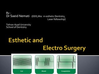
27. 3 – Fully Rectified current
1st half like above
2nd half repeat the same flow
Incise and coagulate at
the same time
28.
29. 4- Fully Rectified Filtered Current
The same properties with reduced changes ( the pick of hills
eliminated)
Fine cut
The current of choice
For esthetic gingival
Recontouring
(the cleanest incision)
30.
31. ◼ It result from resistance of tissue
◼ To minimize:
1. Controlling electrode size
2. The time of contact with tissue(max.1-2 sec.)
3. The type and intensity of current
(lowest level that work)
4 . Keeping the tissue moist (water or saline)
32. ◼Most ESU units on the market today have
REM technology.
◼REM system continually monitors the heat
build-up under the grounding pad
◼If the system detects excess heat build-up it
will shut off the current flow to prevent
patient injury
33. ◼ A return electrode burn
occurs when the heat
produced, over time, is not
safely dissipated by the
size or conductivity of the
patient return electrode.
34. ◼ In the case of reduced contact area, the current flow is
concentrated in a smaller area.
◼ As the current concentration increases, the temperature at
the return electrode increases.
◼ If the temperature at the return electrode site increases
enough, a patient burn may result.
◼ Surface area impedance can be compromised by: excessive
hair, adipose tissue, bony prominences, fluid invasion,
adhesive failure, scar tissue, and many other variables.
35. report side effect but at least one item uncontrolled
No change in heart wave
No change in pulp (animal study)
Healing comparable with sculple surgery
Choosing a suitable unit
In approximation to bone E.S is CONTRAINDICATED
48. ◼ Although possible to complete E.S and restorative
treatment at the same time
Best result gain within1-2 week healing period
49. 1. Pace maker (some types) 16 ft =4.8 m away
2. Patient with history of radiotherapy(delay healing)
3. Near chemicals like ethanol / chloroform
(explosion)
50. 1. No pressure for tissue separation
2. Smooth incision
3. Easy access in posterior
4. Better visibility due to coagulation
5. Little or no scar
6. Sterility (all bacteria in line of incision are volatilized)
7. Tissue Electro planning (electrode tangent to tissue and
remove or plane off a minimal layer)
8. Completion of treatment in one session if necessary
51. 1. Local anesthesia
2. No metal object in patient’s pocket(cause burn if touch the
plate)
3. Place the passive plate( thigh is preferred , scapula or
vertebral eminence have thin soft tissue)
4. Stabilize the jaw with bite block
5. Place moist cotton roll on either side
6. Hold an straight object parallel to inter papillary line
52.
53. 7 -consider zenith point
8 -penetrate the gingiva with an explorer to determine the
amount of tissue removal
55. 9- set the current
10-make a few practice with inactive electrode
11-the odor : place a 2*2 gauze with pleasant perfume
12- the depth of cut should be reaced in each individual
incision the length can be increased (3-4 short
incision)
13- tissue contact not more than 1-2 sec.
56. 14-remove any tissue accumulation with alcohol moistened
gauze
15- clean the site (tissue tag) with scaler,…..)
16-healing 7-14 days
17-over heating can cause pain , swelling, inflammation,…
18-post operative instruction:
a- 3% H2O2mix with tooth paste
b-rinsing with saline
19-Antibiotic is not necessary
57.
58.
59.
60.
61.
62.
63. 1) Development of Adequate Crown
Preparation
2) Esthetics
Indications for Crown Lengthening
67. ◼ the units cost much less than do lasers;
◼ the electrode cuts on its sides as well as on its tip;
◼ the electrode may be bent to meet the clinical need;
◼ cuts are made with ease when the device is set correctly;
◼ hemostasis is immediate;
◼ cutting is consistent; the wound is nearly painless after the procedure;
◼ the soft tissue has minimal trauma;
◼ the tip is self-disinfecting.
68. ◼ : anesthetic is required for cutting;
◼ both the name and the use of electrosurgery cause fear in some patients;
◼ there is an unavoidable burning-flesh odor;
◼ the operator has only a low tactile sense of exactly what is being cut;
◼ the heat developed by monopolar electrosurgery units does not allow for
their use around implants (careful use of bipolar electrosurgery is acceptable
around implants because it
produces less heat);
◼ bone can be damaged;
◼ electrosurgery is dangerous in an explosive environment;
◼ although this issue is controversial, electrosurgery may disrupt the action of
pacemakers16;
◼ patients who have undergone irradiation, have diabetes or have blood
dyscrasias can experience poor postoperative healing.
70. ◼ : their use requires minimal or no anesthetic;
◼ they do not harm dental hard tissues;
◼ their judicious use does not injure the dental pulp;
◼ because of low or no heat production, they can be used around dental
implants;
◼ they are antimicrobial;
◼ they remove endotoxins from root surfaces;
◼ there is growing evidence that laser use may be positive therapy for
periodontal disease;
◼ laser technology is considered state of the art by the lay public, so patients
are more accepting of its use in their treatment than of electrosurgery.
71. ◼ : the cost of laser is significantly higher than that of electrosurgery units;
◼ most of the techniques suggested for laser overlap with those for the much
less expensive electrosurgery;
◼ because of the potential hazard of laser light, laser use requires a learning
period and strict precautions;
◼ laser can cause eye damage, so protective glasses are required during its
use;
◼ cutting with lasers usually is slower than that with electrosurgery;
◼ there is a burning flesh odor;
◼ some techniques are time consuming;
◼ laser plume requires use of a high-filtration face mask, because of the
possible presence of pathogens in the plume
72.
73. 1. Mucosal – when the frenal fibres are attached up to the
mucogingival junction
2. Gingival – when the fibres are inserted within the attached
gingiva.
3. Papillary – when the fibres are extending into the interdental
papilla.
4. Papilla penetrating – when the frenal fibres cross the alveolar
process and extend up to the palatine papilla.