Part 1 ecg 2019 slide share
•Download as PPT, PDF•
2 likes•278 views

Part 1 ECG 2019 slide share

Recommended
More Related Content
What's hot
What's hot (20)
Similar to Part 1 ecg 2019 slide share
Similar to Part 1 ecg 2019 slide share (20)
More from hospital
More from hospital (20)
Recently uploaded
Recently uploaded (20)
Part 1 ecg 2019 slide share
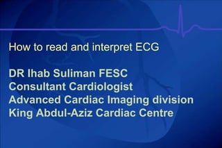
- 1. How to read and interpret ECG DR Ihab Suliman FESC Consultant Cardiologist Advanced Cardiac Imaging division King Abdul-Aziz Cardiac Centre
- 2. The Normal Conduction System
- 4. SA Node Sino Atrial Node • This is a collection of self-excitory (pacemaker) cells that normally fi re at a rate of between 60 and 100 Beats Per Minute (BPM). • The “wave” of Depolarisation moves from the SAN through an intra-atrial tract called Bachmanns bundle into the left atrium and to the Atrioventricular (AV) node. From here the impulse travels down the bundle of His into the right and Left bundle branches and finally into the Purkinje fi bres activating the ventricles.
- 5. Lead Position • A typical ECG report shows the cardiac cycle from 12 different vantage points (I, II, III, aVR, aVL, aVF, V1-V6), like viewing the event electrically from 12 different locations (like a 3D perspective).BUT only 10 electrodes are used. • Lead I represents activity that is going from the right arm to the left arm • Lead II represents activity that is going from the right arm to the left leg • Lead III represents activity that is going from the left arm to the left leg • aVL is placed on the left arm (or shoulder) • aVF is placed on the left leg (or hip) • aVR is placed on the right arm (or shoulder) • V1- 4th intercostal space to the right of sternum • V2- 4th intercostal space to the left of sternum • V3- halfway between V2 and V4 • V4- 5th intercostal space in the left mid-clavicular line • V5- 5th intercostal space in the left anterior axillary line • V6- 5th intercostal space in the left mid axillary line
- 6. NSR
- 7. Normal ECG
- 8. ECG showed ?? LBBB ?? RBBB
- 9. • LBBB in NSR • If acute Chest pain Go to The cath lab
- 10. 55 male with Psychiatry disorder
- 12. •VTACH
- 13. 35 lady with Occasional palpitations 2005 •WPW
- 14. What happened •WPW after ablation needed dual PPM
- 20. • NSR , Juvenile T-wave inversion.
- 21. WPW Syndrome
- 23. AF, Inferior Q waves
- 24. RBBB
- 25. 28 years with palpitations
- 26. • SVT/ AVNRT
- 28. • Atrial Flutter
- 29. 50 years old with mild CAD
- 30. • VT,THIS PT HAD SEVERE DCM,waiting for AICD
- 32. • Paced Rhythm
- 35. The 10 rules for a normal ECG I II III aVR aVL aVF V1 V2 V3 V4 V5 V6 .2
- 36. Rule 1 PR interval Millivolts Milliseconds 0 200 400 600 -0.5 0 0.5 1.0 P R T Q S PR interval should be 120 to 200 milliseconds or 3 to 5 little squares
- 37. Rule 2 Millivolts Milliseconds 0 200 400 600 -0.5 0 0.5 1.0 QRS P R T Q S The width of the QRS complex should not exceed 110 ms, less than 3 little squares
- 38. Rule 3 I II III aVR aVL aVF The QRS complex should be dominantly upright in leads I and II
- 39. Rule 4 I II III aVR aVL aVF QRS and T waves tend to have the same general direction in the limb leads
- 40. Rule 5 P Q T S All waves are negative in lead aVR
- 41. Rule 6 V1 V2 V3 V4 V5 V6 The R wave in the precordial leads must grow from V1 to at least V4
- 42. I II III aVR aVL aVF V1 V2 V3 V4 V5 V6 Rule 7 The ST segment should start isoelectric except in V1 and V2 where it may be elevated
- 43. Rule 8 I II III aVR aVL aVF V1 V2 V3 V4 V5 V6 The P waves should be upright in I, II, and V2 to V6
- 44. Rule 9 I II III aVR aVL aVF V1 V2 V3 V4 V5 V6 There should be no Q wave or only a small q less than 0.04 seconds in width in I, II, V2 to V6
- 45. Rule 10 I II III aVR aVL aVF V1 V2 V3 V4 V5 V6 The T wave must be upright in I, II, V2 to V6
- 47. Necrobiosis Lipoidica Diabeticorum • An inflammatory skin disorder characterized by irregularly shaped, callous lesions with reddish- brown pigmentation and central atrophy — was made on the basis of visual inspection. In necrobiosis lipoidica diabeticorum, the shins, ankles, and feet are typically affected, but 15% of patients may have lesions elsewhere. The disorder is more common among women than men and is more common among persons with diabetes than those without.
- 49. • Idiopathic DCM
- 50. What is the heart rate? •(300 / 6) = 50 bpm •www.uptodate.com
- 51. What is the heart rate? •(300 / ~ 4) = ~ 75 bpm •www.uptodate.com
- 52. What is the heart rate? •(300 / 1.5) = 200 bpm
- 53. 10 Second Rule As most EKGs record 10 seconds of rhythm per page, one can simply count the number of beats present on the EKG and multiply by 6 to get the number of beats per 60 seconds. This method works well for irregular rhythms.
- 54. What is the heart rate? •33 x 6 = 198 bpm •The Alan E. Lindsay ECG Learning Center ; http://medstat.med.utah.edu/kw/ecg/
- 55. Left axis deviation - negative QRS in lead AVF Right axis deviation - negative QRS in lead I Severe Right axis deviation negative QRS in BOTH lead I and AVF Quick & Easy AXIS DETERMINATION AVF AVF AVF AVF AVF AVF I I I I I I
- 56. The QRS Axis By near-consensus, the normal QRS axis is defined as ranging from -30° to +90°. -30° to -90° is referred to as a left axis deviation (LAD) +90° to +180° is referred to as a right axis deviation (RAD)
- 58. The Quadrant Approach 1. Examine the QRS complex in leads I and aVF to determine if they are predominantly positive or predominantly negative. The combination should place the axis into one of the 4 quadrants below.
- 59. Quadrant Approach: Example 1 Negative in I, positive in aVF RAD The Alan E. Lindsay ECG Learning Center http://medstat.med.utah.ed u/kw/ecg/
- 60. Quadrant Approach: Example 2 Positive in I, negative in aVF Predominantly positive in II Normal Axis (non-pathologic LAD) The Alan E. Lindsay ECG Learning Center http://medstat.med.utah.ed u/kw/ecg/
- 61. Thank U Very Much
Editor's Notes
- The 10 rules for a normal ECG For an ECG to be determined as normal, Chamberlain has described 10 rules which must be met.1 The next ten slides will outline these rules.
- Rule 1 As described in Module 3, the PR interval is the time from initiation of depolarisation of the atria to initiation of the depolarisation of the ventricles. The PR interval should be 120 to 200 milliseconds, or 3 to 5 little squares. A longer PR may imply a block in conduction and a shorter interval indicates a vulnerability to arrhythmias.
- Rule 2 The QRS complex is due to depolarisation of the ventricles. The width of the QRS complex should not exceed 110 ms (less than 3 little squares). A wider QRS is sometimes seen in healthy people, but may represent an abnormality of intraventricular conduction.
- Rule 3 The QRS complex should be dominantly upright in leads I and II. Slight disparities are likely to be acceptable.
- Rule 4 The QRS and T waves tend to have the same direction in the standard leads.
- Rule 5 All waves are negative in lead aVR. This has to be so: aVR represents electrical activity as “seen” from the right shoulder. The sinus node is placed top right in the heart nearest the right shoulder, and the electrical activity is moving downwards and leftwards towards the left ventricle.
- Rule 6 The normality of QRS complexes recorded from the precordial leads is dependent on both morphological and dimensional criteria.
- Rule 7 The ST segment should start isoelectric except in V1 and V2 where it may be elevated.
- Rule 8 In leads I, II, and V2 to V6 the P waves should be upright.
- Rule 9 There should be no Q wave or only a small q less than 0.04 seconds in width in I, II, V2 to V6.
- Rule 10 In leads I, II, and V2 to V6 the T wave must be upright.