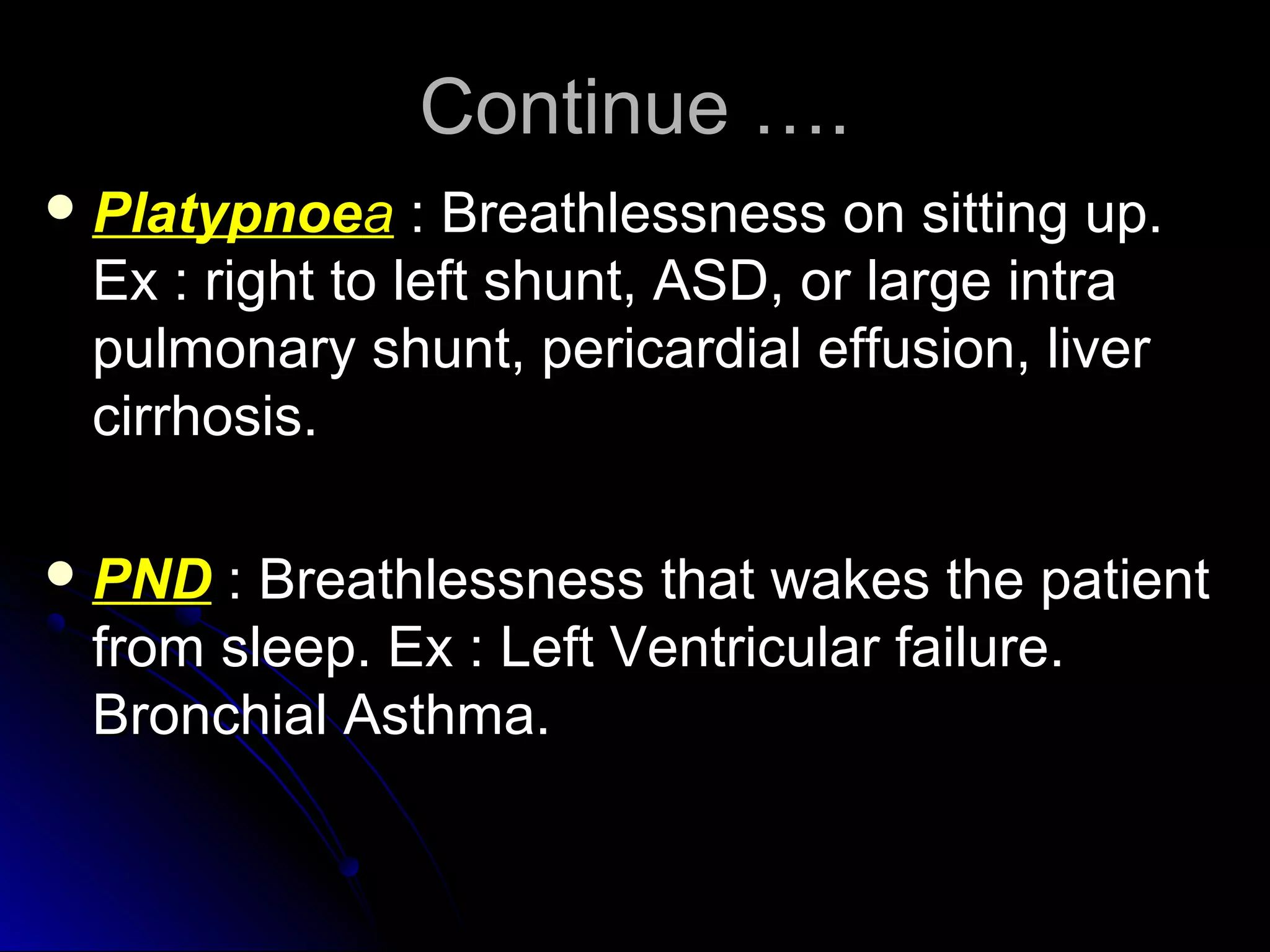
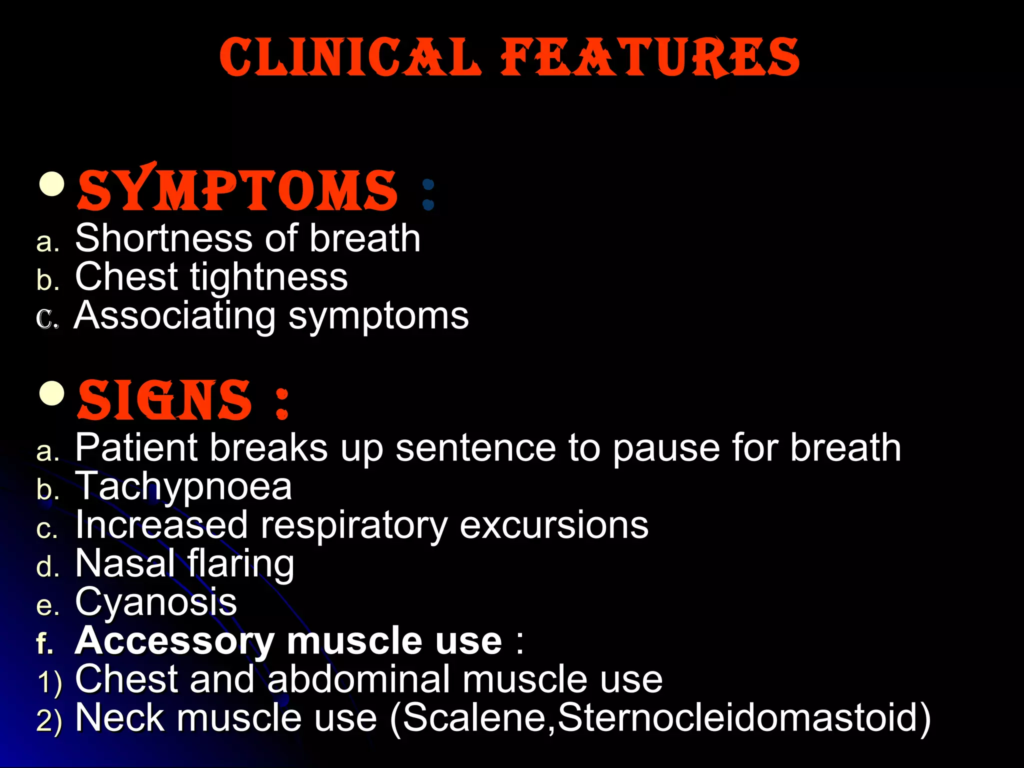
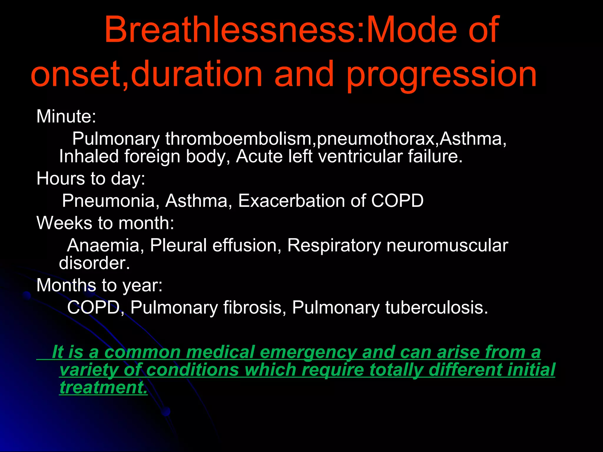
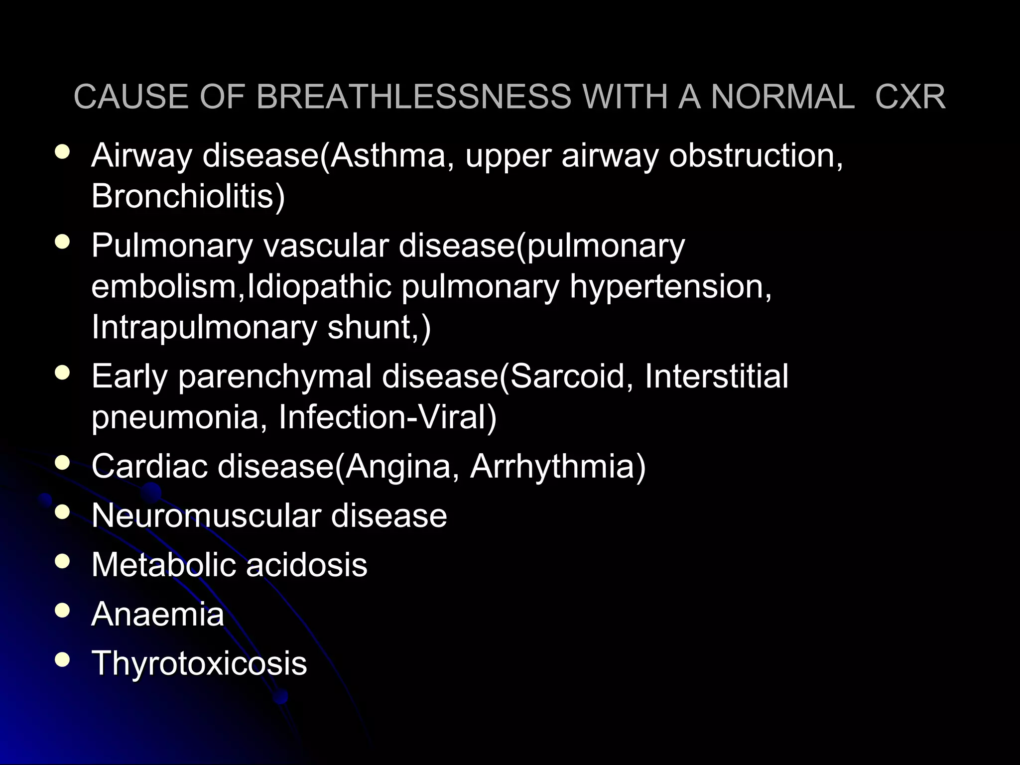
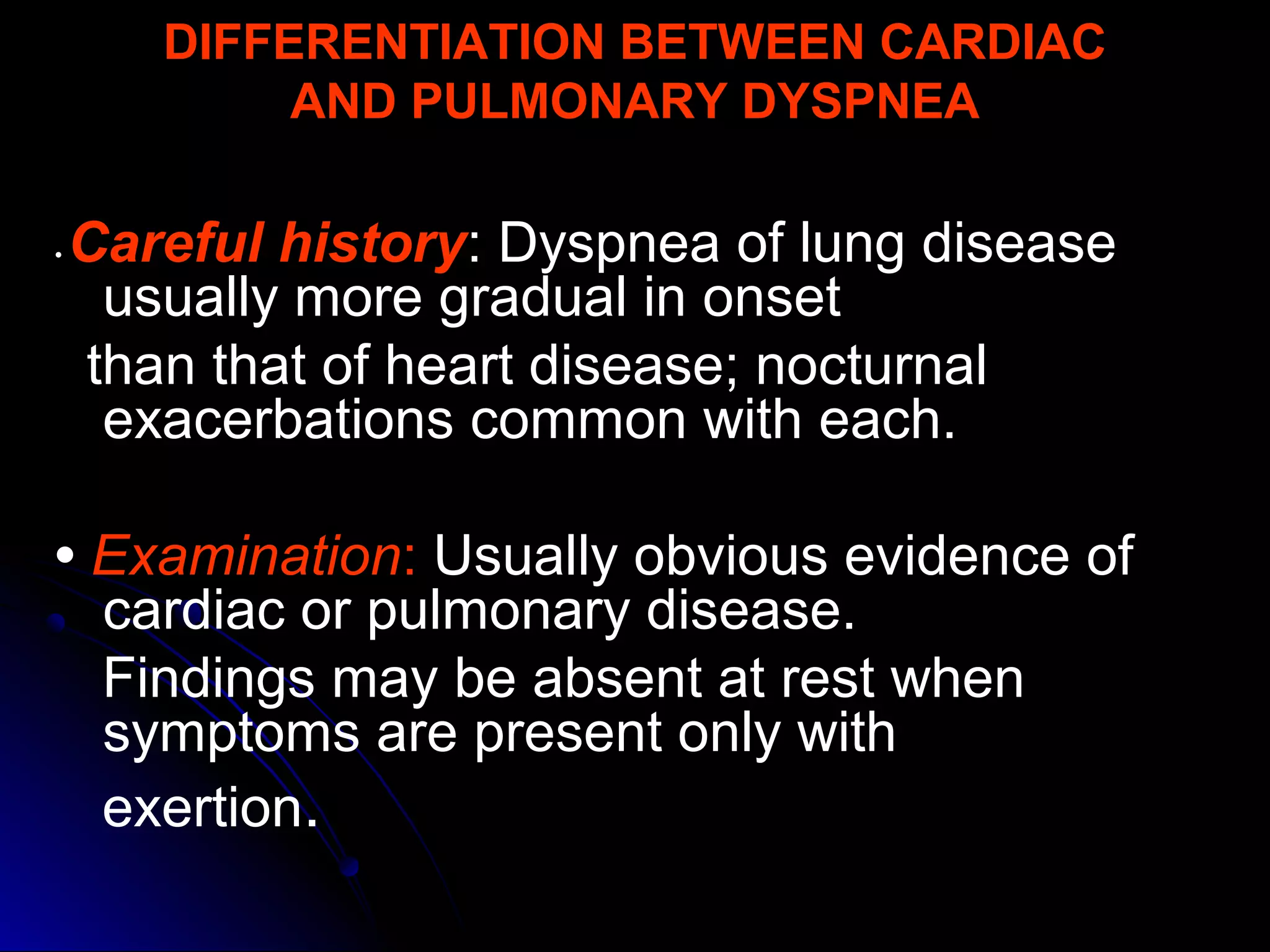
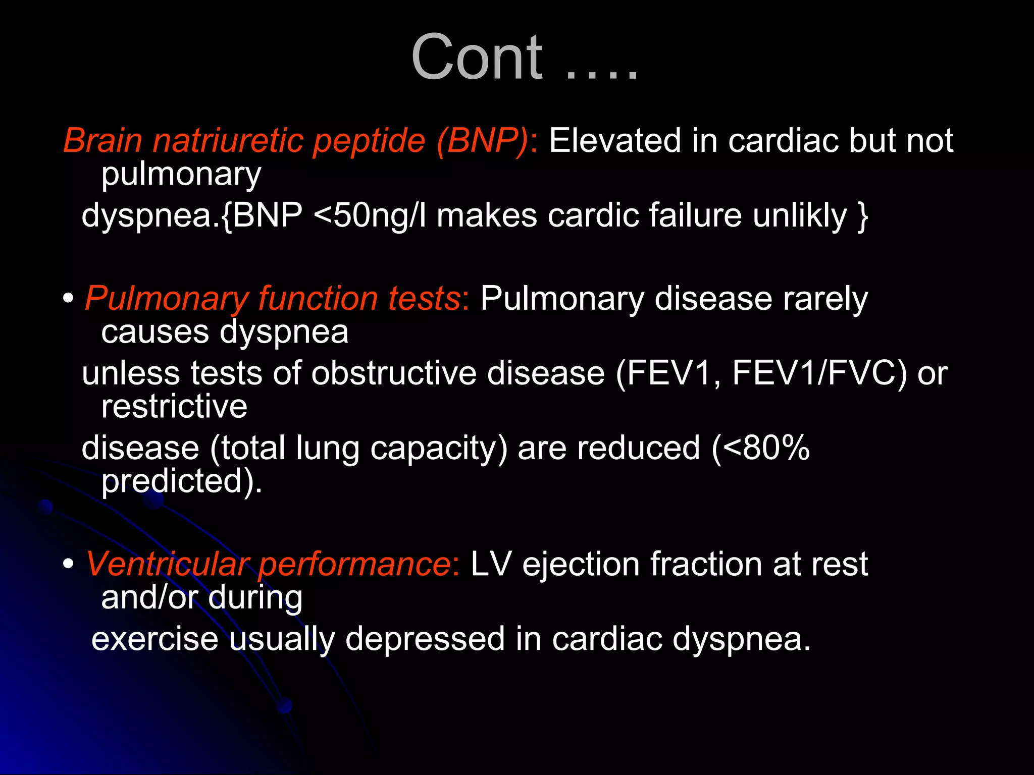
This document provides guidance on evaluating patients presenting with dyspnea (shortness of breath). It defines dyspnea and lists some specific types based on position. Common causes are outlined for pulmonary, cardiac, mixed, and non-cardiopulmonary origins. A clinical approach is described beginning with vital signs and history, followed by physical exam focusing on respiratory, cardiac, and fluid status findings. Initial investigations include chest X-ray, blood gases, ECG, and blood tests. Further tests may include lung function, exercise testing, and biomarkers to differentiate cardiac from pulmonary causes when the chest X-ray is normal. Careful history taking and physical exam remain important to identify underlying conditions.
1. Dyspnea, or shortness of breath, can be caused by conditions affecting the heart, lungs, blood, chest wall, or nerves and muscles controlling breathing.
2. Physical examination may reveal signs related to cardiac causes like pulmonary edema, or pulmonary disorders like emphysema, chronic bronchitis, or restrictive lung diseases.
3. Investigations like chest X-ray, pulmonary function tests, CT scan of the chest and lung biopsy help to evaluate abnormalities and arrive at a diagnosis.
DYSPNOEA IS DEFINED AS THE UNDUE AWARENESS OF UNPLEASANT BREATHING.WHEN THERE IS AMIS MATCH BETWEEN THE AFFERENT VENTILATORY SIGNALS AND THE EFFERENT RESPIRATORY SIGNALS IN THE BRAIN WE MAY GET AN UNIGNORABLE FEELING FOR NEED OF MORE AND MORE OXYGEN.
This document discusses acute dyspnea, defined as an uncomfortable need to breathe. It results from an imbalance between the perceived need and ability to breathe, often due to carbon dioxide buildup or oxygen deprivation. Common causes include pulmonary issues like pneumonia, cardiac issues like pulmonary edema, and metabolic acidosis. Diagnosis involves history, exam, and tests to identify the underlying cause. Treatment focuses on relieving symptoms like with opioids and addressing the specific condition through methods such as antibiotics for pneumonia or oxygen therapy for pulmonary edema.
Pulmonary Oedema is accumulation of fluid in lungs. It can be due to cardiogenic or non-cardiogenic causes. This presentation was a class presentation and discussed its management alongwith diagnosis.
This document provides an overview of dyspnea, or shortness of breath. It defines dyspnea and outlines its physiological and clinical definitions. Common causes of dyspnea are then discussed, including pulmonary issues like COPD, pneumonia, and pulmonary embolism, as well as cardiac issues like heart failure, coronary syndromes, and dysrhythmias. The pathophysiology of how these conditions can stimulate breathing and cause the sensation of dyspnea is explained. Finally, the document discusses assessing and diagnosing patients presenting with dyspnea through clinical exams, investigations like chest x-rays, and determining if the cause is chronic or acute.
This document outlines an approach to evaluating and diagnosing dyspnea. It begins by defining dyspnea and noting its high prevalence. Types of dyspnea like orthopnea and paroxysmal nocturnal dyspnea are described. The diagnostic approach involves obtaining a detailed history regarding onset, duration, patterns and associated symptoms. A physical exam assesses respiratory effort, oxygenation, and signs of heart failure. Initial tests may include EKG, chest x-ray, and bloodwork. Further tests are guided by initial findings and may include echocardiogram, pulmonary function tests, CT, or arterial blood gas. Treatment focuses on the underlying cause identified through diagnosis.
This document provides an overview of the approach to a patient presenting with dyspnea. It begins with definitions of dyspnea and classifications including the modified Medical Research Council dyspnea scale and NYHA classification. It then discusses the receptors involved in the mechanism of dyspnea, common causes of acute, subacute and chronic dyspnea, important parts of the physical examination, red flags, and recommended initial testing.
This document discusses the classification, symptoms, and treatment of heart failure. It begins by classifying heart failure based on its onset (acute or chronic), which side of the heart is affected (left or right), and its severity according to the New York Heart Association stages. The main symptoms of heart failure are then described using the mnemonic "FACES" (fatigue, activities limited, chest congestion, edema, shortness of breath). The document goes on to outline pharmacological treatments including ACE inhibitors, beta blockers, spironolactone, diuretics, and digoxin. Non-pharmacological interventions like diet, exercise, and cardiac rehabilitation are also mentioned.
Pleurisy is inflammation of the moist layer of the lungs. It is caused by diseases like AIDS and cancer, infections like pneumonia and tuberculosis, chest injuries, and drug reactions. The main symptom is a sharp, stabbing pain in the side of the chest that worsens with movement and coughing, along with shortness of breath and fever. Treatments include antibiotics, anti-inflammatory drugs, and pain killers to help relieve symptoms, though they do not provide a cure.
Pulmonary hypertension (2014) dr.tinku josephDr.Tinku Joseph
This document provides information on pulmonary hypertension (PH), including its definition, classification, pathogenesis, diagnosis, and treatment. It begins with defining PH as a mean pulmonary arterial pressure greater than 25 mmHg at rest based on right heart catheterization. PH is classified into 5 groups. The pathogenesis and pathology of each group is described. Diagnostic workup includes labs, imaging like CXR, echocardiogram and right heart catheterization. Treatment involves general measures, diuretics, anticoagulants, oxygen, and PAH-specific therapies like endothelin receptor antagonists, phosphodiesterase inhibitors, prostanoids, and calcium channel blockers in some cases. Prognostic factors and goals of treatment are also discussed.
This document discusses the approach to dyspnea, or shortness of breath. It defines dyspnea as a subjective feeling of breathing discomfort that can vary in intensity. Dyspnea is classified into four stages from exertional to resting. Common causes are then outlined for cardiac, pulmonary, and miscellaneous origins. The differential diagnosis process for acute versus chronic dyspnea is explained. Physical exam signs and investigations are also reviewed to help identify the underlying cause.
This document provides definitions and information about different types of dyspnea (shortness of breath). It discusses the pathophysiology and various causes of dyspnea like asthma, COPD, cardiac failure, pulmonary embolism. The document describes how to take history and examine patients presenting with dyspnea. It outlines investigations like chest imaging and laboratory tests. Differential diagnoses are provided for acute and chronic dyspnea. Management strategies for emergencies and exacerbations of conditions like asthma and COPD are briefly covered.
Obstructive vs. Restrictive Lung diseaseFatima Awadh
This document discusses the differences between restrictive and obstructive lung diseases. Restrictive lung diseases are characterized by a reduction in total lung capacity below 80% of predicted value, while obstructive lung diseases are characterized by a reduction in airflow, seen through a decreased forced expiratory volume in 1 second and ratio of forced expiratory volume to forced vital capacity. Key lung volumes and capacities such as tidal volume, inspiratory reserve volume, and residual volume are also defined.
Diagnosis and Etiology of Dyspnea (Shortness of breath)Mohammed Alawad
This document discusses the differential diagnosis of dyspnea (shortness of breath). It begins by defining dyspnea and listing potential causes as psychiatric, respiratory, or cardiovascular. It then outlines 6 steps for evaluating dyspnea, including assessing airway, breathing and circulation, considering possible etiologies based on history, performing a physical exam, ordering tests, making a differential diagnosis and treating. Key signs like hypoxia and cyanosis are discussed. Causes of respiratory system dyspnea related to control, ventilation or gas exchange are covered. Finally, cardiovascular causes of dyspnea from pump failure or other issues are addressed before concluding with references.
Pulmonary embolism - Diagnosis and managementDr Vivek Baliga
Pulmonary embolism is a common problem seen in medical practice. This presentation by Dr Vivek Baliga discusses the basic aspects and evidence behind current management.
Dyspnea, also known as breathlessness or shortness of breath, is defined as a subjective experience of breathing discomfort that can vary in intensity. It is caused by stimulation of pulmonary and extra-pulmonary receptors in response to conditions like asthma, COPD, pneumonia, pulmonary embolism, cardiac failure, and more. The document outlines the pathophysiology, stages, differential diagnosis, physical exam findings, and key investigations for evaluating the underlying cause of a patient's dyspnea.
This document provides an overview of the approach to dyspnea. It defines dyspnea and describes related terms like orthopnea. The mechanisms of orthopnea and paroxysmal nocturnal dyspnea are increased venous return when lying flat overwhelming the heart. Receptors involved in the perception of dyspnea are described. Common causes of acute and chronic dyspnea from cardiovascular, pulmonary, and other systems are listed. The approach involves assessing airway, breathing, vital signs, history, and focused physical exam. Key exam findings that can point to different diagnoses are outlined. Important investigations include spirometry, ABG, imaging, and ECG.
This document discusses respiratory failure, defined as inadequate oxygenation, ventilation, or both to meet metabolic demands. It can be classified as type 1 (hypoxemic) or type 2 (hypercapnic) respiratory failure. Risk factors include age, smoking, lung disease, and neurological or muscular disorders. Pathophysiology involves ventilation-perfusion mismatching, right-to-left shunting, or hypoventilation. Causes include pneumonia, pulmonary embolism, neuromuscular disorders, and acute respiratory distress syndrome. The control of breathing and gas exchange physiology are also summarized.
The document summarizes pericardial diseases. It discusses the anatomy and physiology of the pericardium, acute pericarditis including symptoms, diagnosis and treatment, and pericardial effusion and tamponade. Acute pericarditis is usually self-limited and treated with NSAIDs. Larger effusions may require hospitalization. Pericardial effusion can progress to tamponade, where fluid accumulation compresses the heart and impairs filling.
1) Respiratory failure is a condition where the lungs cannot properly oxygenate the blood and remove carbon dioxide, classified as Type I (hypoxemic) or Type II (hypercapnic).
2) It can result from problems affecting gas exchange in the lungs, respiratory control centers in the brain, or the chest wall muscles.
3) Common causes of Type I respiratory failure include pneumonia, ARDS, and severe asthma, while Type II is often due to conditions that decrease breathing, such as COPD.
Examination of cardiovascular system in PediatricsBirhanu Melese
The paediatrics cardiovascular exam can be a logistical minefield, requiring a good understanding of cardiac anatomy and possible congenital anomalies. With babies especially, it’s important to be opportunistic with your examination – doing the three ‘quiet things’ first: auscultation of heart sounds, auscultation of breath sounds and palpation of femoral pulses.
The document discusses the evaluation and differential diagnosis of acute dyspnea in adult patients presenting to the emergency department. It outlines the importance of obtaining a thorough history and physical exam to identify potential life-threatening causes of dyspnea such as heart failure, pulmonary embolism, pneumonia, and asthma. Common signs and symptoms associated with different conditions are described. The emergency clinician must work through a wide range of diagnoses while providing initial treatment for what may be a serious underlying illness.
This document discusses various aspects of examining the respiratory system. It covers topics such as respiratory rates, breath sounds, percussion findings, and signs and symptoms of common respiratory conditions. Key points include different types of breath sounds (bronchial, bronchovesicular, vesicular), adventitious sounds like crackles and wheezes, and physical exam findings associated with conditions like pneumonia, COPD, pleural effusion, and pneumothorax. The document provides detailed guidance on respiratory exam techniques and interpretations.
Acute Respiratory Distress Syndrome (ARDS) is a sudden, progressive form of respiratory failure characterized by severe dyspnea, hypoxemia, and decreased lung compliance. It develops from direct or indirect lung injuries and is thought to be caused by stimulation of the inflammatory and immune systems, resulting in leakage of fluid into the lungs. The clinical progression of ARDS involves exudative, proliferative, and fibrotic phases that can lead to respiratory failure if not promptly treated with oxygen supplementation, mechanical ventilation, and other supportive therapies.
This document provides information on ischemic heart disease (IHD), also known as coronary heart disease. It discusses the pathophysiology and risk factors of IHD and describes the differences between chronic stable angina and acute coronary syndrome (ACS). Guidelines are provided on lifestyle modifications and pharmacological therapies for IHD, including antiplatelet agents, statins, antihypertensives, nitrates, beta-blockers, and calcium channel blockers. The roles of percutaneous coronary intervention (PCI) and coronary artery bypass grafting (CABG) in treating IHD are also summarized.
Heart failure, also known as cardiac decompensation or cardiac insufficiency, occurs when the heart is unable to pump enough blood to meet the body's needs. It can be caused by conditions that impair the heart muscle's ability to contract effectively or limit ventricular filling. Symptoms vary depending on whether the left or right ventricle is primarily affected and include dyspnea, fatigue, edema and others. Diagnostic tests may include echocardiography, ECG, chest x-ray and BNP level. Treatment focuses on managing symptoms, slowing disease progression, and preventing hospitalizations through lifestyle changes and medication.
This document provides information on evaluating and diagnosing acute shortness of breath (dyspnea). It defines dyspnea and lists its potential causes, which include respiratory, cardiovascular, musculoskeletal, central nervous system, and gastrointestinal issues. The approach involves taking a thorough history, examining the patient, and conducting initial tests like chest x-ray, ECG, and arterial blood gas analysis to help determine the underlying cause, such as pneumonia, pulmonary edema, asthma exacerbation, or pulmonary embolism. Further tests like spirometry, echocardiogram, or cardiopulmonary exercise testing may be needed in some cases. The goal is to identify any life-threatening conditions and make a rapid diagnosis to guide treatment.
The patient is a 67-year-old male former smoker presenting with shortness of breath on exertion. Physical examination finds reduced breath sounds and wheezing. Tests show reduced lung function and oxygen levels. The differential diagnosis includes cardiac and pulmonary causes like COPD. Dyspnea is the medical term for shortness of breath and can result from various lung and heart conditions. Treatment focuses on the underlying cause, like using bronchodilators for COPD.
Dyspnea, or shortness of breath, is a common symptom that can be caused by many cardiac and pulmonary conditions. A thorough diagnostic evaluation of dyspnea involves taking a detailed patient history, conducting a physical exam, and obtaining initial tests like an electrocardiogram, chest x-ray, and blood tests to evaluate for conditions involving the heart, lungs, blood, and other potential causes and to guide further testing if needed. Grading scales are used to characterize the severity of a patient's dyspnea. The pathophysiology of dyspnea involves an imbalance between the perceived need to breathe and the ability to breathe.
Dyspnea is a subjective experience of breathing discomfort that results from interactions between physiological, psychological, social, and environmental factors. The document discusses the mechanisms, causes, evaluation, and management of dyspnea. Evaluation involves obtaining a thorough history regarding onset, timing, severity, and relieving/precipitating factors. A physical exam focuses on vital signs, respiratory exam, cardiovascular exam, and neurological exam to help identify potential causes like heart failure, COPD, pneumonia, or asthma.
This document provides information on evaluating and diagnosing shortness of breath. It lists various potential causes of shortness of breath including cardiac, lung, anatomical, trauma, and other issues. Specific conditions that could cause wheezing, stridor, crepitations, or a clear chest are identified. The speed of onset can help determine if the underlying cause is acute, subacute, or chronic. Guidelines for triaging patients with shortness of breath into green, yellow, or red zones based on dyspnea and oxygen saturation are also provided. The evaluation involves assessing severity, examining the chest, providing oxygen support if needed, and getting a chest x-ray.
1) A 45-year-old female presented with progressive breathlessness, swelling of the lower limbs, and hard nodules on her palms and soles.
2) Testing revealed features of multiple autoimmune diseases including a strongly positive ANA, interstitial lung disease, pulmonary hypertension, and calcinosis cutis.
3) She was diagnosed with mixed connective tissue disease based on Sharp's criteria, characterized by overlapping features of systemic lupus erythematosus, systemic sclerosis, rheumatoid arthritis, and polymyositis as well as high titers of anti-U1 RNP antibodies.
1) Dyspnea, or shortness of breath, is a subjective experience that results from interactions between physiological, psychological, and environmental factors. It can be caused by disorders of the ventilatory pump or sensory receptors in the lungs and muscles.
2) Pulmonary edema occurs when fluid accumulates in the lungs, which can be cardiogenic due to increased hydrostatic pressures from heart problems, or noncardiogenic from direct lung injury or diseases affecting the lung barrier.
3) Differentiating cardiogenic and noncardiogenic pulmonary edema involves examining physical exam findings, chest x-rays, and response to supplemental oxygen, as they have distinct mechanisms and presentations.
This document summarizes a seminar on dyspnea and respiratory failure. It defines dyspnea as subjective breathlessness and lists its potential causes such as cardiac, respiratory, or psychogenic issues. It describes different grading scales used to classify breathlessness. Types of dyspnea include orthopnea and paroxysmal nocturnal dyspnea. Respiratory failure is defined as impaired gas exchange shown by low oxygen and potentially high carbon dioxide levels. It discusses the mechanisms, clinical manifestations, differential diagnosis, and management of both dyspnea and respiratory failure.
Dyspnoea refers to undue awareness of one's own breathing and increased drive to breathe. It can be normal with strenuous exercise but is pathological if it occurs at lower thresholds. Dyspnoea has many potential causes including cardiac, respiratory, neuromuscular, metabolic issues or toxins. It is classified based on the cardiovascular and respiratory systems with grades ranging from no limitations to total confinement based on the level of physical activity one can perform without symptoms.
Syncope is a transient loss of consciousness due to decreased blood flow to the brain. It has many potential causes including cardiac arrhythmias, orthostatic hypotension, and vasovagal responses. Management involves stopping any procedures, placing the patient supine with legs elevated, assessing ABCs, providing oxygen, and monitoring vitals. For presyncope, stopping and allowing recovery is usually sufficient while syncope may require interventions like atropine for bradycardia. Thorough history and evaluation of potential causes is important to prevent future episodes.
This document provides an overview of pain management and opioid use for cancer patients. It discusses how cancer pain is common and should be properly assessed and treated. The WHO pain ladder is reviewed as the standard approach for treating pain with non-opioids, weak opioids, and strong opioids. Opioid rotation and treating pain crises are covered, including calculating opioid conversions and administering parenteral opioids. Challenges in treating cancer pain in patients with addiction histories are addressed through transparency, long-acting opioids, and pain contracts. Overall guidelines aim to properly treat pain while avoiding exacerbating addiction issues.
Cancer pain is caused by tumors invading tissues and pressing on nerves. There are three types of pain: nociceptive, inflammatory, and neuropathic. Pain signals travel along nerve pathways from tissues to the spinal cord and brain. Cancer pain management involves detailed assessment, analgesic drugs like opioids, and non-pharmacological treatments. Radiation, chemotherapy, surgery, nerve blocks, and cement injections can help reduce tumor size and pressure causing pain. The goal is comprehensive treatment of physical and psychological distress from cancer.
This document discusses cancer pain, its causes, types, prevalence among cancer patients, diagnosis, and management. It states that moderate to severe pain is experienced by 40-50% of cancer patients, while very severe pain affects 25-30%. Pain management methods discussed include radiation therapy, chemotherapy, hormone therapy, bisphosphonates, surgery, complementary therapies like acupuncture and massage, and use of the WHO pain ladder for pharmacological treatments. It also covers mucositis, chemotherapy-induced peripheral neuropathy, and the use of non-analgesic drugs to manage certain painful conditions in cancer patients.
The patient presents with a chronic cough lasting 2 months with initially whitish sputum and now blood-streaked sputum. He also reports difficulty breathing and fever for the past 2 weeks. The doctor's approach involves taking a thorough history regarding cough onset and characteristics, associated symptoms, and appearance of any sputum to determine potential causes and guide appropriate examination and testing. Key considerations include pneumonia, lung cancer, and other infectious or inflammatory respiratory conditions.
Edema can be caused by increased hydrostatic pressure, decreased plasma oncotic pressure, or lymphatic obstruction. Increased hydrostatic pressure can result from heart failure, liver cirrhosis, or venous obstruction. Decreased plasma oncotic pressure occurs when albumin is lost, such as in nephrotic syndrome, or not produced sufficiently, as in liver disease or malnutrition. Lymphatic obstruction leads to localized edema and can be caused by infection, cancer, or surgery. Edema fluid accumulation in tissues can impair function and be life-threatening in the lungs or brain.
The document discusses various respiratory conditions that can cause dyspnea (difficult or labored breathing). It describes hyperventilation syndrome, which causes anxiety and dizziness due to overbreathing. Foreign body obstruction is a common cause of pediatric airway issues and can cause sudden dyspnea in adults. Pharyngeal edema, epiglottitis, and croup can cause upper airway swelling and obstruction. Asthma is a reversible obstructive lung disease often caused by allergens or irritants. Chronic obstructive pulmonary disease (COPD) includes chronic bronchitis and emphysema from long-term smoking. Pulmonary edema and pulmonary embolism can cause fluid or clots in the lungs resulting in
This document discusses pain in several sections:
1. It defines pain and its subjective nature. Pain is the most common reason people seek medical care and acts as a protective mechanism.
2. It describes pain transmission and the gate control theory of pain. Nociceptors transmit pain impulses and can be modulated by other stimuli.
3. It categorizes acute, chronic, and cancer-related pain and discusses factors influencing individual pain responses. Non-pharmacological and pharmacological pain management strategies are also outlined.
Nausea/Vomiting/Anorexia – Bree Johnston, MD, MPH, FACP
Presented at the 2015 Palliative Care Summer Institute conference at Bellingham Technical College
Chronic obstructive pulmonary disease (COPD) is a progressive lung disease characterized by difficulty breathing. It is caused by long-term exposure to irritating gases and particulate matter, primarily from cigarette smoking. Symptoms include a productive cough, breathlessness, and chest infections. The disease is diagnosed through pulmonary function tests and imaging. Treatment focuses on reducing symptoms through bronchodilators and antibiotics for infections. Nursing care involves assessing symptoms, monitoring diagnostic tests, and teaching patients about prevention, treatment, and managing exacerbations.
This document provides guidance on clinically examining the respiratory system. It describes how to inspect, palpate, percuss, and auscultate the chest. Inspection involves examining chest shape, symmetry, movement, and veins. Palpation checks the trachea position, apex beat, chest expansion, vocal fremitus, and tenderness. Percussion distinguishes lung from liver dullness. Auscultation assesses breath sounds and vocal resonance. The examination evaluates the lungs, pleura, chest wall, and underlying bony structures in a systematic manner to detect abnormalities.
Brief Presentation on clinical examination of Respiratory System with Report of Normal case
references:
macleod's clinical examination 13th edition
hutchinson clinical methods
R Alagappan - Manual of Practical Medicine, 4th Edition
Respiratory failure is characterized by severe dysfunction of pulmonary ventilation and/or oxygenation caused by various diseases, resulting in hypoxia and retention of carbon dioxide. It is defined as a PaO2 of less than 8.0 kPa (60 mmHg), and/or a PaCO2 of greater than 6.67 kPa (50 mmHg). The main causes are ventilation dysfunction due to airway obstruction or limitation, and oxygenation dysfunction due to pulmonary edema, interstitial lung disease, or ARDS. The key pathophysiological changes are hypoxia, retention of carbon dioxide, and acidosis, which can affect multiple organ systems and lead to complications.
Examinating the Resipiratory System.pptxssuser504dda
This document provides guidance on examining the respiratory system through history taking and physical examination. It details what to ask patients regarding symptoms like breathlessness, cough, sputum production, and chest pain. It also explains how to inspect, palpate, percuss and auscultate the chest. Specific tests are described like measuring chest expansion, examining neck veins, and evaluating breath sounds and vocal fremitus. A thorough respiratory exam provides clues to underlying cardiopulmonary conditions.
This document provides information on chronic obstructive pulmonary disease (COPD) including its definition, risk factors, pathophysiology, clinical manifestations, medical management, nursing management, and surgical options. COPD is characterized by persistent airflow limitation that is not fully reversible. It is mostly caused by smoking and results in changes like thickening of the airways and inflammation that narrow the lungs over time. Management involves treatments to improve ventilation and remove secretions, as well as strategies to prevent complications and promote overall health.
The document discusses various topics related to the respiratory and cardiovascular systems including:
1. Common respiratory disturbances like restrictive lung disease, COPD, and pulmonary vascular disease.
2. Measures that promote respiratory function such as adequate oxygen supply, deep breathing, coughing exercises, and chest physiotherapy.
3. Common cardiovascular conditions like coronary artery disease, angina, myocardial infarction, and congestive heart failure.
4. Risk factors for coronary artery disease and strategies for controlling cholesterol levels.
This document discusses dyspnea (shortness of breath). It begins by defining dyspnea as a subjective sensation of breathing that can range from mild discomfort to feelings of suffocation. It is a sign of various disorders that generally indicate inadequate ventilation or insufficient oxygen in the blood. The document then covers the causes, history, physical exam findings, investigations and management of dyspnea.
Breathlessness, also known as dyspnea, is an unpleasant sensation of uncomfortable, difficult, or labored breathing. It has various potential causes, both acute and chronic, including conditions affecting the heart, lungs, and other systems. A medical history, physical exam, and tests can help identify potential differential diagnoses and assess severity. Treatment depends on the underlying cause but may involve oxygen supplementation, bronchodilators, opioids, anxiolytics, breathing exercises, and addressing any anxiety components.
1. Chronic obstructive pulmonary disease (COPD) is a preventable and treatable lung disease characterized by airflow limitation caused by cigarette smoking.
2. COPD symptoms include cough, sputum production, and breathlessness. It is a major cause of death and disability worldwide and is expected to be the third leading cause of death by 2020.
3. COPD is diagnosed based on symptoms and spirometry showing airflow limitation that is not fully reversible. The two main phenotypes are chronic bronchitis and emphysema.
The document discusses considerations for transporting patients by air, including assessing their cardiovascular, respiratory and general health status prior to flight and taking precautions to address issues like hypoxia, changes in barometric pressure, thermal stress, noise and vibration during transport. It provides guidance on stabilizing patients, securing airways, providing oxygen, monitoring vital signs, and positioning patients comfortably for air travel.
This document provides an overview of cough, including its definition, mechanism, causes, management, and classification. It begins by defining cough and describing its four step mechanism: 1) inspiratory gasp, 2) valsalva maneuver, 3) expiratory blast, and 4) post-tussive inspiration. Cough can be triggered by stimuli in various structures in the lungs, airways, and chest. Acute cough is classified as lasting less than 3 weeks, sub-acute 3-8 weeks, and chronic over 8 weeks. Common causes of chronic cough include lung diseases like asthma, GERD, post-nasal drip, and medications like ACE inhibitors. Investigation of chronic cough involves ruling out structural
The document summarizes chronic obstructive pulmonary disease (COPD). It covers the general considerations, epidemiology, risk factors, pathogenesis, clinical findings, differential diagnosis, diagnostic testing including spirometry and imaging, and treatment including smoking cessation, oxygen therapy, bronchodilators, corticosteroids, and antibiotics. COPD is characterized by airflow obstruction due to chronic bronchitis or emphysema and is generally progressive. Cigarette smoking is the most important risk factor.
This document provides an overview of respiratory medicine, covering topics such as anatomy and physiology of the lungs, respiratory failure, pleural diseases, asthma, COPD, and more. It defines conditions, lists their typical causes and presentations, and outlines recommended diagnostic tests and treatment approaches. Key areas covered include the mechanics of breathing, definitions of respiratory failure types, common pleural effusions and their management, asthma diagnosis and treatment guidelines, risk factors and management of COPD, and criteria for exacerbation treatment.
1. The document discusses respiratory diseases, focusing on pleural effusions and pneumothorax.
2. For pleural effusions, it describes etiology, pathogenesis, symptoms, diagnostic tests including thoracentesis, and treatment approaches. Transudative and exudative effusions are distinguished.
3. Pneumothorax is defined as gas accumulation in the pleural cavity. Causes include spontaneous rupture of alveoli or chest wall injury. Classification includes spontaneous, traumatic, and nosocomial pneumothorax.
This document discusses several pulmonary conditions including pleural effusion, acute respiratory distress syndrome (ARDS), bronchial asthma, chronic obstructive pulmonary disease (COPD), and lung abscess. For pleural effusion, it describes the causes, signs and symptoms, investigations including pleural fluid analysis, and treatments including thoracentesis. For ARDS, it provides the definition, precipitating factors, symptoms, investigations, and management including supportive care and treating the underlying cause. It also summarizes the definitions, common triggers, signs and symptoms, diagnosis, investigations, and treatment including bronchodilators and corticosteroids for bronchial asthma and COPD.
This document discusses bronchial asthma. It defines asthma as a chronic inflammatory disorder of the airways characterized by recurrent episodes of wheezing, breathlessness, and reversible airflow obstruction. It outlines the signs, triggers, diagnostic testing including spirometry, and goals of treatment. Treatment involves both short-acting relievers and long-term controllers, with classes including beta-agonists, corticosteroids, leukotriene modifiers, and methylxanthines. The document provides details on specific medications and their mechanisms and roles in asthma management.
Gender and Mental Health - Counselling and Family Therapy Applications and In...PsychoTech Services
A proprietary approach developed by bringing together the best of learning theories from Psychology, design principles from the world of visualization, and pedagogical methods from over a decade of training experience, that enables you to: Learn better, faster!
Andreas Schleicher presents PISA 2022 Volume III - Creative Thinking - 18 Jun...EduSkills OECD
Andreas Schleicher, Director of Education and Skills at the OECD presents at the launch of PISA 2022 Volume III - Creative Minds, Creative Schools on 18 June 2024.
THE SACRIFICE HOW PRO-PALESTINE PROTESTS STUDENTS ARE SACRIFICING TO CHANGE T...indexPub
The recent surge in pro-Palestine student activism has prompted significant responses from universities, ranging from negotiations and divestment commitments to increased transparency about investments in companies supporting the war on Gaza. This activism has led to the cessation of student encampments but also highlighted the substantial sacrifices made by students, including academic disruptions and personal risks. The primary drivers of these protests are poor university administration, lack of transparency, and inadequate communication between officials and students. This study examines the profound emotional, psychological, and professional impacts on students engaged in pro-Palestine protests, focusing on Generation Z's (Gen-Z) activism dynamics. This paper explores the significant sacrifices made by these students and even the professors supporting the pro-Palestine movement, with a focus on recent global movements. Through an in-depth analysis of printed and electronic media, the study examines the impacts of these sacrifices on the academic and personal lives of those involved. The paper highlights examples from various universities, demonstrating student activism's long-term and short-term effects, including disciplinary actions, social backlash, and career implications. The researchers also explore the broader implications of student sacrifices. The findings reveal that these sacrifices are driven by a profound commitment to justice and human rights, and are influenced by the increasing availability of information, peer interactions, and personal convictions. The study also discusses the broader implications of this activism, comparing it to historical precedents and assessing its potential to influence policy and public opinion. The emotional and psychological toll on student activists is significant, but their sense of purpose and community support mitigates some of these challenges. However, the researchers call for acknowledging the broader Impact of these sacrifices on the future global movement of FreePalestine.
🔥🔥🔥🔥🔥🔥🔥🔥🔥
إضغ بين إيديكم من أقوى الملازم التي صممتها
ملزمة تشريح الجهاز الهيكلي (نظري 3)
💀💀💀💀💀💀💀💀💀💀
تتميز هذهِ الملزمة بعِدة مُميزات :
1- مُترجمة ترجمة تُناسب جميع المستويات
2- تحتوي على 78 رسم توضيحي لكل كلمة موجودة بالملزمة (لكل كلمة !!!!)
#فهم_ماكو_درخ
3- دقة الكتابة والصور عالية جداً جداً جداً
4- هُنالك بعض المعلومات تم توضيحها بشكل تفصيلي جداً (تُعتبر لدى الطالب أو الطالبة بإنها معلومات مُبهمة ومع ذلك تم توضيح هذهِ المعلومات المُبهمة بشكل تفصيلي جداً
5- الملزمة تشرح نفسها ب نفسها بس تكلك تعال اقراني
6- تحتوي الملزمة في اول سلايد على خارطة تتضمن جميع تفرُعات معلومات الجهاز الهيكلي المذكورة في هذهِ الملزمة
واخيراً هذهِ الملزمة حلالٌ عليكم وإتمنى منكم إن تدعولي بالخير والصحة والعافية فقط
كل التوفيق زملائي وزميلاتي ، زميلكم محمد الذهبي 💊💊
🔥🔥🔥🔥🔥🔥🔥🔥🔥
BÀI TẬP BỔ TRỢ TIẾNG ANH LỚP 9 CẢ NĂM - GLOBAL SUCCESS - NĂM HỌC 2024-2025 - ...
Dyspnoea (2)
1. APPROACHAPPROACH TO THETO THE
PATIENTPATIENT WITHWITH
DYSPNOEADYSPNOEA
Dr. AL TARIQUE
IMO, Department of Medicine,
EMCH
2. DEFINITIONDEFINITION
A subjective experience of breathing
discomfort that consists of qualitatively
distinct sensations that vary in intensity.
(American Thoracic Society )
Other definitions describe it as "difficulty in
breathing, disordered or inadequate
breathing, uncomfortable awareness of
breathing. And It may be acute or chronic.
3. Some definitionsSome definitions
TrepopnoeaTrepopnoea :Breathlessness when lying on:Breathlessness when lying on
one side as a result of ipsilateral pulmonaryone side as a result of ipsilateral pulmonary
disease . Causes are dilateddisease . Causes are dilated
cardiomyopathy, pleural effusion.cardiomyopathy, pleural effusion.
OrthoponeaOrthoponea : Breathlessness when lying: Breathlessness when lying
flat. Ex : Left Ventricular Failure, diaphramaticflat. Ex : Left Ventricular Failure, diaphramatic
weakness, massive pleural effusion, hugeweakness, massive pleural effusion, huge
ascitis, any severe lung disease.ascitis, any severe lung disease.
Breathlessness during swimming isBreathlessness during swimming is
characteristic of bilateral diaphragmcharacteristic of bilateral diaphragm
paralysisparalysis..
4. Continue ….Continue ….
PlatypnoePlatypnoeaa : Breathlessness on sitting up.: Breathlessness on sitting up.
Ex : right to left shunt, ASD, or large intraEx : right to left shunt, ASD, or large intra
pulmonary shunt, pericardial effusion, liverpulmonary shunt, pericardial effusion, liver
cirrhosis.cirrhosis.
PNDPND : Breathlessness that wakes the patient: Breathlessness that wakes the patient
from sleep. Ex : Left Ventricular failure.from sleep. Ex : Left Ventricular failure.
Bronchial Asthma.Bronchial Asthma.
5. Etiology / Differential DiagnosisEtiology / Differential Diagnosis
(Dyspnoea)(Dyspnoea)
Composed of four general categoriesComposed of four general categories
CardiacCardiac
PulmonaryPulmonary
Mixed cardiac or pulmonaryMixed cardiac or pulmonary
non-cardiac or non-pulmonarynon-cardiac or non-pulmonary
9. Noncardiac or Nonpulmonary EtiologyNoncardiac or Nonpulmonary Etiology
It is a normal symptom of heavy exertionIt is a normal symptom of heavy exertion
Normal pregnancy (around 2/3rd
)
Metabolic acidosisMetabolic acidosis
PainPain
TraumaTrauma
Neuromuscular disordersNeuromuscular disorders
FunctionalFunctional (anxiety, panic disorders, hyperventilation)(anxiety, panic disorders, hyperventilation)
Chemical exposureChemical exposure
Obesity
Psychogenic
10. Life Threatening Causes of DyspneaLife Threatening Causes of Dyspnea
Pulmonary EmbolismPulmonary Embolism
Tension PneumothoraxTension Pneumothorax
Severe metabolic acidosisSevere metabolic acidosis
Pulmonary EdemaPulmonary Edema
Status asthmaticusStatus asthmaticus
Hypercapneic Respiratory FailureHypercapneic Respiratory Failure
Severe upper airway obstructionSevere upper airway obstruction
ARDSARDS
12. CLINICAL FEATURESCLINICAL FEATURES
SympTomSSympTomS ::
a.a. Shortness of breathShortness of breath
b.b. Chest tightnessChest tightness
C.C. Associating symptomsAssociating symptoms
SIgNS :SIgNS :
a.a. Patient breaks up sentence to pause for breathPatient breaks up sentence to pause for breath
b.b. TachypnoeaTachypnoea
c.c. Increased respiratory excursionsIncreased respiratory excursions
d.d. Nasal flaringNasal flaring
e.e. CyanosisCyanosis
f.f. Accessory muscle useAccessory muscle use ::
1)1) Chest and abdominal muscle useChest and abdominal muscle use
2)2) Neck muscle use (Scalene,Sternocleidomastoid)Neck muscle use (Scalene,Sternocleidomastoid)
14. Breathlessness:Mode of
onset,duration and progression
Minute:Minute:
Pulmonary thromboembolism,pneumothorax,Asthma,Pulmonary thromboembolism,pneumothorax,Asthma,
Inhaled foreign body, Acute left ventricular failure.Inhaled foreign body, Acute left ventricular failure.
Hours to day:Hours to day:
Pneumonia, Asthma, Exacerbation of COPDPneumonia, Asthma, Exacerbation of COPD
Weeks to month:Weeks to month:
Anaemia, Pleural effusion, Respiratory neuromuscularAnaemia, Pleural effusion, Respiratory neuromuscular
disorder.disorder.
Months to year:Months to year:
COPD, Pulmonary fibrosis, Pulmonary tuberculosis.COPD, Pulmonary fibrosis, Pulmonary tuberculosis.
It is a common medical emergency and can arise from aIt is a common medical emergency and can arise from a
variety of conditions which require totally different initialvariety of conditions which require totally different initial
treatment.treatment.
15. MRC Breathlessness Scale
Grade Degree of dysponea
1 Breathless when hurrying on the level or
walking up a slight hill
2 Breathlessness when walking with people of
own age or on level ground
3 Walks slower than peers or stops when
walking on the flat at own pace
4 Stops after walking 100 meters or a few
minuters on the level
5
[5b]
Too breathless to leave the house
Too breathless to wash or dress
16. CoNT.CoNT.
KNowN RESpIRAToRy dISEASE:
1. Previous acute exacerbation of COPD
requiring hospital management
2.2. Previous lung function test and arterialPrevious lung function test and arterial
blood gas analysis:blood gas analysis:
3.3. RequirementRequirement for home nebulizedfor home nebulized
bronchodialator and/or oxygen therapy.bronchodialator and/or oxygen therapy.
KNowN CARdIAC dISEASE
1. CoRoNARy dISEASE
2. myoCARdIAL oR vALvE dISEASE
RISK FACToRS FoR vENoUS
THRomboEmboLISm
17. APPROACH TO THE PATIENTAPPROACH TO THE PATIENT
The initial approach for evaluation begins byThe initial approach for evaluation begins by
assessment of the airway, breathing, andassessment of the airway, breathing, and
circulation followed by a medical history andcirculation followed by a medical history and
physical examinationphysical examination
Physical findingsPhysical findings::
General appearance: Speak in full sentences?
Accessory muscles? Color?
BP-
Nose and sinus examNose and sinus exam
Fluid status exam :Fluid status exam :
Jugular Venous DistentionJugular Venous Distention
Hepatojugular ReflexHepatojugular Reflex
Peripheral EdemaPeripheral Edema
18. CONT.CONT.
Respiratory ExamRespiratory Exam
I.I. Increased AP Chest diameterIncreased AP Chest diameter
II.II. Wheezing, stridorWheezing, stridor
III.III.CracklesCrackles
IV.IV.CyanosisCyanosis
V.V. ClubbingClubbing
VI.VI.Accessory muscle use (Neck, chest,Accessory muscle use (Neck, chest,
abdomen)abdomen)
VII.VII.Speaking in phrases to catch breathSpeaking in phrases to catch breath
20. INVESTIGATIONSINVESTIGATIONS
FIRST LINE (INITIAL) INV :FIRST LINE (INITIAL) INV :
CHEST X-RAYCHEST X-RAY
ARTERIAL BLOOD GASES AND pHARTERIAL BLOOD GASES AND pH
FULL BLOOD COUNTFULL BLOOD COUNT
ECGECG
TSH levelTSH level
BLOOD GLUCOSEBLOOD GLUCOSE
SODIUM ,POTASSIUM AND CREATININESODIUM ,POTASSIUM AND CREATININE
BIOMARKERS:BIOMARKERS:
D-dimer if pulmonary embolism isD-dimer if pulmonary embolism is
suspectedsuspected
Troponin if acute coronary syndrome isTroponin if acute coronary syndrome is
suspectedsuspected
BNP( brain natriuretic peptide) if heartBNP( brain natriuretic peptide) if heart
failure is suspectedfailure is suspected
21. CONT.CONT.
SECOND LINE INV.( WHEN STABLE):SECOND LINE INV.( WHEN STABLE):
LUNG FUNCTION TESTSLUNG FUNCTION TESTS
(SPIROMETRY)(SPIROMETRY)
Cardiopulmonary exercise testingCardiopulmonary exercise testing
( CPET)IF difficult to differentiate cardiac( CPET)IF difficult to differentiate cardiac
or respiratory cause.or respiratory cause.
22. CAUSE OF BREATHLESSNESS WITH A NORMAL CXRCAUSE OF BREATHLESSNESS WITH A NORMAL CXR
Airway disease(Asthma, upper airway obstruction,Airway disease(Asthma, upper airway obstruction,
Bronchiolitis)Bronchiolitis)
Pulmonary vascular disease(pulmonaryPulmonary vascular disease(pulmonary
embolism,Idiopathic pulmonary hypertension,embolism,Idiopathic pulmonary hypertension,
Intrapulmonary shunt,)Intrapulmonary shunt,)
Early parenchymal disease(Sarcoid, InterstitialEarly parenchymal disease(Sarcoid, Interstitial
pneumonia, Infection-Viral)pneumonia, Infection-Viral)
Cardiac disease(Angina, Arrhythmia)Cardiac disease(Angina, Arrhythmia)
Neuromuscular diseaseNeuromuscular disease
Metabolic acidosisMetabolic acidosis
AnaemiaAnaemia
ThyrotoxicosisThyrotoxicosis
23. DIFFERENTIATION BETWEEN CARDIAC
AND PULMONARY DYSPNEA
• Careful history: Dyspnea of lung disease
usually more gradual in onset
than that of heart disease; nocturnal
exacerbations common with each.
• Examination: Usually obvious evidence of
cardiac or pulmonary disease.
Findings may be absent at rest when
symptoms are present only with
exertion.
24. Cont ….Cont ….
Brain natriuretic peptide (BNP): Elevated in cardiac but not
pulmonary
dyspnea.{BNP <50ng/l makes cardic failure unlikly }
• Pulmonary function tests: Pulmonary disease rarely
causes dyspnea
unless tests of obstructive disease (FEV1, FEV1/FVC) or
restrictive
disease (total lung capacity) are reduced (<80%
predicted).
• Ventricular performance: LV ejection fraction at rest
and/or during
exercise usually depressed in cardiac dyspnea.
26. CONT.CONT.
C.C. Immediately triage unstable patientsImmediately triage unstable patients ::ifif
a)a) HypotensionHypotension
b)b) Altered Level of ConsciousnessAltered Level of Consciousness
c)c) Hypoxia (decreased Oxygen Saturation)Hypoxia (decreased Oxygen Saturation)
d)d) ArrhythmiaArrhythmia
e)e) Stridor or other signs of upper airwayStridor or other signs of upper airway
obstructionobstruction
f)f) Unilateral breath sounds or other PneumothoraxUnilateral breath sounds or other Pneumothorax
signssigns
g)g) Respiratory Rate >40 breaths per minuteRespiratory Rate >40 breaths per minute
h)h) Accessory muscle use with retractionsAccessory muscle use with retractions
i)i) CyanosisCyanosis
27. CONT.CONT.
D.D. Initial management of acute distressInitial management of acute distress
a)a) Obtain Intravenous Access (when appropriate)Obtain Intravenous Access (when appropriate)
b)b) Administer High Flow OxygenAdminister High Flow Oxygen
c)c) Evaluate and treat Hypoxia if presentEvaluate and treat Hypoxia if present
d)d) Consider Pulmonary Embolism DiagnosisConsider Pulmonary Embolism Diagnosis
E.E. Initiate disease specific managementInitiate disease specific management
a)a) Emergency Management of Acute severe AsthmaEmergency Management of Acute severe Asthma
b)b) COPD Exacerbation ManagementCOPD Exacerbation Management
c)c) Acute Pulmonary Edema ManagementAcute Pulmonary Edema Management
d)d) Tension Pneumothorax -Needle ThoracentesisTension Pneumothorax -Needle Thoracentesis
28. Palliative care
Along with the measure above, systemic
immediate release(IR) opioids are
beneficial in reducing the symptom of
shortness of breath due to both cancer
and non cancer causes.
There is a lack of evidence to recommend
midazolam, nebulised opioids, the use of
gas mixtures, or cognitive-behavioral
therapy