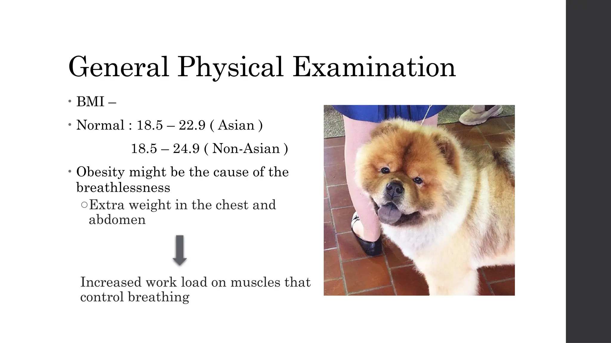
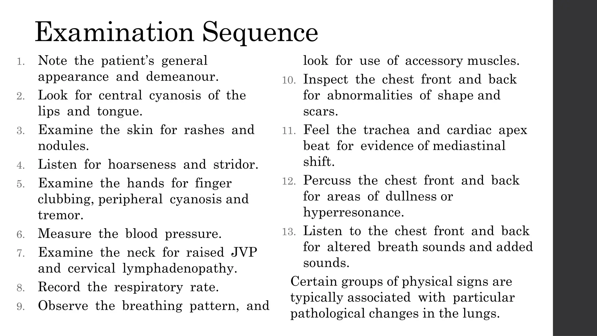
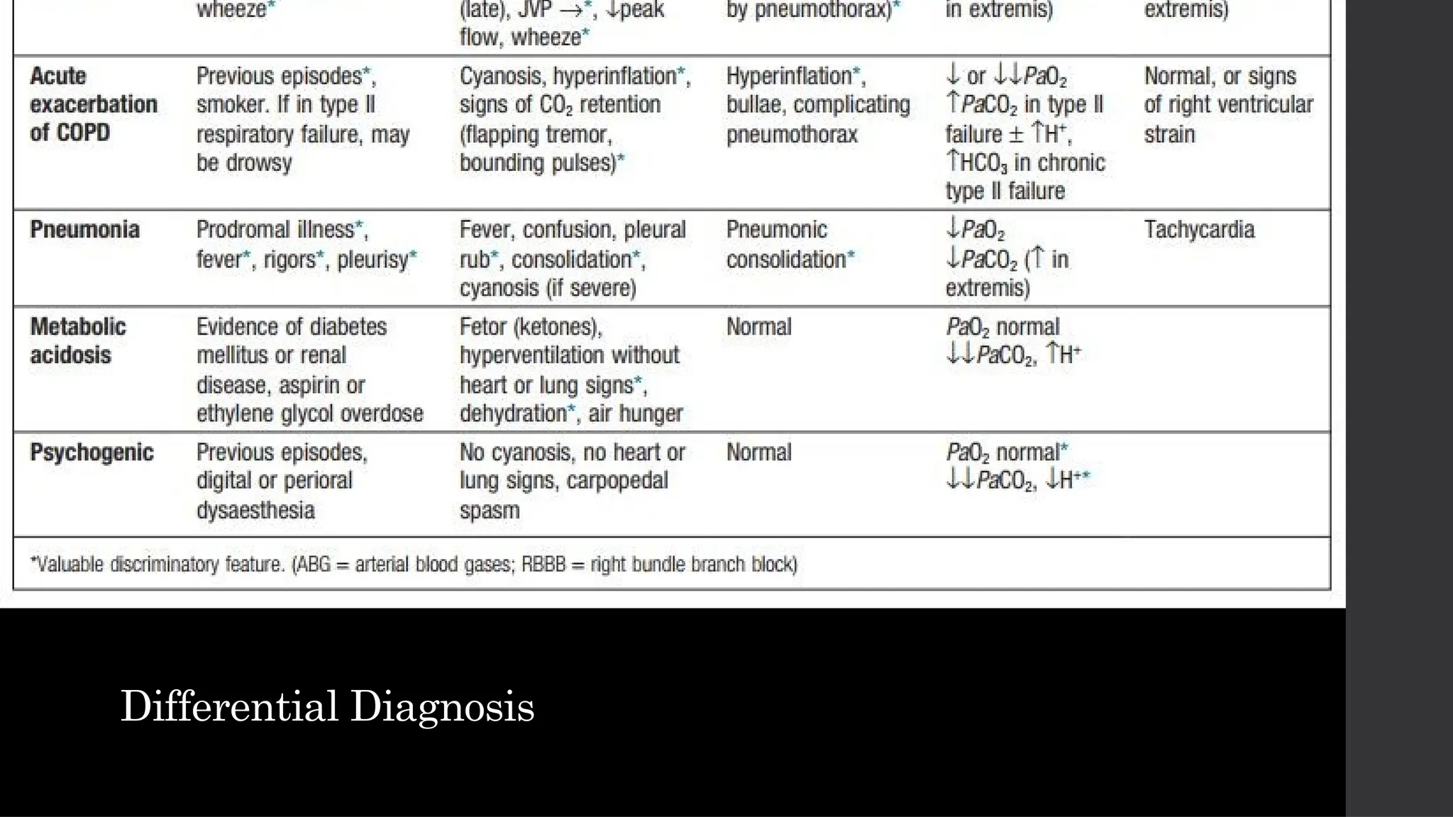
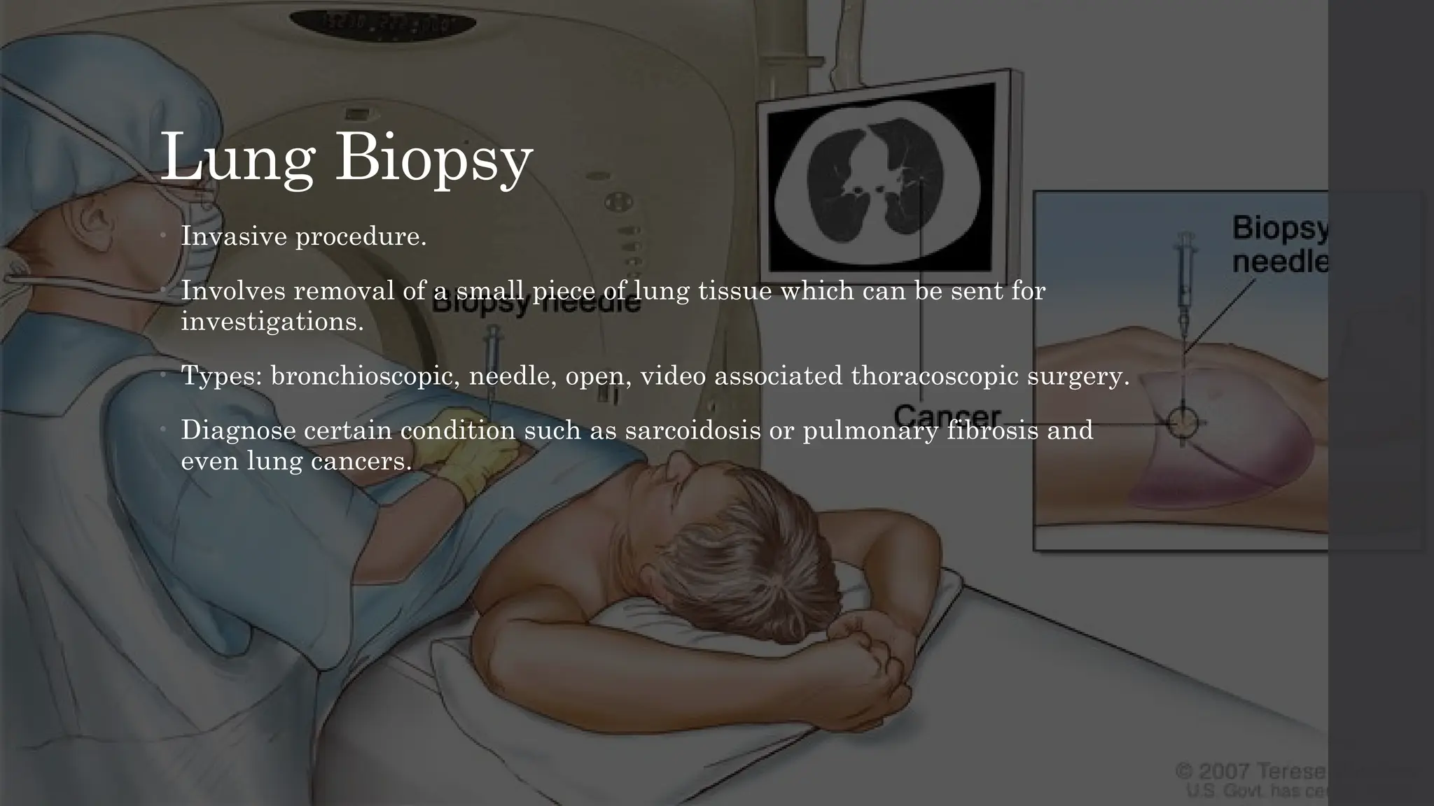
Shortness
of Breath
Photo
by
James
Heilman,
MD
/
CC
BY-SA
3.0
Contents • Definition
• Grades of breathlessness
• Common causes
• History taking
• Abnormalities on physical examination
• Differential diagnosis
• Initial investigations
• Basic management
Definition • Undue awareness of breathing and is normal with
strenuous physical exercise
• Sense of awareness of increased respiratory effort
that is unpleasant and that is recognized by the
patient as being inappropriate
• Breathlessness or dyspnoea can be defined as the
feeling of an uncomfortable need to breathe.
• It is unusual among sensations, as it has no defined
receptors, no localized representation in the brain,
and multiple causes both in health (e.g. exercise)
and in diseases of the lungs, heart or muscles.
• Patients use terms such as ‘shortness of breath’,
difficulty getting enough air in’, ‘tiredness’,
‘difficult, laboured, uncomfortable breathing;
it is an unpleasant type of breathing, though it is
not painful in the usual sense of the word’
MRC Dyspnoea Scale
Grade Degree of breathlessness related to activity
1 Not troubled by breathlessness except on strenuous exercise
2
Short of breath when hurrying on a level or when walking up
a slight hill
3
Walks slower than most people on the level, stops after a
mile (1.6 km) or so, or stops after 15 minutes walking at own
pace
4
Stops for breath after walking 100 yards (90 m), or after a
few minutes on level ground
5
Too breathless to leave the house, or breathless when
dressing/undressing
New York Heart Association (NYHA)
Functional Classification
Grade Symptoms
Grade 1 ( Mild )
No limitation on physical activity. Ordinary physical activity doesn't
cause undue fatigue, palpitation, dyspnoea
Grade 2 ( Mild )
Slight limitation of physical activity. Comfortable at rest but
ordinary physical activity results in fatigue, palpitation, dyspnoea.
Grade 3 ( Moderate )
Marked limitation of physical activity. Comfortable at rest. Less
than ordinary activity causes fatigue, palpitation, or dyspnea.
Grade 4 ( Severe )
Unable to carry out any physical activity without discomfort.
Dyspnoea present at rest, if any physical activity is undertaken the
discomfort increases.
Common Causes
System Acute dyspnoea
Cardiovascular Acute pulmonary oedema
Respiratory
Acute severe asthma
Acute exacerbation of COPD
Pneumothorax
Pneumonia
Pulmonary embolus
ARDS
Inhaled foreign body
Lobar collapse
Laryngeal oedema
Others
Metabolic acidosis
Psychogenic
hyperventilation
System Chronic exertional dyspnoea
Cardiovascular
Chronic heart failure
Myocardial ischaemia
Respiratory
COPD
Chronic asthma
Bronchial carcinoma
Interstitial lung disease
Chronic pulmonary thromboembolism
Lymphatic carcinomatosis
Large pleural effusions
Others
Severe anaemia
Obesity
Deconditioning
Common Causes
History taking
• Patient's details :
 Name
 Age
 Sex
 Occupation : paint sprayers,
rubber industry workers are
prone to lung disease
 Address
 Date of Admission
History taking • Chief Complaints
oShortness of breath for how many
days / weeks
• History of Presenting Illness
oOnset : gradual/sudden
oProgression : worsen/better
oSeverity : NYCA/MRCA grading
oDiurnal variability : worsen at night /
morning- asthma
oPostural variability : orthopnoea , PND-
CVS
oAggravating factors : pollen, dust, cold
climate - asthma, sinusitis
oRelieving factors : rest, medication
History taking • Associated symptoms :
a) Cough : yes/no
oIf yes , onset/duration/progression - chronic
cough - bronchiectasis
oFrequency, severity
oDiurnal variability - might indicate asthma
oPostural variability - might indicate
bronchiectasis
oProductive/ dry cough
b) Sputum :
oHow much? Frequency? - Copious sputum
suggestive of bronchiectasis
oColour? – White (viral) , yellowish
(bacteria), rusty (pneumonia)
oConsistency
oFoul smell? - Suggestive of bronchiectasis,
suppurative lung disease
History taking • Associated symptoms :
c) Haemoptysis :
oDuration, onset, progression
oAmount of blood?
oAssociated with melena or epistaxis?
oMight be due to malignancy
d) Chest pain :
oOnset, duration, progression
oSite? Bilateral/ unilateral
oType of pain
oRadiating - CVS pathology
oPain on deep inspiration, pain relief
when lie on same side- pleuritic chest
pain
History taking • Associated symptoms:
e) Fever :
oOnset, progression, duration
oAssociated with chills or rigor,
night sweats
oHigh / low grade
oEvening rise of temp - TB
f) Wheezing – asthma
g) Weight lost - TB, malignancy
h) Palpitation - CVS pathology
i) Sinusitis
j) Rhinitis
History taking • Past History :
oHistory of TB, Measle/whopping cough,
Asthma/ allergy, IHD, chest trauma/
surgery , similar complains, DM/HTN
• Personal History :
oDiet, Addiction, Bowel/Bladder
movement, Sleep, Appetite
oSmoking- highly ass with Emphysema
oAlcoholism - Aspiration Pneumonia
oLost of appetite & weight - TB,
Malignancy
• Family History :
oAsthma, TB, Cystic Fibrosis, Malignancy
oAnyone with similar complaints
History taking • Social history :
oEconomic status, income, type of house,
water source, no. of people staying in
one house
oCongested homes/ low economic status -
risk of lung pathology
• Menstrual history :
oTo rule out pregnancy
Abnormalities on
Physical Examination
General Physical Examination
• BMI –
• Normal : 18.5 – 22.9 ( Asian )
18.5 – 24.9 ( Non-Asian )
• Obesity might be the cause of the
breathlessness
oExtra weight in the chest and
abdomen
Increased work load on muscles that
control breathing
Vital Signs :
a) Pulse Rate – Normal
(60-90 beats/min)
 Tachycardia : Pneumonia ,
Atrial Fibrillation
 Bradycardia : Myocardial
Infarction
b) Blood Pressure – Normal
(120/80 mmHg)
 Hypotension : Pneumothorax ,
Myocardial Infarction
 Hypertension : Cardiac Failure
c) Temperature
 High : Tuberculosis, Pneumonia
d) Respiratory Rate – Normal
(14-16 breaths/min)
 Tachypnoea : Pulmonary
Embolism , Anxiety
 Bradypnoea : Myocardial
Infarction
Head to Toe Examination
1. Nails and Hands
 Pallor ( Left Heart Failure ,COPD)
 Cyanosis ( Cardiac Failure , COPD , Pulmonary oedema )
 Clubbing ( Carcinoma of Bronchus , Pulmonary
Fibrosis ,Bronchiectasis , Lung Abscess , Pleural Empyema )
 Koilonychia ( Anaemia )
2. Neck :
 JVP increased ( Right Heart Failure caused by Chronic
Pulmonary Hypertension in severe lung disease such as COPD )
Head to Toe Examination
3. Face :
 Eyes – Pallor ( Anaemia )
 Lips and Tongue – Cyanosis ( Cardiac Failure , COPD )
oIndicate poor oxygenation of blood
4. Lower limb :
 Pitting Pedal Oedema
oUnilateral – DVT
oBilateral – Congestive Heart Failure
Respiratory System Examination
 Auscultation
• Stridor ( Laryngeal Oedema , Foreign
bodies )
• Diminished Vesicular Breath Sounds
( Obesity , Pleural Effusion ,
Pneumothorax , COPD , Lung collapse )
• Bronchial Breath Sounds ( Lung
consolidation in pneumonia )
• Vocal Resonance ( over consolidated
lung , the spoken numbers are clearly
audible but over an effusion or collapse
they are muffled )
• Rhonchi / Wheeze ( COPD , Bronchial
Asthma )
• Pleural Rub ( Pneumonia , TB )
• Crackles
Examination Sequence
1. Note the patient’s general
appearance and demeanour.
2. Look for central cyanosis of the
lips and tongue.
3. Examine the skin for rashes and
nodules.
4. Listen for hoarseness and stridor.
5. Examine the hands for finger
clubbing, peripheral cyanosis and
tremor.
6. Measure the blood pressure.
7. Examine the neck for raised JVP
and cervical lymphadenopathy.
8. Record the respiratory rate.
9. Observe the breathing pattern, and
look for use of accessory muscles.
10. Inspect the chest front and back
for abnormalities of shape and
scars.
11. Feel the trachea and cardiac apex
beat for evidence of mediastinal
shift.
12. Percuss the chest front and back
for areas of dullness or
hyperresonance.
13. Listen to the chest front and back
for altered breath sounds and added
sounds.
Certain groups of physical signs are
typically associated with particular
pathological changes in the lungs.
Differential diagnosis
Develop differential diagnosis in a patient with
breathlessness based on history and
examination
Differential Diagnosis
Evaluation
Plan initial investigations and basic
management based on the differential diagnosis
Bedside investigations
Pulse Oximetry
• A spectrophotometric device that
measures arterial oxygen saturation
by determining the differential
absorption of light by oxyhaemoglobin
and deoxyhaemoglobin .
• This allows detection and ongoing
monitoring of hypoxaemia with
initiation of oxygen supplementation
as necessary, while undertaking
diagnostic work-up for its cause
Peak Flow Rate
• Person's maximum speed of expiration.
• Normal test results depends on age
year gender and sometimes occupation.
• Helps to differentiate between
pulmonary and cardiac causes of
dyspnoea. Low peak flow rates are
associated with obstructive lung
disease such as asthma, COPD, and
cystic fibrosis.
• Normal: 80-100 per cent of usual flow
rate.
Bedside investigations
Pulse Oximetry Peak Flow Rate
Radiology
Chest X-Ray
• To determine the lobe / area in which
the lung is affected.
• Make out any cardiomegaly in the PA
view of the x-ray
• Check for any fluid / consolidation of
the lungs.
• Check for any structural damage such
as a broken rib
Chest fluoroscopy / Sniff test
• Determines how well lungs,
diaphragm, or other parts of your
chest are working.
• Uses more radiation than a standard
chest X-ray.
• It detects the movement of the
diaphragm when the patient
breathes.
• Hence able to detect any abnormal
diaphragmatic conditions.
Radiology
Chest X-Ray Chest fluoroscopy / Sniff test
Electrocardiogram
• To detect underlying causes of cardiac origin that causes breathlessness.
• Coronary artery diseases can be a major causes of breathlessness.
• If needed an angiogram can be done to make out any blockage to the
coronary arteries.
Lung Biopsy
• Invasive procedure.
• Involves removal of a small piece of lung tissue which can be sent for
investigations.
• Types: bronchioscopic, needle, open, video associated thoracoscopic surgery.
• Diagnose certain condition such as sarcoidosis or pulmonary fibrosis and
even lung cancers.
Treatment
Management of breathlessness
Pharmacological
• The treatment of breathlessness is complex. It depends on the
underlying causes.
• Opioids-either oral or parenteral-are now considered to be the gold
standard in reducing ventilator demand. A slow release preparation
of morphine has been found to be beneficial
• Anxiolytics such as benzodiazepines may assist in the anxiety
component of breathlessness. However, these may be poorly
tolerated in some patients, especially in those with liver failure.
• Long acting beta agonists may be beneficial in breathlessness due to
COPD in reducing the work of breathing.
• Bronchodilators help in relaxing muscles and improving muscle tone
in the airways.
Oxygen Treatment
• Evidence have shown oxygen treatment is of no
use in a patient with breathlessness WITHOUT
hypoxia.
• Otherwise, high flow oxygen of >60 percent is
used except in COPD patients.
Non-pharmacological
• There does seem to be a relationship between anxiety
and breathlessness, however, which one comes first is
difficult to tell. Strategies such as relaxation training
and distraction do seem to help as do cognitive
behavioural therapies.
• Controlled breathing exercises and techniques such as
an upright leaning forward position and pursed lip
breathing are also beneficial therapies.
• Chest wall vibration, neuro-electrical muscle
stimulation, walking aides, and breathing training.
Thank you!

breathlessness-180310190951.pptx nnnnnnnn

  • 1.
  • 2.
    Contents • Definition •Grades of breathlessness • Common causes • History taking • Abnormalities on physical examination • Differential diagnosis • Initial investigations • Basic management
  • 3.
    Definition • Undueawareness of breathing and is normal with strenuous physical exercise • Sense of awareness of increased respiratory effort that is unpleasant and that is recognized by the patient as being inappropriate • Breathlessness or dyspnoea can be defined as the feeling of an uncomfortable need to breathe. • It is unusual among sensations, as it has no defined receptors, no localized representation in the brain, and multiple causes both in health (e.g. exercise) and in diseases of the lungs, heart or muscles. • Patients use terms such as ‘shortness of breath’, difficulty getting enough air in’, ‘tiredness’, ‘difficult, laboured, uncomfortable breathing; it is an unpleasant type of breathing, though it is not painful in the usual sense of the word’
  • 4.
    MRC Dyspnoea Scale GradeDegree of breathlessness related to activity 1 Not troubled by breathlessness except on strenuous exercise 2 Short of breath when hurrying on a level or when walking up a slight hill 3 Walks slower than most people on the level, stops after a mile (1.6 km) or so, or stops after 15 minutes walking at own pace 4 Stops for breath after walking 100 yards (90 m), or after a few minutes on level ground 5 Too breathless to leave the house, or breathless when dressing/undressing
  • 5.
    New York HeartAssociation (NYHA) Functional Classification Grade Symptoms Grade 1 ( Mild ) No limitation on physical activity. Ordinary physical activity doesn't cause undue fatigue, palpitation, dyspnoea Grade 2 ( Mild ) Slight limitation of physical activity. Comfortable at rest but ordinary physical activity results in fatigue, palpitation, dyspnoea. Grade 3 ( Moderate ) Marked limitation of physical activity. Comfortable at rest. Less than ordinary activity causes fatigue, palpitation, or dyspnea. Grade 4 ( Severe ) Unable to carry out any physical activity without discomfort. Dyspnoea present at rest, if any physical activity is undertaken the discomfort increases.
  • 6.
    Common Causes System Acutedyspnoea Cardiovascular Acute pulmonary oedema Respiratory Acute severe asthma Acute exacerbation of COPD Pneumothorax Pneumonia Pulmonary embolus ARDS Inhaled foreign body Lobar collapse Laryngeal oedema Others Metabolic acidosis Psychogenic hyperventilation
  • 7.
    System Chronic exertionaldyspnoea Cardiovascular Chronic heart failure Myocardial ischaemia Respiratory COPD Chronic asthma Bronchial carcinoma Interstitial lung disease Chronic pulmonary thromboembolism Lymphatic carcinomatosis Large pleural effusions Others Severe anaemia Obesity Deconditioning Common Causes
  • 8.
    History taking • Patient'sdetails :  Name  Age  Sex  Occupation : paint sprayers, rubber industry workers are prone to lung disease  Address  Date of Admission
  • 9.
    History taking •Chief Complaints oShortness of breath for how many days / weeks • History of Presenting Illness oOnset : gradual/sudden oProgression : worsen/better oSeverity : NYCA/MRCA grading oDiurnal variability : worsen at night / morning- asthma oPostural variability : orthopnoea , PND- CVS oAggravating factors : pollen, dust, cold climate - asthma, sinusitis oRelieving factors : rest, medication
  • 10.
    History taking •Associated symptoms : a) Cough : yes/no oIf yes , onset/duration/progression - chronic cough - bronchiectasis oFrequency, severity oDiurnal variability - might indicate asthma oPostural variability - might indicate bronchiectasis oProductive/ dry cough b) Sputum : oHow much? Frequency? - Copious sputum suggestive of bronchiectasis oColour? – White (viral) , yellowish (bacteria), rusty (pneumonia) oConsistency oFoul smell? - Suggestive of bronchiectasis, suppurative lung disease
  • 11.
    History taking •Associated symptoms : c) Haemoptysis : oDuration, onset, progression oAmount of blood? oAssociated with melena or epistaxis? oMight be due to malignancy d) Chest pain : oOnset, duration, progression oSite? Bilateral/ unilateral oType of pain oRadiating - CVS pathology oPain on deep inspiration, pain relief when lie on same side- pleuritic chest pain
  • 12.
    History taking •Associated symptoms: e) Fever : oOnset, progression, duration oAssociated with chills or rigor, night sweats oHigh / low grade oEvening rise of temp - TB f) Wheezing – asthma g) Weight lost - TB, malignancy h) Palpitation - CVS pathology i) Sinusitis j) Rhinitis
  • 13.
    History taking •Past History : oHistory of TB, Measle/whopping cough, Asthma/ allergy, IHD, chest trauma/ surgery , similar complains, DM/HTN • Personal History : oDiet, Addiction, Bowel/Bladder movement, Sleep, Appetite oSmoking- highly ass with Emphysema oAlcoholism - Aspiration Pneumonia oLost of appetite & weight - TB, Malignancy • Family History : oAsthma, TB, Cystic Fibrosis, Malignancy oAnyone with similar complaints
  • 14.
    History taking •Social history : oEconomic status, income, type of house, water source, no. of people staying in one house oCongested homes/ low economic status - risk of lung pathology • Menstrual history : oTo rule out pregnancy
  • 15.
  • 16.
    General Physical Examination •BMI – • Normal : 18.5 – 22.9 ( Asian ) 18.5 – 24.9 ( Non-Asian ) • Obesity might be the cause of the breathlessness oExtra weight in the chest and abdomen Increased work load on muscles that control breathing
  • 17.
    Vital Signs : a)Pulse Rate – Normal (60-90 beats/min)  Tachycardia : Pneumonia , Atrial Fibrillation  Bradycardia : Myocardial Infarction b) Blood Pressure – Normal (120/80 mmHg)  Hypotension : Pneumothorax , Myocardial Infarction  Hypertension : Cardiac Failure c) Temperature  High : Tuberculosis, Pneumonia d) Respiratory Rate – Normal (14-16 breaths/min)  Tachypnoea : Pulmonary Embolism , Anxiety  Bradypnoea : Myocardial Infarction
  • 18.
    Head to ToeExamination 1. Nails and Hands  Pallor ( Left Heart Failure ,COPD)  Cyanosis ( Cardiac Failure , COPD , Pulmonary oedema )  Clubbing ( Carcinoma of Bronchus , Pulmonary Fibrosis ,Bronchiectasis , Lung Abscess , Pleural Empyema )  Koilonychia ( Anaemia ) 2. Neck :  JVP increased ( Right Heart Failure caused by Chronic Pulmonary Hypertension in severe lung disease such as COPD )
  • 19.
    Head to ToeExamination 3. Face :  Eyes – Pallor ( Anaemia )  Lips and Tongue – Cyanosis ( Cardiac Failure , COPD ) oIndicate poor oxygenation of blood 4. Lower limb :  Pitting Pedal Oedema oUnilateral – DVT oBilateral – Congestive Heart Failure
  • 20.
    Respiratory System Examination Auscultation • Stridor ( Laryngeal Oedema , Foreign bodies ) • Diminished Vesicular Breath Sounds ( Obesity , Pleural Effusion , Pneumothorax , COPD , Lung collapse ) • Bronchial Breath Sounds ( Lung consolidation in pneumonia ) • Vocal Resonance ( over consolidated lung , the spoken numbers are clearly audible but over an effusion or collapse they are muffled ) • Rhonchi / Wheeze ( COPD , Bronchial Asthma ) • Pleural Rub ( Pneumonia , TB ) • Crackles
  • 21.
    Examination Sequence 1. Notethe patient’s general appearance and demeanour. 2. Look for central cyanosis of the lips and tongue. 3. Examine the skin for rashes and nodules. 4. Listen for hoarseness and stridor. 5. Examine the hands for finger clubbing, peripheral cyanosis and tremor. 6. Measure the blood pressure. 7. Examine the neck for raised JVP and cervical lymphadenopathy. 8. Record the respiratory rate. 9. Observe the breathing pattern, and look for use of accessory muscles. 10. Inspect the chest front and back for abnormalities of shape and scars. 11. Feel the trachea and cardiac apex beat for evidence of mediastinal shift. 12. Percuss the chest front and back for areas of dullness or hyperresonance. 13. Listen to the chest front and back for altered breath sounds and added sounds. Certain groups of physical signs are typically associated with particular pathological changes in the lungs.
  • 22.
    Differential diagnosis Develop differentialdiagnosis in a patient with breathlessness based on history and examination
  • 23.
  • 24.
  • 25.
    Evaluation Plan initial investigationsand basic management based on the differential diagnosis
  • 26.
    Bedside investigations Pulse Oximetry •A spectrophotometric device that measures arterial oxygen saturation by determining the differential absorption of light by oxyhaemoglobin and deoxyhaemoglobin . • This allows detection and ongoing monitoring of hypoxaemia with initiation of oxygen supplementation as necessary, while undertaking diagnostic work-up for its cause Peak Flow Rate • Person's maximum speed of expiration. • Normal test results depends on age year gender and sometimes occupation. • Helps to differentiate between pulmonary and cardiac causes of dyspnoea. Low peak flow rates are associated with obstructive lung disease such as asthma, COPD, and cystic fibrosis. • Normal: 80-100 per cent of usual flow rate.
  • 27.
  • 28.
    Radiology Chest X-Ray • Todetermine the lobe / area in which the lung is affected. • Make out any cardiomegaly in the PA view of the x-ray • Check for any fluid / consolidation of the lungs. • Check for any structural damage such as a broken rib Chest fluoroscopy / Sniff test • Determines how well lungs, diaphragm, or other parts of your chest are working. • Uses more radiation than a standard chest X-ray. • It detects the movement of the diaphragm when the patient breathes. • Hence able to detect any abnormal diaphragmatic conditions.
  • 29.
    Radiology Chest X-Ray Chestfluoroscopy / Sniff test
  • 30.
    Electrocardiogram • To detectunderlying causes of cardiac origin that causes breathlessness. • Coronary artery diseases can be a major causes of breathlessness. • If needed an angiogram can be done to make out any blockage to the coronary arteries.
  • 31.
    Lung Biopsy • Invasiveprocedure. • Involves removal of a small piece of lung tissue which can be sent for investigations. • Types: bronchioscopic, needle, open, video associated thoracoscopic surgery. • Diagnose certain condition such as sarcoidosis or pulmonary fibrosis and even lung cancers.
  • 32.
  • 33.
    Pharmacological • The treatmentof breathlessness is complex. It depends on the underlying causes. • Opioids-either oral or parenteral-are now considered to be the gold standard in reducing ventilator demand. A slow release preparation of morphine has been found to be beneficial • Anxiolytics such as benzodiazepines may assist in the anxiety component of breathlessness. However, these may be poorly tolerated in some patients, especially in those with liver failure. • Long acting beta agonists may be beneficial in breathlessness due to COPD in reducing the work of breathing. • Bronchodilators help in relaxing muscles and improving muscle tone in the airways.
  • 34.
    Oxygen Treatment • Evidencehave shown oxygen treatment is of no use in a patient with breathlessness WITHOUT hypoxia. • Otherwise, high flow oxygen of >60 percent is used except in COPD patients.
  • 35.
    Non-pharmacological • There doesseem to be a relationship between anxiety and breathlessness, however, which one comes first is difficult to tell. Strategies such as relaxation training and distraction do seem to help as do cognitive behavioural therapies. • Controlled breathing exercises and techniques such as an upright leaning forward position and pursed lip breathing are also beneficial therapies. • Chest wall vibration, neuro-electrical muscle stimulation, walking aides, and breathing training.
  • 36.

Editor's Notes

  • #22 Consider talking about: Acute coronary syndrome Congestive heart failure Chronic obstructive pulmonary disease Asthma Pneumothorax Pneumonia Pulmonary embolism Anaemia Other
  • #25 Consider talking about: Blood tests Imaging
  • #32 Consider talking about: Physiotherapy Palliative