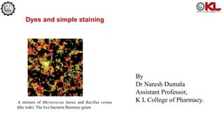
Dyes and simple staining
By
Dr Naresh Dumala
Assistant Professor,
K L College of Pharmacy.
A mixture of Micrococcus luteus and Bacillus cereus
(the rods). The live bacteria fluoresce green
Dyes
A natural or synthetic substance used to add a colour or change the colour
of something/organism.
The many types of dyes used to stain microorganisms have two features in common.
1. They have chromophore groups, groups with conjugated double bonds that give
the dye its color.
2. They can bind with cells by ionic, covalent, or hydrophobic bonding.
For example, a positively charged dye binds to negatively charged structures on the
cell.
chromophore groups
• A chromophore is the part of a molecule responsible for its color. The color that is seen by our eyes
is the one not absorbed by the reflecting object within a certain wavelength spectrum of visible
light.
• The chromophore is a region in the molecule where the energy difference between two
separate molecular orbitals falls within the range of the visible spectrum.
• Visible light that hits the chromophore can thus be absorbed by exciting an electron from its ground
state into an excited state.
• In biological molecules that serve to capture or detect light energy, the chromophore is
the moiety that causes a conformational change in the molecule when hit by light
Ionizable dyes may be divided into two general classes based on the nature of their charged group.
1. Basic dyes
• methylene blue, basic fuchsin, crystal violet, safranin, malachite green—have positively charged
groups (usually some form of pentavalent nitrogen) and are generally sold as chloride salts.
• Basic dyes bind to negatively charged molecules like nucleic acids and many proteins.
• Because the surfaces of bacterial cells also are negatively charged, basic dyes are most often used
in bacteriology.
2. Acid dyes
• eosin, rose bengal, and acid fuchsin—possess negatively charged groups such as carboxyls (—COOH)
and phenolic hydroxyls (—OH).
• Acid dyes, because of their negative charge, bind to positively charged cell structures.
The pH may alter staining effectiveness since the nature and degree of the charge on cell components
change with pH.
Thus, anionic dyes stain best under acidic conditions when proteins and many other molecules carry a
positive charge; basic dyes are most effective at higher pHs.
• Although ionic interactions are probably the most common means of
attachment, dyes also bind through covalent bonds or because of their
solubility characteristics.
• For instance, DNA can be stained by the Feulgen procedure in which
Schiff’s reagent is covalently attached to its deoxyribose sugars after
hydrochloric acid treatment.
• Sudan III (Sudan Black) selectively stains lipids because it is lipid
soluble but will not dissolve in aqueous portions of the cell.
Microorganisms often can be stained very satisfactorily by simple
staining, in which a single staining agent is used.
• Simple staining’s value lies in its simplicity and ease of use.
• One covers the fixed smear with stain for the proper length of time,
washes the excess stain off with water, and blots the slide dry.
• Basic dyes like crystal violet, methylene blue, and carbol-fuchsin are
frequently used to determine the size, shape, and arrangement of bacteria.
Simple Staining
Dyes and Staining.pptx

Dyes and Staining.pptx

  • 1.
    Dyes and simplestaining By Dr Naresh Dumala Assistant Professor, K L College of Pharmacy. A mixture of Micrococcus luteus and Bacillus cereus (the rods). The live bacteria fluoresce green
  • 2.
    Dyes A natural orsynthetic substance used to add a colour or change the colour of something/organism. The many types of dyes used to stain microorganisms have two features in common. 1. They have chromophore groups, groups with conjugated double bonds that give the dye its color. 2. They can bind with cells by ionic, covalent, or hydrophobic bonding. For example, a positively charged dye binds to negatively charged structures on the cell.
  • 3.
    chromophore groups • Achromophore is the part of a molecule responsible for its color. The color that is seen by our eyes is the one not absorbed by the reflecting object within a certain wavelength spectrum of visible light. • The chromophore is a region in the molecule where the energy difference between two separate molecular orbitals falls within the range of the visible spectrum. • Visible light that hits the chromophore can thus be absorbed by exciting an electron from its ground state into an excited state. • In biological molecules that serve to capture or detect light energy, the chromophore is the moiety that causes a conformational change in the molecule when hit by light
  • 4.
    Ionizable dyes maybe divided into two general classes based on the nature of their charged group. 1. Basic dyes • methylene blue, basic fuchsin, crystal violet, safranin, malachite green—have positively charged groups (usually some form of pentavalent nitrogen) and are generally sold as chloride salts. • Basic dyes bind to negatively charged molecules like nucleic acids and many proteins. • Because the surfaces of bacterial cells also are negatively charged, basic dyes are most often used in bacteriology.
  • 5.
    2. Acid dyes •eosin, rose bengal, and acid fuchsin—possess negatively charged groups such as carboxyls (—COOH) and phenolic hydroxyls (—OH). • Acid dyes, because of their negative charge, bind to positively charged cell structures. The pH may alter staining effectiveness since the nature and degree of the charge on cell components change with pH. Thus, anionic dyes stain best under acidic conditions when proteins and many other molecules carry a positive charge; basic dyes are most effective at higher pHs.
  • 6.
    • Although ionicinteractions are probably the most common means of attachment, dyes also bind through covalent bonds or because of their solubility characteristics. • For instance, DNA can be stained by the Feulgen procedure in which Schiff’s reagent is covalently attached to its deoxyribose sugars after hydrochloric acid treatment. • Sudan III (Sudan Black) selectively stains lipids because it is lipid soluble but will not dissolve in aqueous portions of the cell.
  • 7.
    Microorganisms often canbe stained very satisfactorily by simple staining, in which a single staining agent is used. • Simple staining’s value lies in its simplicity and ease of use. • One covers the fixed smear with stain for the proper length of time, washes the excess stain off with water, and blots the slide dry. • Basic dyes like crystal violet, methylene blue, and carbol-fuchsin are frequently used to determine the size, shape, and arrangement of bacteria. Simple Staining