CANCER: A group of disease involving abnormal cell growth with the potential to invade or spread to other part of the body.
CHEMOTHERAPY: the term chemotherapy is describe as the use of chemicals or drugs to treat cancer.
CYTOTOXIC DRUG: lysis both normal and cancer cells
In this presentation i have tried to thoroughly discuss about the concept of Drug induced kidney disease or injury, the mechanism behind it, its classification and how to access it.
conversion from INTRAVENOUS TO ORAL DOSING----- design of dosage regimenpavithra vinayak
conversion from INTRAVENOUS TO ORAL DOSING----- TYPES OF IV TO PO THERAPY CONVERSIONS: MEDICATIONS INCLUDED IN AN IV TO PO CONVERSION PROGRAM: SELECTION OF PATIENTS FOR IV TO PO THERAPY CONVERSION: design of dosage regimen--clinical pharmacokinetics and therapeutic drug monitoring-- fifth pharm D notes
Clinical pharmacokinetics and its application--
1)definition
2) APPLICATIONS OF CLINICAL PHARMACOKINETICS
Design of dosage regimens:
a) Nomograms and Tabulations in designing dosage regimen,
b) Conversion from intravenous to oral dosing,
c) Determination of dose and dosing intervals,
d) Drug dosing in the elderly and pediatrics and obese patients.
Pharmacokinetics of Drug Interaction:
a) Pharmacokinetic drug interactions
b) Inhibition and Induction of Drug metabolism
c) Inhibition of Biliary Excretion.
Therapeutic Drug monitoring:
a) Introduction
b) Individualization of drug dosage regimen (Variability – Genetic, Age and Weight, disease, Interacting drugs).
c) Indications for TDM. Protocol for TDM.
d) Pharmacokinetic/Pharmacodynamic Correlation in drug therapy.
e) TDM of drugs used in the following disease conditions: cardiovascular disease, Seizure disorders, Psychiatric conditions, and Organ transplantations
Dosage adjustment in Renal and Hepatic Disease.
a. Renal impairment
b. Pharmacokinetic considerations
c. General approach for dosage adjustment in renal disease.
d. Measurement of Glomerular Filtration rate and creatinine clearance.
e. Dosage adjustment for uremic patients.
f. Extracorporeal removal of drugs.
g. Effect of Hepatic disease on pharmacokinetics.
Population Pharmacokinetics.
a) Introduction to Bayesian Theory.
b) Adaptive method or Dosing with feedback.
c) Analysis of Population pharmacokinetic Data
A complete drug profile of Tacrolimus an immunosuppressant used for organ transplant. It consist of PK/PD, MOA, Indication & Uses, Contraindications, Warnings & Precautions, Drug-interaction, Doses & Administration, Dosage forms, Chemical Formula, Side-Effects, Adverse Drug Reactions, Therapeutic Drug Monitoring (TDM).
Presentation for Medical undergraduates for teaching pharmacology. It deals with Physiology of steroid hormones and their action along with agents which are used therapeutically with their action, adverse effects and therapeutic uses.
Management of acute lymphoblatic leukemia with light on etiology, clinical features, diagnosis and different aspects of management including chemotherapy and radiation therapy
Tom Selleck Health: A Comprehensive Look at the Iconic Actor’s Wellness Journeygreendigital
Tom Selleck, an enduring figure in Hollywood. has captivated audiences for decades with his rugged charm, iconic moustache. and memorable roles in television and film. From his breakout role as Thomas Magnum in Magnum P.I. to his current portrayal of Frank Reagan in Blue Bloods. Selleck's career has spanned over 50 years. But beyond his professional achievements. fans have often been curious about Tom Selleck Health. especially as he has aged in the public eye.
Follow us on: Pinterest
Introduction
Many have been interested in Tom Selleck health. not only because of his enduring presence on screen but also because of the challenges. and lifestyle choices he has faced and made over the years. This article delves into the various aspects of Tom Selleck health. exploring his fitness regimen, diet, mental health. and the challenges he has encountered as he ages. We'll look at how he maintains his well-being. the health issues he has faced, and his approach to ageing .
Early Life and Career
Childhood and Athletic Beginnings
Tom Selleck was born on January 29, 1945, in Detroit, Michigan, and grew up in Sherman Oaks, California. From an early age, he was involved in sports, particularly basketball. which played a significant role in his physical development. His athletic pursuits continued into college. where he attended the University of Southern California (USC) on a basketball scholarship. This early involvement in sports laid a strong foundation for his physical health and disciplined lifestyle.
Transition to Acting
Selleck's transition from an athlete to an actor came with its physical demands. His first significant role in "Magnum P.I." required him to perform various stunts and maintain a fit appearance. This role, which he played from 1980 to 1988. necessitated a rigorous fitness routine to meet the show's demands. setting the stage for his long-term commitment to health and wellness.
Fitness Regimen
Workout Routine
Tom Selleck health and fitness regimen has evolved. adapting to his changing roles and age. During his "Magnum, P.I." days. Selleck's workouts were intense and focused on building and maintaining muscle mass. His routine included weightlifting, cardiovascular exercises. and specific training for the stunts he performed on the show.
Selleck adjusted his fitness routine as he aged to suit his body's needs. Today, his workouts focus on maintaining flexibility, strength, and cardiovascular health. He incorporates low-impact exercises such as swimming, walking, and light weightlifting. This balanced approach helps him stay fit without putting undue strain on his joints and muscles.
Importance of Flexibility and Mobility
In recent years, Selleck has emphasized the importance of flexibility and mobility in his fitness regimen. Understanding the natural decline in muscle mass and joint flexibility with age. he includes stretching and yoga in his routine. These practices help prevent injuries, improve posture, and maintain mobilit
Basavarajeeyam is an important text for ayurvedic physician belonging to andhra pradehs. It is a popular compendium in various parts of our country as well as in andhra pradesh. The content of the text was presented in sanskrit and telugu language (Bilingual). One of the most famous book in ayurvedic pharmaceutics and therapeutics. This book contains 25 chapters called as prakaranas. Many rasaoushadis were explained, pioneer of dhatu druti, nadi pareeksha, mutra pareeksha etc. Belongs to the period of 15-16 century. New diseases like upadamsha, phiranga rogas are explained.
These simplified slides by Dr. Sidra Arshad present an overview of the non-respiratory functions of the respiratory tract.
Learning objectives:
1. Enlist the non-respiratory functions of the respiratory tract
2. Briefly explain how these functions are carried out
3. Discuss the significance of dead space
4. Differentiate between minute ventilation and alveolar ventilation
5. Describe the cough and sneeze reflexes
Study Resources:
1. Chapter 39, Guyton and Hall Textbook of Medical Physiology, 14th edition
2. Chapter 34, Ganong’s Review of Medical Physiology, 26th edition
3. Chapter 17, Human Physiology by Lauralee Sherwood, 9th edition
4. Non-respiratory functions of the lungs https://academic.oup.com/bjaed/article/13/3/98/278874
Lung Cancer: Artificial Intelligence, Synergetics, Complex System Analysis, S...Oleg Kshivets
RESULTS: Overall life span (LS) was 2252.1±1742.5 days and cumulative 5-year survival (5YS) reached 73.2%, 10 years – 64.8%, 20 years – 42.5%. 513 LCP lived more than 5 years (LS=3124.6±1525.6 days), 148 LCP – more than 10 years (LS=5054.4±1504.1 days).199 LCP died because of LC (LS=562.7±374.5 days). 5YS of LCP after bi/lobectomies was significantly superior in comparison with LCP after pneumonectomies (78.1% vs.63.7%, P=0.00001 by log-rank test). AT significantly improved 5YS (66.3% vs. 34.8%) (P=0.00000 by log-rank test) only for LCP with N1-2. Cox modeling displayed that 5YS of LCP significantly depended on: phase transition (PT) early-invasive LC in terms of synergetics, PT N0—N12, cell ratio factors (ratio between cancer cells- CC and blood cells subpopulations), G1-3, histology, glucose, AT, blood cell circuit, prothrombin index, heparin tolerance, recalcification time (P=0.000-0.038). Neural networks, genetic algorithm selection and bootstrap simulation revealed relationships between 5YS and PT early-invasive LC (rank=1), PT N0—N12 (rank=2), thrombocytes/CC (3), erythrocytes/CC (4), eosinophils/CC (5), healthy cells/CC (6), lymphocytes/CC (7), segmented neutrophils/CC (8), stick neutrophils/CC (9), monocytes/CC (10); leucocytes/CC (11). Correct prediction of 5YS was 100% by neural networks computing (area under ROC curve=1.0; error=0.0).
CONCLUSIONS: 5YS of LCP after radical procedures significantly depended on: 1) PT early-invasive cancer; 2) PT N0--N12; 3) cell ratio factors; 4) blood cell circuit; 5) biochemical factors; 6) hemostasis system; 7) AT; 8) LC characteristics; 9) LC cell dynamics; 10) surgery type: lobectomy/pneumonectomy; 11) anthropometric data. Optimal diagnosis and treatment strategies for LC are: 1) screening and early detection of LC; 2) availability of experienced thoracic surgeons because of complexity of radical procedures; 3) aggressive en block surgery and adequate lymph node dissection for completeness; 4) precise prediction; 5) adjuvant chemoimmunoradiotherapy for LCP with unfavorable prognosis.
NVBDCP.pptx Nation vector borne disease control programSapna Thakur
NVBDCP was launched in 2003-2004 . Vector-Borne Disease: Disease that results from an infection transmitted to humans and other animals by blood-feeding arthropods, such as mosquitoes, ticks, and fleas. Examples of vector-borne diseases include Dengue fever, West Nile Virus, Lyme disease, and malaria.
Title: Sense of Taste
Presenter: Dr. Faiza, Assistant Professor of Physiology
Qualifications:
MBBS (Best Graduate, AIMC Lahore)
FCPS Physiology
ICMT, CHPE, DHPE (STMU)
MPH (GC University, Faisalabad)
MBA (Virtual University of Pakistan)
Learning Objectives:
Describe the structure and function of taste buds.
Describe the relationship between the taste threshold and taste index of common substances.
Explain the chemical basis and signal transduction of taste perception for each type of primary taste sensation.
Recognize different abnormalities of taste perception and their causes.
Key Topics:
Significance of Taste Sensation:
Differentiation between pleasant and harmful food
Influence on behavior
Selection of food based on metabolic needs
Receptors of Taste:
Taste buds on the tongue
Influence of sense of smell, texture of food, and pain stimulation (e.g., by pepper)
Primary and Secondary Taste Sensations:
Primary taste sensations: Sweet, Sour, Salty, Bitter, Umami
Chemical basis and signal transduction mechanisms for each taste
Taste Threshold and Index:
Taste threshold values for Sweet (sucrose), Salty (NaCl), Sour (HCl), and Bitter (Quinine)
Taste index relationship: Inversely proportional to taste threshold
Taste Blindness:
Inability to taste certain substances, particularly thiourea compounds
Example: Phenylthiocarbamide
Structure and Function of Taste Buds:
Composition: Epithelial cells, Sustentacular/Supporting cells, Taste cells, Basal cells
Features: Taste pores, Taste hairs/microvilli, and Taste nerve fibers
Location of Taste Buds:
Found in papillae of the tongue (Fungiform, Circumvallate, Foliate)
Also present on the palate, tonsillar pillars, epiglottis, and proximal esophagus
Mechanism of Taste Stimulation:
Interaction of taste substances with receptors on microvilli
Signal transduction pathways for Umami, Sweet, Bitter, Sour, and Salty tastes
Taste Sensitivity and Adaptation:
Decrease in sensitivity with age
Rapid adaptation of taste sensation
Role of Saliva in Taste:
Dissolution of tastants to reach receptors
Washing away the stimulus
Taste Preferences and Aversions:
Mechanisms behind taste preference and aversion
Influence of receptors and neural pathways
Impact of Sensory Nerve Damage:
Degeneration of taste buds if the sensory nerve fiber is cut
Abnormalities of Taste Detection:
Conditions: Ageusia, Hypogeusia, Dysgeusia (parageusia)
Causes: Nerve damage, neurological disorders, infections, poor oral hygiene, adverse drug effects, deficiencies, aging, tobacco use, altered neurotransmitter levels
Neurotransmitters and Taste Threshold:
Effects of serotonin (5-HT) and norepinephrine (NE) on taste sensitivity
Supertasters:
25% of the population with heightened sensitivity to taste, especially bitterness
Increased number of fungiform papillae
micro teaching on communication m.sc nursing.pdfAnurag Sharma
Microteaching is a unique model of practice teaching. It is a viable instrument for the. desired change in the teaching behavior or the behavior potential which, in specified types of real. classroom situations, tends to facilitate the achievement of specified types of objectives.
Pharynx and Clinical Correlations BY Dr.Rabia Inam Gandapore.pptx
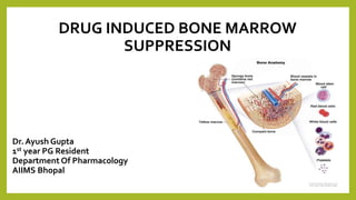
Drug induced bone marrow suppression
1. DRUG INDUCED BONE MARROW
SUPPRESSION
Dr. Ayush Gupta
1st year PG Resident
Department Of Pharmacology
AIIMS Bhopal
2. OUTLINE
INTRODUCTION
• BONE MARROW
• BONE MARROW BLOODVASCULATURE
• NORMAL HEMATOPOIESIS
MYELOSUPPRESSION
CATEGORIES OF DRUG INDUCED MYELOSUPPRESSION
MECHANISM OF DRUG INDUCED MYELOSUPPRESSION
TIMING AND EXTENT OF CHEMOTHERAPY INDUCED
MYELOSUPPRESSION
PATHOPHYSIOLOGY
DIAGNOSIS OF DRUG INDUCED MYELOSUPPRESSION
CONCLUSION
3. BONE MARROW
Bone marrow is highly cellular, spongy or viscous tissue that fills the inside of your bones.
Two types of bone marrow :
Red bone marrow &Yellow bone marrow
Pattern of distribution
Human marrow produces approximately 500 billion
blood cells per day in adults
On average, bone marrow comprises approximately
5% of total body weight
4. BLOODVASCULATURE
• Bone receive up to about 10% of cardiac
output
• The blood supply of bone is delivered to
endosteal cavity by
• The marrow cavity afford a range of
vascular niches that regulate the growth
and differentiation of hematopoietic and
stromal cells
• Metaphyseal and Epiphyseal flow
• The blood vessels between the
haematopoietic compartment and the
circulation form a barrier, referred to as
the marrow-blood barrier (MBB)
5. NORMAL HEMATOPOIESIS
Normal haematopoiesis
involves the development of
various cell lineages –
mediated by various growth
and cytokines in the marrow
environment
multipotent progenitor
cells become differentiated
and committed to specific
developmental pathway
best known CSF are IL-3,
GM-CSF, G-CSF,
erythropoietin and M-CSF.
IL-3 is active throughout
the hemopoietic
cascade
6. Myelosuppression
Myelosuppression is caused by the destruction of the proliferating progenitor cells that
produce the mature red and white blood cells and platelets found in the peripheral
circulation
Myelosuppression is a common and anticipated adverse effect of cytotoxic chemotherapy
It is a potential but rare idiosyncratic effect with any other drug
there is a recognised association with a number of higher-risk agents which justify
additional vigilance
Genetic risk factors are being identified which may predispose individuals to this reaction
with particular drugs – e:g- mercaptopurine
7. Contin..
• Myelosuppression is potentially life threatening because of the infection and
bleeding complications of neutropenia and thrombocytopenia
• Immediate concern for patients undergoing cancer therapy, its management has
been improved significantly in recent years by the use of various hematopoietic
growth factors
• However, many patients receiving chemotherapy and/or ionizing radiation (IR) also
develop residual (or long-term) BM injury (a sustained decrease in HSC reserves due
to an impairment in HSC self-renewal) after the recovery from acute
myelosuppression
8. CATEGORIES OF DRUG INDUCED
MYELOSUPPRESSION
ONTHE BASIS OF MARROW CELLULARITY ONTHE BASIS OF PERIPHERAL CELL DISTRUCTION
Reducing the cellularity of marrow
bi or tricytopenia due to hypoplasia/aplasia of the bone
marrow :
AplasticAnaemia
Selective marrow hypoplasia/ aplasia :
1. pure red cell aplasia
2. drug induced neutropenia/Agranulocytosis
3. drug induced non immune thrombocytopenia
Without reducing the cellularity of marrow
( interfering with marrow cell maturation)
MegaloblasticAnaemia
SideroblasticAnaemia
Drug induced Haemolytic Anaemia
drug induced oxidative haemolytic anaemia
drug induced immune mediated haemolytic anaemia
drug induced immune thrombocytopenia
9. MECHANISMS OF DRUG INDUCED BONE MARROW
SUPPRESSION
TYPE A, DIRECT DOSE RELATEDTOXICITY
1. Acute
myelosuppression
2. Residual bone
marrow injury
TYPE B, IDIOSYNCRATIC MEDIATED
1. Metabolite driven
toxicity
2. Genetic
polymorphisms
IMMUNE MEDIATED TOXICITY
1. Hapten mechanism(drug
adsorption mechanism)
2. Immune complex
mechanism(innocent
bystander mechanism)
3. Autoimmune mechanism
10. PATHOPHYSIOLOGY OF CYTOTOXIC DRUG
Stem cells have two cardinal functions: self-renewal
and differentiation
HSCs serve as reserves to protect the hematopoietic
system from exhaustion under various stress
conditions
HPCs are rapidly proliferating cells with limited self-
renewal ability.
HSCs can undergo self-renewing proliferation and
differentiation
11. PREDICTING MYELOSUPPRESSION
Three main factors will determine when and how much myelosuppression will occur for any
patient about to embark on a course of chemotherapy
Factor 1 : Blood cell life cycle
primary responsible for the timing of myelosuppression
• This is a static factor, applies to all patients, and will be the same no matter which drug is
being used
• Differences in the length and kinetics of the life cycle of particular blood cells account for the
frequency of granulocytopenia, thrombocytopenia and anaemia
E:g. difference in half lives of red blood cell and neutrophils
WBC – 6-8 hours circulating in blood and 2-3 days in tissues
lymphocyte- 100-300+ days
RBC- 120 days Platelets- 5-10 days
12. Contin…
Factor 2 : Drug Characteristics
A) Pharmacokinetics factor
ADME of anticancer drugs is important and have to
be considered.
Drug administration-
The anti tumour effect of 5 – fluorouracil can be
enhanced when treating liver metastasis by direct
infusion through an arterial catheter into the liver ,
Much larger doses can be administered.
•
13. Drug Distribution –
The blood brain barrier can lead to a “ sanctuary effect” where the majority of lipid
soluble antineoplastic agents are unable to effectively reach target malignant cells
despite drug doses that produce life threatening toxicity.
Excretion –
e.g. Methotrexate is primarily excreted by kidneys , can cause major
myelosuppression when administered to a patient with elevated serum creatinine.
14. B) Phase specificity- cell cycle specific and cell cycle non specific
• Drug that are phase specific lead to a fairly rapid
cytopenia (mostly granulocytopenia followed by
thrombocytopenia).
• Recovery- quicker, especially from drugs that are
active in the S and M phase
• Non specific drug – leads to delayed, prolonged
and cumulative myelosuppression
15. Timing and Extent of Chemotherapy induced Myelosuppression
WBC Nadir (Days) WBC Recovery
(Days)
Platelet Nadir (Days)
Comment
Asparaginase 4-7 10-14 5-10
Myelosuppression is rarely a problem
Hydroxyurea
5-Fluorouracil
Cytarabine
7
7-14
12-14
14-21
20-30
22-24
NA
7-17
22-24 Somewhat platelet sparing
6-Mercaptopurine 7-14 14-21 10-14
Methotrexate 7-14 14-21 5-12
Bleomycin
Etoposide
NS
7-14
NS
21
NS
9-16
Vinblastine
Vincristine
Vindesine
5-9
3-5
7
14-21
7
14
4-10
NA- marrow sparing
7- platelet sparing
16. WBC Nadir
(Days)
WBC Recovery
(Days)
Platelet Nadir
(Days) Comment
Busulfan
Carboplatin
Cisplatin
Cyclophosphamide
Procarbazine
7-10
21
18-23
8-14
25-36
24-25
28
29
18-25
35-50+
10-30
21
14
10-25
21
Dose limiting toxicity: thrombocytopenia can be severe
Anaemia can be severe
Platelet sparing
Prolonged, delayed myelosuppression
Dactinomycin
Daunorubicin
Doxorubicin
Mitomycin
14-21
8-10
10-14
21-25
22-25
21
22
28-42
10-14
10-14
14
30
Profound myelosuppression
Cumulative, prolonged myelosuppression
Carmustine
Losmustine
35-42
42
42-56
60
28-35
28
Cumulative, delayed and prolonged myelosuppression
Thrombocytopenia more common than leukopenia
Dacarbazine 10-14 24 14-28
17. Factor 3: Characteristics of the patient
The degree of myelosuppression expected from a specific treatment will be influenced by:
• Patient’s age- older patients have a less cellular marrow with more fat space, and possible
aplasia
• Patient’s Health- debilitation may increase the severity and unpredictability of
myelosuppression
• Nutritional status- the greater the negative nitrogen balance and weight loss, the less
tolerant the patient will be to the drug’s toxic effect on the marrow because there are less
nutritional resources for building new blood cells.
18. Contin..
• Degree of bone marrow reserve- cisplatin, carmustine and busulfan
• Adequacy of liver and kidney function- methotrexate , primarily excreted by kidney,
can cause major myelosuppression
• Fibrosis due to prior radiation therapy decreases bone marrow reserves
• Ascites and pleural effusion create a third space which can prolong drug toxicity.
19. DRUGS ASSOCIATEDWITH IDIOSYNCRATIC(TYPE B)
MYELOSUPPRESSIOM
• Drug reactions that occur rarely and unpredictably amongst the population
• They frequently occur with exposure to new drugs, as they have not been fully tested and the full
range of possible side-effects have not been discovered
• Idiosyncratic drug reactions appear to not be concentration dependent
20. • The proposed mechanism of most idiosyncratic drug reactions is immune-mediated
toxicity and reactive metabolites of the offending drugs
• There is new evidence that drugs that cause IDRs including IDIAG can activate
inflammasome
Genetic polymorphism
• mercaptopurine – inactivated by enzyme thiopurine methyltransferase(TPMT) –
genetically variation inTPMT activity associated with myelosuppression
• mutation in methylenetetrahydrofolate reductase(MTHFR) gene
21.
22. DRUG INDUCED APLASTIC ANAEMIA
Definition:
“Condition, in which body is unable to produce enough new blood cells”
Characterized by a bi- or tricytopenia (thrombocytopenia, anaemia
and granulocytopenia) due to hypoplasia or aplasia of the bone marrow
It was initially reported in the 1930 associated with arsenicals and
aminopyrines.
Bimodal risk distribution when it comes to age
peak incidence between 10-25 years and age >60 years
It is the most serious acquired blood dyscrasia because of its associated high mortality which
averages about 50%
23. PATHOPHYSIOLOGY
• The cause of drug-induced aplastic anaemia is damage to the pluripotential
hematopoietic stem cells before their differentiation to committed stem
cells.
• There are three major etiologies of acquired aplastic anaemia
i. Direct, dose-related drug toxicity
ii. Idiosyncratic mechanisms
iii. Drug-induced autoimmune aplastic anaemia
24. i. DIRECT, DOSE RELATEDTOXICITY :
The majority of chemotherapeutic agents can cause myelosuppression in a dose-
dependent manner.
Among these compounds, alkylating agents, pyrimidine analogues,
methotrexate, hydroxyurea and mitomycin C are highly cytotoxic to BM
Example: Among women with breast cancer, patients receiving CMF regimens and CAF
regimen, were strongly associated with risk of aplastic anaemia
ACUTE MYELOSUPPRESSION (< 3 month) RESIDUAL BONE MARROW INJURY ( >3 months)
• Due to depletion of HPCs • Impaired self-renewal ability of HSCs
25. • The main effect of cyclophosphamide
is due to its active metabolite
• alcohol, rifampicin and phenytoin
• corticosteroids, allopurinol
26. Methotrexate induced Myelosuppression
• Methotrexate has higher affinity than DHF
for DHFR
• The frequency of pancytopenia may
increase if other drugs, such as NSAIDS,
PPI and antidiabetics are co-administered
• Polymorphism in the MTHFR gene have
been associated with toxicity of mtx in RA
pt
27.
28. TREATMENT
Rapid diagnosis and immediate therapy initiation is important because of the high mortality
rate associated with severe and very SAA.
• First step is to remove the suspected offending agent
• Supportive care
• Recombinant human erythropoietin and granulocyte colony-stimulating factor (G-CSF) has not
been shown to improve outcome
• Current treatment guidelines for aplastic anaemia recommend the use of prophylactic
antibiotic and antifungal agents when neutrophil counts are below 500
• The two major treatment options for patients with drug-induced aplastic anaemia are
allogeneic hematopoietic stem cell transplantation (HSCT) and immunosuppressive therapy
a). For age ≤45 years-TOC allogeneic HSCT
b). For age >45 years-TOC IST- antithymocyte globulin and cyclosposrine
29. • Current treatment guidelines for aplastic anaemia recommend the use of prophylactic
antibiotic and antifungal agents when neutrophil counts are below 500
• The two major treatment options for patients with drug-induced aplastic anaemia are
allogeneic hematopoietic stem cell transplantation (HSCT) and immunosuppressive
therapy
• a). For age ≤45 years-TOC allogeneic HSCT
• b). For age >45 years-TOC IST- antithymocyte globulin and cyclosposrine
30. Drug Induced Neutropenia/Agranulocytosis
• Many drugs can cause agranulocytosis and neutropenia by bone marrow suppression
• Agranulocytosis is used to describe a more severe subcategory of neutropenia, applied to cases
in which the ANC is lower than 500/ml
• older patients –greater risk
• women>men
• The highest risk drug groups are antithyroid drugs, macrolides , and procainamides
31. MECHANISMS
The cause of drug induced agranulocytosis by two mechanism
• a). Direct toxicity to myeloid cells, particularly neutrophils
The toxicity may be due to either parent drug or a toxic metabolites
• b). Immune mediated reaction
i. Hapten mechanism
ii. Immune complex mechanism
iii. Complement mediated mechanism(Innocent bystander mechanism)
32. CLOZAPINE INDUCED AGRANULOCYTOSIS
• The mechanism of CIAG is dose independent, with a significant genetic predisposition
without well established pathological background (so-called idiosyncratic)
• Annually, the incidence of drug-induced agranulocytosis, excluding cytotoxic agents, is
estimated to be approximately seven cases per one million people
• The mortality rate from drug-induced agranulocytosis is approximately 5 to 10 percent but
decreases with early identification and treatment
• Clozapine can induce two clinically distinct types of neutropenia
1st- mild to moderate- neutrophils count between 500-1500, which occurs in 1.8% of
treated patients.
2nd- severe- neutrophil count <500, which occurs in 0.78% of treated patients.
• There is an age-related increase in risk of 53% per decade
33. • The pathogenesis, despite multiple
experiment , is not fully cleared
• The current theory suggests reactive oxygen
species- nitrenium ion as an important factor
for CIAG
.
• This metabolite covalently binds to cellular
proteins, run down intracellular glutathione
and leads to cell toxicity.
• Co treatment with CYP1A2 inhibitors
• Specific allele of HLA-38/B39/B67 and HLA
DQB1 showed significant association with
CIAG
35. CONT…..
Class of drugs having higher risk of agranulocytosis are-
• Antithyroid, ticlopidine, clozapine, phenothiazine, chlorpromazine,
sulfasalazine and beta lactam antibiotics.
• Iron chelator – deferiprone
The most serious adverse reaction reported in clinical trial with
FERRIPROX was agranulocytosis
Significant risk of:
Neutropenia- 8.5%
Agranulocytosis- 0.5%
36. TREATMENT
Withdrawal of offending dug- with WBC returning to normal within 2-3 weeks
Granulocyte colony-stimulating factor
• Sargramostim (granulocyte-macrophage colony-stimulating factor [GM-CSF]) and
filgrastim (G-CSF) have been shown to shorten the duration of neutropenia, length of
antibiotic therapy, and hospital length of stay
• Drug-induced agranulocytosis usually resolves over time with supportive care and
management of infection
• Restarting the drug is not usually recommended.
• In the case of penicillin-induced agranulocytosis, the patient can often begin taking
penicillin again, at a lower dosage, after the neutropenia has resolved without any
recurrence of drug-induced agranulocytosis.
37. DRUG INDUCED MEGALOBLASTIC ANAEMIA
More than 50 years ago,Victor Herbert first described the concept that defective nucleoprotein
synthesis, attributable to various causes, results in the development of megaloblastic anaemia
Definition:
“Condition, in which there is abnormal development of RBC precursors(Megaloblasts), in bone
marrow”.
These abnormal megaloblasts, were first described by Paul Ehrlich in 1880.
Drugs cause megaloblastic anaemia by impairing the cellular availability or use of folic acid or
vitamin B12 and by directly affecting the DNA synthesis
38. Drug that interfere with absorption of
folic acid
• Both folic acid and vitamin B12 play a
critical role as cofactors in the pathway
that leads to the synthesis of thymidylate
• methyl group is added to 5 carbon of
uridylate to form thymidylate
• Accumulation of one of the metabolites of
the vitamin in an unusable form, giving
rise to a megaloblastic anaemia
• Many drugs interfere with the absorption
or proper distribution of folic acid.These
include alcohol, antiepileptic agents,
contraceptive drugs, and antibiotics
5-FU
Mtx
39. Drug that decrease the absorption of vitamin B12
Cycloserine, Isoniazid, Metformin, Colchicine, Proton-pump inhibitors, H2 blockers
Increases excretion of vitamin B12
Sodium nitroprusside
Destroys vitamin B12
Nitric oxide
40. TREATMENT
When drug-induced megaloblastic anaemia occurs following chemotherapy, the
anaemia is considered an accepted side effect of therapy.
• Results from cotrimoxazole- folinic acid, 5 to 10 mg up to four times a day,
correct the anaemia.
•
• Folic acid supplementation of 1 mg daily often corrects the drug-induced
megaloblastic anaemia produced by either phenytoin or phenobarbital
41. DRUG INDUCED HEMOLYTIC ANAEMIA
• After their release from the bone marrow, normal RBCs survive for about 120 days before
they are removed by phagocytic cells of the spleen and liver.
• The process of premature RBC destruction is referred to as haemolysis, which can occur
because of either defective RBCs or abnormal changes in the intravascular environment.
• Drugs can promote haemolysis by both processes
• The incidence of drug induced haemolytic anaemia is estimated to be about one in 1 to 2
million individuals
42. The causes of drug-induced haemolytic anaemia divided into two categories
1). Immune mediated
2). Induction of haemolysis by metabolic abnormalities in the RBCs
Patients with drug-induced haemolytic anaemia can present with signs of intravascular or
extravascular haemolysis.
43. Drug induced immune haemolytic anaemia
• Drug-induced immune haemolytic anaemia has been estimated to occur in
approximately 1-4/ million/year
• It is a rare complication of drugs in which immunoglobulin M (IgM) or IgG binds to the
surface of RBCs and initiates haemolysis through mononuclear phagocytic cells or the
complement system
• > 130 drugs are associated with the development of drug-induced immune haemolytic
anaemia
• The most common classes are platinum based chemotherapies and the second and
third generation cephalosporins
• it involves the formation of antibodies directly against RBC.
• drug dependent antibodies and drug independent antibodies
44. Mechanism of drug induced immune haemolytic anaemia
• Hapten mechanism of drug-induced immune
haemolytic anaemia has been reported in
patients who received high doses of penicillin
and cephalosporin derivatives.
• Streptomycin and minocycline tolbutamide
• The anti-hypertensive drug methyldopa was the
first known drug associated with production of
true autoantibodies attacking RBCs and causing
hemolysis
• Cladribine and fludarabine
45. Drug induced oxidative haemolytic anaemia
A hereditary condition
drug induced oxidative haemolytic anaemia, most often accompanies a
glucose-6-phosphate dehydrogenase deficiency (G6PD deficiency)
• HMP shunt- responsible for NADPH IN RBC- glutathione in reduced state-
glutathione peroxidase - protecting from oxidative stress
• oxidative drugs can oxidize the sulfhydryl group of haemoglobin- removing
them from circulation
Drug – Dapsone, metformin, nitrofurantoin
46. TREATMENT
immediate removal of the offending agent and supportive care
Immune haemolytic anaemia immune complex mediated
and auto immune mediated
mild to moderate in severity severe haemolysis
Indications for transfusions - given for severe, symptomatic anaemia or anaemia that is rapidly
progressing
Therapies for drug induced AIHA- glucocorticoids and/or intravenous immune globulin
Ascorbic acid – Ascorbic acid (vitamin C) is an alternative treatment for symptomatic
methemoglobinemia; this is the treatment of choice in individuals with G6PD deficiency.
47. DRUG INDUCEDTHROMBOCYTOPENIA
• Thrombocytopenia is usually defined as a platelet count below 100,000/ml or greater
than 50% reduction from baseline values.
• The annual incidence of drug-induced thrombocytopenia is about 10 cases per
1,000,000 population (excluding cases associated with heparin)
• Drug-induced thrombocytopenia typically presents 1 to 2 weeks after a new drug is
initiated
• but may present immediately after a dose when an agent has been used
intermittently in the past
• Rapid onset may also occur with the GPIIb/IIIa inhibitor class of drugs
48. Cause of drug induced thrombocytopenia
There are two types of drug-induced thrombocytopenia:
immune and nonimmune
• If a medicine causes your body to produce antibodies, which seek and destroy your platelets, the
condition is called drug-induced immune thrombocytopenia. Heparin, a blood thinner, is the most
common cause of drug-induced immune thrombocytopenia.
• If a medicine prevents your bone marrow from making enough platelets, the condition is called
drug-induced nonimmune thrombocytopenia. Chemotherapy drugs and valproic acid may lead to
this problem.
49. Nonimmune-mediated mechanisms
• Nonimmune-mediated mechanisms, such as direct-toxicity-type reactions, are
associated with medications that cause bone marrow suppression
• This results in suppressed thrombopoiesis and a decreased number of megakaryocytes.
• This type of reaction is dose-dependent and takes weeks to manifest.
50. Immune mediated drug induced thrombocytopenia
Several mechanism have been proposed for the development of immune mediated
1). Hapten Mechanism- drug + certain platelet GPs – abs are generated + these drug bound GPs
lysis occur through complement activation or through clearance from the
circulation by macrophages
• Hapten mediated immune thrombocytopenia usually occurs at least 7 days after the initiation
of the drugs
• it can occur sooner if the exposure is actually a reexposure t0 a previous administered drugs
Example : penicillin's and cephalosporins
51. Conti…
2). Drug dependent antibody mechanism
Quinine, anticonvulsants and NSAIDS
3). Immune complex induced thrombocytopenia
example- Heparin induced thrombocytopenia type II
• two types of HIT have been identified.
Type I- occur in 10-20% of patients – it is mild, reversible, nonimmune-mediated reaction
occurs within the first 2 days of therapy
Type II- less common but more severe- 1-5% of patients receiving UFH
0.8% of patients receiving LMWH
platelet declines 5-10 days after therapy
if recently received- decline occurs within an hour of receiving heparin
52. Other medicines that cause drug-induced thrombocytopenia include:
• Furosemide
• Gold, used to treat arthritis
• Nonsteroidal anti-inflammatory drugs (NSAIDs)
• Penicillin
• Quinidine
• Quinine
• Ranitidine
• Sulfonamides
• Linezolid and other antibiotics platelet transfusions
• Statins
53. TREATMENT
Drug discontinuation
Decision to hospitalize- no bleeding or only minor purpura
bleeding more than minor purpura (eg, if there is epistaxis, heavy
menstrual bleeding, or other bleeding),
Steroids are often given because the distinction of DITP from ITP is often initially unclear.
Treatment of bleeding/severe thrombocytopenia
DITP due to a GP IIb/IIIa inhibitor who have severe bleeding- platelet transfusions
no evidence for the efficacy of immunosuppression in treating DITP
54. Drug Induced Sideroblastic Anaemia
Accumulation of perinuclear siderotic granules in the mitochondria of nucleated red cells,
producing ‘ring sideroblasts’
• Drugs causing Sideroblastic Anaemia by :
Inhibiting amino levulinate synthase – depletion of haem synthesis
Pyridoxine act as cofactor for synthesis of amino levulinate
Example chloramphenicol and cycloserine, alcohol, isoniazid and linezolid
55. Treatment
In drug-induced sideroblastic anaemia, the anaemia are reversible and disappear
upon drug withdrawal.
isoniazid - anaemia can also be reversed by administering large doses of vitamin B6
(up to 200 mg/day orally) while continuing the drug, if needed.
56. Few examples of drugs associated with a variety of toxic effects and
their likely mechanism of action
Drug Effect Mechanism of action
Chloramphenicol, benzene,
sulfonamide, diclofenac
Bone marrow aplasia
Trimethoprim-sulfadiazine,
cephalosporin, phenobarbital
Pancytopenia Possibly immune-mediated destruction
of stem cells
Benzene idiosyncratic marrow aplasia Stem cell defect
Estrogen Anaemia; bone marrow suppression Stem cell damage and decreased EPO
Amphotericin B, insulin, isoniazid,
cisplatin, rifampicin, naproxen,
sulfonamide
Immune-mediated hemolytic
anaemia (IMHA)
Antibody-mediated destruction of
erythrocytes
Heparin, gentamycin, aspirin,
acetazolamide, cephalexin, gold salts
Thrombocytopenia Immune-mediated platelet destruction
57. DIAGNOSIS OF DRUG INDUCED MYELOSUPPRESSION
1. Recognition and confirmation of consequent peripheral blood cytopenia
2. whether it is due to a reduction in output of cells from the bone marrow or to a
shortened survival of the affected cell types in peripheral blood.
3. Important cause for shortened survival include haemolysis, immune neutropenia,
immune thrombocytopenia or platelet consumption
4. If myelosuppression is suspected, a drug induced aetiology must be differentiated
from other marrow pathology or marrow infiltration with secondaries
59. Bone marrow biopsy in aplastic anaemia.
no hematopoietic cells, and the marrow
space consists of fat and stroma.
Normal bone marrow biopsy at
low power
61. Conclusion
• Strategies for monitoring, early detection, diagnostic confirmation and appropriate
supportive care are well developed for cytotoxic therapy.
• Developments in antimicrobial chemotherapy, blood product transfusion support and
growth factor therapy have improved outcomes.These advances are largely applicable to
idiosyncratic drug-induced myelosuppression, reinforcing the importance of early
recognition and referral to appropriate expertise
• Because of the seriousness of drug-induced hematologic disorders, it is necessary to track
the development of these disorders to predict their occurrence and to estimate their
incidence.
62. Conti..
• Reporting during post marketing surveillance of a drug is the most common method of
establishing the incidence of adverse drug reactions.The MedWatch program supported by the
Food and Drug Administration is one such program.
• Furthermore, pharmacogenetic research to identify patients who may be slow or normal
metabolizers of drugs can increase the clinician’s ability to predict the development of aplastic
anaemia.
• The problem of polypharmacy is of particular concern in an aging society because elderly
patients tends to have many underlying disease as well as conditions that require medications.