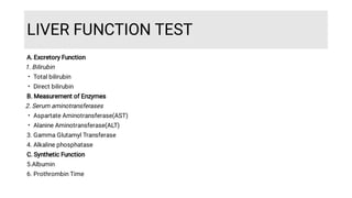
APPROACH TO ABNORMAL
LIVER FUNCTION TEST
DR SOURAV PATRA
GUIDED BY: PROFESSOR
DR SOUMYA SARATHI MONDAL
DEPARTMENT OF GENERAL MEDICINE
MEDICAL COLLEGE, KOLKATA
OVERVIEW
•
•
•
Liver- Perform various kinds of biochemical ,synthetic ,excretory
functions
No single test can detect global liver dysfunction
Clinical history, physical examination most important in
interpretation of liver function test
History
•
•
•
•
•
•
Exposure to any chemical or medication (prescription/OTC/
herbal)
Accompanying symptoms: fever, jaundice, arthralgias, myalgias,
rash, anorexia, weight loss, abdominal pain, pruritus, blood in
stool.
Parenteral exposures: Blood transfusions, IV drug use, tattoos
High risk sexual behaviour
Alcohol consumption
Occupational exposure to hepatotoxins
LIVER FUNCTION TEST
•
•
•
•
A. Excretory Function
1. Bilirubin
Total bilirubin
Direct bilirubin
B. Measurement of Enzymes
2. Serum aminotransferases
Aspartate Aminotransferase(AST)
Alanine Aminotransferase(ALT)
3. Gamma Glutamyl Transferase
4. Alkaline phosphatase
C. Synthetic Function
5.Albumin
6. Prothrombin Time
Uses
•
•
•
1. Detect the presence of liver disease
2. Distinguish among different types of liver disorders
Hepatocellular
Cholestatic
Mixed
3. Gauge the extent of known liver disease
4. Follow the response to treatment
Patterns of abnormal Liver Function TEST
Pattern Cause
AST , ALT > ALP
Bilirubin
Hepatocellular Injury
ALP > AST , ALT
Bilirubin
Cholestasis
Albumin Chronic process
Albumin Acute process
PT/INR Vitamin K deficiency, Obstructive Jaundice
R value
•
•
•
•
•
•
To differentiate hepatocellular-mixed-cholestatic pattern
R = (ALT value / ALT ULN) ÷ (ALP value / ALP ULN)
R value:
>5 hepatocellular pattern
Between 2-5 mixed pattern
<2 cholestatic pattern
Hepatocellular Damage
Markers of Hepatocellular Damage
•
•
•
•
•
•
•
•
•
ALT & AST most frequently used
Released when hepatocytes damaged
Normal values:
ALT: <35 IU/L (Clinical biochemistry laboratory, MCK)
AST: <45 IU/L (Clinical biochemistry laboratory, MCK)
Patterns to note:
AST:ALT ratio
Level of elevation
Rate of decline
ALT AST
Cytosol
• Cytosol 20%, mitochondria 80%
•
More specific for liver
• Other tissues: Cardiac muscle, smooth muscle, kidney,
brain, pancreas, lung, leucocytes, erythrocytes
•
SGPT (Serum Glutamic Pyruvate Transaminase) SGOT (Serum Glutamic Oxaloacetic Transaminase)
Ratio of AST to ALT
A.
•
•
The ratio of AST to ALT in serum is helpful in few specific circumstances--
Alcoholic liver disease: AST level is less than 300 U/L , a ratio –
>2 suggests alcohol associated liver disease
>3 highly suggestive of alcohol- associated liver disease
B. <1: Chronic viral hepatitis and NAFLD. As cirrhosis develops the ratio rises and may
become greater than 1
Level of Elevation
Serum aminotransferase levels are typically elevated in all forms of liver injury
Chronic Mild Elevations, ALT>AST(
<150U/L 0r 5 x Normal)
Hepatic Nonhepatic
Wilson Disease Celiac disease
Autoimmune hepatitis Hyperthyroidism
Chronic viral hepatitis(hepatitis B, hepatitis C)
Hemochromatosis
Steatosis
Alpha1-Antitrypsin deficiency
Severe, Acute Elevations,
ALT>AST(>1000U/L or >20-25 x Normal)
•
•
•
Acute Viral Hepatitis
Drugs and Toxins
Ischemic Hepatitis
Severe Acute Elevations, AST>ALT
(>1000U/L or >20-25 x Normal)
Hepatic Nonhepatic
Medications or toxin in a patient with underlying
alcohol-associated liver injury
Acute rhabdomyolysis
Chronic mild Elevations, AST>ALT(<300
U/L)
Hepatic Non hepatic
AST/ALT > 2 is suggestive and >3 is highly
suggestive of alcohol associated liver disease
Strenuous exercise
Cirrhosis
Rate of Decline
Fast Slow
Drugs Acute Viral Hepatitis
Ischemic Hepatitis Autoimmune Hepatitis
Acute biliary tract obstruction Metabolic Disorders
Elevation of serum Aminotransferase
levels
Drug history, including
Ayurvedic and
homeopathy,
Alcohol history
Stop potential toxins
Follow response of
Aminotransferase
levels
Measure CK to
exclude muscle
injury
Scrub typhus Ig M
antibody test
HBsAg, Anti-HCV, anti HAV
Anti HEV antibody
History of fever
jaundice
HCV RNA
Anti-
HCV+
HBV DNA ,HBeAg
HBsAg+
+drug
history
Continued--
Iron profile,
ANA,SMA,LKM-1
Ceruloplasmin
Ultrasonography
Alpha-AT phenotype
TTG antibody
HFE gene
testing
Liver Biopsy
Liver Biopsy
ANA+,SMA+,
Globulin
Increased iron,
increased ferritin
Ophthalmoscopic
examination
24-hr urine copper
Liver biopsy
Recommend
Weight loss
Liver biopsy if
aminotransferase
levels elevated >2
times ULN
Fatty liver
on USG
Ceruloplasmi
n
Evaluation
unrevealing
Evaluation
unrevealing
Cholestasis
Alkaline Phosphatase (ALP)
•
•
•
•
•
•
•
•
Marker of cholestasis
Half life: 7days
Sources:
Canalicular membrane of hepatocytes
Osteoblasts
Gut
Kidney
Placenta
Raised ALP Causes
Physiologic Causes Pathologic Causes
Age > 60 years Intrahepatic cholestasis
Children, adolescents Extrahepatic cholestasis
Pregnancy
Blood group O and B
Post fatty meal
Raised ALP: Pathologic Causes
Intra hepatic obstruction Extra hepatic obstruction
Drugs Intraluminal: Choledocholithiasis
Sepsis Bile duct wall: Cholangiocarcinoma
Infiltrative liver disease Extraluminal: Pancreatic cancer, metastases,
Mirizzi syndrome
Primary Biliary Cholangitis
Primary Sclerosing Cholangitis
Infections (Brucellosis, Tb, histoplasmosis)
Gamma Glutamyl Transpeptidase(GGTP)
•
•
•
•
•
•
Sources:Liver, Kidney, pancreas, spleen ,Heart, Brain and seminal vesicles
The primary use of serum GGTP level is to identify the source of an isolated elevation in the serum
ALP level
Not found in bone and not raised in pregnancy
Serum level are not different between men and women
Drugs like antiepileptics, barbiturates and some antiretroviral drugs elevates GGTP
Serum GGTP levels are elevated in alcoholics
Evaluation of Alkaline phosphatase
elevation with biochemical and radiological
investigations Fractionate ALP or
measure GGTP Bone Evaluation
Bone origin
Ultrasonography
Liver
origin
CT, ERCP
Dilated
intrahepatic
Bile ducts
Drug history, viral
serologies
AMA, ACE level
Intrahepatic
Bile ducts
Not dilated
Liver
Biopsy
MRCP
Liver Biopsy
AMA+ Evaluation
negative
Biosynthetic Function
Albumin
•
•
•
•
•
•
Albumin is the most important plasma protein, accounts for 75% of the plasma colloid
oncotic pressure
Synthesis: 15g/day
Half life: 14 -21 days
The long half life of albumin in serum accounts for its unreliability as a marker of hepatic
synthetic function in acute liver injury.
Not specific:
Depends on nutrition, volume status, vascular integrity, catabolism, stool/urine losses
Prothrombin time
•
•
•
•
•
•
Measurement of the rate at which prothrombin is converted to thrombin
The extrinsic pathway depends on the activity of clotting factors II, V, VI
& X– all of which are synthesized in liver.
Therefore prothrombin Time is the reflection of synthetic function of
liver
In acute liver diseases, it is a reliable indicator of synthetic function due
to shorter half lives of the clotting factors.
Prothrombin Time more than 5 second of control is poor prognostic
sign in acute viral hepatitis and other acute and chronic liver disease
PT(II,V,VII,X) is dependent on vit K, can be increased in vit K
deficiency(obstructive jaundice or fat malabsorption)
Bilirubin
•
•
•
•
Normal value of total bilirubin is (.1- 1.5) mg/dl and half life 4 hrs
If direct bilirubin is less than 15% of total, the bilirubin can be considered
to be entirely indirect
Increase in total bilirubin level correlates with poor outcomes in
alcoholic hepatitis and greater hepatocellular damage in viral hepatitis
It is a critical component of MELD score, which is used to estimate
survival of patients with end-stage liver disease
Direct Bilirubin
•
•
•
•
•
Conjugated
Water soluble
Polar
Seen in urine
Elevated with biliary
obstruction and
hepatocellular disease
Indirect Bilirubin
•
•
•
•
•
Unconjugated
Lipid soluble
Non-polar
Not in urine
Elevated with haemolysis,
ineffective erythropoiesis ,
drugs
Causes of isolated hyper bilirubinaemia
•
1.
•
•
•
2.
•
3.
•
•
•
A Unconjugated
Increased bilirubin production
Haemolysis
Ineffective erythropoiesis
Hypersplenism
Decreased hepatic uptake
Drugs-rifampicin
Decreased conjugation
Gilbert’s syndrome
Criggler-Najjar syndrome
B Conjugated
1. Dubin-Johnson syndrome
2. Rotor’s syndrome
Limitations
•
•
•
•
Lack sensitivity ( may be normal in cirrhosis )
Lack specificity ( aminotransferase levels may be elevated in
musculoskeletal or cardiac disease)
Aminotransferases and ALP do not measure liver function at all,
rather they detect liver cell damage or interference with the bile flow
Result suggest general category of liver disease, not a specific
diagnosis
•
•
•
Interpretation of laboratory values in patients with
abnormalities in liver function testing is critical to developing
a differential diagnosis and initiation an adequate work-up.
The initial step is determination of an acute, chronic or acute-
on-chronic process
Upon establishment of disorder, the role of us is to establish
the severity of hepatic dysfunction, potential for reversibility
and the need for treatment
Conclusio
n
Sources
THANK YOU

DOC-20230920-WA0002..pdf

  • 1.
    APPROACH TO ABNORMAL LIVERFUNCTION TEST DR SOURAV PATRA GUIDED BY: PROFESSOR DR SOUMYA SARATHI MONDAL DEPARTMENT OF GENERAL MEDICINE MEDICAL COLLEGE, KOLKATA
  • 2.
    OVERVIEW • • • Liver- Perform variouskinds of biochemical ,synthetic ,excretory functions No single test can detect global liver dysfunction Clinical history, physical examination most important in interpretation of liver function test
  • 3.
    History • • • • • • Exposure to anychemical or medication (prescription/OTC/ herbal) Accompanying symptoms: fever, jaundice, arthralgias, myalgias, rash, anorexia, weight loss, abdominal pain, pruritus, blood in stool. Parenteral exposures: Blood transfusions, IV drug use, tattoos High risk sexual behaviour Alcohol consumption Occupational exposure to hepatotoxins
  • 4.
    LIVER FUNCTION TEST • • • • A.Excretory Function 1. Bilirubin Total bilirubin Direct bilirubin B. Measurement of Enzymes 2. Serum aminotransferases Aspartate Aminotransferase(AST) Alanine Aminotransferase(ALT) 3. Gamma Glutamyl Transferase 4. Alkaline phosphatase C. Synthetic Function 5.Albumin 6. Prothrombin Time
  • 5.
    Uses • • • 1. Detect thepresence of liver disease 2. Distinguish among different types of liver disorders Hepatocellular Cholestatic Mixed 3. Gauge the extent of known liver disease 4. Follow the response to treatment
  • 6.
    Patterns of abnormalLiver Function TEST Pattern Cause AST , ALT > ALP Bilirubin Hepatocellular Injury ALP > AST , ALT Bilirubin Cholestasis Albumin Chronic process Albumin Acute process PT/INR Vitamin K deficiency, Obstructive Jaundice
  • 7.
    R value • • • • • • To differentiatehepatocellular-mixed-cholestatic pattern R = (ALT value / ALT ULN) ÷ (ALP value / ALP ULN) R value: >5 hepatocellular pattern Between 2-5 mixed pattern <2 cholestatic pattern
  • 8.
  • 9.
    Markers of HepatocellularDamage • • • • • • • • • ALT & AST most frequently used Released when hepatocytes damaged Normal values: ALT: <35 IU/L (Clinical biochemistry laboratory, MCK) AST: <45 IU/L (Clinical biochemistry laboratory, MCK) Patterns to note: AST:ALT ratio Level of elevation Rate of decline
  • 10.
    ALT AST Cytosol • Cytosol20%, mitochondria 80% • More specific for liver • Other tissues: Cardiac muscle, smooth muscle, kidney, brain, pancreas, lung, leucocytes, erythrocytes • SGPT (Serum Glutamic Pyruvate Transaminase) SGOT (Serum Glutamic Oxaloacetic Transaminase)
  • 11.
    Ratio of ASTto ALT A. • • The ratio of AST to ALT in serum is helpful in few specific circumstances-- Alcoholic liver disease: AST level is less than 300 U/L , a ratio – >2 suggests alcohol associated liver disease >3 highly suggestive of alcohol- associated liver disease B. <1: Chronic viral hepatitis and NAFLD. As cirrhosis develops the ratio rises and may become greater than 1
  • 12.
    Level of Elevation Serumaminotransferase levels are typically elevated in all forms of liver injury
  • 13.
    Chronic Mild Elevations,ALT>AST( <150U/L 0r 5 x Normal) Hepatic Nonhepatic Wilson Disease Celiac disease Autoimmune hepatitis Hyperthyroidism Chronic viral hepatitis(hepatitis B, hepatitis C) Hemochromatosis Steatosis Alpha1-Antitrypsin deficiency
  • 14.
    Severe, Acute Elevations, ALT>AST(>1000U/Lor >20-25 x Normal) • • • Acute Viral Hepatitis Drugs and Toxins Ischemic Hepatitis
  • 15.
    Severe Acute Elevations,AST>ALT (>1000U/L or >20-25 x Normal) Hepatic Nonhepatic Medications or toxin in a patient with underlying alcohol-associated liver injury Acute rhabdomyolysis
  • 16.
    Chronic mild Elevations,AST>ALT(<300 U/L) Hepatic Non hepatic AST/ALT > 2 is suggestive and >3 is highly suggestive of alcohol associated liver disease Strenuous exercise Cirrhosis
  • 17.
    Rate of Decline FastSlow Drugs Acute Viral Hepatitis Ischemic Hepatitis Autoimmune Hepatitis Acute biliary tract obstruction Metabolic Disorders
  • 18.
    Elevation of serumAminotransferase levels Drug history, including Ayurvedic and homeopathy, Alcohol history Stop potential toxins Follow response of Aminotransferase levels Measure CK to exclude muscle injury Scrub typhus Ig M antibody test HBsAg, Anti-HCV, anti HAV Anti HEV antibody History of fever jaundice HCV RNA Anti- HCV+ HBV DNA ,HBeAg HBsAg+ +drug history
  • 19.
    Continued-- Iron profile, ANA,SMA,LKM-1 Ceruloplasmin Ultrasonography Alpha-AT phenotype TTGantibody HFE gene testing Liver Biopsy Liver Biopsy ANA+,SMA+, Globulin Increased iron, increased ferritin Ophthalmoscopic examination 24-hr urine copper Liver biopsy Recommend Weight loss Liver biopsy if aminotransferase levels elevated >2 times ULN Fatty liver on USG Ceruloplasmi n Evaluation unrevealing Evaluation unrevealing
  • 20.
  • 21.
    Alkaline Phosphatase (ALP) • • • • • • • • Markerof cholestasis Half life: 7days Sources: Canalicular membrane of hepatocytes Osteoblasts Gut Kidney Placenta
  • 22.
    Raised ALP Causes PhysiologicCauses Pathologic Causes Age > 60 years Intrahepatic cholestasis Children, adolescents Extrahepatic cholestasis Pregnancy Blood group O and B Post fatty meal
  • 23.
    Raised ALP: PathologicCauses Intra hepatic obstruction Extra hepatic obstruction Drugs Intraluminal: Choledocholithiasis Sepsis Bile duct wall: Cholangiocarcinoma Infiltrative liver disease Extraluminal: Pancreatic cancer, metastases, Mirizzi syndrome Primary Biliary Cholangitis Primary Sclerosing Cholangitis Infections (Brucellosis, Tb, histoplasmosis)
  • 24.
    Gamma Glutamyl Transpeptidase(GGTP) • • • • • • Sources:Liver,Kidney, pancreas, spleen ,Heart, Brain and seminal vesicles The primary use of serum GGTP level is to identify the source of an isolated elevation in the serum ALP level Not found in bone and not raised in pregnancy Serum level are not different between men and women Drugs like antiepileptics, barbiturates and some antiretroviral drugs elevates GGTP Serum GGTP levels are elevated in alcoholics
  • 25.
    Evaluation of Alkalinephosphatase elevation with biochemical and radiological investigations Fractionate ALP or measure GGTP Bone Evaluation Bone origin Ultrasonography Liver origin CT, ERCP Dilated intrahepatic Bile ducts Drug history, viral serologies AMA, ACE level Intrahepatic Bile ducts Not dilated Liver Biopsy MRCP Liver Biopsy AMA+ Evaluation negative
  • 26.
  • 27.
    Albumin • • • • • • Albumin is themost important plasma protein, accounts for 75% of the plasma colloid oncotic pressure Synthesis: 15g/day Half life: 14 -21 days The long half life of albumin in serum accounts for its unreliability as a marker of hepatic synthetic function in acute liver injury. Not specific: Depends on nutrition, volume status, vascular integrity, catabolism, stool/urine losses
  • 28.
    Prothrombin time • • • • • • Measurement ofthe rate at which prothrombin is converted to thrombin The extrinsic pathway depends on the activity of clotting factors II, V, VI & X– all of which are synthesized in liver. Therefore prothrombin Time is the reflection of synthetic function of liver In acute liver diseases, it is a reliable indicator of synthetic function due to shorter half lives of the clotting factors. Prothrombin Time more than 5 second of control is poor prognostic sign in acute viral hepatitis and other acute and chronic liver disease PT(II,V,VII,X) is dependent on vit K, can be increased in vit K deficiency(obstructive jaundice or fat malabsorption)
  • 29.
    Bilirubin • • • • Normal value oftotal bilirubin is (.1- 1.5) mg/dl and half life 4 hrs If direct bilirubin is less than 15% of total, the bilirubin can be considered to be entirely indirect Increase in total bilirubin level correlates with poor outcomes in alcoholic hepatitis and greater hepatocellular damage in viral hepatitis It is a critical component of MELD score, which is used to estimate survival of patients with end-stage liver disease
  • 30.
    Direct Bilirubin • • • • • Conjugated Water soluble Polar Seenin urine Elevated with biliary obstruction and hepatocellular disease Indirect Bilirubin • • • • • Unconjugated Lipid soluble Non-polar Not in urine Elevated with haemolysis, ineffective erythropoiesis , drugs
  • 31.
    Causes of isolatedhyper bilirubinaemia • 1. • • • 2. • 3. • • • A Unconjugated Increased bilirubin production Haemolysis Ineffective erythropoiesis Hypersplenism Decreased hepatic uptake Drugs-rifampicin Decreased conjugation Gilbert’s syndrome Criggler-Najjar syndrome B Conjugated 1. Dubin-Johnson syndrome 2. Rotor’s syndrome
  • 33.
    Limitations • • • • Lack sensitivity (may be normal in cirrhosis ) Lack specificity ( aminotransferase levels may be elevated in musculoskeletal or cardiac disease) Aminotransferases and ALP do not measure liver function at all, rather they detect liver cell damage or interference with the bile flow Result suggest general category of liver disease, not a specific diagnosis
  • 34.
    • • • Interpretation of laboratoryvalues in patients with abnormalities in liver function testing is critical to developing a differential diagnosis and initiation an adequate work-up. The initial step is determination of an acute, chronic or acute- on-chronic process Upon establishment of disorder, the role of us is to establish the severity of hepatic dysfunction, potential for reversibility and the need for treatment Conclusio n
  • 35.
  • 36.