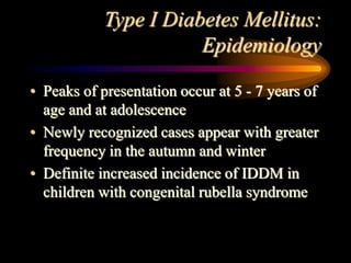
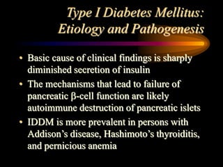
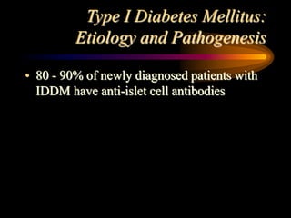
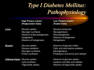
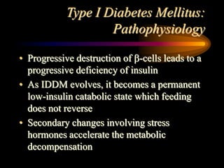
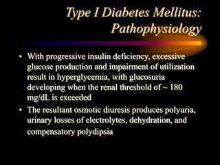
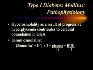
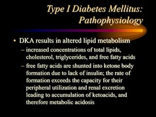
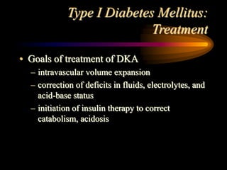
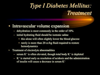
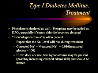
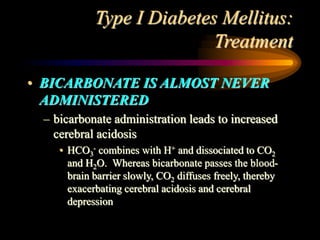
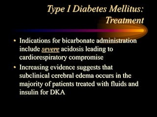
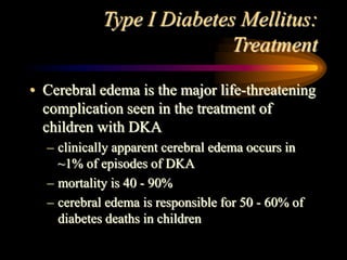
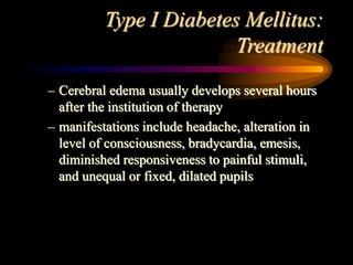
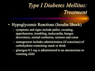
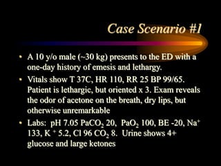
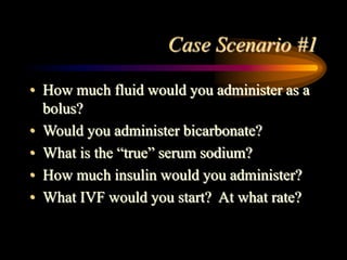
Diabetic Ketoacidosis
Gary David Goulin, MD
Goals & Objectives
• Understand the action of insulin on the
metabolism of carbohydrates, protein, and
fat
• Understand the pathophysiology of IDDM
and DKA
• Understand the management approach to
the patient with DKA
• Appreciate the complications that can occur
during treatment of DKA
Introduction
• Diabetes mellitus is a syndrome of
disturbed energy homeostasis caused by a
deficiency of insulin or of its action
resulting in abnormal metabolism of
carbohydrate, protein, and fat
• Diabetes mellitus is the most common
endocrine-metabolic disorder of childhood
and adolescence
Introduction
• Individuals affected by insulin-dependent
diabetes confront serious burdens that
include an absolute daily requirement for
exogenous insulin, the need to monitor their
own metabolic control, and the need to pay
constant attention to dietary intake
Introduction
• Morbidity and mortality stem from
metabolic derangements and from long-
term complications that affect small and
large vessels and result in retinopathy,
nephropathy, neuropathy, ischemic heart
disease, and arterial obstruction with
gangrene of the extremities
Classification
• Type I Diabetes (insulin-dependent diabetes
mellitus, IDDM)
– characterized by severe insulinopenia and
dependence on exogenous insulin to prevent
ketosis and to preserve life
– onset occurs predominantly in childhood
– probably has some genetic predisposition and is
likely autoimmune-mediated
Classification
• Type II Diabetes (non-insulin-dependent
diabetes mellitus, NIDDM)
– patients are not insulin dependent and rarely develop ketosis
– generally occurs after age 40, and there is a high incidence of
associated obesity
– As the prevalence of childhood obesity increases, more adolescents
are presenting with NIDDM
– insulin secretion is generally adequate; insulin resistance is present
– no associated genetic predisposition
Classification
• Secondary Diabetes
– occurs in response to other disease processes:
• exocrine pancreatic disease (cystic fibrosis)
• Cushing syndrome
• poison ingestion (rodenticides)
Type I Diabetes Mellitus:
Epidemiology
• Prevalence of IDDM among school-age
children in the US is 1.9 per 1000
• The annual incidence in the US is about 12 -
15 new cases per 100,000
• Male to female ratio is equal
• Among African-Americans, the occurrence
of IDDM is about 20 - 30% of that seen in
Caucasian-Americans
Type I Diabetes Mellitus:
Epidemiology
• Peaks of presentation occur at 5 - 7 years of
age and at adolescence
• Newly recognized cases appear with greater
frequency in the autumn and winter
• Definite increased incidence of IDDM in
children with congenital rubella syndrome
Type I Diabetes Mellitus:
Etiology and Pathogenesis
• Basic cause of clinical findings is sharply
diminished secretion of insulin
• The mechanisms that lead to failure of
pancreatic -cell function are likely
autoimmune destruction of pancreatic islets
• IDDM is more prevalent in persons with
Addison’s disease, Hashimoto’s thyroiditis,
and pernicious anemia
Type I Diabetes Mellitus:
Etiology and Pathogenesis
• 80 - 90% of newly diagnosed patients with
IDDM have anti-islet cell antibodies
Type I Diabetes Mellitus:
Pathophysiology
High Plasma Insulin Low Plasma Insulin
(Postprandial State) (Fasted State)
Liver: Glucose uptake Glucose production
Glycogen synthesis Glycogenolysis
Absence of gluconeogenesis Gluconeogenesis
Lipogenesis Absence of lipogenesis
Absence of ketogenesis Ketogenesis
Muscle: Glucose uptake Absence of glucose intake
Glucose oxidation Fatty acid and ketone oxidation
Glycogen synthesis Glycogenolyss
Protein synthesis Proteolysis and amino acid release
Adipose tissue: Glucose uptake Absence of glucose uptake
Lipid synthesis Lipolysis and fatty acid release
Triglyceride uptake Absence of triglyceride uptake
Type I Diabetes Mellitus:
Pathophysiology
• Progressive destruction of -cells leads to a
progressive deficiency of insulin
• As IDDM evolves, it becomes a permanent
low-insulin catabolic state which feeding
does not reverse
• Secondary changes involving stress
hormones accelerate the metabolic
decompensation
Type I Diabetes Mellitus:
Pathophysiology
• With progressive insulin deficiency, excessive
glucose production and impairment of utilization
result in hyperglycemia, with glucosuria
developing when the renal threshold of ~ 180
mg/dL is exceeded
• The resultant osmotic diuresis produces polyuria,
urinary losses of electrolytes, dehydration, and
compensatory polydipsia
Type I Diabetes Mellitus:
Pathophysiology
• Hyperosmolality as a result of progressive
hyperglycemia contributes to cerebral
obtundation in DKA
• Serum osmolality:
– {Serum Na+ + K+} x 2 + glucose + BUN
18 3
Type I Diabetes Mellitus:
Pathophysiology
• DKA results in altered lipid metabolism
– increased concentrations of total lipids,
cholesterol, triglycerides, and free fatty acids
– free fatty acids are shunted into ketone body
formation due to lack of insulin; the rate of
formation exceeds the capacity for their
peripheral utilization and renal excretion
leading to accumulation of ketoacids, and
therefore metabolic acidosis
Type I Diabetes Mellitus:
Pathophysiology
• With progressive dehydration, acidosis,
hyperosmolality, and diminished cerebral
oxygen utilization, consciousness becomes
impaired, and the patient ultimately
becomes comatose
Type I Diabetes Mellitus:
Clinical Manifestations
• Classic presentation of diabetes in children
is a history of polyuria, polydipsia,
polyphagia, and weight loss, usually for up
to one month
• Laboratory findings include glucosuria,
ketonuria, hyperglycemia, ketonemia, and
metabolic acidosis. Serum amylase may be
elevated. Leukocytosis is common
Type I Diabetes Mellitus:
Clinical Manifestations
• Keotacidosis is responsible for the initial
presentation of IDDM in up to 25% of
children
– early manifestations are mild and include vomiting,
polyuria, and dehydration
– More severe cases include Kussmaul respirations, odor
of acetone on the breath
– abdominal pain or rigidity may be present and mimic
acute appendicitis or pancreatitis
– cerebral obtundation and coma ultimately ensue
Type I Diabetes Mellitus:
Diagnosis
• Diagnosis of IDDM is dependent on the
demonstration of hyperglycemia in
association with glucosuria with or without
ketonuria
• DKA must be differentiated from acidosis
and coma due to other causes:
– hypoglycemia, uremia, gastroenteritis with
metabolic acidosis, lactic acidosis, salicylate
intoxication, encephalitis
Type I Diabetes Mellitus:
Diagnosis
• DKA exists when there is hyperglycemia (>
300 mg/dL), ketonemia, acidosis,
glucosuria, and ketonuria
Type I Diabetes Mellitus:
Treatment
• Treatment is divided into 3 phases
– treatment of ketoacidosis
– transition period
– continuing phase and guidance
Type I Diabetes Mellitus:
Treatment
• Goals of treatment of DKA
– intravascular volume expansion
– correction of deficits in fluids, electrolytes, and
acid-base status
– initiation of insulin therapy to correct
catabolism, acidosis
Type I Diabetes Mellitus:
Treatment
• Intravascular volume expansion
– dehydration is most commonly in the order of 10%
– initial hydrating fluid should be isotonic saline
• this alone will often slightly lower the blood glucose
• rarely is more than 20 cc/kg fluid required to restore
hemodynamics
• Treatment of electrolyte abnormalities
– serum K+ is often elevated, though total body K+ is depleted
– K+ is started early as resolution of acidosis and the administration
of insulin will cause a decrease in serum K+
Type I Diabetes Mellitus:
Treatment
• Phosphate is depleted as well. Phosphate may be added as
KPO4 especially if serum chloride becomes elevated
• “Pseudohyponatremia” is often present
– Expect that the Na+ level will rise during treatment
– Corrected Na+ = Measured Na+ + 0.016(measured
glucose - 100)
– If Na+ does not rise, true hyponatremia may be present
(possibly increasing cerebral edema risk) and should be
treated
Type I Diabetes Mellitus:
Treatment
• BICARBONATE IS ALMOST NEVER
ADMINISTERED
– bicarbonate administration leads to increased
cerebral acidosis
• HCO3
- combines with H+ and dissociated to CO2
and H2O. Whereas bicarbonate passes the blood-
brain barrier slowly, CO2 diffuses freely, thereby
exacerbating cerebral acidosis and cerebral
depression
Type I Diabetes Mellitus:
Treatment
• Indications for bicarbonate administration
include severe acidosis leading to
cardiorespiratory compromise
• Increasing evidence suggests that
subclinical cerebral edema occurs in the
majority of patients treated with fluids and
insulin for DKA
Type I Diabetes Mellitus:
Treatment
• Cerebral edema is the major life-threatening
complication seen in the treatment of
children with DKA
– clinically apparent cerebral edema occurs in
~1% of episodes of DKA
– mortality is 40 - 90%
– cerebral edema is responsible for 50 - 60% of
diabetes deaths in children
Type I Diabetes Mellitus:
Treatment
– Cerebral edema usually develops several hours
after the institution of therapy
– manifestations include headache, alteration in
level of consciousness, bradycardia, emesis,
diminished responsiveness to painful stimuli,
and unequal or fixed, dilated pupils
Type I Diabetes Mellitus:
Treatment
• Excessive use of fluids, large doses of insulin, and
especially the use of bicarbonate have been linked to the
increased formation of cerebral edema
– fluids are generally limited to ~ 3 L/m2/24 hours
• Children who present with elevated BUN, PaCO2 < 15 torr,
or who demonstrate a lack of an increase in serum Na+
during therapy have an increased probability of cerebral
edema
• Therapy of cerebral edema may include treatment with
mannitol, hypertonic saline and hyperventilation
Type I Diabetes Mellitus:
Treatment
• Insulin Therapy
– continuous infusion of low-dose insulin IV (~
0.1 U/kg/hr) is effective, simple, and
physiologically sound
– goal is to slowly decrease serum glucose (< 100
mg/dL/hr
– frequent laboratory and blood gas analyses are
obtained to ensure ongoing resolution of
metabolic acidosis
Type I Diabetes Mellitus:
Treatment
• “Maintenance” IV fluid at a rate of 2000 -
2400 cc/m2/day consists of 2/3 NS (0.66%)
or NS
– 5% Dextrose is added to IVF when blood
glucose is ~ 300 mg/dL
– 10% Dextrose is added when blood glucose is ~
200 mg/dL
Type I Diabetes Mellitus:
Treatment
• Insulin is used to treat acidosis, not hyperglycemia
– insulin should never be stopped if ongoing acidosis
persists
• When the acidosis is corrected, the continuous insulin
infusion may be discontinued and subcutaneous insulin
initiated
• With this regimen, DKA usually is usually fully corrected
in 36 to 48 hours
Type I Diabetes Mellitus:
Treatment
• Hypoglycemic Reactions (Insulin Shock)
– symptoms and signs include pallor, sweating,
apprehension, trembling, tachycardia, hunger,
drowsiness, mental confusion, seizures and coma
– management includes administration (if conscious) of
carbohydrate-containing snack or drink
– glucagon 0.5 mg is administered to an unconscious or
vomiting child
Suggested Reading
• Glaser N, et al. Risk factors for cerebral edema in children with diabetic ketoacidosis.
NEJM 355;4:264-269.
• Menon RK, Sperling MA. Diabetic Ketoacidosis. In: Fuhrman BP, Zimmerman JJ, ed.
Pediatric Critical Care. Second Edition. St. Louis: Mosby-Year Book, Inc., 1998:844-
52.
• Kohane DS, Tobin JR, Kohane IS. Endocrine, Mineral, and Metabolic Disease in
Pediatric Intensive Care. In: Rogers, ed. Textbook of Pediatric Intensive Care. Third
Edition. Baltimore: Williams & Wilkins, 1996:1261-72.
• Magee MF, Bhatt BA. Management of Decompensated Diabetes: Diabetic
Ketoacidosis and Hyperglycemic Hyperosmolar Syndrome. In: Zaloga GP, Marik P, ed.
Critical Care Clinics: Endocrine and Metabolic Dysfunction Syndromes in the
Critically Ill. Volume 17:1. Philadelphia: W.B. Saunders Company, 2001: 75-106.
Case Scenario #1
• A 10 y/o male (~30 kg) presents to the ED with a
one-day history of emesis and lethargy.
• Vitals show T 37C, HR 110, RR 25 BP 99/65.
Patient is lethargic, but oriented x 3. Exam reveals
the odor of acetone on the breath, dry lips, but
otherwise unremarkable
• Labs: pH 7.05 PaCO2 20, PaO2 100, BE -20, Na+
133, K + 5.2, Cl 96 CO2 8. Urine shows 4+
glucose and large ketones
Case Scenario #1
• How much fluid would you administer as a
bolus?
• Would you administer bicarbonate?
• What is the “true” serum sodium?
• How much insulin would you administer?
• What IVF would you start? At what rate?
Case Scenario #2
• A 4 y/o female in the PICU is undergoing
treatment for new onset IDDM and DKA. She is
on an insulin infusion at 0.1 u/kg/hr, and fluids are
running at 2400 cc/m2/day.
• Over the last hour, she has been complaining
about increasing headache. She is now found to
be unresponsive with bilateral fixed and dilated
pupils, HR is 50 with BP 150/100.
• What is your next step in management?

dka.ppt

  • 1.
  • 2.
    Goals & Objectives •Understand the action of insulin on the metabolism of carbohydrates, protein, and fat • Understand the pathophysiology of IDDM and DKA • Understand the management approach to the patient with DKA • Appreciate the complications that can occur during treatment of DKA
  • 3.
    Introduction • Diabetes mellitusis a syndrome of disturbed energy homeostasis caused by a deficiency of insulin or of its action resulting in abnormal metabolism of carbohydrate, protein, and fat • Diabetes mellitus is the most common endocrine-metabolic disorder of childhood and adolescence
  • 4.
    Introduction • Individuals affectedby insulin-dependent diabetes confront serious burdens that include an absolute daily requirement for exogenous insulin, the need to monitor their own metabolic control, and the need to pay constant attention to dietary intake
  • 5.
    Introduction • Morbidity andmortality stem from metabolic derangements and from long- term complications that affect small and large vessels and result in retinopathy, nephropathy, neuropathy, ischemic heart disease, and arterial obstruction with gangrene of the extremities
  • 6.
    Classification • Type IDiabetes (insulin-dependent diabetes mellitus, IDDM) – characterized by severe insulinopenia and dependence on exogenous insulin to prevent ketosis and to preserve life – onset occurs predominantly in childhood – probably has some genetic predisposition and is likely autoimmune-mediated
  • 7.
    Classification • Type IIDiabetes (non-insulin-dependent diabetes mellitus, NIDDM) – patients are not insulin dependent and rarely develop ketosis – generally occurs after age 40, and there is a high incidence of associated obesity – As the prevalence of childhood obesity increases, more adolescents are presenting with NIDDM – insulin secretion is generally adequate; insulin resistance is present – no associated genetic predisposition
  • 8.
    Classification • Secondary Diabetes –occurs in response to other disease processes: • exocrine pancreatic disease (cystic fibrosis) • Cushing syndrome • poison ingestion (rodenticides)
  • 9.
    Type I DiabetesMellitus: Epidemiology • Prevalence of IDDM among school-age children in the US is 1.9 per 1000 • The annual incidence in the US is about 12 - 15 new cases per 100,000 • Male to female ratio is equal • Among African-Americans, the occurrence of IDDM is about 20 - 30% of that seen in Caucasian-Americans
  • 10.
    Type I DiabetesMellitus: Epidemiology • Peaks of presentation occur at 5 - 7 years of age and at adolescence • Newly recognized cases appear with greater frequency in the autumn and winter • Definite increased incidence of IDDM in children with congenital rubella syndrome
  • 11.
    Type I DiabetesMellitus: Etiology and Pathogenesis • Basic cause of clinical findings is sharply diminished secretion of insulin • The mechanisms that lead to failure of pancreatic -cell function are likely autoimmune destruction of pancreatic islets • IDDM is more prevalent in persons with Addison’s disease, Hashimoto’s thyroiditis, and pernicious anemia
  • 12.
    Type I DiabetesMellitus: Etiology and Pathogenesis • 80 - 90% of newly diagnosed patients with IDDM have anti-islet cell antibodies
  • 13.
    Type I DiabetesMellitus: Pathophysiology High Plasma Insulin Low Plasma Insulin (Postprandial State) (Fasted State) Liver: Glucose uptake Glucose production Glycogen synthesis Glycogenolysis Absence of gluconeogenesis Gluconeogenesis Lipogenesis Absence of lipogenesis Absence of ketogenesis Ketogenesis Muscle: Glucose uptake Absence of glucose intake Glucose oxidation Fatty acid and ketone oxidation Glycogen synthesis Glycogenolyss Protein synthesis Proteolysis and amino acid release Adipose tissue: Glucose uptake Absence of glucose uptake Lipid synthesis Lipolysis and fatty acid release Triglyceride uptake Absence of triglyceride uptake
  • 14.
    Type I DiabetesMellitus: Pathophysiology • Progressive destruction of -cells leads to a progressive deficiency of insulin • As IDDM evolves, it becomes a permanent low-insulin catabolic state which feeding does not reverse • Secondary changes involving stress hormones accelerate the metabolic decompensation
  • 15.
    Type I DiabetesMellitus: Pathophysiology • With progressive insulin deficiency, excessive glucose production and impairment of utilization result in hyperglycemia, with glucosuria developing when the renal threshold of ~ 180 mg/dL is exceeded • The resultant osmotic diuresis produces polyuria, urinary losses of electrolytes, dehydration, and compensatory polydipsia
  • 16.
    Type I DiabetesMellitus: Pathophysiology • Hyperosmolality as a result of progressive hyperglycemia contributes to cerebral obtundation in DKA • Serum osmolality: – {Serum Na+ + K+} x 2 + glucose + BUN 18 3
  • 17.
    Type I DiabetesMellitus: Pathophysiology • DKA results in altered lipid metabolism – increased concentrations of total lipids, cholesterol, triglycerides, and free fatty acids – free fatty acids are shunted into ketone body formation due to lack of insulin; the rate of formation exceeds the capacity for their peripheral utilization and renal excretion leading to accumulation of ketoacids, and therefore metabolic acidosis
  • 18.
    Type I DiabetesMellitus: Pathophysiology • With progressive dehydration, acidosis, hyperosmolality, and diminished cerebral oxygen utilization, consciousness becomes impaired, and the patient ultimately becomes comatose
  • 19.
    Type I DiabetesMellitus: Clinical Manifestations • Classic presentation of diabetes in children is a history of polyuria, polydipsia, polyphagia, and weight loss, usually for up to one month • Laboratory findings include glucosuria, ketonuria, hyperglycemia, ketonemia, and metabolic acidosis. Serum amylase may be elevated. Leukocytosis is common
  • 20.
    Type I DiabetesMellitus: Clinical Manifestations • Keotacidosis is responsible for the initial presentation of IDDM in up to 25% of children – early manifestations are mild and include vomiting, polyuria, and dehydration – More severe cases include Kussmaul respirations, odor of acetone on the breath – abdominal pain or rigidity may be present and mimic acute appendicitis or pancreatitis – cerebral obtundation and coma ultimately ensue
  • 21.
    Type I DiabetesMellitus: Diagnosis • Diagnosis of IDDM is dependent on the demonstration of hyperglycemia in association with glucosuria with or without ketonuria • DKA must be differentiated from acidosis and coma due to other causes: – hypoglycemia, uremia, gastroenteritis with metabolic acidosis, lactic acidosis, salicylate intoxication, encephalitis
  • 22.
    Type I DiabetesMellitus: Diagnosis • DKA exists when there is hyperglycemia (> 300 mg/dL), ketonemia, acidosis, glucosuria, and ketonuria
  • 23.
    Type I DiabetesMellitus: Treatment • Treatment is divided into 3 phases – treatment of ketoacidosis – transition period – continuing phase and guidance
  • 24.
    Type I DiabetesMellitus: Treatment • Goals of treatment of DKA – intravascular volume expansion – correction of deficits in fluids, electrolytes, and acid-base status – initiation of insulin therapy to correct catabolism, acidosis
  • 25.
    Type I DiabetesMellitus: Treatment • Intravascular volume expansion – dehydration is most commonly in the order of 10% – initial hydrating fluid should be isotonic saline • this alone will often slightly lower the blood glucose • rarely is more than 20 cc/kg fluid required to restore hemodynamics • Treatment of electrolyte abnormalities – serum K+ is often elevated, though total body K+ is depleted – K+ is started early as resolution of acidosis and the administration of insulin will cause a decrease in serum K+
  • 26.
    Type I DiabetesMellitus: Treatment • Phosphate is depleted as well. Phosphate may be added as KPO4 especially if serum chloride becomes elevated • “Pseudohyponatremia” is often present – Expect that the Na+ level will rise during treatment – Corrected Na+ = Measured Na+ + 0.016(measured glucose - 100) – If Na+ does not rise, true hyponatremia may be present (possibly increasing cerebral edema risk) and should be treated
  • 27.
    Type I DiabetesMellitus: Treatment • BICARBONATE IS ALMOST NEVER ADMINISTERED – bicarbonate administration leads to increased cerebral acidosis • HCO3 - combines with H+ and dissociated to CO2 and H2O. Whereas bicarbonate passes the blood- brain barrier slowly, CO2 diffuses freely, thereby exacerbating cerebral acidosis and cerebral depression
  • 28.
    Type I DiabetesMellitus: Treatment • Indications for bicarbonate administration include severe acidosis leading to cardiorespiratory compromise • Increasing evidence suggests that subclinical cerebral edema occurs in the majority of patients treated with fluids and insulin for DKA
  • 29.
    Type I DiabetesMellitus: Treatment • Cerebral edema is the major life-threatening complication seen in the treatment of children with DKA – clinically apparent cerebral edema occurs in ~1% of episodes of DKA – mortality is 40 - 90% – cerebral edema is responsible for 50 - 60% of diabetes deaths in children
  • 30.
    Type I DiabetesMellitus: Treatment – Cerebral edema usually develops several hours after the institution of therapy – manifestations include headache, alteration in level of consciousness, bradycardia, emesis, diminished responsiveness to painful stimuli, and unequal or fixed, dilated pupils
  • 31.
    Type I DiabetesMellitus: Treatment • Excessive use of fluids, large doses of insulin, and especially the use of bicarbonate have been linked to the increased formation of cerebral edema – fluids are generally limited to ~ 3 L/m2/24 hours • Children who present with elevated BUN, PaCO2 < 15 torr, or who demonstrate a lack of an increase in serum Na+ during therapy have an increased probability of cerebral edema • Therapy of cerebral edema may include treatment with mannitol, hypertonic saline and hyperventilation
  • 32.
    Type I DiabetesMellitus: Treatment • Insulin Therapy – continuous infusion of low-dose insulin IV (~ 0.1 U/kg/hr) is effective, simple, and physiologically sound – goal is to slowly decrease serum glucose (< 100 mg/dL/hr – frequent laboratory and blood gas analyses are obtained to ensure ongoing resolution of metabolic acidosis
  • 33.
    Type I DiabetesMellitus: Treatment • “Maintenance” IV fluid at a rate of 2000 - 2400 cc/m2/day consists of 2/3 NS (0.66%) or NS – 5% Dextrose is added to IVF when blood glucose is ~ 300 mg/dL – 10% Dextrose is added when blood glucose is ~ 200 mg/dL
  • 34.
    Type I DiabetesMellitus: Treatment • Insulin is used to treat acidosis, not hyperglycemia – insulin should never be stopped if ongoing acidosis persists • When the acidosis is corrected, the continuous insulin infusion may be discontinued and subcutaneous insulin initiated • With this regimen, DKA usually is usually fully corrected in 36 to 48 hours
  • 35.
    Type I DiabetesMellitus: Treatment • Hypoglycemic Reactions (Insulin Shock) – symptoms and signs include pallor, sweating, apprehension, trembling, tachycardia, hunger, drowsiness, mental confusion, seizures and coma – management includes administration (if conscious) of carbohydrate-containing snack or drink – glucagon 0.5 mg is administered to an unconscious or vomiting child
  • 36.
    Suggested Reading • GlaserN, et al. Risk factors for cerebral edema in children with diabetic ketoacidosis. NEJM 355;4:264-269. • Menon RK, Sperling MA. Diabetic Ketoacidosis. In: Fuhrman BP, Zimmerman JJ, ed. Pediatric Critical Care. Second Edition. St. Louis: Mosby-Year Book, Inc., 1998:844- 52. • Kohane DS, Tobin JR, Kohane IS. Endocrine, Mineral, and Metabolic Disease in Pediatric Intensive Care. In: Rogers, ed. Textbook of Pediatric Intensive Care. Third Edition. Baltimore: Williams & Wilkins, 1996:1261-72. • Magee MF, Bhatt BA. Management of Decompensated Diabetes: Diabetic Ketoacidosis and Hyperglycemic Hyperosmolar Syndrome. In: Zaloga GP, Marik P, ed. Critical Care Clinics: Endocrine and Metabolic Dysfunction Syndromes in the Critically Ill. Volume 17:1. Philadelphia: W.B. Saunders Company, 2001: 75-106.
  • 37.
    Case Scenario #1 •A 10 y/o male (~30 kg) presents to the ED with a one-day history of emesis and lethargy. • Vitals show T 37C, HR 110, RR 25 BP 99/65. Patient is lethargic, but oriented x 3. Exam reveals the odor of acetone on the breath, dry lips, but otherwise unremarkable • Labs: pH 7.05 PaCO2 20, PaO2 100, BE -20, Na+ 133, K + 5.2, Cl 96 CO2 8. Urine shows 4+ glucose and large ketones
  • 38.
    Case Scenario #1 •How much fluid would you administer as a bolus? • Would you administer bicarbonate? • What is the “true” serum sodium? • How much insulin would you administer? • What IVF would you start? At what rate?
  • 39.
    Case Scenario #2 •A 4 y/o female in the PICU is undergoing treatment for new onset IDDM and DKA. She is on an insulin infusion at 0.1 u/kg/hr, and fluids are running at 2400 cc/m2/day. • Over the last hour, she has been complaining about increasing headache. She is now found to be unresponsive with bilateral fixed and dilated pupils, HR is 50 with BP 150/100. • What is your next step in management?