Diseases of female genital tract
•Download as PPTX, PDF•
12 likes•8,230 views
The document describes images from a pathology lab showing abnormal tissue from the female genital tract. The images show grayish nodular tissue with enlarged fish eggs and chorionic villi that have undergone hydrophilic degeneration. Additional images show enlarged chorionic villi, syncytiotrophoblast proliferation, hair shafts and a protruding tooth emerging from the wall of a cyst lined with ectodermal stratified squamous epithelium, sweat glands and a hair follicle.
Report
Share
Report
Share

Recommended
Microprop (1)
Micropropagation is the process of multiplying plants in vitro or in laboratory conditions. It allows for rapid multiplication of plant clones from a single explant sample. Micropropagation has several advantages over conventional propagation including producing many propagules quickly in controlled conditions year-round. However, it also has limitations such as being a more equipment-intensive process requiring specialized technical expertise. Micropropagation is useful for rapidly increasing stocks of new varieties, eliminating diseases, and propagating plants with certain growth characteristics.
chlamydia and EYE
This document discusses Chlamydia, an obligate intracellular parasite bacteria. It has two morphological forms, the elementary body and reticulate body, which undergo a developmental cycle within host cells. Three species are highlighted: C. trachomatis which infects humans, C. psittaci which infects birds, and C. pneumoniae. C. trachomatis is further discussed, as it can cause trachoma, a leading cause of preventable blindness. Trachoma is characterized by recurrent conjunctivitis and scarring. The SAFE strategy of surgery, antibiotics, facial cleanliness and environmental improvement is used to treat and prevent trachoma. C. trachomatis can also cause
Ecological relationship
There are different types of ecological relationships between organisms. Commensalism benefits one organism without affecting the other. Mutualism benefits both organisms. Parasitism benefits one organism and harms the host. Predation benefits the predator and harms the prey. Competition occurs when organisms compete for limited resources. Examples of these relationships in nature were provided such as lichens, remora fish, and tapeworms.
Symbiosis Powerpoint Presentation
please give a comment on our Power Point Presentation. Give your suggestion, and recommendation. echos. :)
Relationships in an ecosystem: mutualism, comensalism and parasitism.
This document describes different types of relationships between individuals. Within a species, there are gregarious associations (unorganized groups), families (small related groups), and societies (organized groups with distributed work). Between species there are mutualism (benefits both), parasitism (benefits one at the expense of the other), and commensalism (benefits one without affecting the other). Examples include birds eating ticks off mammals (mutualism), fleas living on mammals (parasitism), and orchids growing on trees (commensalism).
Common Disorders Of Male Female Reproductive Systems Ppt Sept 2006
Common disorders of the male and female reproductive systems include benign prostatic hyperplasia (BHP) and prostate cancer in men, and pelvic inflammatory disease (PID), endometriosis, uterine prolapse, cystocele, rectocele, and ectopic pregnancy in women. BPH causes urinary obstruction and symptoms like difficulty initiating urination and frequent urination. Prostate cancer is usually asymptomatic early on but can cause urinary symptoms, bone pain, and infertility. PID is an infection of the female pelvic cavity that can lead to ectopic pregnancy and infertility if left untreated. Endometriosis involves endometrial tissue growing outside the uterus and causes pelvic pain and infertility.
Applied nutrition 3 rd presentation - diseases of liver, gall bladder, and ...
The document provides detailed information about diseases of the liver, gallbladder, and pancreas. It discusses the anatomy and functions of the liver, signs and symptoms of various hepatitis types, cirrhosis, and hepatic coma. It also covers cholecystitis, including causes, clinical manifestations, and dietary management for related conditions.
Git diseases presentn no. 2 applied nutrition
This document discusses the dietary management of various gastrointestinal diseases including diarrhea, constipation, gastritis, and peptic ulcers. It describes the anatomy and functions of the digestive system. It defines different types of constipation and diarrhea and their causes. Dietary recommendations are provided for different GI conditions, focusing on fluid and fiber intake, meal patterns, and avoiding irritating foods. Medical treatments including drugs and H. pylori eradication therapies are also summarized.
Getting ahead of headaches
The document discusses headaches, their prevalence, types, symptoms, evaluation, treatment and management. It notes that 90% of individuals experience at least one headache per year, with 40% experiencing severe headaches. It describes the most common primary headache types as migraine (16%), tension-type (69%) and cluster (0.1%). Secondary headaches are often caused by infection, injury or vascular issues. Evaluation of headaches involves considering quality, severity, location, duration and time course. Serious underlying causes may present with worst-ever headaches, new headaches in older patients, or symptoms like fever or neurological issues. Treatment involves identifying and avoiding triggers, managing symptoms, and using medications like triptans, NSAIDs or preventive drugs.
Acute brain attack 911
The document provides guidelines for diagnosing and managing different types and severities of acute brain attacks or strokes. It discusses classifying strokes as TIA, mild, moderate or severe based on symptoms. For TIA and mild strokes, the guidelines recommend emergent diagnostic tests like CT scan and treating conditions like high blood pressure. For moderate strokes, the priorities are supportive care, monitoring vitals, diagnostics like blood tests and CT scan. The guidelines provide recommendations for diagnosing the type of stroke and identifying underlying causes through further diagnostic testing.
Hema intro anemia
This document provides an introduction to hematology and summarizes key topics including:
1. The components of blood and cellular elements such as red blood cells, white blood cells, and platelets.
2. Principles of hematologic diagnosis including medical history, physical examination, and laboratory evaluations like complete blood count and peripheral blood smear.
3. Causes of anemia including hypoproliferative anemias like anemia of chronic disease and anemia of renal disease.
4. Aplastic anemia, its definition, epidemiology, etiology including acquired, inherited, and secondary causes.
EXCRETA, SEWAGE, REFUSE DISPOSAL
The document discusses human excreta (feces and urine), sewage, and refuse disposal. It describes:
- Nutrients contained in human excreta and their importance as fertilizer.
- Public health risks of improper excreta and sewage disposal such as disease transmission.
- Approved methods and systems for excreta, sewage, and refuse disposal including pit latrines, septic tanks, and sewage treatment to remove pathogens and nutrients.
EXCRETA, SEWAGE, REFUSE DISPOSAL
The document provides information on excreta, sewage, and refuse disposal. It discusses the nutrients contained in human feces and urine. It describes various methods for the disposal of human excreta including pit latrines, septic tanks, and composting toilets. It also covers the treatment and reuse of sewage and graywater. The document emphasizes the importance of properly disposing of human waste to prevent disease and pollution, while highlighting the potential for waste to be used as a resource.
Fever & id diet final
1. The document discusses dietary recommendations for various infectious diseases and conditions including fever, malaria, emphysema, rheumatic fever, tuberculosis, and others.
2. For fever and infections, the recommendations are to increase calories to meet increased metabolic demands and avoid starvation, increase protein intake to correct nitrogen balance, and increase carbohydrates and fluids.
3. For specific conditions like emphysema, recommendations include high calorie, low carbohydrate, high protein diets as well as vitamin supplements and regular exercise to promote strength and efficiency.
Eating disorders
Up to 4% of adolescents and young adults suffer from eating disorders like anorexia nervosa and bulimia nervosa. Anorexia nervosa is characterized by refusal to maintain a healthy body weight and an intense fear of gaining weight. Bulimia nervosa involves recurrent binge eating and compensatory behaviors like purging. Both disorders involve biological factors like changes in neurotransmitters and hormones as well as psychological and social factors like perfectionism, low self-esteem, and troubled family relationships. They have varying mortality rates, symptoms, comorbidities, and treatments depending on the specific type and characteristics of each case.
Hepatocellular carcinoma
Hepatocellular carcinoma is one of the most common malignancies worldwide. Its incidence varies significantly between regions, from low rates in the United States and Africa to very high rates in parts of Asia. Major risk factors include chronic hepatitis B and C infections. Clinical features can include abdominal pain, weight loss, jaundice, and hepatomegaly. Diagnosis involves blood tests, ultrasound, CT scan, MRI and sometimes liver biopsy. High-risk groups are screened regularly through alpha-fetoprotein testing and ultrasound. Treatment options depend on the stage but may include surgical resection, ablation, chemotherapy, and transplantation.
Eating disorders
Up to 4% of adolescents and young adults suffer from eating disorders like anorexia nervosa and bulimia nervosa. Anorexia nervosa is characterized by refusal to maintain a healthy body weight and an intense fear of gaining weight. Bulimia nervosa involves recurrent binge eating and compensatory behaviors like purging. Both disorders involve biological factors like changes in neurotransmitters and hormones as well as psychological and social factors like perfectionism, low self-esteem, and troubled family relationships. They have varying mortality rates, symptoms, comorbidities, and treatments depending on the specific type and characteristics of each case.
Normal sleep and sleep disorders
This document summarizes the stages and neurophysiology of sleep. It describes the four stages of non-REM sleep characterized by different brain wave patterns. REM sleep is characterized by low voltage mixed frequency brain waves. Various brain regions are involved in regulating the sleep-wake cycle. Sleep serves important restorative functions and deficiencies can cause problems like daytime sleepiness. Insomnia and other sleep disorders are also summarized.
Tic disorder
This document discusses tic disorders such as Tourette's disorder. It defines tics as involuntary muscle contractions or vocalizations. Tourette's is characterized by both multiple motor and at least one vocal tic over a period of more than one year. Onset is before age 18. It occurs more in males than females and is associated with ADHD and OCD. Treatment includes haloperidol. Differential diagnoses include chronic motor or vocal tic disorder and transient tic disorder.
Thyroid
The document discusses the benefits of exercise for mental health. It notes that regular physical activity can help reduce anxiety and depression and improve mood and cognitive function. Exercise has also been shown to enhance self-esteem and serve as a healthy distraction from daily stressors.
Dermatology dr.n.ramos
The document outlines a dermatology syllabus covering various common skin conditions organized into 18 topics. Some of the key conditions discussed include eczema (its classification and types), urticaria, acne/rosacea, psoriasis, infections (bacterial, viral, fungal), sexually transmitted diseases, tumors (benign and malignant), and connective tissue diseases. For each condition, the syllabus provides details on pathogenesis, clinical features, diagnostic criteria where relevant, and treatment approaches.
Tic disorder
This document discusses tic disorders, including Tourette's disorder. It defines tics as involuntary muscle contractions or vocalizations. There are simple and complex motor and vocal tics. Tourette's disorder is characterized by both multiple motor and at least one vocal tic for over a year. Onset is before age 18. It occurs more in males than females and is associated with ADHD and OCD. While the cause is unknown, dopamine and endogenous opioids may play a role. Treatment includes haloperidol. Transient tic disorder involves single or multiple tics for less than a year.
Toxic and drug induced hepatitis
The document discusses various causes of drug-induced liver injury including direct toxicity from drugs like acetaminophen and carbon tetrachloride as well as idiosyncratic reactions. Certain drugs are more likely to cause hepatotoxicity through both direct toxicity and idiosyncratic mechanisms. Supportive treatment measures for acetaminophen overdose-induced liver injury are also outlined. Herbal and dietary supplements can also potentially cause liver injury through mechanisms like pyrrolizidine alkaloid contamination.
Pancreatic disease
The document discusses acute and chronic pancreatitis, including causes such as alcohol abuse, gallstones, and trauma. It describes clinical features such as severe epigastric pain and elevated serum amylase and lipase levels. Diagnostic tests include blood tests, imaging like CT scans and MRCP, and endoscopic ultrasound. Treatment depends on the severity and includes IV fluids, analgesics, antibiotics, and surgery for complications like pseudocysts or obstruction.
Liver disease
This document provides an overview of evaluating and diagnosing liver disease. It discusses classifying liver diseases as hepatocellular, cholestatic, or mixed based on etiology and evaluating disease severity and stage. Common symptoms, diagnostic tests, clinical findings, and classifications such as Child-Pugh staging for cirrhosis are outlined.
Complications of cirrhosis
The document discusses various complications that can arise from cirrhosis of the liver. It describes portal hypertension, ascites, spontaneous bacterial peritonitis, hepatorenal syndrome, hepatic encephalopathy, coagulation abnormalities, and other hematologic abnormalities as common complications. Diagnosis and treatment approaches are mentioned for several of these conditions.
Cirrhosis
This document discusses chronic hepatitis, including its classification by cause and grade/stage. It describes chronic forms of hepatitis B and C, the clinical features and survival rates associated with chronic hepatitis B at different stages of disease. It also discusses cirrhosis, including its clinical features, causes such as alcoholic cirrhosis, and complications. Therapies for various chronic liver conditions are briefly mentioned.
Chronic hep b
This document discusses chronic hepatitis, which is hepatic inflammation and necrosis that continues for at least 6 months. It can be classified by cause such as hepatitis B, C, autoimmune hepatitis, or drug-associated hepatitis. It can also be classified by grade based on the extent of necrosis and inflammation visible on biopsy. Chronic hepatitis B has varying likelihood of chronicity depending on age at infection and may present asymptomatically or with fatigue, jaundice, ascites, or bleeding varices. Laboratory findings include elevated liver enzymes and hypoalbuminemia. Chronic hepatitis B clinical forms are differentiated based on presence of HBeAg and levels of ALT and HBVDNA.
BÀI TẬP DẠY THÊM TIẾNG ANH LỚP 7 CẢ NĂM FRIENDS PLUS SÁCH CHÂN TRỜI SÁNG TẠO ...
BÀI TẬP DẠY THÊM TIẾNG ANH LỚP 7 CẢ NĂM FRIENDS PLUS SÁCH CHÂN TRỜI SÁNG TẠO ...Nguyen Thanh Tu Collection
https://app.box.com/s/qhtvq32h4ybf9t49ku85x0n3xl4jhr15More Related Content
More from MD Specialclass
Getting ahead of headaches
The document discusses headaches, their prevalence, types, symptoms, evaluation, treatment and management. It notes that 90% of individuals experience at least one headache per year, with 40% experiencing severe headaches. It describes the most common primary headache types as migraine (16%), tension-type (69%) and cluster (0.1%). Secondary headaches are often caused by infection, injury or vascular issues. Evaluation of headaches involves considering quality, severity, location, duration and time course. Serious underlying causes may present with worst-ever headaches, new headaches in older patients, or symptoms like fever or neurological issues. Treatment involves identifying and avoiding triggers, managing symptoms, and using medications like triptans, NSAIDs or preventive drugs.
Acute brain attack 911
The document provides guidelines for diagnosing and managing different types and severities of acute brain attacks or strokes. It discusses classifying strokes as TIA, mild, moderate or severe based on symptoms. For TIA and mild strokes, the guidelines recommend emergent diagnostic tests like CT scan and treating conditions like high blood pressure. For moderate strokes, the priorities are supportive care, monitoring vitals, diagnostics like blood tests and CT scan. The guidelines provide recommendations for diagnosing the type of stroke and identifying underlying causes through further diagnostic testing.
Hema intro anemia
This document provides an introduction to hematology and summarizes key topics including:
1. The components of blood and cellular elements such as red blood cells, white blood cells, and platelets.
2. Principles of hematologic diagnosis including medical history, physical examination, and laboratory evaluations like complete blood count and peripheral blood smear.
3. Causes of anemia including hypoproliferative anemias like anemia of chronic disease and anemia of renal disease.
4. Aplastic anemia, its definition, epidemiology, etiology including acquired, inherited, and secondary causes.
EXCRETA, SEWAGE, REFUSE DISPOSAL
The document discusses human excreta (feces and urine), sewage, and refuse disposal. It describes:
- Nutrients contained in human excreta and their importance as fertilizer.
- Public health risks of improper excreta and sewage disposal such as disease transmission.
- Approved methods and systems for excreta, sewage, and refuse disposal including pit latrines, septic tanks, and sewage treatment to remove pathogens and nutrients.
EXCRETA, SEWAGE, REFUSE DISPOSAL
The document provides information on excreta, sewage, and refuse disposal. It discusses the nutrients contained in human feces and urine. It describes various methods for the disposal of human excreta including pit latrines, septic tanks, and composting toilets. It also covers the treatment and reuse of sewage and graywater. The document emphasizes the importance of properly disposing of human waste to prevent disease and pollution, while highlighting the potential for waste to be used as a resource.
Fever & id diet final
1. The document discusses dietary recommendations for various infectious diseases and conditions including fever, malaria, emphysema, rheumatic fever, tuberculosis, and others.
2. For fever and infections, the recommendations are to increase calories to meet increased metabolic demands and avoid starvation, increase protein intake to correct nitrogen balance, and increase carbohydrates and fluids.
3. For specific conditions like emphysema, recommendations include high calorie, low carbohydrate, high protein diets as well as vitamin supplements and regular exercise to promote strength and efficiency.
Eating disorders
Up to 4% of adolescents and young adults suffer from eating disorders like anorexia nervosa and bulimia nervosa. Anorexia nervosa is characterized by refusal to maintain a healthy body weight and an intense fear of gaining weight. Bulimia nervosa involves recurrent binge eating and compensatory behaviors like purging. Both disorders involve biological factors like changes in neurotransmitters and hormones as well as psychological and social factors like perfectionism, low self-esteem, and troubled family relationships. They have varying mortality rates, symptoms, comorbidities, and treatments depending on the specific type and characteristics of each case.
Hepatocellular carcinoma
Hepatocellular carcinoma is one of the most common malignancies worldwide. Its incidence varies significantly between regions, from low rates in the United States and Africa to very high rates in parts of Asia. Major risk factors include chronic hepatitis B and C infections. Clinical features can include abdominal pain, weight loss, jaundice, and hepatomegaly. Diagnosis involves blood tests, ultrasound, CT scan, MRI and sometimes liver biopsy. High-risk groups are screened regularly through alpha-fetoprotein testing and ultrasound. Treatment options depend on the stage but may include surgical resection, ablation, chemotherapy, and transplantation.
Eating disorders
Up to 4% of adolescents and young adults suffer from eating disorders like anorexia nervosa and bulimia nervosa. Anorexia nervosa is characterized by refusal to maintain a healthy body weight and an intense fear of gaining weight. Bulimia nervosa involves recurrent binge eating and compensatory behaviors like purging. Both disorders involve biological factors like changes in neurotransmitters and hormones as well as psychological and social factors like perfectionism, low self-esteem, and troubled family relationships. They have varying mortality rates, symptoms, comorbidities, and treatments depending on the specific type and characteristics of each case.
Normal sleep and sleep disorders
This document summarizes the stages and neurophysiology of sleep. It describes the four stages of non-REM sleep characterized by different brain wave patterns. REM sleep is characterized by low voltage mixed frequency brain waves. Various brain regions are involved in regulating the sleep-wake cycle. Sleep serves important restorative functions and deficiencies can cause problems like daytime sleepiness. Insomnia and other sleep disorders are also summarized.
Tic disorder
This document discusses tic disorders such as Tourette's disorder. It defines tics as involuntary muscle contractions or vocalizations. Tourette's is characterized by both multiple motor and at least one vocal tic over a period of more than one year. Onset is before age 18. It occurs more in males than females and is associated with ADHD and OCD. Treatment includes haloperidol. Differential diagnoses include chronic motor or vocal tic disorder and transient tic disorder.
Thyroid
The document discusses the benefits of exercise for mental health. It notes that regular physical activity can help reduce anxiety and depression and improve mood and cognitive function. Exercise has also been shown to enhance self-esteem and serve as a healthy distraction from daily stressors.
Dermatology dr.n.ramos
The document outlines a dermatology syllabus covering various common skin conditions organized into 18 topics. Some of the key conditions discussed include eczema (its classification and types), urticaria, acne/rosacea, psoriasis, infections (bacterial, viral, fungal), sexually transmitted diseases, tumors (benign and malignant), and connective tissue diseases. For each condition, the syllabus provides details on pathogenesis, clinical features, diagnostic criteria where relevant, and treatment approaches.
Tic disorder
This document discusses tic disorders, including Tourette's disorder. It defines tics as involuntary muscle contractions or vocalizations. There are simple and complex motor and vocal tics. Tourette's disorder is characterized by both multiple motor and at least one vocal tic for over a year. Onset is before age 18. It occurs more in males than females and is associated with ADHD and OCD. While the cause is unknown, dopamine and endogenous opioids may play a role. Treatment includes haloperidol. Transient tic disorder involves single or multiple tics for less than a year.
Toxic and drug induced hepatitis
The document discusses various causes of drug-induced liver injury including direct toxicity from drugs like acetaminophen and carbon tetrachloride as well as idiosyncratic reactions. Certain drugs are more likely to cause hepatotoxicity through both direct toxicity and idiosyncratic mechanisms. Supportive treatment measures for acetaminophen overdose-induced liver injury are also outlined. Herbal and dietary supplements can also potentially cause liver injury through mechanisms like pyrrolizidine alkaloid contamination.
Pancreatic disease
The document discusses acute and chronic pancreatitis, including causes such as alcohol abuse, gallstones, and trauma. It describes clinical features such as severe epigastric pain and elevated serum amylase and lipase levels. Diagnostic tests include blood tests, imaging like CT scans and MRCP, and endoscopic ultrasound. Treatment depends on the severity and includes IV fluids, analgesics, antibiotics, and surgery for complications like pseudocysts or obstruction.
Liver disease
This document provides an overview of evaluating and diagnosing liver disease. It discusses classifying liver diseases as hepatocellular, cholestatic, or mixed based on etiology and evaluating disease severity and stage. Common symptoms, diagnostic tests, clinical findings, and classifications such as Child-Pugh staging for cirrhosis are outlined.
Complications of cirrhosis
The document discusses various complications that can arise from cirrhosis of the liver. It describes portal hypertension, ascites, spontaneous bacterial peritonitis, hepatorenal syndrome, hepatic encephalopathy, coagulation abnormalities, and other hematologic abnormalities as common complications. Diagnosis and treatment approaches are mentioned for several of these conditions.
Cirrhosis
This document discusses chronic hepatitis, including its classification by cause and grade/stage. It describes chronic forms of hepatitis B and C, the clinical features and survival rates associated with chronic hepatitis B at different stages of disease. It also discusses cirrhosis, including its clinical features, causes such as alcoholic cirrhosis, and complications. Therapies for various chronic liver conditions are briefly mentioned.
Chronic hep b
This document discusses chronic hepatitis, which is hepatic inflammation and necrosis that continues for at least 6 months. It can be classified by cause such as hepatitis B, C, autoimmune hepatitis, or drug-associated hepatitis. It can also be classified by grade based on the extent of necrosis and inflammation visible on biopsy. Chronic hepatitis B has varying likelihood of chronicity depending on age at infection and may present asymptomatically or with fatigue, jaundice, ascites, or bleeding varices. Laboratory findings include elevated liver enzymes and hypoalbuminemia. Chronic hepatitis B clinical forms are differentiated based on presence of HBeAg and levels of ALT and HBVDNA.
More from MD Specialclass (20)
Recently uploaded
BÀI TẬP DẠY THÊM TIẾNG ANH LỚP 7 CẢ NĂM FRIENDS PLUS SÁCH CHÂN TRỜI SÁNG TẠO ...
BÀI TẬP DẠY THÊM TIẾNG ANH LỚP 7 CẢ NĂM FRIENDS PLUS SÁCH CHÂN TRỜI SÁNG TẠO ...Nguyen Thanh Tu Collection
https://app.box.com/s/qhtvq32h4ybf9t49ku85x0n3xl4jhr15BÀI TẬP BỔ TRỢ TIẾNG ANH 8 CẢ NĂM - GLOBAL SUCCESS - NĂM HỌC 2023-2024 (CÓ FI...
BÀI TẬP BỔ TRỢ TIẾNG ANH 8 CẢ NĂM - GLOBAL SUCCESS - NĂM HỌC 2023-2024 (CÓ FI...Nguyen Thanh Tu Collection
https://app.box.com/s/y977uz6bpd3af4qsebv7r9b7s21935vdLeveraging Generative AI to Drive Nonprofit Innovation
In this webinar, participants learned how to utilize Generative AI to streamline operations and elevate member engagement. Amazon Web Service experts provided a customer specific use cases and dived into low/no-code tools that are quick and easy to deploy through Amazon Web Service (AWS.)
LAND USE LAND COVER AND NDVI OF MIRZAPUR DISTRICT, UP
This Dissertation explores the particular circumstances of Mirzapur, a region located in the
core of India. Mirzapur, with its varied terrains and abundant biodiversity, offers an optimal
environment for investigating the changes in vegetation cover dynamics. Our study utilizes
advanced technologies such as GIS (Geographic Information Systems) and Remote sensing to
analyze the transformations that have taken place over the course of a decade.
The complex relationship between human activities and the environment has been the focus
of extensive research and worry. As the global community grapples with swift urbanization,
population expansion, and economic progress, the effects on natural ecosystems are becoming
more evident. A crucial element of this impact is the alteration of vegetation cover, which plays a
significant role in maintaining the ecological equilibrium of our planet.Land serves as the foundation for all human activities and provides the necessary materials for
these activities. As the most crucial natural resource, its utilization by humans results in different
'Land uses,' which are determined by both human activities and the physical characteristics of the
land.
The utilization of land is impacted by human needs and environmental factors. In countries
like India, rapid population growth and the emphasis on extensive resource exploitation can lead
to significant land degradation, adversely affecting the region's land cover.
Therefore, human intervention has significantly influenced land use patterns over many
centuries, evolving its structure over time and space. In the present era, these changes have
accelerated due to factors such as agriculture and urbanization. Information regarding land use and
cover is essential for various planning and management tasks related to the Earth's surface,
providing crucial environmental data for scientific, resource management, policy purposes, and
diverse human activities.
Accurate understanding of land use and cover is imperative for the development planning
of any area. Consequently, a wide range of professionals, including earth system scientists, land
and water managers, and urban planners, are interested in obtaining data on land use and cover
changes, conversion trends, and other related patterns. The spatial dimensions of land use and
cover support policymakers and scientists in making well-informed decisions, as alterations in
these patterns indicate shifts in economic and social conditions. Monitoring such changes with the
help of Advanced technologies like Remote Sensing and Geographic Information Systems is
crucial for coordinated efforts across different administrative levels. Advanced technologies like
Remote Sensing and Geographic Information Systems
9
Changes in vegetation cover refer to variations in the distribution, composition, and overall
structure of plant communities across different temporal and spatial scales. These changes can
occur natural.
South African Journal of Science: Writing with integrity workshop (2024)
South African Journal of Science: Writing with integrity workshop (2024)Academy of Science of South Africa
A workshop hosted by the South African Journal of Science aimed at postgraduate students and early career researchers with little or no experience in writing and publishing journal articles.How to deliver Powerpoint Presentations.pptx
"How to make and deliver dynamic presentations by making it more interactive to captivate your audience attention"
PCOS corelations and management through Ayurveda.
This presentation includes basic of PCOS their pathology and treatment and also Ayurveda correlation of PCOS and Ayurvedic line of treatment mentioned in classics.
Wound healing PPT
This document provides an overview of wound healing, its functions, stages, mechanisms, factors affecting it, and complications.
A wound is a break in the integrity of the skin or tissues, which may be associated with disruption of the structure and function.
Healing is the body’s response to injury in an attempt to restore normal structure and functions.
Healing can occur in two ways: Regeneration and Repair
There are 4 phases of wound healing: hemostasis, inflammation, proliferation, and remodeling. This document also describes the mechanism of wound healing. Factors that affect healing include infection, uncontrolled diabetes, poor nutrition, age, anemia, the presence of foreign bodies, etc.
Complications of wound healing like infection, hyperpigmentation of scar, contractures, and keloid formation.
RHEOLOGY Physical pharmaceutics-II notes for B.pharm 4th sem students
Physical pharmaceutics notes for B.pharm students
Chapter wise All Notes of First year Basic Civil Engineering.pptx
Chapter wise All Notes of First year Basic Civil Engineering
Syllabus
Chapter-1
Introduction to objective, scope and outcome the subject
Chapter 2
Introduction: Scope and Specialization of Civil Engineering, Role of civil Engineer in Society, Impact of infrastructural development on economy of country.
Chapter 3
Surveying: Object Principles & Types of Surveying; Site Plans, Plans & Maps; Scales & Unit of different Measurements.
Linear Measurements: Instruments used. Linear Measurement by Tape, Ranging out Survey Lines and overcoming Obstructions; Measurements on sloping ground; Tape corrections, conventional symbols. Angular Measurements: Instruments used; Introduction to Compass Surveying, Bearings and Longitude & Latitude of a Line, Introduction to total station.
Levelling: Instrument used Object of levelling, Methods of levelling in brief, and Contour maps.
Chapter 4
Buildings: Selection of site for Buildings, Layout of Building Plan, Types of buildings, Plinth area, carpet area, floor space index, Introduction to building byelaws, concept of sun light & ventilation. Components of Buildings & their functions, Basic concept of R.C.C., Introduction to types of foundation
Chapter 5
Transportation: Introduction to Transportation Engineering; Traffic and Road Safety: Types and Characteristics of Various Modes of Transportation; Various Road Traffic Signs, Causes of Accidents and Road Safety Measures.
Chapter 6
Environmental Engineering: Environmental Pollution, Environmental Acts and Regulations, Functional Concepts of Ecology, Basics of Species, Biodiversity, Ecosystem, Hydrological Cycle; Chemical Cycles: Carbon, Nitrogen & Phosphorus; Energy Flow in Ecosystems.
Water Pollution: Water Quality standards, Introduction to Treatment & Disposal of Waste Water. Reuse and Saving of Water, Rain Water Harvesting. Solid Waste Management: Classification of Solid Waste, Collection, Transportation and Disposal of Solid. Recycling of Solid Waste: Energy Recovery, Sanitary Landfill, On-Site Sanitation. Air & Noise Pollution: Primary and Secondary air pollutants, Harmful effects of Air Pollution, Control of Air Pollution. . Noise Pollution Harmful Effects of noise pollution, control of noise pollution, Global warming & Climate Change, Ozone depletion, Greenhouse effect
Text Books:
1. Palancharmy, Basic Civil Engineering, McGraw Hill publishers.
2. Satheesh Gopi, Basic Civil Engineering, Pearson Publishers.
3. Ketki Rangwala Dalal, Essentials of Civil Engineering, Charotar Publishing House.
4. BCP, Surveying volume 1
Main Java[All of the Base Concepts}.docx
This is part 1 of my Java Learning Journey. This Contains Custom methods, classes, constructors, packages, multithreading , try- catch block, finally block and more.
clinical examination of hip joint (1).pdf
described clinical examination all orthopeadic conditions .
BBR 2024 Summer Sessions Interview Training
Qualitative research interview training by Professor Katrina Pritchard and Dr Helen Williams
What is Digital Literacy? A guest blog from Andy McLaughlin, University of Ab...
What is Digital Literacy? A guest blog from Andy McLaughlin, University of Aberdeen
Recently uploaded (20)
BÀI TẬP DẠY THÊM TIẾNG ANH LỚP 7 CẢ NĂM FRIENDS PLUS SÁCH CHÂN TRỜI SÁNG TẠO ...
BÀI TẬP DẠY THÊM TIẾNG ANH LỚP 7 CẢ NĂM FRIENDS PLUS SÁCH CHÂN TRỜI SÁNG TẠO ...
Digital Artefact 1 - Tiny Home Environmental Design
Digital Artefact 1 - Tiny Home Environmental Design
BÀI TẬP BỔ TRỢ TIẾNG ANH 8 CẢ NĂM - GLOBAL SUCCESS - NĂM HỌC 2023-2024 (CÓ FI...
BÀI TẬP BỔ TRỢ TIẾNG ANH 8 CẢ NĂM - GLOBAL SUCCESS - NĂM HỌC 2023-2024 (CÓ FI...
Leveraging Generative AI to Drive Nonprofit Innovation
Leveraging Generative AI to Drive Nonprofit Innovation
Film vocab for eal 3 students: Australia the movie
Film vocab for eal 3 students: Australia the movie
LAND USE LAND COVER AND NDVI OF MIRZAPUR DISTRICT, UP
LAND USE LAND COVER AND NDVI OF MIRZAPUR DISTRICT, UP
South African Journal of Science: Writing with integrity workshop (2024)
South African Journal of Science: Writing with integrity workshop (2024)
spot a liar (Haiqa 146).pptx Technical writhing and presentation skills
spot a liar (Haiqa 146).pptx Technical writhing and presentation skills
RHEOLOGY Physical pharmaceutics-II notes for B.pharm 4th sem students
RHEOLOGY Physical pharmaceutics-II notes for B.pharm 4th sem students
Chapter wise All Notes of First year Basic Civil Engineering.pptx
Chapter wise All Notes of First year Basic Civil Engineering.pptx
What is Digital Literacy? A guest blog from Andy McLaughlin, University of Ab...
What is Digital Literacy? A guest blog from Andy McLaughlin, University of Ab...
Diseases of female genital tract
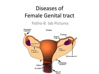
- 1. Diseases of Female Genital tract Patho-B lab Pictures
- 5. Grayish Nodular Tissue FISH EGGS- they are enlarged Chorionic villi which have undergone Hydrophic degeneration
- 6. At the pointer Enlarged Chorionic VIlli Syncitio-Trophoblast Proliferation
- 14. Hair Shafts & Protruding incisor Tooth from the wall
- 15. Cystic Wall Ectodermal stratified Sqamous epithelium Sweat glands Hair Follicle