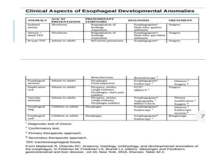
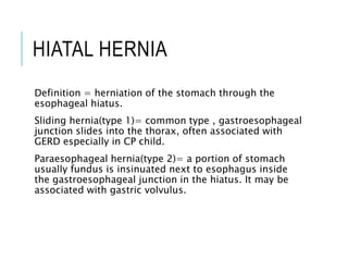
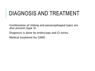
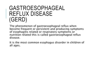
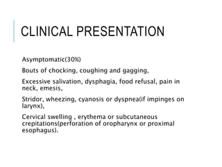
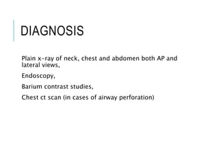
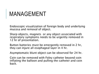
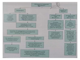
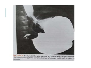
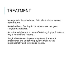
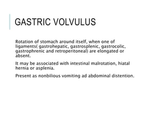
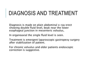
The summary of Cleft Lip/Palate is explained in very simple wording and style by the help of a scenario. Easy to remember and present due to interesting pictures. Helpful for medical students, parents having child with cleft lip/cleft palate and knowledge seekers.
The most common congenital craniofacial anomaly is cleft lip and palate. It is a separation that occurs in the lip or palate or both. Cleft occurs when the lip and/or the palate do not completely fuse during fetal development between the 6th and 9th week of pregnancy. While many factors have been associated with clefts, the cause of this condition seems complex and most cases of cleft lip and palate are thought to occur by an interaction of genetic and environmental factors or as a part of a genetic syndrome. Children with cleft lip and palate often have problems with feeding, speech, dentition, hearing, and aesthetics.
There are lots of diseases and condition that can occur in oral oral cavity which has huge effect in our overall heath . here are the few condition discussed along with there etiology , sign , symptoms and their treatment .
cleft lip and palate are the most common type of congenital anomalies. the worldwide prevalence of cleft lip and cleft palate ranges from 0.8 to 2.7 cases per 1000 live births. cleft lip is called cheiloschisis and cleft plate is called palatoschisis. Cleft lip is a gap or indentation in the lip or split continued up to the nostril due to the failure of fusion of the maxillary and medial nasal process.
Cleft palate is the condition in which the two plates of the skull that forms hard palate are not completely joined due to the failure of fusion of the lateral palatine processes, nasal septum and medial palatine process. EMBRYOLOGYPrimary palate forms during the 4-7th week of gestation when two maxillary processes and two medial nasal processes fuse.
Secondary palate forms in 6-9th weeks of gestation when palatal shelves change from vertical to horizontal position and fuse. Tongue migrates Antero-inferiorly.
Cleft lip occurs when an epithelial bridge fails.
Clefts of primary palate occur anterior to incisive foramen and clefts of secondary palate occur posterior to the incisive foramen.
ETIOLOGY Genetic: Non-syndromic inheritance (risk increases with parents or siblings or both affected); chromosome aberrations, associated with other syndromes like Van der Woude syndrome.
Environmental teratogens: Intrauterine exposure to the anticonvulsant phenytoin, alcohol, retinoic acid, maternal smoking, Rubella virus, thalidomide, aminopterin.
Maternal/intrauterine condition: Maternal diabetes mellitus and amniotic band syndrome.
Advanced paternal age
Unknown
CLASSIFICATION Prof. Balakrishnan (1975) classified cleft lip and palate according to the Indian context and divided them into three groups.
Group 1: Only cleft lip, which may be unilateral (right/ left), bilateral, or midline.
Group 2: Only cleft palate, which may be which also can be unilateral (right/left), bilateral, or submucosa.
Group 3: Includes cleft lip, alveolus, and cleft palate, which can be unilateral, bilateral, or midline. LAHSAL system for the classification of cleft lip and/or palate (2005) modified by Royal College of Surgeons Britain: LAHSAL system is a diagrammatic classification of cleft lip and palate. According to this classification, the mouth is divided into six parts. LAHSAL code indicates a complete cleft with a capital letter and an incomplete cleft with a small letter.
CLINICAL FEATURES Cleft lip: Notched vermilion border and may involve alveolar ridge.
Cleft palate: Nasal distortion, exposed nasal cavities.
Misaligned teeth.
Passage of milk through nasal passages during feeding.
Recurrent ear infection.
Speech difficulties.
Poor weight gain and failure to thrive.
DIAGNOSIS Newborn examination at birth
Palpate with a gloved finger or visual examination flashlight
In-utero ultrasonography
PROBLEMS OR COMPLICATIONS OF A CHILD WITH CLEFT LIP AND CLEFT PALATE Immediate Problems:
Feeding difficulty:
Infant with an unrepaired cleft palate will have ...
Children have oral mucosal conditions and other head and neck medical problems which have both similarities and differences to those found in adults .
A wide variety of oral lesions and soft tissue anomalies are detected in children, but the low frequency at which many of these entities occur makes them challenging to clinically diagnose.
developmentally disabled child
downs syndrome
cerebral palsy
introduction
incidence and prevalance
symptoms
precautions to be taken during treatments or during dental treatment or dental checkup
intra oral symptoms which are more commonly seen in these childrens
these children have early or late erruption of teeth
general manifestation of patients with downs syndrome are brachycephelic skull,flat nose bridge
eyes: oblique palpebral fissure , scanty eye lashes
dysplastic ear
shoert neck
congenital heart disease
dudenal atresia
umblical hernia
oral menifestation : such as drooping mouth , open mouth
protusive fissured tongue
dry fissured lips
retarded erruption of teeth
cerebral palsy : classification
spastic cerebral palsy
athetosis
ataxia
rigidity
tremors
atonia
mixed
dental problems : dental caries
periodontal diseases
malocclusion
bruxism
trauma
treatment of these patients
at last conclusion
referance from shobha tandon (pedodontics)
Management of Orofacial Cleft Dr. Sunil (2).pptx Management of Orofacial Clef...ssuser12303b
Management of Orofacial Cleft Dr. Sunil (2).pptxManagement of Orofacial Cleft Dr. Sunil (2).pptxManagement of Orofacial Cleft Dr. Sunil (2).pptxManagement of Orofacial Cleft Dr. Sunil (2).pptx
Approach to Short Stature
Dr Raheel Ahmed
FCPS in Paediatric Medicine
Children Hospital, Chanka Medical College, Larkana
Topics
Definition.
Etiology
Measurements.
Examination.
Investigations.
Management.
Take home message.
Who is short child?
Short stature is defined as height that is two standard deviations below the mean height for age and sex (less than the third percentile).
OR
more than two standard deviations below the mid-parental height.
Etiology
Proportionate Short Stature
1) Normal Variants:
i) Familial
ii) Constitutional Growth Delay
2) Prenatal Causes:
i) Intra-uterine Growth Restriction-
Placental causes, Infections, Teratogens
ii) Intra-uterine Infections
iii) Genetic Disorders (Chromosomal
& Metabolic Disorders)
Postnatal Causes:
i) Undernutrition
ii) Chronic Systemic Illness
- Cardiopulmonary: CHD, Chronic Asthma,
Cystic Fibrosis
- Renal: RTA, CRF, Steroid dependent
Nephrotic Syndrome
- GI and Hepatic: Malabsorption, IBD, chronic
liver disease
- Chronic Severe Infections
- Hematological : Thalassemia, Sickle cell
anemia
iii) Psychosocial Short Stature
(emotional deprivation)
iv) Endocrine Causes:
- Growth Hormone Deficiency/ insensitivity
- Hypothyroidism
- hypopituitrism
- Diabetes Mellitus
- Cushing Syndrome
- Pseudohypoparathyroidism
- Precocious/ delayed puberty
Disproportionate Short Stature
1) With Short Limbs:
Achondroplasia,
Hypochondroplasia,
Chondrodysplasia punctata,
Chondroectodermal Dysplasia,
Diastrophic dysplasia,
Metaphyseal Chondrodysplasia
Osteogenesis Imperfecta,
Refractory Rickets
2) With Short Trunk:
Spondyloepiphyseal dysplasia,
Mucolipidosis
Mucopolysaccharidosis
Mid Parental Height
TCR
Calculated by MPH +-10
How to measure upper and lowersegments?
You should measure the upper segment( US ) then by using the total height you will obtain LS.
Upper segment is the sitting height.
Disproportionate short statue with short LS:-
Achondroplesia
Osteogenesis imperfecta.
Refractory rickets.
Disproportionate short stature with short US:-
Spondyloepiphysial dysplasia.
Mucopolysaccharidosis.
Growth velocity
0-1 year : 25cm/year
1-2 year: 12cm/year
2-3 year: 8cm/year
3-4 year: 7cm/year
4-9 year : 5-6 cm/year
As a rule any growth rate <4.5cm/year between 2-12 year is pathological.
Dysentery
Dr. Raheel Ahmed
FCPS Pediatric Medicine
Children Hospital, Chandka Medical College, Larkana
On a global scale, of the estimated 165 million Shigella diarrhoeal episodes estimated to occur each year, 99% occur in developing countries, mainly in children.
1999, reported Shigella to be responsible for 1.1 million deaths per year, 61% of which in children less than 5 years of age
In 2013, estimates suggesting between 28,000 and 48,000 deaths annually amongst children under 5 years due to Shigellosis
Dysentry occurs predominantly in developing countries due to overcrowding and poor sanitation.
Infants,
non-breast fed children,
children recovering from measles,
malnourished children, and
adults older than 50 years
have a more severe illness and a greater risk of death.
Bascillary Dysentery
Shigella is a Gram-negative, non-motile bacillus belonging to the Enterobacteriaceae family.
There are four species of Shigellae:
S. dysenteriae, S. flexneri, S. boydii and S. sonnei
(designated as serogroups A, B, C and D respectively).
S. boydii and S. sonnei usually cause a relatively mild illness (watery or bloody diarrhoea only),
S. flexneri and S. dysenteriae are chiefly responsible for endemic and epidemic shigellosis (respectively) in developing countries, with high transmission rates and significant case fatality rates.
Transmission occurs via the faecal-oral route, person-to-person contact, household flies, infected water, and inanimate objects.
Shigella species can survive in gastric acid, and infection can occur following exposure to as few as 10-100 organisms.
Once infected, all Shigella species multiply invading the colonic epithelium where pro-inflammatory cytokines are released, and the subsequent inflammatory reaction destroys the epithelial cells lining the gut mucosa, allowing for further direct invasion by Shigella.
The resultant infectious diarrhoea is associated with loss of water and electrolytes and a clinical picture of abdominal cramping, fever, and bloody/mucoid stools.
History
Examination
Investigation
Case Definitions
Suspected case: a case with gastroenteritis, bloody mucoid diarrhea, abdominal cramps, fever and rectal pain.
Probable case: A clinical compatible case thatis epidermiologically linked i.e. Is a contact toa confirmed case or a member of risk group defined by public health authorities during an outbreak.
Confirmed case: a case that meets the confirmed laboratory criteria for diagnosis i.e. ISOLATION of Shigella species from a clinical specimen.
Period of Communicability: shed in feces 4 weeks after infection then as long as organisms present in faeces.
COMMON Pediatrics' SURGICAL EMERGENCIES
Presented By: Dr. Raheel Ahmed
FCPS – Pediatrics Medicine
Children hospital, Chandka Medical College, Larkana
Topics we will be discussing today are:
Tracheoesophageal Fistula.
Duodenal Atresia.
Meckel’s Diverticulum.
Hirschprung’s Disease.
Appendicitis.
Biliary Atresia.
Adenoids
Definition
The adenoids are enlarged and hypertrophied nasopharyngeal tonsils, sufficient to produce symptoms
It is disease of infancy and childhood.
Adenoids are subjected to physiological enlargement in childhood hence nasopharyngeal tonsils are commonly called Adenoids.
Nasopharyngeal Tonsil
Single pyramidal mass of sub-epithelial lymphoid tissue, present in nasopharynx at the junction of its roof and posterior wall.
The pharyngeal tonsil is composed of vertical ridges of lymphoid tissues separated by deep cleft and covered by Pseudostraitified ciliated columinar epithelium.
The free surface has 6 folds
It has no capsule
These lymphoid tissues consits of T and B lymphocytes.
It forms roof of waledeyer’s ring.
Can't normally see them because they are above and behind the uvula.
Arterial Supply
Ascending branch of facial artery
Ascending pharyngeal branch of external carotid
Pharyngeal branch of third part of maxillary artery.
Ascending cervical branch of inferior thyroid artery of thyrocervial trunk
Development
Adenoids begin forming in 3rd month of fetal development
Glandular primordia on posterior pharynx are infiltrated by lymphocytes.
Covered by pseudostratified ciliated epithelium
Fully formed by 7 month
Growth
They are not visible on X-ray in infants under age of one month.
50% of cases, it is visible at 6 month.
At the age of 2 years undergo hypertrophy and hyperplasia.
Can become nearly the size of a Table Tennis ball
Hypertrophy continues up to puberty (12 years)
Then, undergoes atrophy after puberty
Finally disappears in adults
Why does adenoid physiologically enlarge?
Poorly develop at birth.
Grows rapidly during childhood.
Generalized lymphoid hyperplasia occurs in children
Among the first aggregative lymphoid tissues in respiratory tract.
Physiology
Part of secondary immune system
No afferent lymphatics
Exposed to inspired antigens passed through the epithelial layer
Membrane cells and antigen presenting cells are involved in transport of antigen from the surface to the lymphoid follicle
Antigen is presented to T-helper cells
T-helper cells induce B cells in germinal center to produce antibody
Secretory IgA is primary antibody produced
Involved in local immunity
Etiology
Age : 3 -12 years
Season: winter
Food: Cold, sour, oily food
General lymphoid hyperplasia
Infection in tonsils alone or associated with
Rhinitis, Sinusitis, Tonsillitis
(esp. chronic maxillary sinusitis)
Recurrent attacks of rhinitis, sinusitis or tonsillitis may causes chronic adenoid infection
Allergy of respiratory tract.
Clinical features
Symptoms occur most commonly between ages of
3-7 years.
Depending on size of adenoid mass and space
3 types
Nasal symptoms
Aural symptoms
General symptoms
Nasal symptoms
Bilateral Nasal obstruction
Mouth breathing
interfere
Continents
Definitions
EPI
Current EPI Schedule
Different EPI Vaccines
Cold Chain
Non-EPI vaccines
Immunization
It is process where by a person is made immune or resistant to an infection, typically by administration of vaccine
It is proven tool for controlling and elimination life-threatening infectious.
Types of immunity
Innate or natural Immunity
Immunity with birth
Acquired Immunity
Develops during life time
Acquired naturally or artificially
Vaccine
A vaccine is a non- pathogenic antigen that mimics a particular pathogen in order to elicit an immune response as if that actual pathogen were in the body
Types
Live, attenuated Vaccine
Inactive Vaccine
Whole cell vaccine
Protein based: Toxoid, subunit
Polysaccharide based: Conjugate, pure
EPI: Extended Program of Immunization
EPI was established in 1974.
Built on success of the global smallpox eradication
It ensures that all children in worldwide benefited from life saving vaccines.
AIM
It focuses on following items
Standard Immunization Schedule
Supplemental immunization activities
Disease survillance
Mopping up
Promoting safe injection techniques
Improving the stocking and availability of vaccines
Protecting vaccines’ potency through cold chain management
To prepare for introduction of new vaccines
Objectives
To increase coverage of immunization for eligible children
To reduce incidence of immunizable diseases among children below five years of age
Eradication of polio, measles, neonatal tetanus, diphtheria
Reduce incidence of hepatitis B, whooping cough, bacterial meningitis
Prevention of severe forms of TB
Management of Preterm And Low Birth Weight
Dr. Raheel Ahmed FCPS Pediatrics
Children Hospital, Chandka Medical College Larkana
Definitions
Prevalent
Etiology
Assessment of gestational age
Problems of prematurity
Management
Antenatal (Prevention)
Natal (Delivery room care)
Post natal (after birth care)
Prognosis
Discharge criteria
Definitions
Term?
Preterm?
Immature?
LBW? VLBW?ELBW? ILBW?
SGA?
IUGR?
Gestational Age
Full-term
infant born after 37 completed menstrual weeks of pregnancy
Preterm (or premature) infant
infant born before 37 completed weeks of gestation
Late preterm infant (a recently identified category)
infant born between 34 and 36 weeks gestation
Moderately preterm infant
infant born between 32 and 34 completed weeks of gestation
Very preterm infant/ Early preterm
infant born before 32 completed weeks of gestation
Immature < 28 weeks
ELGAN: Extremely Low Gestational Age Newborn < 26 weeks
Weight
Low birth weight (LBW)
infant who weighs less than 2,500 grams at delivery
Very low birth weight (VLBW)
infant who weighs less than 1,500 grams at delivery
Extremely low birth weight (ELBW)
infant who weighs less than 1,000 grams at delivery
Incredible Low birth weight
infant who weighs less than 750 grams at delivery
APPROACH TO AKI IN CHILDREN
ACUTE KIDNEY INJURY
It is defined as abrupt loss of kidney function leading to rapid decline in GFR , accumulation of waste products BUN and creatinine and dysregulation of extracellular volume and electrolyte homeostasis.
AKI can ranges from small increase in creatinine to complete anuric renal failure .
INCIDENCE
2-5 % of all hospitalization.
>25% in critically ill children .
CLASSIFICATION OF AKI
CAUSES
CLINICAL MANIFESTATION
DIAGNOSTIC TEST
HISTORY AND PHYSICAL
EXAMINATION
IDENTIFICATION OF PRECIPTATING CAUSE
COMPLICATION
MANAGEMENT
MANAGEMENT
There is no definitive therapy for AKI, supportive care is mainstay of management regardless of aetiology.
Goal of treatment is :
Minimize degree of insult.
Reduce extrarenal complication.
Restoration of AKI.
Optimize the systemic and renal hemodynamic(fluid resuscitation or use of vasopressor).
Avoid the nephrotoxic drugs (e.g aminoglycoside, NSAIDs, ACE inhibitor, ARB blocker, acyclovir) or adjust the dose .
Catheterize the patient in case of obstruction like PUV, UPJ obstruction
POST-RENAL AKI
Prompt relieve of urinary tract obstruction.
Relief of obstruction is usually followed by an appropriate diuresis and may require continue administration of iv fluids and electrolyte.
RENAL REPLACEMENT THERAPY
The purpose of RRT is to prevent morbidity.
It may be necessary for days or upto 12 weeks.
Mostly require dialysis support for 1-3 weeks.
Indication Of RRT :
A= ACIDOSIS, ANURIA
E= ELECTROLYTE DISTURBANCE (hypokalemia)
I= INTOXICATION
O= OVERLOAD(hypertension, pulmonary edema)
U= UREMIA
PROGNOSIS
Pre-renal and post-renal have better prognosis.
In case of post-infectious glomerulonephritis is 1%
In case of multi organ failure >50%.
Kidney may recover even after dialysis .
10% cases requiring dialysis develop CKD.
CARRY HOME MESSAGE
Diagnose early- biomarkers have great potential.
Look for aetiology.
Prevent rather than treat.
No role of low dose dopamine prevention and treatment .
Initiate RRT when indicated.
CP
Non-specific term that include disorders characterized by early onset and impaired movement and posture.
Non-progressive and may include perceptual problems, language deficits, and intellectual involvement.
Incidence
Most common physical disability of childhood.
Incidence has increased since the 60’s, maybe due to improved survival of VLBW infants.
Etiology
Variety of perinatal, prenatal, and postnatal factors contribute, either singly or multifactorily to CP.
Commonly thought to be due to birth asphyxia; now known to be due to existing prenatal brain abnormalities.
Premature delivery is the single most important determinant of CP.
In 24% of cases, no cause is found.
Approach and Management of Malabsorption Syndromes in children.pptxRaheelAhmed210939
Approach and Management of Malabsorption Syndromes in children
Dr. Raheel Ahmed MBBS, FCPS
Children Hospital, Chandka Medical College, Larkana
Etiology
Common causes
Coeliac Disease
Cystic Fibrosis
Post gastroenteritis syndrome
Bacterial overgrowth
Tuberculosis
HIV (immunodeficiency)
Giardiasis
Rare Causes
IBD;
Crohn’s disease (CD); Ulcerative colitis (UC)
Short Bowel Syndrome
CLD with cholestasis
Immunodeficiency syndromes
Intestinal lymphangiectasia
Abetalipoproteinemia
NVBDCP.pptx Nation vector borne disease control programSapna Thakur
NVBDCP was launched in 2003-2004 . Vector-Borne Disease: Disease that results from an infection transmitted to humans and other animals by blood-feeding arthropods, such as mosquitoes, ticks, and fleas. Examples of vector-borne diseases include Dengue fever, West Nile Virus, Lyme disease, and malaria.
Knee anatomy and clinical tests 2024.pdfvimalpl1234
This includes all relevant anatomy and clinical tests compiled from standard textbooks, Campbell,netter etc..It is comprehensive and best suited for orthopaedicians and orthopaedic residents.
Report Back from SGO 2024: What’s the Latest in Cervical Cancer?bkling
Are you curious about what’s new in cervical cancer research or unsure what the findings mean? Join Dr. Emily Ko, a gynecologic oncologist at Penn Medicine, to learn about the latest updates from the Society of Gynecologic Oncology (SGO) 2024 Annual Meeting on Women’s Cancer. Dr. Ko will discuss what the research presented at the conference means for you and answer your questions about the new developments.
ARTIFICIAL INTELLIGENCE IN HEALTHCARE.pdfAnujkumaranit
Artificial intelligence (AI) refers to the simulation of human intelligence processes by machines, especially computer systems. It encompasses tasks such as learning, reasoning, problem-solving, perception, and language understanding. AI technologies are revolutionizing various fields, from healthcare to finance, by enabling machines to perform tasks that typically require human intelligence.
Flu Vaccine Alert in Bangalore Karnatakaaddon Scans
As flu season approaches, health officials in Bangalore, Karnataka, are urging residents to get their flu vaccinations. The seasonal flu, while common, can lead to severe health complications, particularly for vulnerable populations such as young children, the elderly, and those with underlying health conditions.
Dr. Vidisha Kumari, a leading epidemiologist in Bangalore, emphasizes the importance of getting vaccinated. "The flu vaccine is our best defense against the influenza virus. It not only protects individuals but also helps prevent the spread of the virus in our communities," he says.
This year, the flu season is expected to coincide with a potential increase in other respiratory illnesses. The Karnataka Health Department has launched an awareness campaign highlighting the significance of flu vaccinations. They have set up multiple vaccination centers across Bangalore, making it convenient for residents to receive their shots.
To encourage widespread vaccination, the government is also collaborating with local schools, workplaces, and community centers to facilitate vaccination drives. Special attention is being given to ensuring that the vaccine is accessible to all, including marginalized communities who may have limited access to healthcare.
Residents are reminded that the flu vaccine is safe and effective. Common side effects are mild and may include soreness at the injection site, mild fever, or muscle aches. These side effects are generally short-lived and far less severe than the flu itself.
Healthcare providers are also stressing the importance of continuing COVID-19 precautions. Wearing masks, practicing good hand hygiene, and maintaining social distancing are still crucial, especially in crowded places.
Protect yourself and your loved ones by getting vaccinated. Together, we can help keep Bangalore healthy and safe this flu season. For more information on vaccination centers and schedules, residents can visit the Karnataka Health Department’s official website or follow their social media pages.
Stay informed, stay safe, and get your flu shot today!
MANAGEMENT OF ATRIOVENTRICULAR CONDUCTION BLOCK.pdfJim Jacob Roy
Cardiac conduction defects can occur due to various causes.
Atrioventricular conduction blocks ( AV blocks ) are classified into 3 types.
This document describes the acute management of AV block.
Ethanol (CH3CH2OH), or beverage alcohol, is a two-carbon alcohol
that is rapidly distributed in the body and brain. Ethanol alters many
neurochemical systems and has rewarding and addictive properties. It
is the oldest recreational drug and likely contributes to more morbidity,
mortality, and public health costs than all illicit drugs combined. The
5th edition of the Diagnostic and Statistical Manual of Mental Disorders
(DSM-5) integrates alcohol abuse and alcohol dependence into a single
disorder called alcohol use disorder (AUD), with mild, moderate,
and severe subclassifications (American Psychiatric Association, 2013).
In the DSM-5, all types of substance abuse and dependence have been
combined into a single substance use disorder (SUD) on a continuum
from mild to severe. A diagnosis of AUD requires that at least two of
the 11 DSM-5 behaviors be present within a 12-month period (mild
AUD: 2–3 criteria; moderate AUD: 4–5 criteria; severe AUD: 6–11 criteria).
The four main behavioral effects of AUD are impaired control over
drinking, negative social consequences, risky use, and altered physiological
effects (tolerance, withdrawal). This chapter presents an overview
of the prevalence and harmful consequences of AUD in the U.S.,
the systemic nature of the disease, neurocircuitry and stages of AUD,
comorbidities, fetal alcohol spectrum disorders, genetic risk factors, and
pharmacotherapies for AUD.
Tom Selleck Health: A Comprehensive Look at the Iconic Actor’s Wellness Journeygreendigital
Tom Selleck, an enduring figure in Hollywood. has captivated audiences for decades with his rugged charm, iconic moustache. and memorable roles in television and film. From his breakout role as Thomas Magnum in Magnum P.I. to his current portrayal of Frank Reagan in Blue Bloods. Selleck's career has spanned over 50 years. But beyond his professional achievements. fans have often been curious about Tom Selleck Health. especially as he has aged in the public eye.
Follow us on: Pinterest
Introduction
Many have been interested in Tom Selleck health. not only because of his enduring presence on screen but also because of the challenges. and lifestyle choices he has faced and made over the years. This article delves into the various aspects of Tom Selleck health. exploring his fitness regimen, diet, mental health. and the challenges he has encountered as he ages. We'll look at how he maintains his well-being. the health issues he has faced, and his approach to ageing .
Early Life and Career
Childhood and Athletic Beginnings
Tom Selleck was born on January 29, 1945, in Detroit, Michigan, and grew up in Sherman Oaks, California. From an early age, he was involved in sports, particularly basketball. which played a significant role in his physical development. His athletic pursuits continued into college. where he attended the University of Southern California (USC) on a basketball scholarship. This early involvement in sports laid a strong foundation for his physical health and disciplined lifestyle.
Transition to Acting
Selleck's transition from an athlete to an actor came with its physical demands. His first significant role in "Magnum P.I." required him to perform various stunts and maintain a fit appearance. This role, which he played from 1980 to 1988. necessitated a rigorous fitness routine to meet the show's demands. setting the stage for his long-term commitment to health and wellness.
Fitness Regimen
Workout Routine
Tom Selleck health and fitness regimen has evolved. adapting to his changing roles and age. During his "Magnum, P.I." days. Selleck's workouts were intense and focused on building and maintaining muscle mass. His routine included weightlifting, cardiovascular exercises. and specific training for the stunts he performed on the show.
Selleck adjusted his fitness routine as he aged to suit his body's needs. Today, his workouts focus on maintaining flexibility, strength, and cardiovascular health. He incorporates low-impact exercises such as swimming, walking, and light weightlifting. This balanced approach helps him stay fit without putting undue strain on his joints and muscles.
Importance of Flexibility and Mobility
In recent years, Selleck has emphasized the importance of flexibility and mobility in his fitness regimen. Understanding the natural decline in muscle mass and joint flexibility with age. he includes stretching and yoga in his routine. These practices help prevent injuries, improve posture, and maintain mobilit
Ozempic: Preoperative Management of Patients on GLP-1 Receptor Agonists Saeid Safari
Preoperative Management of Patients on GLP-1 Receptor Agonists like Ozempic and Semiglutide
ASA GUIDELINE
NYSORA Guideline
2 Case Reports of Gastric Ultrasound
2. INTRODUCTION
A fetus can swallow amniotic fluid as early as 12 weeks
of gestation.
Nutritive sucking appear first at about 34 weeks of
gestation.
Initially tongue movement is upward and outward to
express milk from nipples.
By 1 month of age infant appear to show preference for
sweet and salty foods.
3. Infants interest in solid increase at about 4 month of age
.
The recommendation to begin solid food at 6 is based on
nutritional and cultural concepts rather than maturation
of swallowing process.
4. ANATOMIC VARIATIONS
Short lingual frenulum (tongue tie)= it dose not interfere
with nursing, bottle feeding , eating or speech, may be
worrisome to parents.
Bifid uvula= may be isolated or associated with cleft of
soft palate .
5. Regurgitation = regurgitation mostly resolves in 80% of
infant by 6 mo of age , 90% by 12 mo.
Stool frequency = can vary in newborn from none to 7
per day.
Toddler’s diarrhea = occurs btw 1 and 3 yr age.
Intermittent lose stools containing vegetable matter, due
to consuming increase quantity of carbohydrate
containing beverages.
6. Blood loss= blood loss from GIT is never normal , but
swallowed blood may be misinterpreted as
gastrointestinal bleeding. Nasal or oropharyngeal
bleeding is occasionally mistaken for gastrointestinal
bleeding.
Jaundice = normal in neonates, especially in preterm
infants and usually results from the inability of an
immature liver to conjugate bilirubin. Direct bilirubin
fraction should account no more than 15-20% of total.
7. ORAL CAVITY
The primary dentition starts with eruption of first
primary tooth, all 20 primary teeth erupts b 3 yr of age .
The permanent teeth start erupting around age of 6 yr
and completed by 13 yr of age .
The transition time btw primary and permanent dentition
, when a mix teeth are present , is referred to as mixed
dentition .
8.
9. SOME ANOMALIES
Natal teeth= present in 1 In 2000 newborn infants, are
present at birth usually in position of mandibular central
incisors. Natal teeth are associated with cleft palate,
Pierre Robin syndrome, Hallermann-Streiff syndrome and
other anomalies .
Neonatal teeth= erupts in 1 mo of life.
10. Anodontia= absence of teeth , occurs when no tooth
buds form (ectodermal dysplasia, or familial missing
teeth) or when there is disturbance of normal site of
initiation (the area of palate cleft).
Supernumerary teeth = more than normal number of
buds, more often in area btw maxillary central incisors.
11. Fluorosis(molted enamel) = occurs when systemic
fluoride consumption is > 0.05mg/dl during enamel
formation. Excessive fluoride during enamel formation
results in white , lacy patches on the enamel to severe
brownish discoloration and hypoplasia. These are seen
with fluoride concentration in drinking water >5.0 ppm.
12. ANOMALIES OF COLOR
Neonatal hyperbilirubinemia can produce blue to black
discoloration.
Porphyria produces red brown discoloration.
Tetracycline result in brown yellow discoloration.
Period of risk extends btw 4 mo of gestation to 7 yr of
life.
13. Teething = associated with primary tooth eruption. Have
benign symptoms such as gingival hyperemia, irritability,
sucking fingers and drooling. Some even have no
symptoms at all.
Treatment of symptoms is often unnecessary but could
include oral analgesics and iced teeth rings.
14. CLEFT LIP AND PALATE
Cleft lip appears due to hypoplasia of mesenchymal
layers, resulting in failure of medial nasal and maxillary
processes to join.
Cleft palate results due to failure of palatal shelves to
fuse.
Incidence of cleft lip is 1 in 750 white births, while that
of palate is 1 in 2500 white births.
Cleft lip is more common in males.
15.
16. CAUSES AND INCIDENCE
Causes are maternal drug exposure, genetic factors ,
may associated with syndromes( app 400) , may be
sporadically and inherited as dominant fashion(van der
Woude syndrome).
Incidence is highest among Asians(1 in 500) and Native
Americans(1 in 300) and lowest among blacks(1 in
2500).
Cleft lip can be associated with other facial anomalies ,
cleft palate is associated with central nervous anomalies.
17. CLINCAL MANIFESTATIONS
Cleft lip may be unilateral(mostly on left side)or bilateral.
Cleft lip can be a small notch or complete separation
involving skin, mucosa, muscle, tooth and bone.
Cleft palate isolated occurs in midline might involve only
uvula or extend into through the soft and hard palates
to incisive foramen.
18. TREATMENT
Multidisciplinary approach involving pediatrician, plastic
surgeon, otolaryngologist, oral and maxillofacial
surgeon, pediatric dentist, prosthodontist, orthodontist,
speech therapist, geneticist, medical and social worker,
psychologist and public health nurse.
Feeding can be achieved by construction of plastic
obturator, artificial nipples with large openings,
squeezable bottle and proper instructions.
19. Surgical closure of cleft lip performed by 3 months of
age .
Commonly used is modified Millard rotation-
advancement technique.
Cleft palate closure is usually done before 1 year of age.
Missing teeth that are nonfunctional are replaced by
prosthetic devices.
20. Postoperative care= aspiration of nasopharynx, avoid
tension on sutures, keep toys away from infant,
semisolid food is advised for 3 weeks.
Complication= recurrent otitis media, hearing loss,
speech defect , malposition of teeth are some of
sequelae of cleft lip and palate.
21. SYNDROMES
Ectodermal dysplasias= it’s a heterogeneous group of
condition with oral manifestations. Teeth can be totally
or partially absent or malformed, alveolar process is
absent, overclosure of mandible causes lips to protrude,
teeth are small conical , dryness of oral mucosa occur.
Treatment= full dentures are needed.
22.
23. Pierre Robin syndrome= consist of micrognathia , high
arced palate or cleft palate. Floor of mouth is
foreshortened.
Treatment = prevent suffocation, prone position ,
tracheostomy may be needed, Mandibular distraction
procedures.
Stickler syndrome= Autosomal dominant, 30-50%
associated Pierre Robin syndrome, include prominent
joints, arthritis , hypotonia, hypermobile joints, mitral
valve prolapse, spine and ocular problems and hearing
loss.
24.
25. ESOPHAGEAL ATRESIA AND
TRACHEOESOPHAGEAL
FISTULA
Incidence= 1.7 PER 10,000 live births mostly associated
with tracheoesophageal fistula.
Causes = advance maternal age, obesity, low
socioeconomic status, tobacco smoking, European
ethnicity, genetic mutations(syndrome cases) Feingold
and CHARGE.
Associated with VACTERAL.
26. CLINICAL PRESENTATION
Frothing, bubbling from mouth and nose, episodes of
coughing, cyanosis and respiratory distress.
Isolated TEF come in medical attention later in life with
chronic respiratory problems(recurrent pneumonia,
refractory bronchospasm).
28. DIAGNOSIS
Inability to pass NG or OG tube .
Plain x-ray shows coiled feeding tube, air distended
stomach(coexisting TEF), airless scaphoid
abdomen(isolated EA).
Esophagogram with contrast medium demonstrate the
defect (isolated TEF).
Endoscopy and Bronchoscopy.
29.
30.
31. TREATMENT
Nonspecific = maintain airway, prone position
decompress to prevent aspiration of secretions,
antibiotic cover for pneumonia.
Specific= surgical ligation of TEF and primary end to end
anastomosis of esophagus via right sided thoracotomy
is standard approach.
Complications= anastomic leak and stricture
refistulization and GERD.
32.
33. HIATAL HERNIA
Definition = herniation of the stomach through the
esophageal hiatus.
Sliding hernia(type 1)= common type , gastroesophageal
junction slides into the thorax, often associated with
GERD especially in CP child.
Paraesophageal hernia(type 2)= a portion of stomach
usually fundus is insinuated next to esophagus inside
the gastroesophageal junction in the hiatus. It may be
associated with gastric volvulus.
34. DIAGNOSIS AND TREATMENT
Combination of sliding and paraesophageal types are
also present (type 3).
Diagnosis is done by endoscopy and GI series.
Medical treatment for GERD .
35. GASTROESOPHAGEAL
REFLUX DISEASE
(GERD)
The phenomenon of gastroesophageal reflux when
become frequent or persistent and producing symptoms
of esophagitis related or respiratory symptoms or
nutrition related this is called gastroesophageal reflux
disease.
It is the most common esophagus disorder in children of
all ages.
36. CLINICAL FEATURES
INFANTS= postprandial regurgitation, irritability, arching,
choking, gagging, failure to thrive.
CHILDREN= regurgitation, abdominal pain, chest pain,
refusal to food, neck contortions(arching or turning of
head) known as Sandifer syndrome.
GERD can be present with respiratory symptoms, stridor,
apnea, asthma symptoms .
37. FACTORS ASSOCIATED TO
GERD
Duration of exposure,
Causticity of reflux,
Straining,
Obesity,
Large volume or hyperosmolar meals,
Sliding hernia,
Increase respiratory efforts(coughing wheezing).
38. DIAGNOSIS
Clinical = a thorough history and examination.
Barium radiographic= for esophagus and upper GI tract
to evaluate achalasia, esophageal strictures, stenosis,
hiatal hernia, gastric and intestinal obstruction.
Esophageal pH monitoring= for assessing acid
suppression during treatment, evaluating apneic
episodes.
39. Endoscopy=for erosive esophagitis, strictures or barret
esophagus.
Intraluminal impedance=potential for diagnosing GERD,
understanding esophageal function and in combination
with manometry in motor patterns associated with GERD
Laryngotracheobronchoscopy= for airway signs
associated with extraesophageal GERD.
Empirical antireflux therapy=not evaluated in children,
trial of PPI for time limited duration.
40. MANAGEMENT
Lifestyle modifications= dietary measures for
infants(thickened feed), position measures(prone).
Histamine-2 receptor antagonists=(cimetidine,
famotidine, ranitidine) used in mild to moderate reflux
esophagitis. First line therapy.
PPIs=(omeprazole, lansoprazole, pantoprazole) most
potent antireflux. Superior to H2RAs in treatment of
severe and erosive esophagitis. Side effects respiratory
infections, clostridium difficile infection, bone fracture,
kidney damage and hypomagnesemia.
41. Prokinetic agents=(metoclopramide, bethanecol,
erythromycin) inc LES pressure, improve gastric
emptying.
In 2009 metoclopramide is announced by FDA as black
box warning, side effect is tardive dyskinesia.
Surgery =fundoplication is effective for intractable GERD.
It can be performed as open procedure, by laparoscopy
or by endoluminal techniques. Pediatric is limited with
endoscopic application of radiofrequency therapy(Stretta
procedure) to create high pressure zone.
43. FOREIGN BODIES IN
ESOPHAGUS
Foreign body ingestion occurs mostly accidentally .
In majority 5 year or younger.
Coins are most commonly ingested foreign body.
Most esophageal foreign body lodge at level of
cricopharyngeus, aortic arch or just superior to
diaphragm at gastroesophageal junction.
44. CLINICAL PRESENTATION
Asymptomatic(30%)
Bouts of chocking, coughing and gagging,
Excessive salivation, dysphagia, food refusal, pain in
neck, emesis,
Stridor, wheezing, cyanosis or dyspnea(if impinges on
larynx),
Cervical swelling , erythema or subcutaneous
crepitations(perforation of oropharynx or proximal
esophagus).
45. DIAGNOSIS
Plain x-ray of neck, chest and abdomen both AP and
lateral views,
Endoscopy,
Barium contrast studies,
Chest ct scan (in cases of airway perforation)
46. MANAGEMENT
Endoscopic visualization of foreign body and underlying
mucosa and removal of object.
Sharp objects, magnets or any object associated with
respiratory symptoms needs to be urgently removed in
12 hr of presentation.
Button batteries must be emergently removed in 2 hr,
they can injure all esophageal layer in 4 hr.
Asymptomatic blunt object can be observed for 24 hr.
Coin can be removed with Foley catheter beyond coin
inflating the balloon and pulling the catheter and coin
back.
47.
48. PYLORIC STENOSIS
Occurs in 1-3/1000 infants, more common in whites.
More in males, especially first born.
Infants with blood group O and B.
High risk in those neonates using erythromycin.
Associated with eosinophilic gastroenteritis, Apert
syndrome, Zellweger syndrome, trisomy 18.
Also associated in infants of mothers treated with
macrolide during pregnancy and lactation.
Reduced level of nitric oxide also contribute to
pathogenesis of pyloric stenosis.
49. CLINICAL PRESENTATION
Non-bilious vomiting is initial symptom, may or not be
projectile, occur after feed.
After vomiting infant is hungry and wants to feed again,
May present as early as 1st week or as late as 5th month
of age.
Hyperbilirubinemia (icteropyloric syndrome)
unconjugated bilirubin.
Olive mass, firm about 2cm located above the umbilicus
at mid epigastrium region. Palpated after an episode of
vomiting.
Visible gastric peristaltic wave progress across abdomen.
50. DIAGNOSIS
ABGs shows hypochloremic metabolic alkalosis.
Ultrasound is used for diagnosis(criteria pyloric
thickness=3-4mm, pyloric length=15-19mm and pyloric
diameter=10-14mm.
Contrast studies shows elongated pyloric channel string
sign.
Bulge of pyloric muscle into antrum, shoulder sign.
51.
52.
53. TREATMENT
Manage acid base balance, fluid electrolytes, correct
dehydration.
Nasoduodenal feeding in those who are not good
surgical candidates.
Atropine sulphate at a dose of 0.01mg/kg i/v 6 times a
day 5 min before feeding .
Surgical treatment is pyloromyotomy (ramstedt
procedure), the underlying pyloric mass is cut
longitudinally and incision is closed.
54. GASTRIC VOLVULUS
Rotation of stomach around itself, when one of
ligaments( gastrohepatic, gastrosplenic, gastrocolic,
gastrophrenic and retroperitoneal) are elongated or
absent.
It may be associated with intestinal malrotation, hiatal
hernia or asplenia.
Present as nonbilious vomiting ad abdominal distention.
55. DIAGNOSIS AND TREATMENT
Diagnosis is made on plain abdominal x-ray erect
showing double fluid level, beak near the lower
esophageal junction in mesenteric volvulus,
In organoaxial the single fluid level is seen.
Treatment is emergent laparoscopic gastropexy surgery
after stabilization of patient.
For chronic volvulus and older patients endoscopic
correction is suggestive.
56. DUODENAL ATRESIA
Occurs in 1/10,000 live births and account for 20-40%
for all intestinal atresias.
Duodenal atresia result from failure of recanalization of
intestinal lumen during gestation.
Mostly occur in premature infants and associated with
other anomalies(congenital renal, esophageal atresia,
skeletal malformations, trisomy 21).
57. CLINICAL PRESENTATION
Bilious vomiting usually noted on first day of life.
Jaundice is present.
Peristaltic wave may be visualized.
History of polyhydrominas is present.
58. DIAGNOSIS
Diagnosed by plain abdominal radiograph showing
double bubble sign.
Contrast studies are used to exclude malrotation and
volvulus.
Prenatal diagnosis by fetal ultrasound showing double
bubble.
ECHO, renal ultrasonograpy, x-ray chest to evaluate
associated anomalies.
59.
60. TREATMENT
NG decompression
Iv fluids electrolyte balance.
Duodenoduodenostomy is the surgical procedure.
Prognosis is excellent , 90% survival .
61. MALROTATION
Intestinal rotation and attachment begins in 5th week of
gestation, when the mid bowel begins to elongate, and
completed by 12 weeks of gestation.
Nonrotation occurs when bowel fails to rotate after it
returns to abdominal cavity.
1st and 2nd portion of duodenum are in normal position ,
but the remainder of duodenum, jejunum, and ileum
occupy the right side of abdomen and colon is located to
left.
Malrotation is often associated with gastrochisis,
diaphragmatic hernia and omphalocele.
62. CLINICAL FEATURES
Mostly present in 1st year of life, of which 50% present
within first month of life.
Vomiting is most common symptom, infant present in 1st
week with bilious emesis and bowel obstruction.
Can present with recurrent abdominal pain and
postprandial bloating.
Occasionally present as malabsorption or protein-
loosing enteropathy.
HETEROPAXY SYNDROME= congenital heart
malformations, malrotation, biliary atresia, asplenia or
polysplenia.
63.
64. DIAGNOSIS
Upper GI series is imaging test of choice and gold
standard in evaluation and diagnosis of malrotation and
volvulus.
Ultrasound demonstrate inversion of superior mesenteric
artery and vein.
SMV located to left of SMA is suggestive of malrotation.
66. MECKEL DIVERTICULUM
Meckel diverticulum is most common residual structure
of failure of involution of the omphalomesentric duct.
Meckel diverticulum is out pouching of ileum along the
antimesentric border of 50-75cm(app 2 feet ) from
ileocecal valve.
The ‘rule of 2s’ is often used for Meckel’s diverticulum:
within 2 feet of the ileocecal valve, 2 inches in length,
occurring in about 2% of the population, two times more
symptomatic in males, symptomatic by 2 years of age
and potentially containing 2 heterotopic tissues, gastric
and pancreatic.
67.
68. CLINICAL FEATURES
The common presenting problems of a Meckel
diverticulum are bleeding, obstruction, pain
(inflammation) and umbilical discharge.
The bleeding is due to ulcer formation from the acid
secreted from the ectopic gastric mucosa.
The stool is typically describes as brick color or current
jelly colored.
Occasionally became inflamed (diverticulitis) manifest as
appendicitis.
69. DIAGNOSIS
Technetium-99m pertechnetate scintigraphy (Meckel’s
scan) is the investigation of choice. It is used to detect
heterotrophic gastric mucosa. Pentagastrin, histamine
blockers and glucagon may enhance the accuracy of
diagnosis.
Mesenteric Angiography, radiolabeled red blood cells
scan, abdominal ultrasound, abdominal CT are other
methods to evaluate.
70. TREATMENT
In symptomatic patients, the diverticulum is removed
using a laparoscopic, laparoscopic-assisted or open
technique.
In an asymptomatic patients , there is debate for excision
vs. observation.
71. ENCOPRESIS
It is defined as voluntary or involuntary passage of feces
into inappropriate place at least once a month for
3consecutive months once a chronologic or
developmental age of 4years has been reached.
72. ETIOLOGY
Developmental and behavior factors= Lack of adequate
school bathroom facilities, harsh toilet training, Sexual
abuse, negative defecation experience(associated with
prior diarrhea)
Anatomic factors= hirschsprung’s disease, anal stenosis.
73. SUBTYPES
Retentive encopresis= defined as constipation with
overflow incontinence.
Nonretentive encopresis= defined as no evidence of fecal
retention (impaction) >1episode per week in previous
2months.
Primary or continuous encopresis = persist from infancy
onward.
Secondary or discontinuous encopresis= can appear
after successful toilet training In response to stressful
event, such as separation or family illness.
75. APPROACH
A detailed bowel history
Age of child
Frequency of bowel movement
h/o constipation
Description of stool.
EXAMINATION=Growth pattern, Neurologic examination
as spine, lower limb deep reflexes and abdominal
reflexes. Rectal examination as sphincter tone, presence
of stool in rectal ampulla.
Thyroid testing.
76. TREATMENT
Education =postprandial toilet training, no punishment
to patient, reward for healthy bowel movements.
Relief of impaction= enema
Softening of stools = polyethylene glycol, lactulose,
mineral oil.
Psychologist and behavior management
Neurostimulation and pelvic physiotherapy.
77. HIRSCHSPRUNG DISEASE
Also known as Congenital Megacolon or Aganglionic
Megacolon.
It is a developmental disorder of enteric nervous system
characterized by absence of ganglion cells in the
submucosal and myentric plexus.
It is the most common cause of lower intestinal
obstruction.
78. SPECTRUM OF DISEASE
Small segment: Disease limited to rectosigmoid colon
80%.
Long segment: Disease proximal to sigmoid colon 10-
15%.
Total bowel aganglionosis is rare and accounts for 5%.
79. CLINICAL FEATURES
Hirschsprung disease should be suspected in any full
term infant with delayed passage of stool with in 48
hours of life.
Constipation, Abdominal distention, poor feeding and
failure to thrive.
In older children large fecal mass palpable in left lower
quadrant while rectum is empty.
Stool character: small pellets, ribbon like, fluid
consistency.
80. DIAGNOSIS
Abdominal Straight X-ray= Air fluid levels in colon and
distended loops of intestine.
Barium Contrast Enema= Classic findings are based on
the presence of an abrupt narrow transition zone b/w the
normal dilated proximal colon and a smaller-caliber
obstructed aganglionic segment.
Rectosigmoid Index= Measurement of diameter of the
rectum to that of sigmoid colon during contrast enema.
Rectal diameter that is same or smaller than the sigmoid
colon suggests Hirschsprung disease.
81.
82. Anorectal manometery= Shows absence of relaxation
reflex after distention of balloon in rectum.(Means
absence of rectoanal inhibitory reflex”RIAR” which is
normally present).
Rectal Suction Biopsy= It is GOLD STANDARD for
diagnosing Hirschspung disease.
The biopsy material should contain an adequate amount
of submucosa to evaluate for the presence of ganglion
cells
Biopsy sample should be 2 to 3 cm above the dentate
line (line dividing the upper 2/3rd and lower 3rd of the
anal canal) to avoid obtaining biopsies in the normal area
of hypoganglionosis.
83. TREATMENT
Pre-operative interventions= i/v fluids and antibiotics
and Decompression of the colon.
Surgical procedures=
Swenson’s procedure= Excision of aganglionic segment
and anastomosis of the normal proximal bowel with the
rectum 1-2 cm above the dentate line (Enterocolitis).
Duhamel’s procedure= Neorectum created= normally
innervated bowel is brought down aganglionic rectum
(Constipation).
84. Soave’s procedure= Endorectal pull-through procedure,
Mucosa striped from the aganglionic rectum and
normally innervated colon is brought through the
residual muscular cuff (Diarrhea).
Laparoscopic assisted transanal pull-through(LATEP)=
Advance procedure, removal of the aganglionic colon
segment and coloanal anastomosis using normally
innervated colon.
85. INTUSSUSCEPTION
When a portion of alimentary tract is telescoped into an
adjacent segment is called intussusception.
1 to 4 in 1000 infants and children, more males than
females ( 2:1 or 3:2 ratio) 90% in children within 3 years
of age. More than 40% are seen between 3 and 9 months
of age .
Associated with HSP , Rotavirus vaccine , adenovirus and
seasonal variations (peak in fall and winter).
86. LEAD POINTS
Meckel diverticulum
Intestinal polyp
Neurofibroma
Periappendicitis
Appendiceal stump
Inversion appendectomy
Appendiceal mucocele
Local suture line
Massive local lymphoid hyperplasia
Benign tumors (adenoma, leiomyoma, carcinoid,
neurofibroma, hemangioma); and malignant tumors
(lymphoma, sarcoma, leukemia).
87. CLINICAL FEATURES
Severe abdominal pain, vomiting bile stained
Lethargy, shock like state, weak pulses
Fever and peritonitis,
Currant jelly stools,
Classic triad= pain, palpable sausage shaped mass and
currant jelly stools.
88. DIAGNOSIS
Plain radiograph shows density in area of
intussusception.
Ultrasound= high sensitivity and specificity, doughnut or
target appearance.
Contrast enema= demonstrate filling defect or cupping
where its advance is obstructed. Or coiled spring sign.
89. TREATMENT
Evaluation by surgeon if there is need for emergent
operation
Fluid resuscitation, NG tube decompression Broad-
spectrum antibiotic, radiologic reduction or operative
reduction, resection, closure of an enema perforation
excision of by laparotomy or laparoscopy.
In prolonged intussusception and signs of peritonitis,
hydrostatic reduction is contraindicated.
90. CHRONIC ULCERATIVE
COLITIS
Ulcerative colitis is an idiopathic chronic inflammatory
disorder, is localized to the colon and spares the upper
GI tract.
It is a form of IBD.
Men are slightly more likely to acquire than women .
91. CLINICAL FEATURES
Blood(5 bloody stools per day for 5 days fulminant
colitis) mucus, pus in stool, diarrhea or constipation
(proctitis).
Fever, severe anemia, hypoalbunemia, leukocytosis.
Anorexia, weight loss and growth failure.
It is marked by remission and relapse.
92. EXTRAINTESTINAL
MANIFESTATIONS
UC (IBD) is not restricted to GI tract, can involve almost
any organ system.
Pyoderma gangrenosum.
Sclerosing cholangitis.
Chronic active hepatitis.
Ankylosing spondylitis.
Secondary amenorrhea.
Iron deficiency anemia, folate deficiency (Salfasalazine).
93.
94.
95. DIAGNOSIS
CBC= anemia, wbc count raised
Elevated CRP
Hypoalbunemia
Elevated fecal calprotectin(specific for GI inflammation)
P-ANCA(Positive in 60%-80% of patients, associated with
an earlier need for surgery)
Barium enema
96. Colonoscopy/ Sigmoidoscopy(to establish diagnosis,
exclude alternate diagnosis like ischemic and infectious
colitis, to determine the extent and severity of disease)
Endoscopic findings of ulcerative colitis from microulcers
to pseudopolyps may be seen.
Biopsy (histologic findings are cryptitis, crypt abscesses,
separation of crypts by inflammatory cells, edema,
mucus depletion, branching of crypts).
97. TREATMENT
Medical
Mild to moderate= Aminosalicyclate(sulfasalazine 30-
100mg/kg/24 divided 2-4 doses, mesalamine 50-
100mg/kg/day, balsalazide 2.25-6.75g/day).
Moderate to severe= corticosteroids (prednisone 1-
2mg/day)
Immunomodulators (azthioprine 2-2.5mg/kg/day, 6-
mercaptopurine 1-1.5mg/kg/day).
98. Surgical =About 10% to 20% of patients with UC.
Indications:
1) Chronic intractable disease ,not controlled with
medications or drug side effects are too severe.
2) Severe acute colitis requiring an urgent procedure.
3) Presence of dysplasia or cancer.
4) Colonic perforation.
Surgery is total colectomy.
99. CROHN DISEASE
It can affect any area of the GI tract, from the mouth to
the anus.
The inflammatory process tends to be eccentric and
segmental often with skip areas.
GI involvement is transmural.
Bimodal age distribution with first peak beginning in
teenage.
100. CLINICAL MENIFESTATION
Crohn disease is granulomatous inflammation, resulting
in ulceration, stricturing, fistula & abscess formation.
Crampy abdominal pain
Fatigue
Pain with passing stool (Tenesmus) ,Persistent watery
diarrhea
Fever, Loss of appetite , Unintentional weight loss
General malaise
Other symptoms are Constipation , Eye inflammation,
fistulas , fissures , abscess, mouth ulcer, rectal bleeding.
Perianal disease is common.
101. EXTRA INTESTINAL
Oral apthous ulcers
Peripheral arthritis(non deforming)
Erythema nodosum
Digital clubbing
Episcleritis
Renal stones and gallstones
102. DIAGNOSIS
Blood tests to check for anemia, WBC count
ESR, CRP
Fecal calprotectin
Stool test
Barium x-ray
esophagogastroduodenoscopy/Colonoscopy (patchy
nonspecific inflammatory changes, erythema, friability,
loss of vascular pattern)
Biopsy (noncaseating granulomas).
104. Surgery
The surgical approach to Chron disease is to remove
limited length of bowel as possible.
Intraabdominal or liver abscess is treated by
ultrasonographyic or CT guided catheter drainage and
antibiotic treatment.
Perianal abscess often requires drainage.
105. CELIAC DISEASE
It is an immune mediated systemic disorder elicited by
gluten.
Environmental factors may affect risk of celiac disease .
Earlier introduction of gluten is associated with earlier
development of CD.
Rotavirus is shown to associate with inc risk of CD.
106.
107. Strongest association is with HLA-DQ2.5.
Other association with
Autoimmune thyroiditis,
Type I diabetes
Selective IgA deficiency
Down Syndrome
Williams Syndrome
Turner Syndrome
Addison disease
Sjogren syndrome
108. CLINICAL PRESENTATION
Recurrent diarrhea, malabsorption
Abdominal distension
Anemia refractory to iron
Failure to thrive, poor weight gain, short stature
Delayed puberty
Osteopenia / Osteoporosis
Aphthous stomatitis
Dental Enamel Hypoplasia
Dermatitis Herpetiformis
Peripheral Neuropathy
109. DIAGNOSIS
Serological Antibody tests on Gluten diet – Anti-
transglutaminase IgA - 10 X ULN
HLA detection – DQ2 ,DQ8
Small Bowel Biopsy – Villous atrophy
Clinical Response to Gluten free diet
Anti-transglutaminase – IgA= most sensitive and specific
test, child should be on Gluten containing diet.
110. Antibody titer correlates with Villous atrophy > 10 X ULN
is taken as positive evidence
Small bowel Histology needed if strong clinical suspicion
and Values < 10 X ULN
Antibody titer decreases to normal after strict Gluten free
diet.
Biopsy should be taken at least 4 fragments from
descending part of duodenum and at least 1 from
duodenal bulb.
111. VILOUS ATROPHY-FLAT
MUCOSA
Protein Energy Malnutrition
Celiac Disease
Post-enteritis malabsorption
Giardiasis
Tropical Enteropathy (Tropical Sprue)
Lactose intolerance
Cow Milk Protein Allergy
HIV enteropathy.
112. CLINICAL SPECTRUM OF
CELIAC DISEASE
Silent Celiac Disease=No or minimal symptoms,
damaged mucosa, positive serology Identified by
screening asymptomatic individuals from groups at risk.
Latent Celiac Disease= No or minimal symptoms,
“normal” mucosa, normal / abnormal serology, may
develop clinical disease or abnormal serology later on.
Potential = subjects with positive serology but without
evidence of altered intestinal histology.
113. TREATMENT
Gluten free diet (avoid Wheat, Barley)
Vitamin A, Vitamin D, Micronutrients, Iron therapy (Oral /
IV).
Gluten free Foods= Cereals, Rice, Corn, Millet, Meat, Egg,
Milk, Fruits and Vegetables.
Non celiac gluten sensitive(NCGS)= it is poorly defined,
diagnosed in patients who do not have CD or wheat
allergy but shown GI and non GI symptoms upon
ingestion of gluten containing food.
114. BILIARY ATRESIA
Progressive obliterative cholangiopathy
Incidence 1:10,000 to 1:15,000
Infant with new onset or persistent jaundice beyond 2
weeks of life should be screened with total and
conjugated bilirubin level.
Stool color card helps detect acholic stools, have been
used.
115. TYPES
1ST TYPE(correctable biliary atresia)= patent proximal
extrahepatic bile duct with atresia of distal bile duct.
Present in 7% cases.
2nd TYPE= present in 15% of cases, there is atresia of
common hepatic at different level. Patent gallbladder,
cystic duct and common bile duct may be present.
3rd TYPE(most common)=entire extra and intrahepatic
biliary system is non patent.
116.
117. CLASSIFICATION
3 categories.
Perinatal biliary atresia(70%)= patient is jaundiced at
birth, not associated with other malformations.
Biliary atresia splenic malformation syndrome(BASM)=
15%, associated with heterotaxia malformation including
situs inversus, malrotation, polysplenia, interrupted
inferior vena cava and congenital heart disease.
3rd category= 15% associated with choledochal cysts,
kidney malformation.
118. IDIOPATHIC NEONATAL
HEPATITIS VS. BILIARY
ATRESIA
Idiopathic neonatal hepatitis has familial incidence,
biliary atresia is unlikely to occur in family.
Congenital anomalies are associated with biliary atresia.
Acholic stools in biliary atresia , consistently pigmented
stools rule out biliary atresia.
Abnormal size liver or consistency in biliary atresia, this
is uncommon in neonatal hepatitis.
119. DIAGNOSIS
Liver function tests= indirect hyperbilirubin.
US= Triangular Cord Sign (cone shaped fibrotic mass
cranial to bifurcation of portal vein).
HIDA Isotope study=sensitive but not specific, 5 day
Phenobarbital preloading.
Percutaneous liver biopsy=bands of fibrous tissue, bile
duct proliferation. lobular architecture intact.
Cholangiography= Site of obstruction.
120.
121. TREATMENT
Hepatoportoenterostomy= The Kasai procedure involves
removing the blocked bile ducts and gallbladder and
replacing them with a segment of your child's own small
intestine. This segment of intestine is sewn to the liver
and functions as a new extrahepatic bile duct system.
122.
123. WILSON DISEASE
Wilson disease (WD or hepatolenticular
degeneration) is an autosomal recessive
disorder that can be associated with
degenerative changes in the brain, liver
and Kayser – Fleisher rings in cornea.
The abnormal gene for Wilson disease is
localized to long arm of chromosome
13.(13q14.3) that encodes a copper
transporter.
With the time liver become overloaded
and copper is redistributed to other
tissue including brain, kidneys and
cornea causing toxicity.
124. CLINICAL PRESENTATION
Liver disease= The liver manifestations may be of almost
any variety and severity.
Asymptomatic
Hepatomegaly(with or without spleenomegaly)
Abnormal LFT even as early as the first year of life
Fatty liver
Acute hepatitis
Hepatic dysfunction(delayed puberty, amenorrhea)
Cirrhosis: compensated or decompensated
Acute liver failure (female > males).
125. Kayser–Fleischer ring=The clinical hallmark of WD.
Caused by deposition of copper in Desçemet’s
membrane of cornea
Characterized by greenish-brown discoloration of the
corneal margin appearing first at the upper periphery
Present in 95% of patients with neurologic symptoms and
in >50% of those without neurologic symptoms
Sometimes visible only by slit-lamp examination. They
disappear with treatment.
126.
127. Neurological disease = May be first clinical
manifestation, appearing simultaneously with hepatic
signs, or years later. Can be extremely subtle, and inter-
mingled for many years. May also develop very rapidly,
leading within a few months to complete disability.
Movement disorders (tremor, involuntary movements)
Dysarthria ,
Rigid dystonia.
128. Psychiatric disease= Upto 30% of patients initially
present with psychiatric abnormalities.
Depression
Neurotic behavior
Personality changes
Psychosis
In children declining school performance, personality
changes, impulsiveness, labile mood and inappropriate
behavior are observed.
129. Blood= Coombs-negative hemolytic anemia
Marked hemolysis is commonly associated with severe
liver disease.
Decay of liver cells may result in the release of large
amounts of stored copper, which further aggravates
hemolysis.
Others= Fanconi syndrome, nephrolithiasis, arthritis,
recurrent miscarriages, cardiomyopathy.
130. DIAGNOSIS
Most patient with Wilson have decreased cerruloplasmin level
<20mg/dl.
Serum copper may be elevated in early Wilson >1.6micromol/L.
Urinary copper excretion is increased to >100microg/day.
KF rings should be diagnosed on slit lamp examination by
ophthalmologist may resolve with adequate treatment.
Liver biopsy determine the severity and extent of disease.
copper content Normally <10micrgram/g dry weight.
In Wilson hepatic copper content may exceed >250microgram/g
dry weight .
Hepatic copper accumulation is hallmark of Wilson disease and
measurement of hepatic parenchyma copper concentration is
method of choice of diagnosis.
131. TREATMENT
Chelation therapy= Penicillamine (20mg/kg/day), side
effects hypersensitivity, Goodpasture syndrome.
Ammonium tetrathiomolybdate , for neurological
disease(120mg/day).
Zinc = zinc acetate(25mg 3 times a day)Acts by blocking
the absorption of copper in the intestinal tract. This
action both depletes accumulated copper and prevents
its reaccumulation.
Liver transplantation =May require in patients with
severe hepatitis or liver failure. The value of liver
transplantation in severe neurological Wilson’s disease is
unclear.
Prognosis is Excellent, provided treatment is started
before there is irreversible damage.