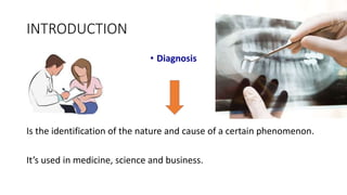
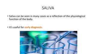
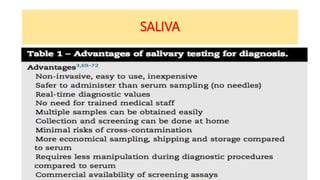
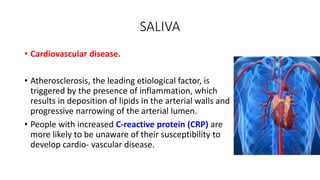
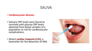
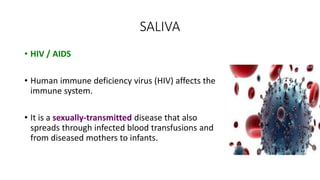
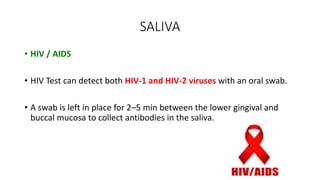
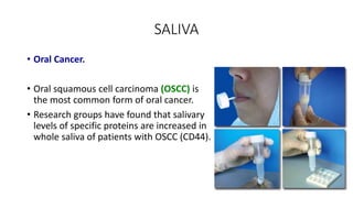
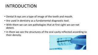
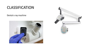
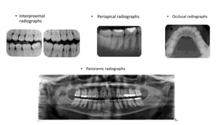
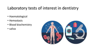
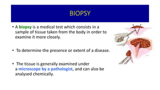
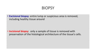
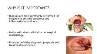
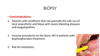
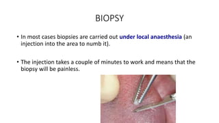
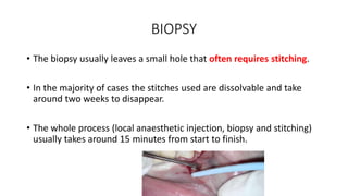
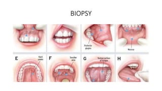
This lecture, which oriented to the level of mind of undergraduate students, discuss the topic of pulpectomy, its indications, contraindications, and procedural steps.
Visit us on Facebook:
https://www.facebook.com/iraqi.Dental.Academy
In this lecture I explain the basic concept of root canal filling or what called obturation. The lectures discuss different techniques used in that matter in step-by-step fashion and explanatory pictures.
It is directed to the level of undergraduate mind.
Diagnostic aids in endodontics /certified fixed orthodontic courses by India...Indian dental academy
Welcome to Indian Dental Academy
The Indian Dental Academy is the Leader in continuing dental education , training dentists in all aspects of dentistry and offering a wide range of dental certified courses in different formats.
Indian dental academy has a unique training program & curriculum that provides students with exceptional clinical skills and enabling them to return to their office with high level confidence and start treating patients
State of the art comprehensive training-Faculty of world wide repute &Very affordable.
Here are some key resources on pain and anxiety control in dentistry:
- Sturdvent - A leading manufacturer of dental equipment for pain and anxiety control.
- Ada's journal on anxiety and pain control - The American Dental Association's journal focused on non-pharmacological approaches.
- Journal on pain management by the American Society of Endodontists - Focuses on managing endodontic pain.
- Journal on pain control in dentistry - Focused specifically on controlling pain during various dental procedures.
- Pickard's manual of operative dentistry - A comprehensive textbook covering techniques for operative dentistry including pain control.
- Pain control in operative dentistry by Dr Ann Elrich - A
This document discusses techniques for localizing objects using radiography. It describes common reasons for needing to localize foreign bodies or other objects like unerupted teeth, fractures, or tumors. Two main techniques are described: Miller's technique which uses two radiographs at right angles, and Clark's tube-shift technique which analyzes how an object's image shifts when the projection angle is changed. The advantages and disadvantages of each technique are provided.
Tooth mobility refers to loose teeth that can move within their sockets. It is classified on a scale of 0 to 3 based on the degree of horizontal and vertical movement. Physiologic mobility of about 0.25mm is normal, while pathologic mobility over 1mm indicates loose teeth from periodontal disease or trauma. Periodontal disease is a primary cause as it leads to loss of attachment and bone supporting the teeth. Treatment involves splinting loose teeth together, replacing missing teeth, and correcting occlusal surfaces to reduce excessive forces. For advanced periodontal cases, extraction may be necessary.
The document discusses local anesthesia techniques for pediatric patients. It notes that the mandibular foramen is lower in children than adults, so the inferior alveolar nerve block injection must be made slightly lower and more posterior. The technique involves opening the patient's mouth wide, placing the thumb on the coronoid notch and fingers on the posterior mandible, then inserting the needle parallel to the bone at the occlusal plane level between ridges and injecting adjacent to the bone. A long buccal nerve block can also be used to anesthetize molar gingiva by injecting in the mucobuccal fold distal to the most posterior molar.
This document provides an overview of periodontal instruments, including their classification, components, uses, and proper techniques. It discusses:
1) The four main classifications of instruments: diagnostic, scaling/root planing/curettage, cleansing/polishing, and surgical.
2) Specific instruments within each classification like mirrors, probes, scalers, curettes, and their purposes.
3) Factors like proper grasp, finger rests, and sharpness that provide stability and control during instrumentation.
The document provides information on traumatic injuries to teeth, including concussions, luxations, and fractures. It describes the clinical signs, radiographic findings, and treatment approaches for each type of injury. Concussions involve inflammation of the periodontal ligament without tooth displacement. Luxations occur when a tooth is displaced from its socket, sometimes with alveolar bone fractures. Fractures are classified as enamel fractures, enamel-dentin fractures, enamel-dentin-pulp fractures, or root fractures. Treatment depends on the specific injury but may include repositioning displaced teeth, pulpotomies, root canals, extractions, or orthodontic/surgical repositioning.
This document discusses various types of root resorption including their causes, characteristics, diagnosis and treatment. It describes internal and external root resorption, further dividing external resorption into surface, inflammatory, replacement and invasive types. The key causes are trauma, pressure from impacted teeth or tumors, and systemic conditions. Diagnosis involves history, clinical exams, and radiographs to identify patterns of tooth structure loss. Treatment aims to arrest the resorptive process through root canal therapy or surgery depending on the type and severity.
This document discusses early diagnosis of dental caries. It defines dental caries and outlines various diagnostic methods including visual and tactile inspection, caries detection dyes, radiography, fiber optic transillumination, and digital methods. Radiography provides additional information compared to visual examination alone but has limitations in detecting enamel lesions. Digital radiography and subtraction techniques allow comparisons over time. Overall, early and accurate diagnosis is important for determining treatment and prognosis.
An overview of the diagnostic process in endodontics, including information about the pain system, referred pain, non-odontogenic pain, the diagnostic process, tests and treatment planning in endodontics.
This document discusses different impression techniques used in dentistry. It describes stock and custom impression trays, and their advantages and disadvantages. It also explains several impression techniques in detail, including the physiologic, McLean, Hindels modification, fluid wax, and selected pressure techniques. The selected pressure technique aims to equalize support between teeth and soft tissue by directing more force to areas that can absorb stress without damage, while protecting less resilient areas.
In this lecture I explain in step-by-step fashion the basics of Endodontic Diagnosis: Pulp Vitality Tests. a photo guide is attached to the guide to aid in better understanding of the topic
A absolutely minimalist way to describe each and every diagnostic aid in the beautiful stream of endodontics.
one has to understand the topic by going through the bible, "Grossman 13th Edition" along with the slides I've created.
Hope this helps.
by Dr. Ishaan Adhaulia
The document discusses various techniques for cleaning and shaping the root canal system during endodontic treatment. It describes the objectives and basic principles of root canal preparation, including removing debris and maintaining the original shape of the canal. Several techniques are summarized, such as step-back, crown-down, balanced force, and ultrasonic instrumentation. For each technique, the document outlines the steps and discusses advantages and disadvantages.
Radiography is essential for endodontic diagnosis, treatment, and evaluation of treatment outcomes. It helps determine pulpal and periapical pathology, root and canal morphology, working lengths, location of missed canals, and quality of obturation. Key radiographic views include diagnostic, working length, post-treatment, and recall films. Diagnostic films aim to visualize 3-4mm beyond the apex to identify lesions. Angulation and tube shift techniques help differentiate superimposed structures. Features like lamina dura continuity, lesion borders, density and effects on adjacent structures aid diagnosis. Newer technologies include digital radiography and cone beam CT for improved visualization of complex anatomy.
This document discusses various chairside investigations that can be performed to examine dental caries and pulp disease. It describes tests such as caries dyes, transillumination, electrical conductance measurement, laser fluorescence, and endoscopy that can detect dental caries. It also discusses pulp vitality tests involving heat, cold, or electricity to determine pulp status. Additional tests mentioned include lactobacillus colony count, Snyder test, and salivary buffer capacity tests to assess caries activity.
Traumatic Dental Injuries to Permanent TeethDrSusmita Shah
A comprehensive presentation of traumatic injuries to permanent teeth; this includes multiple classifications, risk factors, prevalence and management according to International Association of Dental Traumatology and Adreasen J O.
This document discusses space maintainers, which are appliances used to preserve space in the dental arch after premature loss of primary teeth. It describes different types of space maintainers including band and loop, lingual arch, distal shoe, and removable space maintainers. Indications and contraindications for each type are provided. The document also discusses myofunctional appliances used to correct oral habits like thumb sucking. Various types of clasps and springs used in removable partial dentures are described.
Dental caries is defined as a localized pathological process caused by acids produced by bacteria in dental plaque that leads to demineralization of tooth hard tissues. Diagnosis of caries involves detecting lesions, assessing activity, and determining risk factors to identify lesions requiring treatment and persons at high risk. A variety of tools can be used for caries diagnosis including visual inspection, fiber-optic transillumination, and diagnostic technologies assessing properties like fluorescence. The International Caries Detection and Assessment System (ICDAS) provides a standardized visual method for caries detection and assessment and has demonstrated validity though performance varies depending on tooth type and surface.
This document discusses the management of deep dental caries. It defines affected dentin as softened but not infected, while infected dentin is both softened and contaminated with bacteria. Various treatment modalities are described depending on whether the pulp is exposed and vital or non-vital. Factors like remaining dentin thickness and choice of restorative material influence pulpal response and reactionary dentin deposition. Materials used for pulp capping include calcium hydroxide, MTA, Biodentine and glass ionomer cements. Indirect and direct pulp capping techniques are also outlined.
Endodontic diagnosis could be a difficult task in most occasions, but with clinical assessment and careful history taking this task would be easier and clearer.
This lecture assembled by Osama Asadi, B.D.S, concentrating at the basic science of diagnosing pulpal and periapical diseases and their differential diagnosis and treatment plan. also endodontic case sheet and review-cases attached to the lecture at the end to help proper understanding of the subject.
This document discusses the use of radiography in endodontics. It begins with a brief history of dental radiography and then covers topics like how to obtain good radiographs, relevant findings for endodontists, different types of radiographs used, and techniques for intraoral periapical radiographs. The document emphasizes the importance of radiographs for diagnosing issues, determining working lengths, evaluating treatment outcomes, and providing follow-up assessments. Both advantages and disadvantages of radiographs are outlined. Various anatomical landmarks are also identified on sample radiographs.
Diagnosis and treatment planing in EndodonticsSalem Rekab
This document provides an overview of endodontic diagnosis and treatment planning. It discusses the importance of gathering a chief complaint, health history, dental history, and conducting a subjective and objective examination of the patient. The subjective examination involves questioning the patient about their symptoms such as the intensity, spontaneity, and persistence of any pain. The objective examination includes extraoral and intraoral soft tissue exams, examining the dentition, and performing clinical tests like percussion, palpation, and pulp vitality tests using heat, cold, or electricity. Together, the subjective and objective exams allow the clinician to make a tentative diagnosis which is then confirmed through further examination and testing.
This document discusses differential diagnosis and biopsy for oral lesions. It covers examination methods like health history and clinical examination. Biopsy techniques like incisional, excisional and aspiration are described. The document provides details on surgical biopsy procedure including anesthesia, tissue stabilization, wound closure and specimen handling. Surgical management of oral lesions is also outlined, discussing goals, techniques like enucleation, marsupialization and resection, and factors to consider.
This document provides information on examining radiographic films and interpreting x-rays. It discusses mounting films properly and viewing them as if inside the patient's mouth. When interpreting films, the dentist should do so systematically while considering the patient's age, history and clinical exam findings. Key areas to examine on x-rays include the teeth, bone, sinuses and joints. Terms used in radiographic diagnosis are also defined.
Cancer of the oral cavity can occur in any part of the mouth or throat and is highly curable if discovered early. Risk factors include smoking, alcohol, age over 50, and low fruit/vegetable diet. Diagnosis involves visual exam, biopsy, and imaging tests. Treatment may include surgery to remove the cancer, radiation therapy, chemotherapy, or a combination. Post-operative care focuses on maintaining nutrition, airway patency, and graft monitoring to help restore form and function.
The Indian Dental Academy is the Leader in continuing dental education , training dentists in all aspects of dentistry and
offering a wide range of dental certified courses in different formats.
A 35-year-old female presented with a swelling in her lower right jaw that had been gradually increasing in size over 6 months. Clinical and radiographic examination revealed a cystic lesion associated with missing tooth #45. Histologic examination of the surgically removed cyst confirmed the diagnosis of an odontogenic keratocyst. This type of cyst is known for its high recurrence rate due to the presence of satellite cysts and thin epithelial linings that can be left behind. Close follow-up for at least 5 years is recommended to monitor for recurrence.
This document presents a case report of a rare occurrence of basal cell adenoma in the palate of a 25-year-old male patient. It describes the clinical findings and diagnostic workup including imaging, biopsy, histopathological examination, and immunohistochemical analysis of the excised tumor. The pathological features were consistent with basal cell adenoma. The tumor was successfully removed surgically and the patient recovered well, with the tumor representing an unusual site for this rare minor salivary gland tumor.
This document discusses the investigation profile for acute hematogenous osteomyelitis. It notes that C-reactive protein levels and bacterial cultures from aspirated bone or tissue are important for diagnosis. Imaging plays a key role as well, with X-rays able to detect bone changes within a few days while MRI is most sensitive for detecting early bone marrow changes. The presentation reviews the typical findings and timeline of changes seen on various imaging modalities in the evaluation of acute osteomyelitis.
This document discusses biopsy procedures, including different types of biopsies and factors considered when determining the appropriate biopsy method. A biopsy is a surgical procedure that removes a tissue sample to examine microscopically for diagnosis. The goals are to define lesions, establish prognosis, guide treatment, assess treatment efficacy, and provide medical-legal documentation. Biopsy is indicated when oral abnormalities persist after treatment or irritant removal, or when malignancy is suspected based on characteristics like color, ulceration, induration, or fixation. Contraindications include vascular or intrabony lesions and those near vital structures. The appropriate biopsy technique depends on factors like the site, appearance, size, and number of abnormalities as well as imaging and available systems
Radiographic Features of Normal and Abnormal Salivary Glands Hadi Munib
This document provides an overview of salivary gland diseases and their diagnostic imaging. It defines inflammatory and non-inflammatory salivary gland disorders as well as space-occupying masses. Clinical signs and symptoms are outlined along with differential diagnoses of salivary gland enlargements. Various diagnostic imaging techniques are discussed including plain radiography, sialography, CT, MRI, ultrasound and scintigraphy. Conventional sialography, which involves infusing contrast agent into the ductal system, is described as an important method for evaluating ductal obstructions and the detailed anatomy.
This document provides information about biopsies, including definitions, indications, contraindications, types of biopsies, and how to properly perform and handle specimens. It defines a biopsy as a surgical procedure that obtains a living tissue sample for diagnosis. The main types discussed are incisional, excisional, and punch biopsies. Incisional biopsies remove a small portion of tissue, while excisional biopsies remove the entire lesion. Punch biopsies can be used for small, accessible lesions. Proper biopsy technique and specimen handling are emphasized to avoid errors and obtain an accurate diagnosis.
This document provides an overview of salivary gland disorders. It discusses the anatomy and function of major and minor salivary glands. Common salivary gland disorders are then described, including sialolithiasis, mucoceles, ranulas, infections such as mumps, Sjögren's syndrome, and various neoplasms including pleomorphic adenoma, Warthin's tumor, and mucoepidermoid carcinoma. Diagnosis and treatment approaches are outlined for each condition.
The document provides information about cancer of the larynx (voice box), including its definition, causes, signs and symptoms, diagnostic tests, staging, and treatment options. The main causes are smoking and drinking alcohol. Symptoms depend on the location of the tumor and may include hoarseness, cough, neck mass, difficulty swallowing and breathing. Diagnostic tests include laryngoscopy, CT/MRI scans, and biopsy. Treatment involves surgery such as partial or total laryngectomy, chemotherapy, and radiation therapy. Complications can include airway obstruction, hemorrhage, and fistula formation.
The document discusses salivary gland disorders. It begins with definitions and classifications of salivary glands. It then discusses the anatomy, functions, and disorders of the parotid, submandibular, and sublingual salivary glands. Diagnostic aids are outlined including clinical history, physical examination, imaging such as CT, MRI, ultrasound and sialography. Cystic conditions of the minor salivary glands such as mucoceles are also summarized. Disorders are classified and inflammatory, obstructive, neoplastic and other conditions are described.
Osteomyelitis/certified fixed orthodontic courses by Indian dental academyIndian dental academy
The Indian Dental Academy is the Leader in continuing dental education , training dentists in all aspects of dentistry and
offering a wide range of dental certified courses in different formats.for more details please visit
www.indiandentalacademy.com
1) Parotitis is inflammation of the parotid salivary glands, usually caused by viral infection in children or dehydration in elderly/ill patients. Symptoms include swollen glands, ear pain, difficulty swallowing. Treatment focuses on hydration, antibiotics, and sometimes surgery.
2) Sialadenitis is inflammation of any salivary gland that can be caused by dehydration, radiation, stress or infection. Symptoms are pain, swelling and discharge. Treatment is antibiotics, massage, hydration and sometimes surgery.
3) Salivary stones form from calcium deposits and cause pain from duct obstruction. Diagnosis is by ultrasound/x-rays. Treatment involves extracting stones
Estudio sobre la elevaciòn del seno del maxilar con abordaje crestal. Se compara con la técnica de ventana lateral. Se estudia las cualidades del hueso autologo (AUTOBT).
La relación que existe entre el mundo odontologico y el consumo de alcohol. En el detalle, hemos dado la prioridad al manejo clinico de estas circunstancias especificas.
Técnicas histopatologicas e inmunohistoquimicasFedeVillani
Técnicas frecuentes de diagnostico para patologias de la cavidad bucal. Concretamente se analizan el pénfigo, penfigoide y el liquen plano. Explicaciòn de la biopsia y de la inmunofluorescencia.
Este documento describe las diferentes clases de lesiones cariosas (clases I-VI) según la clasificación de Black, incluyendo definiciones, morfología de la cavidad y materiales de restauración apropiados. Explica los procedimientos clínicos para cada clase, como la preparación de la cavidad y colocación del material restaurador. También resume brevemente algunos estudios sobre técnicas y materiales para restauraciones de clases II y III. El objetivo es proporcionar una guía sobre el enfoque operatorio adecuado para cada
Este documento describe los trastornos convulsivos y las enfermedades neurológicas. Explica que las convulsiones son un síntoma transitorio caracterizado por actividad neuronal anormal en el cerebro. La epilepsia implica la recurrencia crónica de crisis convulsivas. Describe los tipos de convulsiones, las causas y el diagnóstico de la epilepsia, así como los tratamientos disponibles como la cirugía y la medicación. También resume la neuralgia del trigémino, un trastorno doloroso crónico que afecta el nervio
- Los estudios clínicos y médicos han demostrado que la superficie más externa del esmalte participa en la reacción al ácido desde el inicio de la lesión, disolviéndose directamente y agrandando las vías de difusión intercristalina.
- Durante la erupción dental, los dientes están expuestos a placa bacteriana y pueden presentar varios grados de desintegración de la microsuperficie. Estos cambios subclínicos pueden convertirse en lesiones inactivas una vez eliminado el estí
El documento proporciona una introducción a la fitoterapia, incluyendo su historia, comparación entre países, composición de plantas medicinales, y ejemplos de plantas con acciones inmunoestimulante, cardiovascular, digestiva, antirreumática y a nivel del sistema nervioso central. También incluye una bibliografía de recursos adicionales sobre fitoterapia.
alternativas terapeuticas a la depresiónFedeVillani
Este documento describe alternativas terapéuticas naturales para tratar la depresión, incluyendo fitoterapia con hierbas como la hierba de San Juan y el ginkgo biloba, psicoterapia, actividad física, masajes y una dieta saludable. También distingue entre depresión endógena y exógena.
Normal human oral keratinocyte. how to study NHOK. ResearchFedeVillani
This document describes experiments conducted on normal human oral keratinocytes (NHOK). NHOK were isolated from oral tissue, cultured, and infected with retroviruses or lentiviruses. The effects of overexpressing or knocking down the Bmi-1 gene were examined. Various techniques were used, including PCR, western blotting, ELISA, and galactosidase staining. The results provide insights into how Bmi-1 affects proliferation, senescence pathways, and TGF-β secretion in NHOK.
Patologia- Lesiones de la piel frecuentesFedeVillani
Este documento describe diferentes tipos de lesiones cutáneas, incluyendo eritema, angioma, escara, viruela, estrella venosa y quistes. Proporciona definiciones breves para cada lesión, describiendo sus características y causas. El documento fue creado por un grupo de estudiantes como parte de un proyecto sobre lesiones de la piel.
Coordination of mastication, swallowing and breathingFedeVillani
The document discusses the coordination of mastication (chewing), swallowing, and breathing in the pharynx. The pharynx serves multiple functions including breathing, speaking, chewing, and swallowing. During chewing, food accumulates in the pharynx before swallowing. Swallowing requires the pharynx to change from an airway to a food passage and coordination between swallowing and breathing. Future research is needed to better understand control of structures like the soft palate and tongue during these functions as well as preventing aspiration during chewing and swallowing.
Semiologia cancer de pulmones, mama, prostata y colonFedeVillani
Este documento describe varios tipos de cáncer, incluyendo cáncer de mama, pulmón, próstata, colon, cuello uterino, melanoma, y cerebral. Explica las características, causas, síntomas, detección y tratamientos de cada uno. El cáncer se produce cuando las células crecen de forma anormal e invaden otros tejidos. Los factores de riesgo incluyen edad, historia familiar, tabaco, dieta, infecciones virales y exposición a radiación UV o químicos. La detección temp
El documento describe los analgésicos más empleados en odontología, clasificándolos en opiáceos, no opiáceos y antiinflamatorios no esteroideos. Explica que el dolor dental es agudo y puede estar relacionado con terapias como endodoncia o extracciones. Los analgésicos no opiáceos como el ibuprofeno, paracetamol y AINES son los más usados para el dolor leve a moderado, mientras que los opiáceos como la morfina se reservan para el dolor severo. Con
Osteoporosis - Definition , Evaluation and Management .pdfJim Jacob Roy
Osteoporosis is an increasing cause of morbidity among the elderly.
In this document , a brief outline of osteoporosis is given , including the risk factors of osteoporosis fractures , the indications for testing bone mineral density and the management of osteoporosis
Basavarajeeyam is a Sreshta Sangraha grantha (Compiled book ), written by Neelkanta kotturu Basavaraja Virachita. It contains 25 Prakaranas, First 24 Chapters related to Rogas& 25th to Rasadravyas.
TEST BANK For An Introduction to Brain and Behavior, 7th Edition by Bryan Kol...rightmanforbloodline
TEST BANK For An Introduction to Brain and Behavior, 7th Edition by Bryan Kolb, Ian Q. Whishaw, Verified Chapters 1 - 16, Complete Newest Versio
TEST BANK For An Introduction to Brain and Behavior, 7th Edition by Bryan Kolb, Ian Q. Whishaw, Verified Chapters 1 - 16, Complete Newest Version
Muktapishti is a traditional Ayurvedic preparation made from Shoditha Mukta (Purified Pearl), is believed to help regulate thyroid function and reduce symptoms of hyperthyroidism due to its cooling and balancing properties. Clinical evidence on its efficacy remains limited, necessitating further research to validate its therapeutic benefits.
TEST BANK For Basic and Clinical Pharmacology, 14th Edition by Bertram G. Kat...rightmanforbloodline
TEST BANK For Basic and Clinical Pharmacology, 14th Edition by Bertram G. Katzung, Verified Chapters 1 - 66, Complete Newest Version.
Adhd Medication Shortage Uk - trinexpharmacy.comreignlana06
The UK is currently facing a Adhd Medication Shortage Uk, which has left many patients and their families grappling with uncertainty and frustration. ADHD, or Attention Deficit Hyperactivity Disorder, is a chronic condition that requires consistent medication to manage effectively. This shortage has highlighted the critical role these medications play in the daily lives of those affected by ADHD. Contact : +1 (747) 209 – 3649 E-mail : sales@trinexpharmacy.com
1. DIAGNOSTIC TOOLS IN DENTISTRY
• English Activity
Valencia, Spain.
M51
18-5-17
2. INDEX OF WORK
• Introduction
• Saliva
• X-Ray
• Biopsy
• Bibliography
3. INTRODUCTION
• Diagnosis
Is the identification of the nature and cause of a certain phenomenon.
It’s used in medicine, science and business.
4. SALIVA
• Saliva is a watery substance secreted by the salivary glands.
• Comprises:
a. water 99,5%.
b. electrolytes.
c. mucus.
d. glycoproteins.
e. enzymes.
f. antimicrobial agents.
g. epithelial cells.
h. White blood cells.
5. SALIVA
• Saliva can be seen in many cases as a reflection of the physiological
function of the body.
• It’s useful for early diagnosis.
7. SALIVA
• Cardiovascular disease.
• Atherosclerosis, the leading etiological factor, is
triggered by the presence of inflammation, which
results in deposition of lipids in the arterial walls and
progressive narrowing of the arterial lumen.
• People with increased C-reactive protein (CRP) are
more likely to be unaware of their susceptibility to
develop cardio- vascular disease.
8. SALIVA
• Cardiovascular disease.
• Salivary CRP levels were found to
correlate with plasma CRP levels
obtained from blood samples of a
population at risk for cardiovascular
complications.
• Detect cardiac troponin (cTn), a
biomarker for the detection of AMI.
9. SALIVA
• HIV / AIDS
• Human immune deficiency virus (HIV) affects the
immune system.
• It is a sexually-transmitted disease that also
spreads through infected blood transfusions and
from diseased mothers to infants.
10. SALIVA
• HIV / AIDS
• HIV Test can detect both HIV-1 and HIV-2 viruses with an oral swab.
• A swab is left in place for 2–5 min between the lower gingival and
buccal mucosa to collect antibodies in the saliva.
11. SALIVA
• Oral Cancer.
• Oral squamous cell carcinoma (OSCC) is
the most common form of oral cancer.
• Research groups have found that salivary
levels of specific proteins are increased in
whole saliva of patients with OSCC (CD44).
12. SALIVA
• Salivary tests will pave the way for chair-side diagnosis of multiple
oral and systemic diseases at the dental office.
• The advent of sensitive and specific salivary diagnostic tools and the
establishment of defined guidelines will make salivary diagnostics a
reality in the near future.
14. • Dental X-rays are a type of image of the teeth and mouth.
• Are used in dentistry as a fundamental diagnostic tool.
• With them we can see pathologies that at first sight we can not
detect.
• In them we see the structures of the oral cavity reflected according to
their density.
INTRODUCTION
17. OBJECTIVES
• Quantity, size and position of teeth.
• Teeth that have not come out or teeth impacted.
• Dental caries.
• Bone damage (such as that produced by periodontitis).
• Dental abscesses.
• Fractured jaw.
• Problems in the way the upper and lower teeth fit Other anomalies of
mandibular teeth and bones.
18. Parallel technique:
• The radiograph is placed in a position parallel to the longitudinal axis
of the tooth to be examined, in a central beam directed perpendicular
to the radiograph and the longitudinal axis of the tooth
Bisector technique:
• At the point where the radiograph has contact with the tooth, the
plane of the radiograph, and the longitudinal axis of the tooth form
an angle, the radiologist must imagine a plane that divides by half
the angle formed by the film and the longitudinal axis of the tooth,
this plane is called bisector, which creates two equal angles
19. RISKS
• Exposure to radiation from dental
radiographs is very low.
• A lead apron may be used to cover the
body and reduce radiation exposure.
• Pregnant women should not be given x-
rays unless absolutely necessary.
20. Laboratory tests of interest in dentistry
• Haematological
• Hemostasis
• Blood biochemistry
• saliva
21. Blood count
• Erythrocyte series: number of red blood cells, hematocrit,
hemoglobin
• Leukocyte series: Total count, neutrophils, monocytes…
• Platelet series: normal value : 150.000-400.000 mm3
23. Infections diseases:
• VIH
• Hepatitis
• TBC
It is better to prevent a complication through
diagnosis than to resolve it intraoperatively
24. BIOPSY
• A biopsy is a medical test which consists in a
sample of tissue taken from the body in order to
examine it more closely.
• To determine the presence or extent of a disease.
• The tissue is generally examined under
a microscope by a pathologist, and can also be
analysed chemically.
25. BIOPSY
• Excisional biopsy: entire lump or suspicious area is removed,
including healthy tissue around
• Incisional biopsy: only a sample of tissue is removed with
preservation of the histological architecture of the tissue’s cells.
26. WHY IS IT IMPORTANT?
• Biopsies are most commonly performed for
insight into possible cancerous and
inflammatory conditions.
• Lesion with similar clinical or radiological
morphology.
• Provides definitive diagnosis, prognosis and
treatment information.
27. BIOPSY
• Contraindications:
1. Patients with conditions that can preclude the safe use of
local anaesthetic and those with severe bleeding diseases
and coagulopathies.
1. Invasive procedures on the bone: NO in patients with
bisphosphonates treatment.
2. Risk for metastasis.
28. BIOPSY
• In most cases biopsies are carried out under local anaesthesia (an
injection into the area to numb it).
• The injection takes a couple of minutes to work and means that the
biopsy will be painless.
29. BIOPSY
• The biopsy usually leaves a small hole that often requires stitching.
• In the majority of cases the stitches used are dissolvable and take
around two weeks to disappear.
• The whole process (local anaesthetic injection, biopsy and stitching)
usually takes around 15 minutes from start to finish.