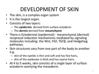
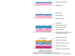
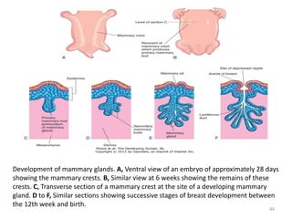
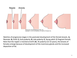
DEVELOPMENT OF SKIN AND
ASSOCIATED STRUCTURES
Dr Peter Gichangi
Nov 12 2012
1
OBJECTIVES
• Understand the development of different
layers of the skin
• Understand development of skin
associated/related structures
• Understand the embryological basis of
congenital malformations of skin development
2
INTRODUCTION
• The integumentary system consists of:
– the skin
– and its appendages:
• sweat glands,
• nails,
• hairs,
• sebaceous glands,
• arrector muscles of hairs (arrector pili muscles),
• mammary glands,
• teeth
3
DEVELOPMENT OF SKIN
• The skin, is a complex organ system
• It is the largest organ.
• Consists of two layers:
– The epidermis derived from surface ectoderm
– The dermis derived from mesenchyme
• There is Ectodermal (epidermal) - mesenchymal (dermal)
reciprocal induction mechanisms mediated by signaling
molecules including: the Wnt, FGF, TGFβ, and Hedgehog
pathways.
• Skin structures vary from one part of the body to another
e.g.
– skin of the eyelids is thin and soft and has fine hairs,
– skin of the eyebrows is thick and has coarse hairs.
• At 4 to 5 weeks, skin consists of a single layer of surface
ectoderm overlying the mesoderm.
4
5
Light micrograph of thick skin (x132). Observe the epidermis and dermis as well as the
dermal papilla interdigitating with the epidermal ridges.
6
Epidermis
• Epidermal growth occurs in stages.
• The primordium is a single layer of surface ectodermal cells.
• These cells proliferate and form a layer of squamous epithelium,
the periderm, and a basal layer.
• The cells of the periderm continually undergo keratinization and
desquamation, and are replaced by cells from the basal layer.
• The exfoliated peridermal cells and sebum from sebaceous
glandsform a white greasy substance - vernix caseosa - that covers
the fetal skin.
• The vernix:
– protects the developing skin from constant exposure to amniotic fluid,
with its high content of urine, bile salts, and sloughed cells, during the
fetal period.
– facilitates birth of the fetus.
7
• The basal layer of the epidermis becomes the stratum
germinativum which produces other layers and epidermal
ridges.
• The transformation of the surface ectoderm into the
multilayered definitive epidermis is induced by the dermis.
• By 11 weeks, the intermediate layer is formed.
• Replacement of peridermal cells continues until
approximately the 21st week; thereafter, the periderm
disappears and the stratum corneum forms from the
stratum lucidum.
• Epidermal ridges:
– appear by 10th week
– permanently established by 19 weeks
– those of the hand appear approximately one week earlier than
in the feet.
8
• The epidermal ridges produce grooves on the
surface of the palms and soles, fingers and toes.
• The type of pattern genetically determined
genetically and is specific for an individual - basis
for fingerprints.
• Abnormal chromosome complements affect the
development of ridge patterns; for instance,
infants with Down syndrome have distinctive
patterns on their hands and feet that are of
diagnostic value.
• Neural crest cells migrate into the mesenchyme
of the developing dermis and differentiate into
melanoblasts.
9
• Melanoblasts migrate to the dermoepidermal
junction and differentiate into melanocytes
which contain pigment granules.
• Melanocytes appear in the developing skin at
40 to 50 days, immediately after the migration
of neural crest cells.
• Only a few melanin - containing cells are
normally present in the dermis.
• The melanocytes begin producing melanin
before birth and distribute it to the epidermal
cells.
10
Drawing of the
successive
stages in the
development
of hairs,
sebaceous
glands, and
arrector
muscles of
hair. Note that
the sebaceous
gland develops
as an
outgrowth
from the side
of the hair
follicle.
11
12
• The relative content of melanin inside the
melanocytes accounts for the different colors
of skin in different races and not the number
of melanocytes.
• Skin is classified as thick or thin based on the
thickness of the epidermis.
– Thick skin covers the palms of the hands and the
soles of the feet; it lacks hair follicles, arrector
muscles of hairs, and sebaceous glands, but it has
sweat glands.
– Thin skin covers most of the rest of the body; it
contains hair follicles, arrector muscles of hairs,
sebaceous glands, and sweat glands.
13
Dermis
• The dermis develops from mesenchyme derived
from:
– somatic layer of lateral mesoderm
– dermatomes of the somites.
• By 11 weeks, the mesenchymal cells have begun
to produce collagenous and elastic connective
tissue fibers.
• The epidermal ridges form the dermis projects
into the epidermis, forming dermal papillae,
which interdigitate with the epidermal ridges.
14
Light micrograph of thick skin (x132). Observe the epidermis and dermis as well as the
dermal papilla interdigitating with the epidermal ridges. (From Gartner LP, Hiatt JL: Color
Textbook of Histology, 2nd ed. Philadelphia, WB Saunders, 2001.) 15
• Some dermal papillae:
– have capillary loops of blood vessels to nourish the
epidermis
– others have sensory nerve endings.
• The developing afferent nerve fibers influence
the spatial and temporal sequence of dermal
ridge formation.
• By 5th week, capillary - like vessels are present.
• These capillary - like structures differentiate to
arterioles, arteries, venules and veins.
• By the end of the first trimester, the major
vascular organization of the fetal dermis is
established.
16
Development of Glands
• The glands of the skin include:
–eccrine sweat glands
–apocrine sweat glands
–sebaceous glands
–mammary glands
• They are derived from the epidermis
and grow into the dermis.
17
Sebaceous Glands
• Cellular buds develop from the sides of the developing
epithelial root sheaths of hair follicles.
• The buds invade the surrounding dermal connective
tissue and branch to form several alveoli and their
associated ducts.
• The central cells of the alveoli break down, forming an
oily substance – sebrum - that protects the skin against
friction and dehydration.
• Sebum is released into the hair follicle and passes to
the surface of the skin to form vernix caseosa.
• Sebaceous glands in the glans of the penis and labia
minora, develop as cellular buds from the epidermis
that invade the dermis.
18
Drawing of the
successive
stages in the
development
of hairs,
sebaceous
glands, and
arrector
muscles of
hair. Note that
the sebaceous
gland develops
as an
outgrowth
from the side
of the hair
follicle.
19
Sweat Glands
• Eccrine sweat glands are located in the skin
throughout most of the body.
• They develop as cellular buds from the epidermis
that grow into the underlying mesenchyme.
• As the buds elongate, their ends coil to form the
bodies of the secretory parts of the glands.
• The epithelial attachments of the developing
glands to the epidermis forms the sweat ducts.
• The peripheral cells of the secretory parts of the
glands differentiate into myoepithelial and
secretory cells.
20
Illustrations of the successive stages of the development of a sweat gland. A and B, The
cellular buds of the glands develop at approximately 20 weeks as a solid growth of
epidermal cells into the mesenchyme. C, Its terminal part coils and forms the body of the
gland. The central cells degenerate to form the lumen of the gland. D, The peripheral cells
differentiate into secretory cells and contractile myoepithelial cells.
21
• Myoepithelial cells are specialized smooth muscle cells
that assist in expelling sweat from the glands.
• Eccrine sweat glands begin to function shortly after
birth.
• Sudoriferous (producing sweat) apocrine sweat glands
are mostly confined to the axillary, pubic and perineal
regions, and the areolae of the breast.
• The glands develop from down growths of the stratum
germinativum of the epidermis .
• The ducts of these glands open, not onto the skin
surface, as do eccrine sweat glands, but into the canals
of the hair follicles superficial to the entry of the
sebaceous gland ducts.
• Secretion by apocrine sweat glands is influenced by
hormones and does not begin until puberty.
22
DISORDERS OF KERATINIZATION
• Ichthyosis is agroup of skin disorders resulting from excessive
keratinization - formation of horny layers of skin.
– The skin is characterized by dryness and scaling, which may involve the entire body
surface.
• A harlequin fetus results from a rare keratinizing disorder that is inherited
as an autosomal recessive trait.
– The skin is markedly thickened, ridged, and cracked.
– Most affected neonates die during the first week of life.
• A collodion infant is covered by a thick, taut membrane that resembles
collodion or parchment.
– The membranous skin cracks and begins to fall off in large sheets.
– Deficiency of transglutaminase-1 is the most common cause.
– Complete shedding may take several weeks, occasionally leaving normal-appearing skin.
• Lamellar ichthyosis is an autosomal recessive disorder. A neonate may
appear to be a collodion baby; however, the scaling persists.
– Growth of hair may be curtailed and development of sweat glands is often impeded.
– Affected infants are unable to sweat.
23
A, A child with congenital hypertrichosis and hyperpigmentation. Note the excessive
hairiness on the shoulders and back. B, A child with severe keratinization of the skin
(ichthyosis) from the time of birth.
24
CONGENITAL ECTODERMAL DYSPLASIA
• A group of rare hereditary disorders involving tissues that are
ectodermal in origin.
• The teeth are completely or partially absent and often the hairs and
nails are affected.
• Ectrodactyly - ectodermal dysplasia - clefting syndrome is a
congenital skin that involves both ectodermal and mesodermal
tissues, consisting of:
– ectodermal dysplasia associated with hypopigmentation of skin and
hair,
– scanty hair and eyebrows,
– absence of eyelashes,
– nail dystrophy,
– hypodontia
– microdontia,
– ectrodactyly, and cleft lip and palate.
25
ANGIOMAS OF SKIN
• Some transitory and/or surplus primitive
blood or lymphatic vessels persist as either:
– arteries,
– venous,
– cavernous angiomas,
– often of a mixed type.
• Angiomas composed of lymphatics are called
cystic lymphangiomas or cystic hygromas.
• True angiomas are benign tumors of
endothelial cells.
26
• Nevus flammeus denotes a flat, pink or red,
flame - like blotch that often appears on the
posterior surface of the neck.
• A port - wine stain hemangioma which is sharply
demarcated is a larger and darker angioma than a
nevus flammeus and is nearly always anterior or
lateral on the face and/or neck.
• Common angioma (pinkish-red blotch) may cross
the median plane.
• A port-wine stain in the area of distribution of the
trigeminal nerve is sometimes associated with a
similar type of angioma of the meninges of the
brain and seizures at birth (Sturge-Weber
syndrome).
27
Hemangioma (port-wine stain) in an infant.
28
ALBINISM
• Generalized albinism, is an autosomal
recessive trait where the skin, hairs, and retina
lack pigment; however, the iris usually shows
some pigmentation.
• Albinism occurs when the melanocytes fail to
produce melanin because of the lack of the
enzyme tyrosinase or other pigment enzymes.
• In localized albinism – piebaldism - an
autosomal dominant trait, there is a lack of
melanin in patches of skin and/or hair.
29
Mammary Glands
• Mammary glands are modified and highly
specialized types of sweat glands.
• Development of mammary glands also called
mammogenesis is similar in male and female
embryos.
• The first evidence of mammary development
appears in the fourth week when mammary
crests (ridges)/line develop along each side of the
ventral surface of the embryo.
• These crests extend from the axillary region to
the inguinal region.
30
• The crests usually disappear except for the
parts at the site of the future breasts.
• Involution of the remaining mammary crests
in the 5th week produces the primary
mammary buds.
• These buds are down growths of the
epidermis into the underlying mesenchyme.
• The changes occur in response to an inductive
influence from the mesenchyme.
31
Development of mammary glands. A, Ventral view of an embryo of approximately 28 days
showing the mammary crests. B, Similar view at 6 weeks showing the remains of these
crests. C, Transverse section of a mammary crest at the site of a developing mammary
gland. D to F, Similar sections showing successive stages of breast development between
the 12th week and birth.
32
• Each primary mammary bud soon gives rise to several
secondary mammary buds, which develop into lactiferous
ducts and their branches.
• Canalization of secondary mammary buds is induced by
placental sex hormones entering the fetal circulation.
• By term, 15 to 19 lactiferous ducts are formed.
• The fibrous connective tissue and fat of the mammary
glands develop from the surrounding mesenchyme.
• During the late fetal period, the epidermis at the site of
origin of the mammary gland becomes depressed, forming
a shallow mammary pit.
• The nipples are poorly formed and depressed in neonates.
• Soon after birth, the nipples usually rise from the
mammary pits because of proliferation of the surrounding
connective tissue of the areola.
33
• The smooth muscle fibers of the nipple and areola
differentiate from surrounding mesenchymal cells.
• The rudimentary mammary glands of newborn males and
females are identical and are often enlarged.
• Some secretion, often called "witch's milk" (galactorrhea),
may be produced caused by maternal hormones.
• The breasts of neonates contain lactiferous ducts but no
alveoli (secretory parts of glands).
• In females, the breasts enlarge rapidly during puberty,
mainly because of development of the mammary glands
and the accumulation of the fibrous stroma and fat
associated with them.
• Full development occurs at approximately 19 years.
• Normally, the lactiferous ducts of males remain
rudimentary throughout life.
34
Sketches of progressive stages in the postnatal development of the female breasts. A,
Neonate. B, Child. C, Early puberty. D, Late puberty. E, Young adult. F, Pregnant female.
Note that the nipple is inverted at birth (A). At puberty (12-15 years), the breasts of
females enlarge because of development of the mammary glands and the increased
deposition of fat.
35
GYNECOMASTIA
• Gynecomastia refers to the development of the
rudimentary lactiferous ducts in the male mammary
tissue.
• During midpuberty, approximately two thirds of boys
develop varying degrees of hyperplasia of the breasts.
• This subareolar hyperplasia may persist for a few
months to 2 years.
• A decreased ratio of testosterone to estradiol is found
in boys with gynecomastia.
• Approximately 80% of males with Klinefelter syndrome
have gynecomastia, which is associated with an XXY
chromosome complement.
36
ABSENCE OF NIPPLES (ATHELIA) OR
BREASTS (AMASTIA)
• These rare birth defects may occur bilaterally or
unilaterally.
• They result from failure of development or
disappearance of the mammary crests.
• These conditions may also result from failure of
mammary buds to form.
• More common is hypoplasia of the breast, often
found in association with gonadal agenesis and
Turner syndrome.
• Poland syndrome is associated with hypoplasia or
absence of the breast or nipple.
37
APLASIA OF BREAST
• The breasts of postpubertal females often
differ somewhat in size.
• Marked differences are regarded as anomalies
because both glands are exposed to the same
hormones at puberty.
• In these cases, there is often associated
rudimentary development of muscles of the
thoracic wall, usually the pectoralis major.
38
SUPERNUMERARY BREASTS AND NIPPLES
• An extra breast (polymastia) or nipple
(polythelia) occurs in approximately 1% of the
female population - inherited.
• An extra breast or nipple usually develops just
inferior to the normal breast.
• Supernumerary nipples are also relatively
common in males; often they are mistaken for
moles.
• Less commonly, supernumerary breasts or
nipples appear in the axillary or abdominal
regions of females.
39
• In these positions, the nipples or breasts develop
from extra mammary buds that develop from
remnants of the mammary crests.
• They usually become more obvious in women
when they are pregnant.
• Approximately one third of affected persons have
two extra nipples or breasts.
• Supernumerary mammary tissue very rarely
occurs in a location other than along the course
of the mammary crests.
• It probably develops from tissue that was
displaced from these crests.
40
Female infant with an extra nipple (polythelia) on the left side.
41
A man with polythelia (extra nipples) in the axillary and thigh regions. Insets are
enlargements of the nipples (arrowheads). The broken line indicates the original
position of the left mammary crests.
42
INVERTED NIPPLES
• Sometimes the nipples fail to elevate above
the skin surface after birth or during puberty,
that is, they remain in their prenatal location.
• Inverted nipples may make breast-feeding of
an infant difficult; however, a number of
breast-feeding techniques can be used to
reduce this difficulty.
43
Development of mammary glands. A, Ventral view of an embryo of approximately 28 days
showing the mammary crests. B, Similar view at 6 weeks showing the remains of these
crests. C, Transverse section of a mammary crest at the site of a developing mammary
gland. D to F, Similar sections showing successive stages of breast development between
the 12th week and birth.
44
Sketches of progressive stages in the postnatal development of the female breasts. A,
Neonate. B, Child. C, Early puberty. D, Late puberty. E, Young adult. F, Pregnant female.
Note that the nipple is inverted at birth (A). At puberty (12-15 years), the breasts of
females enlarge because of development of the mammary glands and the increased
deposition of fat.
45
Development of Hairs
• Hairs begin to develop early in the fetal period,
but they do not become easily recognizable until
approximately the 20th week.
• Hairs are first recognizable on the eyebrows,
upper lip, and chin.
• The hair follicles begin as proliferations of the
stratum germinativum of the epidermis, and
extend into the underlying dermis.
• The hair buds soon become club shaped, forming
hair bulbs.
46
• The epithelial cells of the hair bulbs constitute the
germinal matrix, which later produces the hair shafts.
• The hair bulbs (primordia of hair roots) are soon
invaginated by small mesenchymal hair papillae.
• The peripheral cells of the developing hair follicles
form the epithelial root sheaths, and the surrounding
mesenchymal cells differentiate into the dermal root
sheaths.
• As cells in the germinal matrix proliferate, they are
pushed toward the surface, where they become
keratinized to form hair shafts.
• The hairs grow through the epidermis on the eyebrows
and upper lip by the end of the 12th week.
47
Drawing of the
successive
stages in the
development
of hairs,
sebaceous
glands, and
arrector
muscles of
hair. Note that
the sebaceous
gland develops
as an
outgrowth
from the side
of the hair
follicle.
48
• The first hairs to appear - lanugo - are fine, soft,
and lightly pigmented.
• Lanugo hairs help to hold the vernix caseosa on
the skin.
• Lanugo is replaced by coarser hairs during the
perinatal period.
• This hair persists over most of the body, except in
the axillary and pubic regions, where it is
replaced at puberty by even coarser terminal
hairs.
• In males, similar coarse hairs also appear on the
face and often on the chest - anterior wall of
thorax.
49
• Melanoblasts migrate into the hair bulbs and differentiate
into melanocytes.
• The relative content of melanin accounts for different hair
colors.
• Arrector muscles of hairs, small bundles of smooth muscle
fibers, differentiate from the mesenchyme surrounding the
hair follicles and attach to the dermal root sheaths of the
hair follicles and the papillary layer of the dermis, which
interdigitates with the epidermis.
• Contractions of the arrector muscles depress the skin over
their attachment and elevate the skin around the hair
shafts, causing the hairs to stand up ("goose bumps").
• The arrector muscles are poorly developed in the hairs of
the axillary region and in certain parts of the face.
• The hairs forming the eyebrows and the cilia forming the
eyelashes have no arrector muscles.
50
Light micrograph
of a longitudinal
section of a hair
follicle with its
hair root (R) and
papilla (P) (x132).
51
52
• I-II - about three months, local thickenings of the epidermis appear
(beginning in the region of the forehead and eye-brows) and grow
obliquely into the underlying dermis in the form of solid buds the hair
germs. As the buds continue to elongate they become club-shaped and
the epithelium at the end of each molds itself over a little portion of the
dermis in which the cells have become more numerous and which is
known as the hair papilla.
• III - IV - As the epidermal bud grows deeper, its central cells become
spindle-shaped and undergo keratinization to form the beginning of the
hair shaft; the peripheral layers constitute the anlage of the root sheath.
The hair shaft grows from its basal end, new keratinized cells being added
from the epithelium nearest the papilla as the older cells are pushed
toward the surface of the skin.
• V - The surface cells of the hair shaft become flattened to form the cuticle
of the hair. The hairs appear above the surface about the fifth month. Of
the cells of the root sheath, those nearest the hair become scale-like to
form the cuticle of the root sheath; the next few layers become modified
(keratinized) to form Huxley's and Henle's layers. Outside of these is the
stratum germinativum, the basal layer of which is composed of columnar
cells resting upon a distinct basement membrane. The stratum
germinativum is continued over the tip of the papilla, where its cells give
rise to new cells for the hair shaft.
53
ALOPECIA
• Absence or loss of scalp hairs may occur alone
or with other defects of the skin and its
derivatives.
• Congenital alopecia (hair loss) may be caused
by failure of hair follicles to develop, or it may
result from follicles producing poor - quality
hairs.
54
55
• HYPERTRICHOSIS
– Excessive hairiness results from the development of
supernumerary hair follicles, or from the persistence of
lanugo hairs that normally disappear during the perinatal
period.
– It may be localized (e.g., on the shoulders and back) or
diffuse.
– Localized hypertrichosis is often associated with spina
bifida occulta.
• PILI TORTI
– In this familial disorder, the hairs are twisted and bent
(Latin tortus, twisted).
– Other ectodermal defects (e.g., distorted nails) may be
associated with this condition.
– Pili torti is usually first recognized at 2 to 3 years of age.
56
Development of Nails
• Toe nails and fingernails begin to develop at the
tips of the digits at approximately 10 weeks.
• Development of fingernails precedes that of
toenails by approximately 4 weeks.
• The primordia of nails appear as thickened areas
or nail fields of the epidermis at the tip of each
digit .
• Later these fields migrate onto the dorsal
surfaces of the nails, carrying their innervation
from the ventral surface.
• The nail fields are surrounded laterally and
proximally by folds of epidermis, the nail folds.
57
• Cells from the proximal nail fold grow over the
nail field and become keratinized to form the nail
plate .
• At first, the developing nail is covered by a
narrow band of epidermis, the eponychium.
• Eponychium degenerates, exposing the nail,
except at its base, where it persists as the cuticle.
• The skin under the free margin of the nail is the
hyponychium .
• The fingernails reach the fingertips by
approximately 32 weeks; the toenails reach the
toe tips by approximately 36 weeks.
• Nails that have not reached the tips of the digits
at birth indicate prematurity.
58
Successive stages in the development of a fingernail. A, The first indication of a nail is a
thickening of the epidermis, the nail field, at the tip of the finger. B, As the nail plate
develops, it slowly grows toward the tip of the finger. C, The fingernail reaches the end of
the finger by 32 weeks.
59
60
APLASTIC ANONYCHIA
• Anonychia is congenital absence of nails.
• Anonychia results from failure of nail fields to
form or from failure of the proximal nail folds to
form nail plates.
• The abnormality is permanent.
• Aplastic anonychia (defective development or
absence of nails) may be associated with
extremely poor development of hairs and with
defects of the teeth.
• Anonychia may be restricted to one or more nails
of the digits of the hands and/or feet.
61
Development of Teeth
• Two sets of teeth normally develop:
– primary dentition or deciduous teeth
– secondary dentition or permanent teeth.
• Teeth develop from:
– oral ectoderm,
– mesenchyme,
– neural crest cells.
• The enamel is derived from ectoderm of the oral cavity
• All other tissues differentiate from the surrounding
mesenchyme and neural crest cells.
• As the mandible and maxilla grow to accommodate the
developing teeth, the shape of the face changes.
62
Sketches of sagittal sections through the developing jaws illustrating early development
of the teeth. A, Early in the sixth week, showing the dental laminae. B, Later in the sixth
week, showing tooth buds arising from the laminae.
63
Schematic drawings of sagittal
sections illustrating successive stages
in the development and eruption of
an incisor tooth. A, At 6 weeks,
showing the dental lamina. B, At 7
weeks, showing the tooth bud
developing from the dental lamina. C,
At 8 weeks, showing the cap stage of
tooth development. D, At 10 weeks,
showing the early bell stage of a
deciduous tooth and the bud stage of
a permanent tooth. E, At 14 weeks,
showing the advanced bell stage of
tooth development. Note that the
connection (dental lamina) of the
tooth to the oral epithelium is
degenerating. F, At 28 weeks, showing
the enamel and dentine layers. G, At 6
months postnatally, showing early
stage of tooth eruption. H, At 18
months postnatally, showing a fully
erupted deciduous incisor tooth. The
permanent incisor tooth now has a
well-developed crown. I, Section
through a developing tooth showing
ameloblasts (enamel producers) and
odontoblasts (dentine producers).
64
• Odontogenesis (tooth development) is a property of
the oral epithelium.
• Odontogenesis is a continuous process involving
reciprocal induction between neural crest mesenchyme
and the overlying oral epithelium.
• Odontogenesis is divided into stages for descriptive
purposes based on the appearance of the developing
tooth.
• The first tooth buds appear in the anterior mandibular
region; later, in the anterior maxillary region and then
progresses posteriorly in both jaws.
• Tooth development continues for years after birth.
• The first indication of tooth development occurs early
in the 6th week as a thickening of the oral epithelium.
• These U - shaped bands - dental laminae - follow the
curves of the primitive jaws.
65
TOOTH
USUAL
ERUPTION TIME SHEDDING TIME
Deciduous
Medial incisor 6-8 mo 6-7 yr
Lateral incisor 8-10 mo 7-8 yr
Canine 16-20 mo 10-12 yr
First molar 12-16 mo 9-11 yr
Second molar 20-24 mo 10-12 yr
Permanent*
Medial incisor 7-8 yr
Lateral incisor 8-9 yr
Canine 10-12 yr
First premolar 10-11 yr
Second premolar 11-12 yr
First molar 6-7 yr
Second molar 12 yr
Third molar 13-25 yr
66
Bud Stage of Tooth Development
• Each dental lamina develops 10 centers of proliferation from which
swellings - tooth buds - grow into the underlying mesenchyme.
• These buds develop into the deciduous teeth.
• The tooth buds for permanent teeth that have deciduous
predecessors begin to appear at approximately 10 weeks from deep
continuations of the dental lamina.
• They develop lingual (toward the tongue) to the deciduous tooth
buds.
• The permanent molars have no deciduous predecessors and
develop as buds from posterior extensions of the dental laminae
(horizontal bands).
• The tooth buds for the permanent teeth appear at different times,
mostly during the fetal period.
• The buds for the second and third permanent molars develop after
birth.
• The deciduous teeth have well-developed crowns at birth, whereas
the permanent teeth remain as tooth buds.
67
Cap Stage of Tooth Development
• Invagination of the tooth bud by mesenchyme
that forms the primordium of the dental papilla
and dental follicle converts the tooth bud into a
cap.
• The ectodermal part of the developing tooth, the
enamel organ produces enamel.
• The internal part of each cap - shaped tooth, the
dental papilla, is the primordium of dentin and
dental pulp.
• The outer cell layer of the enamel organ is the
outer enamel epithelium, and the inner cell layer
lining the papilla is the inner enamel epithelium.
68
Photomicrograph of the primordium of a lower incisor tooth. A, A 12-week-old fetus (early
bell stage). A cap-like enamel organ is formed and the dental papilla is developing beneath
it. B, Primordium of a lower incisor tooth in a 15-week-old fetus (late bell stage). Observe
the inner and outer enamel layers, the dental papilla, and bud of the permanent tooth.
69
• The central core of loosely arranged cells between the
layers of enamel epithelium is the enamel reticulum
(stellate reticulum).
• As the enamel organ and dental papilla develop, the
mesenchyme surrounding the developing tooth
condenses to form the dental sac (dental follicle), a
vascularized capsular structure.
• The dental sac is the primordium of the cement and
periodontal ligament.
• The cement is the bone-like, mineralized connective
tissue covering the root of the tooth.
• The periodontal ligament, derived from neural crest
cells, is a specialized vascular connective tissue that
surrounds the root of the tooth, attaching it to the
alveolar bone.
70
Bell Stage of Tooth Development
• As the enamel organ differentiates, the developing tooth assumes
the shape of a bell.
• The mesenchymal cells in the dental papilla adjacent to the internal
enamel epithelium differentiate into odontoblasts, which produce
predentine and deposit it adjacent to the epithelium.
• Later, the predentine calcifies and becomes dentine, the second
hardest tissue in the body.
• As the dentine thickens, the odontoblasts regress toward the
center of the dental papilla; however, their finger - like cytoplasmic
processes - odontoblastic processes - remain embedded in the
dentine.
• Cells of the inner enamel epithelium differentiate into ameloblasts
under the influence of the odontoblasts, which produce enamel in
the form of prisms (rods) over the dentine.
71
Photomicrograph of a
section of the crown
and neck of a tooth
(x17). Observe the
enamel (E), dentine (D),
dental pulp (P), and
odontoblasts (O).
72
• As the enamel increases, the ameloblasts migrate
toward the outer enamel epithelium.
• Enamel is the hardest tissue in the body.
• It overlies and protects the dentine from being
fractured.
• Enamel and dentine formation begins at the cusp (tip)
of the tooth and progresses toward the future root.
• The root of the tooth begins to develop after dentine
and enamel formation are well advanced.
• The inner and outer enamel epithelia come together at
the neck of the tooth (cementoenamel junction),
where they form a fold, the epithelial root sheath.
• This sheath grows into the mesenchyme and initiates
root formation.
73
Photomicr
ograph of
a section
of a lower
incisor
tooth in a
term
fetus. The
enamel
and
dentine
layers and
the pulp
are clearly
demarcate
d.
74
• The odontoblasts adjacent to the epithelial root sheath form
dentine that is continuous with that of the crown.
• As the dentine increases, it reduces the pulp cavity to a narrow
root canal through which the vessels and nerves pass.
• The inner cells of the dental sac differentiate into cementoblasts,
which produce cement that is restricted to the root.
• Cement is deposited over the dentine of the root and meets the
enamel at the neck of the tooth.
• As the teeth develop and the jaws ossify, the outer cells of the
dental sac also become active in bone formation.
• Each tooth soon becomes surrounded by bone, except over its
crown.
• The tooth is held in its alveolus (bony socket) by the strong
periodontal ligament, a derivative of the dental sac.
• Some fibers of this ligament are embedded in the cement of the
root; other fibers are embedded in the bony wall of the alveolus.
75
Tooth Eruption
• As the deciduous teeth develop, they begin a
continuous slow movement toward the oral cavity.
• This process (eruption) results in the emergence of the
tooth from the dental follicle in the jaw to its
functional position in the mouth.
• The mandibular teeth usually erupt before the
maxillary teeth, and girls' teeth usually erupt sooner
than boys' teeth.
• The child's dentition contains 20 deciduous teeth.
• As the root of the tooth grows, its crown gradually
erupts through the oral epithelium.
• The part of the oral mucosa around the erupted crown
becomes the gingiva (gum).
76
A 4-year-old
child's
cranium. Bone
has been
removed from
the mandible
and maxilla to
expose the
relationship of
the developing
permanent
teeth to the
erupted
deciduous
teeth.
77
• Usually eruption of the deciduous teeth occurs
between the 6th and 24th months after birth.
• The mandibular medial or central incisor teeth
typically erupt 6 to 8 months after birth, but this
process may not begin until 12 or 13 months in some
children.
• Despite this, all 20 deciduous teeth are usually present
by the end of the second year in healthy children.
• Delayed eruption of all teeth may indicate a systemic
or nutritional disturbance such as hypopituitarism or
hypothyroidism.
• The complete permanent dentition consists of 32
teeth.
• The permanent teeth develop in a manner similar to
that described for deciduous teeth.
78
• As a permanent tooth grows, the root of the
corresponding deciduous tooth is gradually resorbed
by osteoclasts (odontoclasts).
• Consequently, when the deciduous tooth is shed, it
consists only of the crown and the uppermost part of
the root.
• The permanent teeth usually begin to erupt during the
sixth year and continue to appear until early
adulthood.
• The shape of the face is affected by the development
of the paranasal sinuses and the growth of the maxilla
and mandible to accommodate the teeth.
• It is the lengthening of the alveolar processes (bony
sockets supporting the teeth) that results in the
increase in the depth of the face during childhood.
79
NATAL TEETH
• Natal teeth are erupted at birth.
• There are usually two in the position of the mandibular
incisors.
• Natal teeth are observed in approximately one in 2000
newborn infants.
• Natal teeth may produce maternal discomfort during
breast-feeding.
• In addition, the infant's tongue may be lacerated or the
teeth may detach and be aspirated; for these reasons,
natal teeth are sometimes extracted.
• Because these are prematurely erupting decidual
teeth, spacers may be required to prevent
overcrowding of the other teeth.
80
ENAMEL HYPOPLASIA
• Defective enamel formation causes pits and/or fissures in
the enamel of teeth.
• These defects result from temporary disturbances of
enamel formation.
• Various factors may injure ameloblasts, the enamel builders
(e.g., nutritional deficiency, tetracycline therapy, and
infectious diseases such as measles).
• Rickets occurring during the critical in utero period of tooth
development (6-12 weeks) is a common cause of enamel
hypoplasia.
• Rickets, a disease in children who are deficient in vitamin D,
is characterized by disturbance of ossification of the
epiphysial cartilages and disorientation of cells at the
metaphysis.
81
Some common birth defects of
teeth. A, Enamel pearl
(furcation of a permanent
maxillary third molar). B,
Gemination and tetracyline
staining (maxillary third
molar). C, Fusion (permanent
mandibular central and lateral
incisors). D, Abnormally short
root (microdont permanent
maxillary central incisor). E,
Dens invaginatus (talon cusps
on the lingual surface of the
permanent maxillary central
incisor). F, Taurodont tooth
(radiograph of the mesial
surface of the permanent
maxillary second molar). G,
Fusion (primary mandibular
central and lateral incisors).
82
More common birth
defects of teeth. A,
Amelogenesis imperfecta.
B, Extra root (mandibular
molar). C, Extra root
(mandibular canine). D,
Accessory root (maxillary
lateral incisor). Extra roots
present challenges for
root canal therapy and
extraction. E, Tetracycline
staining (root of maxillary
third molar). F, A midline
supernumerary tooth (M,
mesiodens) located near
the apex of the central
incisor. The prevalence of
supernumerary teeth is
1% to 3% in the general
population
83
Variations of Tooth Shape
• Abnormally shaped teeth are relatively common.
• Occasionally there is a spherical mass of enamel-
enamel pearl - on the root of a tooth that is separate
from the enamel of the crown.
• The pearl is formed by aberrant groups of
ameloblasts.
• In other cases, the maxillary lateral incisor teeth may
have a slender, tapering shape (peg-shaped incisors).
• Congenital syphilis affects the differentiation of the
permanent teeth, resulting in incisors with central
notches in their incisive edges.
• The molars are also affected and are called mulberry
molars because of their characteristic features.
84
NUMERICAL ABNORMALITIES
• One or more supernumerary teeth (mesiodens) may
develop, or the normal number of teeth may fail to form.
• Many studies report a higher prevalence in females.
• Supernumerary teeth usually develop in the area of the
maxillary incisors and can disrupt the position and eruption
of normal teeth.
• The extra teeth commonly erupt posterior to the normal
ones (or remain unerupted) and are asymptomatic in most
cases.
• In partial anodontia, one or more teeth are absent; this is
often a familial trait.
• In total anodontia, no teeth develop; this is an extremely
rare condition.
• It is usually associated with congenital ectodermal
dysplasia.
85
• Occasionally a tooth bud either partially or completely
divides into two separate teeth.
• A partially divided tooth germ is called gemination.
• The result is a macrodontia (large teeth) with a
common root canal system.
• Small teeth (microdontia) also occur.
• If the tooth germ completely divides into two separate
teeth, the result is twinning with one additional tooth
in the dentition.
• Fusion of two teeth results in one fewer tooth in the
dentition.
• This condition can be differentiated radiographically
from gemination by two separate root canal systems
found with fusion.
86
DENTIGEROUS CYST
• A cyst may develop in a mandible, maxilla, or
maxillary sinus that contains an unerupted
tooth.
• The dentigerous (tooth-bearing) cyst develops
because of cystic degeneration of the enamel
reticulum of the enamel organ of an
unerupted tooth.
• Most cysts are deeply situated in the jaw and
are associated with misplaced or malformed
secondary teeth that have failed to erupt.
87
AMELOGENESIS IMPERFECTA
• Amelogenesis imperfecta is a complex group of at least 14 different
clinical entities that involve developmental aberrations in enamel
formation in the absence of any systemic disorder.
• This is a inherited ectodermal birth defect that primarily affects the
enamel only.
• The enamel may be hypoplastic, hypocalcified, or hypomature.
• Depending on the type of amelogenesis imperfecta, the enamel
may be hard or soft, pitted or smooth, and thin or normal in
thickness.
• The incidence of amelogenesis imperfecta ranges from 1 in 700 to 1
in 8000, depending on the population studied.
• Multiple modes of inheritance patterns are involved.
• Classification of this condition is based on clinical and radiographic
findings, as well as mode of inheritance.
88
DENTINOGENESIS IMPERFECTA
• The teeth are brown to gray-blue with an opalescent
sheen because the odontoblasts fail to differentiate
normally and poorly calcified dentine results.
• Both deciduous and permanent teeth are usually
involved.
• The enamel tends to wear down rapidly, exposing the
dentine.
• This anomaly is inherited as an autosomal dominant
trait with the genetic defect in most cases localized on
chromosome 4q and this condition is relatively
common in Caucasian children.
89
The teeth of a child with dentinogenesis imperfecta. 90
DISCOLORED TEETH
• Foreign substances incorporated into the developing
enamel and dentine discolor the teeth.
• The hemolysis associated with erythroblastosis fetalis
or hemolytic disease of the newborn may produce blue
to black discoloration of the teeth.
• All tetracyclines are extensively incorporated into the
teeth.
• The critical period at risk is from approximately 14
weeks of fetal life to the 10th postnatal month for
deciduous teeth, and from approximately 14 weeks of
fetal life to the eighth postnatal year for permanent
teeth.
91
• Tetracycline staining affects both enamel and
dentine because it binds to hydroxyapatite.
• The brownish-yellow discoloration (mottling) of
the teeth, produced by tetracycline, is due to the
conversion of tetracycline to a colored by-product
under the action of light.
• The dentine is probably affected more than the
enamel because it is more permeable than
enamel after tooth mineralization is complete.
• The enamel is completely formed on all but the
third molars by approximately 8 years of age.
• For this reason, tetracyclines should not be
administered to pregnant women or children
younger than 8 years of age.
92

DEVELOPMENT OF SKIN AND ASSOCIATED STRUCTURES.pdf

  • 1.
    DEVELOPMENT OF SKINAND ASSOCIATED STRUCTURES Dr Peter Gichangi Nov 12 2012 1
  • 2.
    OBJECTIVES • Understand thedevelopment of different layers of the skin • Understand development of skin associated/related structures • Understand the embryological basis of congenital malformations of skin development 2
  • 3.
    INTRODUCTION • The integumentarysystem consists of: – the skin – and its appendages: • sweat glands, • nails, • hairs, • sebaceous glands, • arrector muscles of hairs (arrector pili muscles), • mammary glands, • teeth 3
  • 4.
    DEVELOPMENT OF SKIN •The skin, is a complex organ system • It is the largest organ. • Consists of two layers: – The epidermis derived from surface ectoderm – The dermis derived from mesenchyme • There is Ectodermal (epidermal) - mesenchymal (dermal) reciprocal induction mechanisms mediated by signaling molecules including: the Wnt, FGF, TGFβ, and Hedgehog pathways. • Skin structures vary from one part of the body to another e.g. – skin of the eyelids is thin and soft and has fine hairs, – skin of the eyebrows is thick and has coarse hairs. • At 4 to 5 weeks, skin consists of a single layer of surface ectoderm overlying the mesoderm. 4
  • 5.
  • 6.
    Light micrograph ofthick skin (x132). Observe the epidermis and dermis as well as the dermal papilla interdigitating with the epidermal ridges. 6
  • 7.
    Epidermis • Epidermal growthoccurs in stages. • The primordium is a single layer of surface ectodermal cells. • These cells proliferate and form a layer of squamous epithelium, the periderm, and a basal layer. • The cells of the periderm continually undergo keratinization and desquamation, and are replaced by cells from the basal layer. • The exfoliated peridermal cells and sebum from sebaceous glandsform a white greasy substance - vernix caseosa - that covers the fetal skin. • The vernix: – protects the developing skin from constant exposure to amniotic fluid, with its high content of urine, bile salts, and sloughed cells, during the fetal period. – facilitates birth of the fetus. 7
  • 8.
    • The basallayer of the epidermis becomes the stratum germinativum which produces other layers and epidermal ridges. • The transformation of the surface ectoderm into the multilayered definitive epidermis is induced by the dermis. • By 11 weeks, the intermediate layer is formed. • Replacement of peridermal cells continues until approximately the 21st week; thereafter, the periderm disappears and the stratum corneum forms from the stratum lucidum. • Epidermal ridges: – appear by 10th week – permanently established by 19 weeks – those of the hand appear approximately one week earlier than in the feet. 8
  • 9.
    • The epidermalridges produce grooves on the surface of the palms and soles, fingers and toes. • The type of pattern genetically determined genetically and is specific for an individual - basis for fingerprints. • Abnormal chromosome complements affect the development of ridge patterns; for instance, infants with Down syndrome have distinctive patterns on their hands and feet that are of diagnostic value. • Neural crest cells migrate into the mesenchyme of the developing dermis and differentiate into melanoblasts. 9
  • 10.
    • Melanoblasts migrateto the dermoepidermal junction and differentiate into melanocytes which contain pigment granules. • Melanocytes appear in the developing skin at 40 to 50 days, immediately after the migration of neural crest cells. • Only a few melanin - containing cells are normally present in the dermis. • The melanocytes begin producing melanin before birth and distribute it to the epidermal cells. 10
  • 11.
    Drawing of the successive stagesin the development of hairs, sebaceous glands, and arrector muscles of hair. Note that the sebaceous gland develops as an outgrowth from the side of the hair follicle. 11
  • 12.
  • 13.
    • The relativecontent of melanin inside the melanocytes accounts for the different colors of skin in different races and not the number of melanocytes. • Skin is classified as thick or thin based on the thickness of the epidermis. – Thick skin covers the palms of the hands and the soles of the feet; it lacks hair follicles, arrector muscles of hairs, and sebaceous glands, but it has sweat glands. – Thin skin covers most of the rest of the body; it contains hair follicles, arrector muscles of hairs, sebaceous glands, and sweat glands. 13
  • 14.
    Dermis • The dermisdevelops from mesenchyme derived from: – somatic layer of lateral mesoderm – dermatomes of the somites. • By 11 weeks, the mesenchymal cells have begun to produce collagenous and elastic connective tissue fibers. • The epidermal ridges form the dermis projects into the epidermis, forming dermal papillae, which interdigitate with the epidermal ridges. 14
  • 15.
    Light micrograph ofthick skin (x132). Observe the epidermis and dermis as well as the dermal papilla interdigitating with the epidermal ridges. (From Gartner LP, Hiatt JL: Color Textbook of Histology, 2nd ed. Philadelphia, WB Saunders, 2001.) 15
  • 16.
    • Some dermalpapillae: – have capillary loops of blood vessels to nourish the epidermis – others have sensory nerve endings. • The developing afferent nerve fibers influence the spatial and temporal sequence of dermal ridge formation. • By 5th week, capillary - like vessels are present. • These capillary - like structures differentiate to arterioles, arteries, venules and veins. • By the end of the first trimester, the major vascular organization of the fetal dermis is established. 16
  • 17.
    Development of Glands •The glands of the skin include: –eccrine sweat glands –apocrine sweat glands –sebaceous glands –mammary glands • They are derived from the epidermis and grow into the dermis. 17
  • 18.
    Sebaceous Glands • Cellularbuds develop from the sides of the developing epithelial root sheaths of hair follicles. • The buds invade the surrounding dermal connective tissue and branch to form several alveoli and their associated ducts. • The central cells of the alveoli break down, forming an oily substance – sebrum - that protects the skin against friction and dehydration. • Sebum is released into the hair follicle and passes to the surface of the skin to form vernix caseosa. • Sebaceous glands in the glans of the penis and labia minora, develop as cellular buds from the epidermis that invade the dermis. 18
  • 19.
    Drawing of the successive stagesin the development of hairs, sebaceous glands, and arrector muscles of hair. Note that the sebaceous gland develops as an outgrowth from the side of the hair follicle. 19
  • 20.
    Sweat Glands • Eccrinesweat glands are located in the skin throughout most of the body. • They develop as cellular buds from the epidermis that grow into the underlying mesenchyme. • As the buds elongate, their ends coil to form the bodies of the secretory parts of the glands. • The epithelial attachments of the developing glands to the epidermis forms the sweat ducts. • The peripheral cells of the secretory parts of the glands differentiate into myoepithelial and secretory cells. 20
  • 21.
    Illustrations of thesuccessive stages of the development of a sweat gland. A and B, The cellular buds of the glands develop at approximately 20 weeks as a solid growth of epidermal cells into the mesenchyme. C, Its terminal part coils and forms the body of the gland. The central cells degenerate to form the lumen of the gland. D, The peripheral cells differentiate into secretory cells and contractile myoepithelial cells. 21
  • 22.
    • Myoepithelial cellsare specialized smooth muscle cells that assist in expelling sweat from the glands. • Eccrine sweat glands begin to function shortly after birth. • Sudoriferous (producing sweat) apocrine sweat glands are mostly confined to the axillary, pubic and perineal regions, and the areolae of the breast. • The glands develop from down growths of the stratum germinativum of the epidermis . • The ducts of these glands open, not onto the skin surface, as do eccrine sweat glands, but into the canals of the hair follicles superficial to the entry of the sebaceous gland ducts. • Secretion by apocrine sweat glands is influenced by hormones and does not begin until puberty. 22
  • 23.
    DISORDERS OF KERATINIZATION •Ichthyosis is agroup of skin disorders resulting from excessive keratinization - formation of horny layers of skin. – The skin is characterized by dryness and scaling, which may involve the entire body surface. • A harlequin fetus results from a rare keratinizing disorder that is inherited as an autosomal recessive trait. – The skin is markedly thickened, ridged, and cracked. – Most affected neonates die during the first week of life. • A collodion infant is covered by a thick, taut membrane that resembles collodion or parchment. – The membranous skin cracks and begins to fall off in large sheets. – Deficiency of transglutaminase-1 is the most common cause. – Complete shedding may take several weeks, occasionally leaving normal-appearing skin. • Lamellar ichthyosis is an autosomal recessive disorder. A neonate may appear to be a collodion baby; however, the scaling persists. – Growth of hair may be curtailed and development of sweat glands is often impeded. – Affected infants are unable to sweat. 23
  • 24.
    A, A childwith congenital hypertrichosis and hyperpigmentation. Note the excessive hairiness on the shoulders and back. B, A child with severe keratinization of the skin (ichthyosis) from the time of birth. 24
  • 25.
    CONGENITAL ECTODERMAL DYSPLASIA •A group of rare hereditary disorders involving tissues that are ectodermal in origin. • The teeth are completely or partially absent and often the hairs and nails are affected. • Ectrodactyly - ectodermal dysplasia - clefting syndrome is a congenital skin that involves both ectodermal and mesodermal tissues, consisting of: – ectodermal dysplasia associated with hypopigmentation of skin and hair, – scanty hair and eyebrows, – absence of eyelashes, – nail dystrophy, – hypodontia – microdontia, – ectrodactyly, and cleft lip and palate. 25
  • 26.
    ANGIOMAS OF SKIN •Some transitory and/or surplus primitive blood or lymphatic vessels persist as either: – arteries, – venous, – cavernous angiomas, – often of a mixed type. • Angiomas composed of lymphatics are called cystic lymphangiomas or cystic hygromas. • True angiomas are benign tumors of endothelial cells. 26
  • 27.
    • Nevus flammeusdenotes a flat, pink or red, flame - like blotch that often appears on the posterior surface of the neck. • A port - wine stain hemangioma which is sharply demarcated is a larger and darker angioma than a nevus flammeus and is nearly always anterior or lateral on the face and/or neck. • Common angioma (pinkish-red blotch) may cross the median plane. • A port-wine stain in the area of distribution of the trigeminal nerve is sometimes associated with a similar type of angioma of the meninges of the brain and seizures at birth (Sturge-Weber syndrome). 27
  • 28.
  • 29.
    ALBINISM • Generalized albinism,is an autosomal recessive trait where the skin, hairs, and retina lack pigment; however, the iris usually shows some pigmentation. • Albinism occurs when the melanocytes fail to produce melanin because of the lack of the enzyme tyrosinase or other pigment enzymes. • In localized albinism – piebaldism - an autosomal dominant trait, there is a lack of melanin in patches of skin and/or hair. 29
  • 30.
    Mammary Glands • Mammaryglands are modified and highly specialized types of sweat glands. • Development of mammary glands also called mammogenesis is similar in male and female embryos. • The first evidence of mammary development appears in the fourth week when mammary crests (ridges)/line develop along each side of the ventral surface of the embryo. • These crests extend from the axillary region to the inguinal region. 30
  • 31.
    • The crestsusually disappear except for the parts at the site of the future breasts. • Involution of the remaining mammary crests in the 5th week produces the primary mammary buds. • These buds are down growths of the epidermis into the underlying mesenchyme. • The changes occur in response to an inductive influence from the mesenchyme. 31
  • 32.
    Development of mammaryglands. A, Ventral view of an embryo of approximately 28 days showing the mammary crests. B, Similar view at 6 weeks showing the remains of these crests. C, Transverse section of a mammary crest at the site of a developing mammary gland. D to F, Similar sections showing successive stages of breast development between the 12th week and birth. 32
  • 33.
    • Each primarymammary bud soon gives rise to several secondary mammary buds, which develop into lactiferous ducts and their branches. • Canalization of secondary mammary buds is induced by placental sex hormones entering the fetal circulation. • By term, 15 to 19 lactiferous ducts are formed. • The fibrous connective tissue and fat of the mammary glands develop from the surrounding mesenchyme. • During the late fetal period, the epidermis at the site of origin of the mammary gland becomes depressed, forming a shallow mammary pit. • The nipples are poorly formed and depressed in neonates. • Soon after birth, the nipples usually rise from the mammary pits because of proliferation of the surrounding connective tissue of the areola. 33
  • 34.
    • The smoothmuscle fibers of the nipple and areola differentiate from surrounding mesenchymal cells. • The rudimentary mammary glands of newborn males and females are identical and are often enlarged. • Some secretion, often called "witch's milk" (galactorrhea), may be produced caused by maternal hormones. • The breasts of neonates contain lactiferous ducts but no alveoli (secretory parts of glands). • In females, the breasts enlarge rapidly during puberty, mainly because of development of the mammary glands and the accumulation of the fibrous stroma and fat associated with them. • Full development occurs at approximately 19 years. • Normally, the lactiferous ducts of males remain rudimentary throughout life. 34
  • 35.
    Sketches of progressivestages in the postnatal development of the female breasts. A, Neonate. B, Child. C, Early puberty. D, Late puberty. E, Young adult. F, Pregnant female. Note that the nipple is inverted at birth (A). At puberty (12-15 years), the breasts of females enlarge because of development of the mammary glands and the increased deposition of fat. 35
  • 36.
    GYNECOMASTIA • Gynecomastia refersto the development of the rudimentary lactiferous ducts in the male mammary tissue. • During midpuberty, approximately two thirds of boys develop varying degrees of hyperplasia of the breasts. • This subareolar hyperplasia may persist for a few months to 2 years. • A decreased ratio of testosterone to estradiol is found in boys with gynecomastia. • Approximately 80% of males with Klinefelter syndrome have gynecomastia, which is associated with an XXY chromosome complement. 36
  • 37.
    ABSENCE OF NIPPLES(ATHELIA) OR BREASTS (AMASTIA) • These rare birth defects may occur bilaterally or unilaterally. • They result from failure of development or disappearance of the mammary crests. • These conditions may also result from failure of mammary buds to form. • More common is hypoplasia of the breast, often found in association with gonadal agenesis and Turner syndrome. • Poland syndrome is associated with hypoplasia or absence of the breast or nipple. 37
  • 38.
    APLASIA OF BREAST •The breasts of postpubertal females often differ somewhat in size. • Marked differences are regarded as anomalies because both glands are exposed to the same hormones at puberty. • In these cases, there is often associated rudimentary development of muscles of the thoracic wall, usually the pectoralis major. 38
  • 39.
    SUPERNUMERARY BREASTS ANDNIPPLES • An extra breast (polymastia) or nipple (polythelia) occurs in approximately 1% of the female population - inherited. • An extra breast or nipple usually develops just inferior to the normal breast. • Supernumerary nipples are also relatively common in males; often they are mistaken for moles. • Less commonly, supernumerary breasts or nipples appear in the axillary or abdominal regions of females. 39
  • 40.
    • In thesepositions, the nipples or breasts develop from extra mammary buds that develop from remnants of the mammary crests. • They usually become more obvious in women when they are pregnant. • Approximately one third of affected persons have two extra nipples or breasts. • Supernumerary mammary tissue very rarely occurs in a location other than along the course of the mammary crests. • It probably develops from tissue that was displaced from these crests. 40
  • 41.
    Female infant withan extra nipple (polythelia) on the left side. 41
  • 42.
    A man withpolythelia (extra nipples) in the axillary and thigh regions. Insets are enlargements of the nipples (arrowheads). The broken line indicates the original position of the left mammary crests. 42
  • 43.
    INVERTED NIPPLES • Sometimesthe nipples fail to elevate above the skin surface after birth or during puberty, that is, they remain in their prenatal location. • Inverted nipples may make breast-feeding of an infant difficult; however, a number of breast-feeding techniques can be used to reduce this difficulty. 43
  • 44.
    Development of mammaryglands. A, Ventral view of an embryo of approximately 28 days showing the mammary crests. B, Similar view at 6 weeks showing the remains of these crests. C, Transverse section of a mammary crest at the site of a developing mammary gland. D to F, Similar sections showing successive stages of breast development between the 12th week and birth. 44
  • 45.
    Sketches of progressivestages in the postnatal development of the female breasts. A, Neonate. B, Child. C, Early puberty. D, Late puberty. E, Young adult. F, Pregnant female. Note that the nipple is inverted at birth (A). At puberty (12-15 years), the breasts of females enlarge because of development of the mammary glands and the increased deposition of fat. 45
  • 46.
    Development of Hairs •Hairs begin to develop early in the fetal period, but they do not become easily recognizable until approximately the 20th week. • Hairs are first recognizable on the eyebrows, upper lip, and chin. • The hair follicles begin as proliferations of the stratum germinativum of the epidermis, and extend into the underlying dermis. • The hair buds soon become club shaped, forming hair bulbs. 46
  • 47.
    • The epithelialcells of the hair bulbs constitute the germinal matrix, which later produces the hair shafts. • The hair bulbs (primordia of hair roots) are soon invaginated by small mesenchymal hair papillae. • The peripheral cells of the developing hair follicles form the epithelial root sheaths, and the surrounding mesenchymal cells differentiate into the dermal root sheaths. • As cells in the germinal matrix proliferate, they are pushed toward the surface, where they become keratinized to form hair shafts. • The hairs grow through the epidermis on the eyebrows and upper lip by the end of the 12th week. 47
  • 48.
    Drawing of the successive stagesin the development of hairs, sebaceous glands, and arrector muscles of hair. Note that the sebaceous gland develops as an outgrowth from the side of the hair follicle. 48
  • 49.
    • The firsthairs to appear - lanugo - are fine, soft, and lightly pigmented. • Lanugo hairs help to hold the vernix caseosa on the skin. • Lanugo is replaced by coarser hairs during the perinatal period. • This hair persists over most of the body, except in the axillary and pubic regions, where it is replaced at puberty by even coarser terminal hairs. • In males, similar coarse hairs also appear on the face and often on the chest - anterior wall of thorax. 49
  • 50.
    • Melanoblasts migrateinto the hair bulbs and differentiate into melanocytes. • The relative content of melanin accounts for different hair colors. • Arrector muscles of hairs, small bundles of smooth muscle fibers, differentiate from the mesenchyme surrounding the hair follicles and attach to the dermal root sheaths of the hair follicles and the papillary layer of the dermis, which interdigitates with the epidermis. • Contractions of the arrector muscles depress the skin over their attachment and elevate the skin around the hair shafts, causing the hairs to stand up ("goose bumps"). • The arrector muscles are poorly developed in the hairs of the axillary region and in certain parts of the face. • The hairs forming the eyebrows and the cilia forming the eyelashes have no arrector muscles. 50
  • 51.
    Light micrograph of alongitudinal section of a hair follicle with its hair root (R) and papilla (P) (x132). 51
  • 52.
  • 53.
    • I-II -about three months, local thickenings of the epidermis appear (beginning in the region of the forehead and eye-brows) and grow obliquely into the underlying dermis in the form of solid buds the hair germs. As the buds continue to elongate they become club-shaped and the epithelium at the end of each molds itself over a little portion of the dermis in which the cells have become more numerous and which is known as the hair papilla. • III - IV - As the epidermal bud grows deeper, its central cells become spindle-shaped and undergo keratinization to form the beginning of the hair shaft; the peripheral layers constitute the anlage of the root sheath. The hair shaft grows from its basal end, new keratinized cells being added from the epithelium nearest the papilla as the older cells are pushed toward the surface of the skin. • V - The surface cells of the hair shaft become flattened to form the cuticle of the hair. The hairs appear above the surface about the fifth month. Of the cells of the root sheath, those nearest the hair become scale-like to form the cuticle of the root sheath; the next few layers become modified (keratinized) to form Huxley's and Henle's layers. Outside of these is the stratum germinativum, the basal layer of which is composed of columnar cells resting upon a distinct basement membrane. The stratum germinativum is continued over the tip of the papilla, where its cells give rise to new cells for the hair shaft. 53
  • 54.
    ALOPECIA • Absence orloss of scalp hairs may occur alone or with other defects of the skin and its derivatives. • Congenital alopecia (hair loss) may be caused by failure of hair follicles to develop, or it may result from follicles producing poor - quality hairs. 54
  • 55.
  • 56.
    • HYPERTRICHOSIS – Excessivehairiness results from the development of supernumerary hair follicles, or from the persistence of lanugo hairs that normally disappear during the perinatal period. – It may be localized (e.g., on the shoulders and back) or diffuse. – Localized hypertrichosis is often associated with spina bifida occulta. • PILI TORTI – In this familial disorder, the hairs are twisted and bent (Latin tortus, twisted). – Other ectodermal defects (e.g., distorted nails) may be associated with this condition. – Pili torti is usually first recognized at 2 to 3 years of age. 56
  • 57.
    Development of Nails •Toe nails and fingernails begin to develop at the tips of the digits at approximately 10 weeks. • Development of fingernails precedes that of toenails by approximately 4 weeks. • The primordia of nails appear as thickened areas or nail fields of the epidermis at the tip of each digit . • Later these fields migrate onto the dorsal surfaces of the nails, carrying their innervation from the ventral surface. • The nail fields are surrounded laterally and proximally by folds of epidermis, the nail folds. 57
  • 58.
    • Cells fromthe proximal nail fold grow over the nail field and become keratinized to form the nail plate . • At first, the developing nail is covered by a narrow band of epidermis, the eponychium. • Eponychium degenerates, exposing the nail, except at its base, where it persists as the cuticle. • The skin under the free margin of the nail is the hyponychium . • The fingernails reach the fingertips by approximately 32 weeks; the toenails reach the toe tips by approximately 36 weeks. • Nails that have not reached the tips of the digits at birth indicate prematurity. 58
  • 59.
    Successive stages inthe development of a fingernail. A, The first indication of a nail is a thickening of the epidermis, the nail field, at the tip of the finger. B, As the nail plate develops, it slowly grows toward the tip of the finger. C, The fingernail reaches the end of the finger by 32 weeks. 59
  • 60.
  • 61.
    APLASTIC ANONYCHIA • Anonychiais congenital absence of nails. • Anonychia results from failure of nail fields to form or from failure of the proximal nail folds to form nail plates. • The abnormality is permanent. • Aplastic anonychia (defective development or absence of nails) may be associated with extremely poor development of hairs and with defects of the teeth. • Anonychia may be restricted to one or more nails of the digits of the hands and/or feet. 61
  • 62.
    Development of Teeth •Two sets of teeth normally develop: – primary dentition or deciduous teeth – secondary dentition or permanent teeth. • Teeth develop from: – oral ectoderm, – mesenchyme, – neural crest cells. • The enamel is derived from ectoderm of the oral cavity • All other tissues differentiate from the surrounding mesenchyme and neural crest cells. • As the mandible and maxilla grow to accommodate the developing teeth, the shape of the face changes. 62
  • 63.
    Sketches of sagittalsections through the developing jaws illustrating early development of the teeth. A, Early in the sixth week, showing the dental laminae. B, Later in the sixth week, showing tooth buds arising from the laminae. 63
  • 64.
    Schematic drawings ofsagittal sections illustrating successive stages in the development and eruption of an incisor tooth. A, At 6 weeks, showing the dental lamina. B, At 7 weeks, showing the tooth bud developing from the dental lamina. C, At 8 weeks, showing the cap stage of tooth development. D, At 10 weeks, showing the early bell stage of a deciduous tooth and the bud stage of a permanent tooth. E, At 14 weeks, showing the advanced bell stage of tooth development. Note that the connection (dental lamina) of the tooth to the oral epithelium is degenerating. F, At 28 weeks, showing the enamel and dentine layers. G, At 6 months postnatally, showing early stage of tooth eruption. H, At 18 months postnatally, showing a fully erupted deciduous incisor tooth. The permanent incisor tooth now has a well-developed crown. I, Section through a developing tooth showing ameloblasts (enamel producers) and odontoblasts (dentine producers). 64
  • 65.
    • Odontogenesis (toothdevelopment) is a property of the oral epithelium. • Odontogenesis is a continuous process involving reciprocal induction between neural crest mesenchyme and the overlying oral epithelium. • Odontogenesis is divided into stages for descriptive purposes based on the appearance of the developing tooth. • The first tooth buds appear in the anterior mandibular region; later, in the anterior maxillary region and then progresses posteriorly in both jaws. • Tooth development continues for years after birth. • The first indication of tooth development occurs early in the 6th week as a thickening of the oral epithelium. • These U - shaped bands - dental laminae - follow the curves of the primitive jaws. 65
  • 66.
    TOOTH USUAL ERUPTION TIME SHEDDINGTIME Deciduous Medial incisor 6-8 mo 6-7 yr Lateral incisor 8-10 mo 7-8 yr Canine 16-20 mo 10-12 yr First molar 12-16 mo 9-11 yr Second molar 20-24 mo 10-12 yr Permanent* Medial incisor 7-8 yr Lateral incisor 8-9 yr Canine 10-12 yr First premolar 10-11 yr Second premolar 11-12 yr First molar 6-7 yr Second molar 12 yr Third molar 13-25 yr 66
  • 67.
    Bud Stage ofTooth Development • Each dental lamina develops 10 centers of proliferation from which swellings - tooth buds - grow into the underlying mesenchyme. • These buds develop into the deciduous teeth. • The tooth buds for permanent teeth that have deciduous predecessors begin to appear at approximately 10 weeks from deep continuations of the dental lamina. • They develop lingual (toward the tongue) to the deciduous tooth buds. • The permanent molars have no deciduous predecessors and develop as buds from posterior extensions of the dental laminae (horizontal bands). • The tooth buds for the permanent teeth appear at different times, mostly during the fetal period. • The buds for the second and third permanent molars develop after birth. • The deciduous teeth have well-developed crowns at birth, whereas the permanent teeth remain as tooth buds. 67
  • 68.
    Cap Stage ofTooth Development • Invagination of the tooth bud by mesenchyme that forms the primordium of the dental papilla and dental follicle converts the tooth bud into a cap. • The ectodermal part of the developing tooth, the enamel organ produces enamel. • The internal part of each cap - shaped tooth, the dental papilla, is the primordium of dentin and dental pulp. • The outer cell layer of the enamel organ is the outer enamel epithelium, and the inner cell layer lining the papilla is the inner enamel epithelium. 68
  • 69.
    Photomicrograph of theprimordium of a lower incisor tooth. A, A 12-week-old fetus (early bell stage). A cap-like enamel organ is formed and the dental papilla is developing beneath it. B, Primordium of a lower incisor tooth in a 15-week-old fetus (late bell stage). Observe the inner and outer enamel layers, the dental papilla, and bud of the permanent tooth. 69
  • 70.
    • The centralcore of loosely arranged cells between the layers of enamel epithelium is the enamel reticulum (stellate reticulum). • As the enamel organ and dental papilla develop, the mesenchyme surrounding the developing tooth condenses to form the dental sac (dental follicle), a vascularized capsular structure. • The dental sac is the primordium of the cement and periodontal ligament. • The cement is the bone-like, mineralized connective tissue covering the root of the tooth. • The periodontal ligament, derived from neural crest cells, is a specialized vascular connective tissue that surrounds the root of the tooth, attaching it to the alveolar bone. 70
  • 71.
    Bell Stage ofTooth Development • As the enamel organ differentiates, the developing tooth assumes the shape of a bell. • The mesenchymal cells in the dental papilla adjacent to the internal enamel epithelium differentiate into odontoblasts, which produce predentine and deposit it adjacent to the epithelium. • Later, the predentine calcifies and becomes dentine, the second hardest tissue in the body. • As the dentine thickens, the odontoblasts regress toward the center of the dental papilla; however, their finger - like cytoplasmic processes - odontoblastic processes - remain embedded in the dentine. • Cells of the inner enamel epithelium differentiate into ameloblasts under the influence of the odontoblasts, which produce enamel in the form of prisms (rods) over the dentine. 71
  • 72.
    Photomicrograph of a sectionof the crown and neck of a tooth (x17). Observe the enamel (E), dentine (D), dental pulp (P), and odontoblasts (O). 72
  • 73.
    • As theenamel increases, the ameloblasts migrate toward the outer enamel epithelium. • Enamel is the hardest tissue in the body. • It overlies and protects the dentine from being fractured. • Enamel and dentine formation begins at the cusp (tip) of the tooth and progresses toward the future root. • The root of the tooth begins to develop after dentine and enamel formation are well advanced. • The inner and outer enamel epithelia come together at the neck of the tooth (cementoenamel junction), where they form a fold, the epithelial root sheath. • This sheath grows into the mesenchyme and initiates root formation. 73
  • 74.
    Photomicr ograph of a section ofa lower incisor tooth in a term fetus. The enamel and dentine layers and the pulp are clearly demarcate d. 74
  • 75.
    • The odontoblastsadjacent to the epithelial root sheath form dentine that is continuous with that of the crown. • As the dentine increases, it reduces the pulp cavity to a narrow root canal through which the vessels and nerves pass. • The inner cells of the dental sac differentiate into cementoblasts, which produce cement that is restricted to the root. • Cement is deposited over the dentine of the root and meets the enamel at the neck of the tooth. • As the teeth develop and the jaws ossify, the outer cells of the dental sac also become active in bone formation. • Each tooth soon becomes surrounded by bone, except over its crown. • The tooth is held in its alveolus (bony socket) by the strong periodontal ligament, a derivative of the dental sac. • Some fibers of this ligament are embedded in the cement of the root; other fibers are embedded in the bony wall of the alveolus. 75
  • 76.
    Tooth Eruption • Asthe deciduous teeth develop, they begin a continuous slow movement toward the oral cavity. • This process (eruption) results in the emergence of the tooth from the dental follicle in the jaw to its functional position in the mouth. • The mandibular teeth usually erupt before the maxillary teeth, and girls' teeth usually erupt sooner than boys' teeth. • The child's dentition contains 20 deciduous teeth. • As the root of the tooth grows, its crown gradually erupts through the oral epithelium. • The part of the oral mucosa around the erupted crown becomes the gingiva (gum). 76
  • 77.
    A 4-year-old child's cranium. Bone hasbeen removed from the mandible and maxilla to expose the relationship of the developing permanent teeth to the erupted deciduous teeth. 77
  • 78.
    • Usually eruptionof the deciduous teeth occurs between the 6th and 24th months after birth. • The mandibular medial or central incisor teeth typically erupt 6 to 8 months after birth, but this process may not begin until 12 or 13 months in some children. • Despite this, all 20 deciduous teeth are usually present by the end of the second year in healthy children. • Delayed eruption of all teeth may indicate a systemic or nutritional disturbance such as hypopituitarism or hypothyroidism. • The complete permanent dentition consists of 32 teeth. • The permanent teeth develop in a manner similar to that described for deciduous teeth. 78
  • 79.
    • As apermanent tooth grows, the root of the corresponding deciduous tooth is gradually resorbed by osteoclasts (odontoclasts). • Consequently, when the deciduous tooth is shed, it consists only of the crown and the uppermost part of the root. • The permanent teeth usually begin to erupt during the sixth year and continue to appear until early adulthood. • The shape of the face is affected by the development of the paranasal sinuses and the growth of the maxilla and mandible to accommodate the teeth. • It is the lengthening of the alveolar processes (bony sockets supporting the teeth) that results in the increase in the depth of the face during childhood. 79
  • 80.
    NATAL TEETH • Natalteeth are erupted at birth. • There are usually two in the position of the mandibular incisors. • Natal teeth are observed in approximately one in 2000 newborn infants. • Natal teeth may produce maternal discomfort during breast-feeding. • In addition, the infant's tongue may be lacerated or the teeth may detach and be aspirated; for these reasons, natal teeth are sometimes extracted. • Because these are prematurely erupting decidual teeth, spacers may be required to prevent overcrowding of the other teeth. 80
  • 81.
    ENAMEL HYPOPLASIA • Defectiveenamel formation causes pits and/or fissures in the enamel of teeth. • These defects result from temporary disturbances of enamel formation. • Various factors may injure ameloblasts, the enamel builders (e.g., nutritional deficiency, tetracycline therapy, and infectious diseases such as measles). • Rickets occurring during the critical in utero period of tooth development (6-12 weeks) is a common cause of enamel hypoplasia. • Rickets, a disease in children who are deficient in vitamin D, is characterized by disturbance of ossification of the epiphysial cartilages and disorientation of cells at the metaphysis. 81
  • 82.
    Some common birthdefects of teeth. A, Enamel pearl (furcation of a permanent maxillary third molar). B, Gemination and tetracyline staining (maxillary third molar). C, Fusion (permanent mandibular central and lateral incisors). D, Abnormally short root (microdont permanent maxillary central incisor). E, Dens invaginatus (talon cusps on the lingual surface of the permanent maxillary central incisor). F, Taurodont tooth (radiograph of the mesial surface of the permanent maxillary second molar). G, Fusion (primary mandibular central and lateral incisors). 82
  • 83.
    More common birth defectsof teeth. A, Amelogenesis imperfecta. B, Extra root (mandibular molar). C, Extra root (mandibular canine). D, Accessory root (maxillary lateral incisor). Extra roots present challenges for root canal therapy and extraction. E, Tetracycline staining (root of maxillary third molar). F, A midline supernumerary tooth (M, mesiodens) located near the apex of the central incisor. The prevalence of supernumerary teeth is 1% to 3% in the general population 83
  • 84.
    Variations of ToothShape • Abnormally shaped teeth are relatively common. • Occasionally there is a spherical mass of enamel- enamel pearl - on the root of a tooth that is separate from the enamel of the crown. • The pearl is formed by aberrant groups of ameloblasts. • In other cases, the maxillary lateral incisor teeth may have a slender, tapering shape (peg-shaped incisors). • Congenital syphilis affects the differentiation of the permanent teeth, resulting in incisors with central notches in their incisive edges. • The molars are also affected and are called mulberry molars because of their characteristic features. 84
  • 85.
    NUMERICAL ABNORMALITIES • Oneor more supernumerary teeth (mesiodens) may develop, or the normal number of teeth may fail to form. • Many studies report a higher prevalence in females. • Supernumerary teeth usually develop in the area of the maxillary incisors and can disrupt the position and eruption of normal teeth. • The extra teeth commonly erupt posterior to the normal ones (or remain unerupted) and are asymptomatic in most cases. • In partial anodontia, one or more teeth are absent; this is often a familial trait. • In total anodontia, no teeth develop; this is an extremely rare condition. • It is usually associated with congenital ectodermal dysplasia. 85
  • 86.
    • Occasionally atooth bud either partially or completely divides into two separate teeth. • A partially divided tooth germ is called gemination. • The result is a macrodontia (large teeth) with a common root canal system. • Small teeth (microdontia) also occur. • If the tooth germ completely divides into two separate teeth, the result is twinning with one additional tooth in the dentition. • Fusion of two teeth results in one fewer tooth in the dentition. • This condition can be differentiated radiographically from gemination by two separate root canal systems found with fusion. 86
  • 87.
    DENTIGEROUS CYST • Acyst may develop in a mandible, maxilla, or maxillary sinus that contains an unerupted tooth. • The dentigerous (tooth-bearing) cyst develops because of cystic degeneration of the enamel reticulum of the enamel organ of an unerupted tooth. • Most cysts are deeply situated in the jaw and are associated with misplaced or malformed secondary teeth that have failed to erupt. 87
  • 88.
    AMELOGENESIS IMPERFECTA • Amelogenesisimperfecta is a complex group of at least 14 different clinical entities that involve developmental aberrations in enamel formation in the absence of any systemic disorder. • This is a inherited ectodermal birth defect that primarily affects the enamel only. • The enamel may be hypoplastic, hypocalcified, or hypomature. • Depending on the type of amelogenesis imperfecta, the enamel may be hard or soft, pitted or smooth, and thin or normal in thickness. • The incidence of amelogenesis imperfecta ranges from 1 in 700 to 1 in 8000, depending on the population studied. • Multiple modes of inheritance patterns are involved. • Classification of this condition is based on clinical and radiographic findings, as well as mode of inheritance. 88
  • 89.
    DENTINOGENESIS IMPERFECTA • Theteeth are brown to gray-blue with an opalescent sheen because the odontoblasts fail to differentiate normally and poorly calcified dentine results. • Both deciduous and permanent teeth are usually involved. • The enamel tends to wear down rapidly, exposing the dentine. • This anomaly is inherited as an autosomal dominant trait with the genetic defect in most cases localized on chromosome 4q and this condition is relatively common in Caucasian children. 89
  • 90.
    The teeth ofa child with dentinogenesis imperfecta. 90
  • 91.
    DISCOLORED TEETH • Foreignsubstances incorporated into the developing enamel and dentine discolor the teeth. • The hemolysis associated with erythroblastosis fetalis or hemolytic disease of the newborn may produce blue to black discoloration of the teeth. • All tetracyclines are extensively incorporated into the teeth. • The critical period at risk is from approximately 14 weeks of fetal life to the 10th postnatal month for deciduous teeth, and from approximately 14 weeks of fetal life to the eighth postnatal year for permanent teeth. 91
  • 92.
    • Tetracycline stainingaffects both enamel and dentine because it binds to hydroxyapatite. • The brownish-yellow discoloration (mottling) of the teeth, produced by tetracycline, is due to the conversion of tetracycline to a colored by-product under the action of light. • The dentine is probably affected more than the enamel because it is more permeable than enamel after tooth mineralization is complete. • The enamel is completely formed on all but the third molars by approximately 8 years of age. • For this reason, tetracyclines should not be administered to pregnant women or children younger than 8 years of age. 92