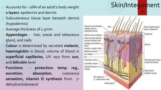
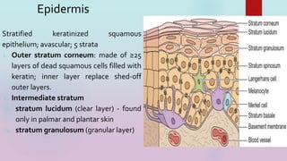
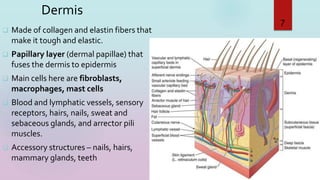
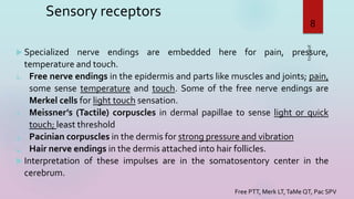
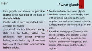
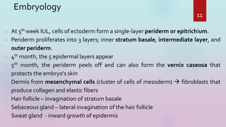
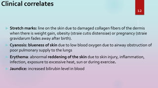
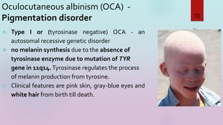
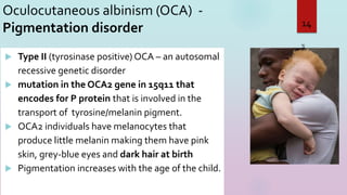
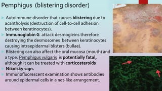
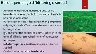
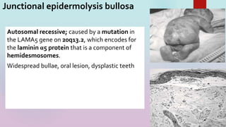
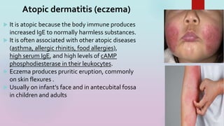
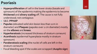
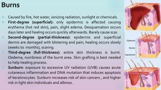
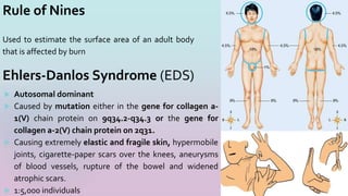
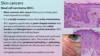
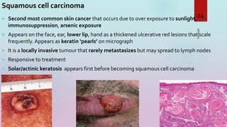
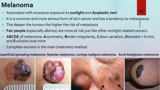
Integumentary
System
MR. UDEH
3/16/2023
1
Learning objectives
 Students should know the layers of the skin and the histology of each layer
 Describe the embryology of the skin and accessory glands
 Describe the pathology of “high-yield” clinical correlates associated with the skin
3/16/2023
2
Skin/Integument
 Accounts for ~16% of an adult’s body weight
 2 layers: epidermis and dermis
 Subcutaneous tissue layer beneath dermis
(hypodermis)
 Average thickness of 1.5mm
 Appendages - hair, sweat and sebaceous
gland, and nails
 Colour is determined by secreted melanin,
haemoglobin in blood, volume of blood in
superficial capillaries, UV rays from sun,
and bilirubin level
 Functions - protection, temp. reg.,
excretion, absorption, cutaneous
sensation, vitamin D synthesis from 7-
dehydrocholesterol
3/16/2023
3
Epidermis
Stratified keratinized squamous
epithelium; avascular; 5 strata
i. Outer stratum corneum: made of ≥25
layers of dead squamous cells filled with
keratin; inner layer replace shed-off
outer layers.
ii. Intermediate stratum
iii. stratum lucidum (clear layer) - found
only in palmar and plantar skin
iv. stratum granulosum (granular layer)
3/16/2023
4
Epidermis
stratum spinosum - spiny multilayer held
by desmosomes; contains Langerhans
cells and keratinocytes (held by
desmosomes).
i. Deep stratum basale (stratum
germinativum/basal layer) - single layer of
cells held by desmosomes and
hemidesmomes to the basement
membrane; undergo mitosis every 19 days:
4 cell types; melanocytes, Merkel (tactile)
cells, most numerous keratinocytes and
Langerhans (dendritic) cells.
3/16/2023
5
6
Crashing Low Grade So Bad
Dermis
 Made of collagen and elastin fibers that
make it tough and elastic.
 Papillary layer (dermal papillae) that
fuses the dermis to epidermis
 Main cells here are fibroblasts,
macrophages, mast cells
 Blood and lymphatic vessels, sensory
receptors, hairs, nails, sweat and
sebaceous glands, and arrector pili
muscles.
 Accessory structures – nails, hairs,
mammary glands, teeth
3/16/2023
7
Sensory receptors
 Specialized nerve endings are embedded here for pain, pressure,
temperature and touch.
1. Free nerve endings in the epidermis and parts like muscles and joints; pain,
some sense temperature and touch. Some of the free nerve endings are
Merkel cells for light touch sensation.
2. Meissner’s (Tactile) corpuscles in dermal papillae to sense light or quick
touch; least threshold
3. Pacinian corpuscles in the dermis for strong pressure and vibration
4. Hair nerve endings in the dermis attached into hair follicles.
 Interpretation of these impulses are in the somatosentory center in the
cerebrum.
3/16/2023
8
Free PTT, Merk LT,TaMe QT, Pac SPV
Hair
 Hair growth starts from the germinal
matrix in the hair bulb at the base of
the hair follicle
 On the side of each embedded hair is
arrector pili muscle
 3 types of hair in a lifetime: Lanugo
(late IUL to birth), vellus hair
(children’s hair except eyebrow,
lashes, scalp hairs; 2/3 of women’s
hair;1/10 of men’s hair) and terminal
hairs in adults.
3/16/2023
9
Sweat glands
• Eccrine and apocrine sweat glands
• Eccrine – narrow (0.4mm) lumen; lined
with stratified cuboidal epithelium;
empties clear and watery sweat unto the
surface; more on the forehead, palm and
sole.
• Apocrine- wide (3-5mm) lumen; more
coiled secretory unit; secretes viscous
and odour producing sweat into hair
follicles; and more in the genital, anal,
areolar of the nipple and the axillary
regions.
Subcutaneous tissue (hypodermis)
 Made of areola connective tissue and adipose tissue
 Stores fat which insulates the body and cushion for projecting
bony parts
 The thickness is dependent on nutritional status. The
distribution also varies in different sites of an individual.
 Fat deposit also show sexual dimorphism
10
 At 5th week IUL, cells of ectoderm form a single-layer periderm or epitrichium.
 Periderm proliferates into 3 layers; inner stratum basale, intermediate layer, and
outer periderm.
 4th month, the 5 epidermal layers appear
 5th month, the periderm peels off and can also form the vernix caseosa that
protects the embryo’s skin
 Dermis from mesenchymal cells (cluster of cells of mesoderm)  fibroblasts that
produce collagen and elastic fibers
 Hair follicle – invagination of stratum basale
 Sebaceous gland – lateral invagination of the hair follicle
 Sweat gland - inward growth of epidermis
11
Embryology
Clinical correlates
 Stretch marks: line on the skin due to damaged collagen fibers of the dermis
when there is weight gain, obesity (straie cutis distensiae) or pregnancy (straie
gravidarum fades away after birth).
 Cyanosis: blueness of skin due to low blood oxygen due to airway obstruction of
poor pulmonary supply to the lungs
 Erythema: abnormal reddening of the skin due to skin injury, inflammation,
infection, exposure to excessive heat, sun or during exercise.
 Jaundice: increased bilirubin level in blood
12
Oculocutaneous albinism (OCA) -
Pigmentation disorder
 Type I or (tyrosinase negative) OCA - an
autosomal recessive genetic disorder
 no melanin synthesis due to the absence of
tyrosinase enzyme due to mutation of TYR
gene in 11q14.Tyrosinase regulates the process
of melanin production from tyrosine.
 Clinical features are pink skin, gray-blue eyes and
white hair from birth till death.
3/16/2023
13
Oculocutaneous albinism (OCA) -
Pigmentation disorder
3/16/2023
14
 Type II (tyrosinase positive) OCA – an autosomal
recessive genetic disorder
 mutation in the OCA2 gene in 15q11 that
encodes for P protein that is involved in the
transport of tyrosine/melanin pigment.
 OCA2 individuals have melanocytes that
produce little melanin making them have pink
skin, grey-blue eyes and dark hair at birth
 Pigmentation increases with the age of the child.
Piebaldism (localized albinism)- an autosomal dominant
genetic disorder; mutation in the KIT gene on 4q11 that
encodes for tyrosine kinase receptor. No melanin in some
parts of the skin and/or hair, seen as patches of depigmented
skin on forehead, neck, chest, abdomen and mid-extremities.
15
Vitiligo -autoimmune disorder; antibodies destroy
melanocytes in different parts of the body causing
depigmentation of skin and hairs in the affected areas.
Melasma (chloasma) – “mask of pregnancy”, common acquired
bilateral hyperpigmentation on the face. Caused by
oestrogen/progesterone surge during pregnancy or women using
oral contraceptives containing the hormones.
Pemphigus (blistering disorder)
 Autoimmune disorder that causes blistering due to
acantholysis (destruction of cell-to-cell adhesion
between keratinocytes).
 Immunoglobin G attack desmogleins therefore
destroying the desmosomes between keratinocytes
causing intraepidermal blisters (bullae).
 Blistering can also affect the oral mucosa (mouth) and
a type. Pemphigus vulgaris is potentially fatal,
although it can be treated with corticosteroids
 Nikolsky sign.
 Immunofluorescent examination shows antibodies
around epidermal cells in a net-like arrangement.
16
17
Bullous pemphigoid (blistering disorder)
 Autoimmune disorder due to IgG destroying
hemidesmosomes that hold the stratum basale to
basement membrane.
 Bullous pemphigoid is less severe than pemphigus
vulgaris, it barely affect the oral mucosa and it can
be drug-induced.
 IgG cluster at the dermal-epidermal junction in the
form of a line is seen using immunofluorescence
technique
 Nikolsky sign is evident here if more pressure is
applied
 Can be treated with corticosteroids
18
Junctional epidermolysis bullosa
Autosomal recessive; caused by a mutation in
the LAMA5 gene on 20q13.2, which encodes for
the laminin α5 protein that is a component of
hemidesmosomes.
Widespread bullae, oral lesion, dysplastic teeth
19
 It is atopic because the body immune produces
increased IgE to normally harmless substances.
 It is often associated with other atopic diseases
(asthma, allergic rhinitis, food allergies),
high serum IgE, and high levels of cAMP
phosphodiesterase in their leukocytes.
 Eczema produces pruritic eruption, commonly
on skin flexures .
 Usually on infant’s face and in antecubital fossa
in children and adults
Atopic dermatitis (eczema)
Psoriasis
 Hyperproliferation of cell in the lower strata (basale and
spinosum) of the epidermis making the epidermis to become
thickened and silvery scaling off.The cause is not fully
understood; not contagious
 S&S: PPHAP
 Papules (elevated solid skin lesion less than 1cm in
diameter) and Plaques (papules over 1 cm diameter) mostly
on the elbows and knees.
 Hyperkeratosis (increased thickness of stratum corneum)
 Acanthosis (epidermal hyperplasia mostly in stratum
spinosum)
 Parakeratosis scaling (the nuclei of cells are left in the
stratum corneum)
 Focal bleeding spot if the scales are scrapped (Auspitz sign)
20
Burns
 Caused by fire, hot water, ionizing radiation, sunlight or chemicals.
 First-degree (superficial): only epidermis is affected causing
erythema (hot red skin), pain, slight edema. Desquamation occurs
days later and healing occurs quickly afterwards. Barely cause scar.
 Second-degree (partial-thickness): epidermis and superficial
dermis are damaged with blistering and pain; healing occurs slowly
(weeks to months), scaring.
 Third-degree (full-thickness): entire skin thickness is burnt.
Oedema, numbness of the burnt area. Skin grafting is best needed
to help healing process.
 Sunburn: exposure to excessive UV radiation (UVB) causes acute
cutaneous inflammation and DNA mutation that induces apoptosis
of keratinocytes. Sunburn increases risk of skin cancers , and higher
risk in light skin individuals and albinos.
21
Rule of Nines
Used to estimate the surface area of an adult body
that is affected by burn
22
 Autosomal dominant
 Caused by mutation either in the gene for collagen a-
1(V) chain protein on 9q34.2-q34.3 or the gene for
collagen a-2(V) chain protein on 2q31.
 Causing extremely elastic and fragile skin, hypermobile
joints, cigarette-paper scars over the knees, aneurysms
of blood vessels, rupture of the bowel and widened
atrophic scars.
 1:5,000 individuals
Ehlers-Danlos Syndrome (EDS)
Skin cancers
Basal cell carcinoma (BCC)
 Most common skin cancer that occurs in body parts
most exposed to sunlight.
 It is a locally invasive tumour that rarely metastasizes
 BCC appears superficially as pearl-shaped nodules that
are waxy and pinkish, and commonly with rolled
borders and telangiectasis and central ulceration. BCC
can also appear as a scaling plaque
 Micrograph shows the tumour cell have oval nuclei and
high nuclei to cytoplasmic ratio.The cluster of the
tumour cells show peripheral palisading or palisading
nuclei. Melanin deposit can also be seen inside or near
the cluster (nest)
 Complete excision can give an excellent cure rate
23
Squamous cell carcinoma
 Second most common skin cancer that occurs due to over exposure to sunlight,
immunosuppression, arsenic exposure
 Appears on the face, ear, lower lip, hand as a thickened ulcerative red lesions that scale
frequently. Appears as keratin ‘pearls’ on micrograph
 It is a locally invasive tumour that rarely metastasizes but may spread to lymph nodes
 Responsive to treatment
 Solar/actinic keratosis appears first before becoming squamous cell carcinoma
3/16/2023
24
Melanoma
 Associated with excessive exposure to sunlight and dysplastic nevi
 It is a common and more serious form of skin cancer and has a tendency to metastasize
 The deeper the tumour the higher the risk of metastasis
 Fair people (especially albinos) are more at risk just like other sunlight related cancers.
 ABCDE of melanoma: Asymmetry, Border irregularity, Colour variation, Diameter > 6 mm,
and Evolution over time
 Complete excision is the main treatment method
3/16/2023
25
Superficial spreading melanoma. Nodular melanoma. Lentigo maligna melanoma. Acral lentiginous melanoma

INTEGUMENTARY.pptx

  • 1.
  • 2.
    Learning objectives  Studentsshould know the layers of the skin and the histology of each layer  Describe the embryology of the skin and accessory glands  Describe the pathology of “high-yield” clinical correlates associated with the skin 3/16/2023 2
  • 3.
    Skin/Integument  Accounts for~16% of an adult’s body weight  2 layers: epidermis and dermis  Subcutaneous tissue layer beneath dermis (hypodermis)  Average thickness of 1.5mm  Appendages - hair, sweat and sebaceous gland, and nails  Colour is determined by secreted melanin, haemoglobin in blood, volume of blood in superficial capillaries, UV rays from sun, and bilirubin level  Functions - protection, temp. reg., excretion, absorption, cutaneous sensation, vitamin D synthesis from 7- dehydrocholesterol 3/16/2023 3
  • 4.
    Epidermis Stratified keratinized squamous epithelium;avascular; 5 strata i. Outer stratum corneum: made of ≥25 layers of dead squamous cells filled with keratin; inner layer replace shed-off outer layers. ii. Intermediate stratum iii. stratum lucidum (clear layer) - found only in palmar and plantar skin iv. stratum granulosum (granular layer) 3/16/2023 4
  • 5.
    Epidermis stratum spinosum -spiny multilayer held by desmosomes; contains Langerhans cells and keratinocytes (held by desmosomes). i. Deep stratum basale (stratum germinativum/basal layer) - single layer of cells held by desmosomes and hemidesmomes to the basement membrane; undergo mitosis every 19 days: 4 cell types; melanocytes, Merkel (tactile) cells, most numerous keratinocytes and Langerhans (dendritic) cells. 3/16/2023 5
  • 6.
  • 7.
    Dermis  Made ofcollagen and elastin fibers that make it tough and elastic.  Papillary layer (dermal papillae) that fuses the dermis to epidermis  Main cells here are fibroblasts, macrophages, mast cells  Blood and lymphatic vessels, sensory receptors, hairs, nails, sweat and sebaceous glands, and arrector pili muscles.  Accessory structures – nails, hairs, mammary glands, teeth 3/16/2023 7
  • 8.
    Sensory receptors  Specializednerve endings are embedded here for pain, pressure, temperature and touch. 1. Free nerve endings in the epidermis and parts like muscles and joints; pain, some sense temperature and touch. Some of the free nerve endings are Merkel cells for light touch sensation. 2. Meissner’s (Tactile) corpuscles in dermal papillae to sense light or quick touch; least threshold 3. Pacinian corpuscles in the dermis for strong pressure and vibration 4. Hair nerve endings in the dermis attached into hair follicles.  Interpretation of these impulses are in the somatosentory center in the cerebrum. 3/16/2023 8 Free PTT, Merk LT,TaMe QT, Pac SPV
  • 9.
    Hair  Hair growthstarts from the germinal matrix in the hair bulb at the base of the hair follicle  On the side of each embedded hair is arrector pili muscle  3 types of hair in a lifetime: Lanugo (late IUL to birth), vellus hair (children’s hair except eyebrow, lashes, scalp hairs; 2/3 of women’s hair;1/10 of men’s hair) and terminal hairs in adults. 3/16/2023 9 Sweat glands • Eccrine and apocrine sweat glands • Eccrine – narrow (0.4mm) lumen; lined with stratified cuboidal epithelium; empties clear and watery sweat unto the surface; more on the forehead, palm and sole. • Apocrine- wide (3-5mm) lumen; more coiled secretory unit; secretes viscous and odour producing sweat into hair follicles; and more in the genital, anal, areolar of the nipple and the axillary regions.
  • 10.
    Subcutaneous tissue (hypodermis) Made of areola connective tissue and adipose tissue  Stores fat which insulates the body and cushion for projecting bony parts  The thickness is dependent on nutritional status. The distribution also varies in different sites of an individual.  Fat deposit also show sexual dimorphism 10
  • 11.
     At 5thweek IUL, cells of ectoderm form a single-layer periderm or epitrichium.  Periderm proliferates into 3 layers; inner stratum basale, intermediate layer, and outer periderm.  4th month, the 5 epidermal layers appear  5th month, the periderm peels off and can also form the vernix caseosa that protects the embryo’s skin  Dermis from mesenchymal cells (cluster of cells of mesoderm)  fibroblasts that produce collagen and elastic fibers  Hair follicle – invagination of stratum basale  Sebaceous gland – lateral invagination of the hair follicle  Sweat gland - inward growth of epidermis 11 Embryology
  • 12.
    Clinical correlates  Stretchmarks: line on the skin due to damaged collagen fibers of the dermis when there is weight gain, obesity (straie cutis distensiae) or pregnancy (straie gravidarum fades away after birth).  Cyanosis: blueness of skin due to low blood oxygen due to airway obstruction of poor pulmonary supply to the lungs  Erythema: abnormal reddening of the skin due to skin injury, inflammation, infection, exposure to excessive heat, sun or during exercise.  Jaundice: increased bilirubin level in blood 12
  • 13.
    Oculocutaneous albinism (OCA)- Pigmentation disorder  Type I or (tyrosinase negative) OCA - an autosomal recessive genetic disorder  no melanin synthesis due to the absence of tyrosinase enzyme due to mutation of TYR gene in 11q14.Tyrosinase regulates the process of melanin production from tyrosine.  Clinical features are pink skin, gray-blue eyes and white hair from birth till death. 3/16/2023 13
  • 14.
    Oculocutaneous albinism (OCA)- Pigmentation disorder 3/16/2023 14  Type II (tyrosinase positive) OCA – an autosomal recessive genetic disorder  mutation in the OCA2 gene in 15q11 that encodes for P protein that is involved in the transport of tyrosine/melanin pigment.  OCA2 individuals have melanocytes that produce little melanin making them have pink skin, grey-blue eyes and dark hair at birth  Pigmentation increases with the age of the child.
  • 15.
    Piebaldism (localized albinism)-an autosomal dominant genetic disorder; mutation in the KIT gene on 4q11 that encodes for tyrosine kinase receptor. No melanin in some parts of the skin and/or hair, seen as patches of depigmented skin on forehead, neck, chest, abdomen and mid-extremities. 15 Vitiligo -autoimmune disorder; antibodies destroy melanocytes in different parts of the body causing depigmentation of skin and hairs in the affected areas. Melasma (chloasma) – “mask of pregnancy”, common acquired bilateral hyperpigmentation on the face. Caused by oestrogen/progesterone surge during pregnancy or women using oral contraceptives containing the hormones.
  • 16.
    Pemphigus (blistering disorder) Autoimmune disorder that causes blistering due to acantholysis (destruction of cell-to-cell adhesion between keratinocytes).  Immunoglobin G attack desmogleins therefore destroying the desmosomes between keratinocytes causing intraepidermal blisters (bullae).  Blistering can also affect the oral mucosa (mouth) and a type. Pemphigus vulgaris is potentially fatal, although it can be treated with corticosteroids  Nikolsky sign.  Immunofluorescent examination shows antibodies around epidermal cells in a net-like arrangement. 16
  • 17.
    17 Bullous pemphigoid (blisteringdisorder)  Autoimmune disorder due to IgG destroying hemidesmosomes that hold the stratum basale to basement membrane.  Bullous pemphigoid is less severe than pemphigus vulgaris, it barely affect the oral mucosa and it can be drug-induced.  IgG cluster at the dermal-epidermal junction in the form of a line is seen using immunofluorescence technique  Nikolsky sign is evident here if more pressure is applied  Can be treated with corticosteroids
  • 18.
    18 Junctional epidermolysis bullosa Autosomalrecessive; caused by a mutation in the LAMA5 gene on 20q13.2, which encodes for the laminin α5 protein that is a component of hemidesmosomes. Widespread bullae, oral lesion, dysplastic teeth
  • 19.
    19  It isatopic because the body immune produces increased IgE to normally harmless substances.  It is often associated with other atopic diseases (asthma, allergic rhinitis, food allergies), high serum IgE, and high levels of cAMP phosphodiesterase in their leukocytes.  Eczema produces pruritic eruption, commonly on skin flexures .  Usually on infant’s face and in antecubital fossa in children and adults Atopic dermatitis (eczema)
  • 20.
    Psoriasis  Hyperproliferation ofcell in the lower strata (basale and spinosum) of the epidermis making the epidermis to become thickened and silvery scaling off.The cause is not fully understood; not contagious  S&S: PPHAP  Papules (elevated solid skin lesion less than 1cm in diameter) and Plaques (papules over 1 cm diameter) mostly on the elbows and knees.  Hyperkeratosis (increased thickness of stratum corneum)  Acanthosis (epidermal hyperplasia mostly in stratum spinosum)  Parakeratosis scaling (the nuclei of cells are left in the stratum corneum)  Focal bleeding spot if the scales are scrapped (Auspitz sign) 20
  • 21.
    Burns  Caused byfire, hot water, ionizing radiation, sunlight or chemicals.  First-degree (superficial): only epidermis is affected causing erythema (hot red skin), pain, slight edema. Desquamation occurs days later and healing occurs quickly afterwards. Barely cause scar.  Second-degree (partial-thickness): epidermis and superficial dermis are damaged with blistering and pain; healing occurs slowly (weeks to months), scaring.  Third-degree (full-thickness): entire skin thickness is burnt. Oedema, numbness of the burnt area. Skin grafting is best needed to help healing process.  Sunburn: exposure to excessive UV radiation (UVB) causes acute cutaneous inflammation and DNA mutation that induces apoptosis of keratinocytes. Sunburn increases risk of skin cancers , and higher risk in light skin individuals and albinos. 21
  • 22.
    Rule of Nines Usedto estimate the surface area of an adult body that is affected by burn 22  Autosomal dominant  Caused by mutation either in the gene for collagen a- 1(V) chain protein on 9q34.2-q34.3 or the gene for collagen a-2(V) chain protein on 2q31.  Causing extremely elastic and fragile skin, hypermobile joints, cigarette-paper scars over the knees, aneurysms of blood vessels, rupture of the bowel and widened atrophic scars.  1:5,000 individuals Ehlers-Danlos Syndrome (EDS)
  • 23.
    Skin cancers Basal cellcarcinoma (BCC)  Most common skin cancer that occurs in body parts most exposed to sunlight.  It is a locally invasive tumour that rarely metastasizes  BCC appears superficially as pearl-shaped nodules that are waxy and pinkish, and commonly with rolled borders and telangiectasis and central ulceration. BCC can also appear as a scaling plaque  Micrograph shows the tumour cell have oval nuclei and high nuclei to cytoplasmic ratio.The cluster of the tumour cells show peripheral palisading or palisading nuclei. Melanin deposit can also be seen inside or near the cluster (nest)  Complete excision can give an excellent cure rate 23
  • 24.
    Squamous cell carcinoma Second most common skin cancer that occurs due to over exposure to sunlight, immunosuppression, arsenic exposure  Appears on the face, ear, lower lip, hand as a thickened ulcerative red lesions that scale frequently. Appears as keratin ‘pearls’ on micrograph  It is a locally invasive tumour that rarely metastasizes but may spread to lymph nodes  Responsive to treatment  Solar/actinic keratosis appears first before becoming squamous cell carcinoma 3/16/2023 24
  • 25.
    Melanoma  Associated withexcessive exposure to sunlight and dysplastic nevi  It is a common and more serious form of skin cancer and has a tendency to metastasize  The deeper the tumour the higher the risk of metastasis  Fair people (especially albinos) are more at risk just like other sunlight related cancers.  ABCDE of melanoma: Asymmetry, Border irregularity, Colour variation, Diameter > 6 mm, and Evolution over time  Complete excision is the main treatment method 3/16/2023 25 Superficial spreading melanoma. Nodular melanoma. Lentigo maligna melanoma. Acral lentiginous melanoma