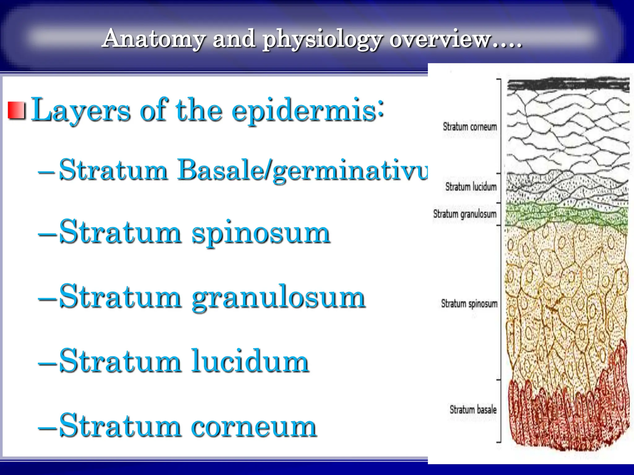
SlideShare a Scribd company logo
1 of 273
1
Anatomy and physiology overview
The integumentary system includes
the skin and its appendages:
–The skin
–The hair
–The nail
–Glands of the skin
2
Anatomy and physiology overview
…
3
Anatomy and physiology overview…..
The skin is the largest organ
system of the body.
The skin has 3 layers:
–Epidermis
–Dermis (true skin)
–Sub cutaneous tissue
4
Anatomy and physiology overview….
1.Epidermis
Outer most layer of the skin.
Made up of stratified epithelial cells.
Composed predominantly of keratinocytes.
Thickness, ranges between 0.1mm (eye lid)
and 1mm (palms and feet sole).
5
Anatomy and physiology overview….
Layers of the epidermis:
–Stratum Basale/germinativum
–Stratum spinosum
–Stratum granulosum
–Stratum lucidum
–Stratum corneum
6
The epidermis is nourished by blood vessels
in the dermis.
The epidermis regenerates with new cells
every 28 days.
The two major types of epidermal cells are
melanocytes (5%) and keratinocytes (90%).
Melanocytes are contained in the deep, basal
layer (stratum germinativum) of the
epidermis.
They contain melanin, a pigment that gives
color to the skin and hair and protects the
body from damaging ultraviolet (UV)
sunlight. 7
Sunlight and hormones stimulate the
melanosome (within the melanocyte) to
increase the production of melanin.
The wide range of skin color is caused by the
amount of melanin produced; more melanin
results in darker skin color.
Keratinocytes are synthesized from epidermal
cells in the basal layer.
8
As they mature (keratinize), they move to the
surface, where they flatten and die to form the
outer skin layer (stratum corneum).
Keratinocytes produce a fibrous protein,
keratin, which is vital to the skin’s protective
barrier function.
The upward movement of keratinocytes from
the basement membrane to the stratum
corneum takes approximately 4 weeks. 9
Anatomy and physiology overview…
2.Dermis (True skin)
Makes up largest portion of the skin
Providing strength and structure of skin.
Dermal thickness varies from 1 to 4 mm.
The dermis is very vascular.
Layers of the dermis:
– Papillary Layers: - layer beneath the epidermis
which composed of fibroblast cells (collagen
fiber).
– Reticular layer: - lie beneath the papillary layer.
Produce collagen and elastic bundles. 10
Collagen forms the greatest part of the dermis
and is responsible for the skin’s mechanical
strength.
The primary cell type in the dermis is the
fibroblast.
Fibroblasts produce collagen and elastin fibers
and are important in wound healing.
Nerves, lymphatic vessels, hair follicles, and
sebaceous glands are also found in the dermis
11
Anatomy and physiology overview….
3.Subcutaneous tissue
Is also called hypodermis.
Is the inner most layer of the skin.
The subcutaneous tissue contains loose connective
tissue and fat cells that provide insulation.
Composed of adipose tissue.
Is deposition site for fat.
Provide cushion between the skin layer, muscle and
bone. 12
Skin Appendages
Appendages of the skin include the hair, nails, and
glands (sebaceous, apocrine, and eccrine).
These structures develop from the epidermal layer
and receive nutrients, electrolytes, and fluids from
the dermis.
Hair and nails form from specialized keratin that
becomes hardened.
Hair grows on most of the body except for the lips,
the palms of the hands, and the soles of the feet.
13
Anatomy and physiology overview…
The hair:
• Parts of the hair.
Root: formed in the dermis.
Hair shaft: projects beyond the skin.
Hair follicle: is a cavity in which the hair
grows.
Hair bulb: part of the hair in the hair follicle
where proliferation of cells takes place. 14
The color of the hair is a result of heredity and
is determined by the type and amount of
melanin in the hair shaft.
Abundance of eumelanin gives people black
and brown hair, while pheomelanin gives red.
Hair grows approximately 1 cm per month.
On average 100 hairs are lost each day.
The absence of hair may be related to disease,
treatment, or heredity.
15
Anatomy and physiology overview….
The Nail
It is a hard, transparent plate of keratin.
Found on dorsal surface of fingers and
toes.
Nails grow from the matrix that is
located at the proximal area of the nail
plate.
The matrix is commonly called the
16
Nail Parts
17
The nail bed that is under the nail matrix and nail
plate is normally pink and contains blood vessels.
The nail plate adheres to and is supported by the
nail bed.
The cuticle is part of the skin that extends a small
distance on the nail plate before being shed (like
the stratum corneum).
The nail root is bordered by the cuticle and hidden
by a fold of skin.
18
Grow through out life (daily 0.1mm)
Function- protection.
A lost fingernail usually regenerates in 3 to 6
months, whereas a lost toenail may require 12
months or longer for regeneration.
19
Anatomy and physiology overview…
Glands of the skin
They are of two types glands associated with
skin:
1.Sebaceous glands
– Associated with hair follicles.
– Produce sebum on to the surface between
the hair follicle and the hair shaft.
– Function: lubricate the hair and render the
skin soft and pliable. 20
Sebum prevents the skin and hair from
becoming dry and somewhat bacteriostatic and
fungistatic and consists mainly of lipids.
They secrete a thick milky substance of
becomes odoriferous when altered by skin
surface bacteria.
These glands enlarge and become active at
puberty because of reproductive hormones.
21
Anatomy and physiology overview….
2.Sweat glands
Found in the skin over most of the body surface.
Sweat glands are devoid at:
– Tip of penis (glans penis)
– Clitoris
– Labia minora
– Margin of the lips
– External ear
– Nail beds
22
Anatomy and physiology overview….
A. Eccrine (small) -The eccrine sweat glands
are widely distributed over the body, except
in a few areas, such as the lips.
– Found in all areas of the skin
– Release thin watery secretion
– Their ducts are open on the skin surface.
23
Anatomy and physiology overview…
B. Apocrine (larger)
Located in breast areolae, umbilical, eyelids, axillae,
anal region, scrotum and labia majora.
Their ducts are directly open on to hair follicles.
They become active at puberty.
Produce milky sweat.
Specialized apocrine glands found in external ear canal
are called Ceruminous Glands (produce cerumen).
24
Anatomy and physiology overview….
Functions of the skin:
Protection
Sensation
Fluid balance
Temperature regulation
Vitamin production (Vit D)
Immune response function
25
Dermatologic Assessment
When caring for patients with dermatologic
disorders, the nurse obtains important
information through the health history and
direct observations.
26
ASSESSMENT (PATIENT HISTORY)
During the health history interview, the nurse asks about
any family and personal history of skin allergies; allergic
reactions to food, medications, and chemicals; previous
skin conditions; and skin cancer.
The names of cosmetics, soaps, shampoos, and other
personal hygiene products are obtained if there have been
any recent skin conditions noticed with the use of these
products.
27
If suspicious areas are noted, the patient is questioned
about nonprescription or herbal preparations that are
being used.
The health history addresses the onset, signs and
symptoms, location, and duration of any pain, itching,
rash, or other discomfort experienced by the patient.
28
ASSESSMENT (PATIENT HISTORY)
History of main symptoms of skin
problems:
–Itching
–Burning sensation
–Crawling sensation
Rash of the skin
History of allergic condition
29
Cont……
Relation with specific events (food, medication, alcohol, …)
History of cosmetics use.
What treatments have been used?
Example: Topical ointment
What precipitates the condition?
History current skin change (Hypo or hyper pigmentation)
30
PHYSICAL EXAMINATION
@ Assessment of the skin involves the entire skin area,
including the mucous membranes, scalp, hair, and nails.
The skin is a reflection of a person’s overall health, and
alterations commonly correspond to disease in other
organ systems.
Inspection and palpation are techniques commonly used
in examining the skin.
The room must be well lighted and warm.
31
PALPATION
For:
Temperature
Moisture
Turgor
Texture of the skin
32
PALPATION….
Temperature and moisture:
– The skin is normally warm & dry.
– Increase in skin temperature and moisture
indicates an elevation of body temperature.
– An excessive amount of perspiration is called
diaphoresis.
– Loss of excess body water is called dehydration
= results in dryness and poor turgor.
33
PALPATION….
Turgor:
– Turgor is the fullness or elasticity of the skin.
Difficulty in lifting a skin fold may
indicate excess fluid in the tissue or
oedema.
Oedema is characterized by:
–Swelling
–Shiny skin of the area.
–Indentation remain after pressing
34
PALPATION….
Texture:
– Vary from smooth & soft to rough & dry.
– In a dehydrated client:
Wrinkled
The mucus membranes are cracked and dry.
35
Diagnosis
The provider will do a skin examination with a
dermatoscope.
Most of the time, the cause can be identified after the
exam.
If needed, the following tests may be ordered:
o Biopsy of the skin
o Allergy testing
o Other tests to find the cause of erythroderma
36
PHYSICAL EXAMINATION SKIN LESIONS ……..
1. Primary lesions:
 are the initial lesions and are characteristic of the disease itself
1. Secondary lesions result from changes in primary lesions
resulting from external causes, such as scratching, trauma,
infections, or changes caused by wound healing
I. Primary skin lesions:
Macule: flat <1cm and non-palpable skin color change.
37
Primary skin lesions:
Patch: flat and >1cm, with irregular border.
• Example: flat mole
38
Primary skin lesions:
Papule:
–< 0.5cm
–Elevated
–Palpable
–Solid mass
–With circumscribed or
regular border. 39
Primary skin lesions:
Plaque:
–>0.5cm
–Elevated
–Palpable solid mass
–Circumscribed with flat top.
Example:
–Eczematous dermatitis
–Psoriasis
40
Primary skin lesions:
Nodule:
–0.5-2cm
–Elevated
–Palpable solid mass
–Extended and deeper
to the dermis than papule
–Circumscribed.
–Example: Lipoma 41
Primary skin lesions:
Tumor:
–1-2cm
–Elevated
–Palpable, solid mass
–Extended deeper
to dermis than papules.
–Do not have sharp border.
42
Primary skin lesions:
Vesicle:
–<0.5cm
–Circumscribed
–Elevated and palpable mass
–Containing serous fluid.
–Vesicles: Herpes simplex/zoster,
chickenpox, second-degree burn
(blister) 43
Primary skin lesions:
Bulla:
–>0.5cm and
–The same as vesicles.
Examples Pemphigus,
large burn blisters
44
Primary skin lesions:
Wheal:
–Elevated mass with irregular border, and
does not contain free fluid.
–Often accompanies
by severe itching.
–Example:
Insect bite
Urticaria
o Caused by movement of
serous fluid into the dermis
45
Primary skin lesions:
Pustule
–Pus-filled vesicles or bulla.
– E.g.
Impetigo
Boil
Acne, Small pox
46
Primary skin lesions:
Cyst
–Encapsulated fluid filled or semi-
solid mass in the subcutaneous or
dermis.
Examples: Sebaceous cyst
47
II. Secondary skin lesions:
Erosion: - loss of
superficial epidermis
does not extend to
dermis, depressed and
moist.
Ulcer: skin loss
extending past
epidermis necrotic tissue
loss, bleeding and
scarring.
Fissure: Linear cracks 48
Scales: flake secondary to desquamated dead
epithelial tissue, thick or thin.
• superficial epidermal cells-
that are dead and cast off from the skin .
•scales are secondary to desquamated, dead
epithelium.
• Flakes may adhere to skin surface
Examples: Dandruff, psoriasis
49
Secondary skin lesions:
Crust: dried residue of
serum, blood, or pus
on the skin surface.
Scar: skin mark after
healing of a wound or
lesion replacement by
connective tissue of
the left one.
50
Secondary skin lesions:
Keloid: hypertrophied
scar tissue, secondary
to extensive collagen
formation during
healing which is
elevated and irregular.
51
Secondary skin lesions:
Lichenification:
thickening and
roughening of the skin
may be secondary to
repeated rubbing,
irritation or scratching.
52
III. Vascular lesions:
Petechiae: 1-3mm, deep red or reddish
purple, fading away over time.
• Round red or purple macule
• Small: 1–2 mm
• Secondary to blood extravasation
• Associated with bleeding tendencies
53
Ecchymosis: purple or purplish blue, larger than
petechiae and flat hematoma.
• Round or irregular macular lesion
• Secondary to blood extravasation
• Associated with trauma, bleeding tendencies
54
Spider angioma- fiery red, very small to 2cm
in size and usually appear on face, neck, arms,
and upper trunk
Red, arteriole lesion
Central body with radiating branches
Associated with liver disease, pregnancy,
vitamin B deficiency.
55
Cherry angioma: bright or ruby red; may
become brownish with age & 1–3 mm in size
Papular and round
Red or purple
Noted on trunk, extremities
May blanch with pressure
Normal age-related skin alteration
56
Secondary skin lesions:
Telangiectasia/spider veins- is a dilated or
brocken superficial blood vessel. Color bluish or red
Does not blanch when pressure is applied
Noted on legs, anterior chest
Secondary to superficial dilation of venous vessels
and capillaries
Associated with increased venous pressure states
Shape varies: spider-like or linear
57
Secondary skin lesions:
Excoriation describes superficial, often linear,
skin erosion caused by scratching.
58
Secondary skin lesions:
Hypopigmentation: is
decreased skin pigment;
Hyperpigmentation: is
increased skin pigment;
and
Depigmentation:- is
total loss of skin
pigment.
59
Secondary skin lesions:
Edema is swelling due to accumulation of
water in tissue.
60
Disorders
of
INTEGUMENTARY
System
61
DERMATITIS
Definition:- is the general term of
inflammation of the skin also called eczema
Types:
1. Contact dermatitis
• Allergic contact dermatitis,
• Irritant contact dermatitis,
2. Exfoliative dermatitis
3. Atopic dermatitis
4. Seborrheic dermatitis and
5. Stasis dermatitis
62
1. CONTACT DERMATITIS
It is caused by contact with substances which is
irritant or to which the individual is sensitive.
The epidermis is damaged by repeated physical or
chemical agents.
Cause:
Physical agents
Chemical agents
Biological agents 63
Clinical manifestations:
Irritation at the site of contact with irritant
Itching
Burning and erythema followed.
Edema
Papules
Vesicles and oozing or weeping.
N.B. if repeated reactions occur:
The patient continually scratches the skin.
Pigmentation occurs
The skin thicken (lichenification)
64
Management:
Objectives:
– Protect the skin from further damage.
– Rest the affected skin.
65
Specific treatment:
1. Avoid using soap until healing occurs.
2. Apply antipruritic agents:
– Promethazine 25 mg po
3. Wet dressing to clear oozing eczematous lesions by GV or
saline compress.
4. Corticosteroid creams or ointments.
– Betamethasone dipropionate 0.05% cream BID for two
weeks or
– Dexamethasone 0.25% cream BID for two weeks or
– Hydrocortison acetate 1 %
– Predinisolone 10 -20mg po/d for 7-10 days (sever
cases)
5. In more wide spread conditions systemic steroids can be
used. 66
……Tx …
“Instruct on”
–Patterns of dermatitis and what causes it.
–Avoid contact with irritant.
–Avoid soap, heat and rubbing.
–Avoid using un prescribed drugs.
–Wash the skin immediately after contact
with irritant.
–Follow the patient for four months.
67
Allergic contact dermatitis (ACD)
ACD is a form of dermatitis/eczema caused by
an allergic reaction to a material, called
an allergen, in contact with the skin.
The allergen is harmless to people that are not
allergic to it.
 ACD is common in metal
workers, hairdressers, beauticians, health care
workers, cleaners, painters and florists. 68
ACD…
69
Causes of ACD
ACD is a type 4 or delayed hypersensitivity reaction
and occurs 48–72 hours after exposure to
the allergen.
The mechanism involves CD4+ T-lymphocytes, which
recognise an antigen on the skin surface,
releasing cytokines that activate the immune system
and cause the dermatitis.
Contact allergy occurs predominantly from an allergen
on the skin rather than from internal sources or food.
70
Clinical features of ACD
ACD arises some hours after contact with the
responsible material.
Allergic contact dermatitis is generally confined
to the site of contact with the allergen,
but it may extend outside the contact area or
become generalized.
Transmission from the fingers can lead to
dermatitis on the eyelids and genitals.
The affected skin may be red and itchy, swollen
and blistered, or dry and bumpy.
71
differential diagnosis of ACD
Irritant contact dermatitis, which is due to irritation
or repetitive injury to the skin.
Irritant contact dermatitis may affect anyone, providing
they have had enough exposure to the irritant, but
those with atopic dermatitis are particularly sensitive.
Irritant contact dermatitis can occur immediately after
a single injury or develop slowly after repeated
exposure to an irritant.
Contact urticaria, in which a rash appears within
minutes of exposure & fades away within minutes to
hours.
The allergic reaction to latex is the best-known
example
72
Diagnosis of ACD
Sometimes it is easy to recognize contact allergy and
no specific tests are necessary.
Taking a very good history including information on
the work environment, hobbies, products in use at
home and work and sun exposure will enhance the
chances of finding a diagnosis.
The rash usually (but not always) completely clears up
if the allergen is no longer in contact with the skin
73
The open application test is used to confirm
contact allergy to a cosmetic, such as a
moisturizer.
The product under suspicion is applied several
times daily for several days to a small area
of sensitive skin
The inner aspect of the upper arm is suitable.
Contact allergy is likely if dermatitis arises in the
treated area.
Fungal scrapings of skin
for microscopy and culture can
exclude fungal infection
Dimethylglyoximes test is available to ‘spot test’ if
a product contains nickel.
74
treatment
Find out precisely what you are allergic to by
having comprehensive patch tests.
Identify where the allergen is found, thus read
labels of all products before use.
Carefully study your environment to locate the
allergen.
Wear appropriate gloves to protect hands from
touching materials to which you react and
remove gloves in the appropriate way.
Some chemicals will penetrate certain gloves …
75
Active dermatitis is usually treated with:
o Emollient creams
o Topical steroids
o Topical or oral antibiotics for secondary infection
o Oral steroids, usually short courses, for severe
cases
o Phototherapy or photochemotherapy.
o Azathioprine, ciclosporin or
another immunosuppressive agent.
o Tacrolimus ointment and pimecrolimus cream are
immune-modulating calcineurin inhibitors and
may prove helpful for allergic contact dermatitis.
76
Irritant contact dermatitis
(ICD)
Irritant contact dermatitis occurs more often than
allergic contact dermatitis. You develop a rash when a
chemical substance irritates the skin’s outer layers.
The rash is more painful than itchy.
77
Common causes of irritant contact dermatitis
include:
– Acids.
– Alkalis like drain cleaners.
– Body fluids, including urine and saliva.
– Certain plants
– Hair dyes.
– Nail polish remover or other solvents.
– Paints and varnishes.
– Harsh soaps or detergents 78
Symptoms of contact dermatitis
–Red
–Swollen, blistering or oozing
–Burning or stinging
–Flaky or scaling.
–Swollen or hive-like
–Itchy
–Painful
79
Management and Treatment
Treatment for both types of contact dermatitis is the
same.
Even with treatment, it can take several weeks for the
rash to go away. Treatments include:
Avoidance: take steps to avoid it or minimize
exposure.
Anti-itch creams: Corticosteroid creams can ease
inflammation and itching.
Oral steroids: Prednisone, a type of steroid, can
relieve rash symptoms that don’t respond to
antihistamines or other treatments.
Immunosuppressive medications: In severe cases
80
How can I prevent contact dermatitis?
Avoiding known allergens and irritants is the
best way to prevent contact dermatitis. But you
can’t always stay away from every possible
irritant. These steps can help:
– Choose fragrance-free moisturizers.
– Use mild, fragrance and dye-free soaps and
cleansers.
– Wash immediately after coming into contact with a
known allergen or irritant. 81
2. EXFOLIATIVE DERMATITIS (erythroderma)
It is a progressive inflammation of the skin which is
characterized by generalized erythema and scaling.
It cause capillary leakage through the excoriated skin.
severe skin condition that causes extreme shedding of
the top layers of your skin.
It may cover most of your body and lead to so much
skin damage that your body can't keep warm.
Dehydration is also a risk due to your skin losing its
ability to hold onto moisture
82
2. EXFOLIATIVE DERMATITIS
(erythroderma)…
Cause: Has variety of causes:
– Secondary to systemic diseases.
– As a part of lymphoma disease.
– Reaction to certain drugs including penicillin and
phenylbutazone.
It may cover most of your body and lead to so much
skin damage that your body can't keep warm.
Dehydration is also a risk due to your skin losing its
ability to hold onto moisture. 83
EXFOLIATIVE DERMATITIS (erythroderma)…
84
Clinical manifestations:
Generalized erythematic eruption
Fever and prostration
Itchy scaling skin lesions
Loss of outer layer of the skin
Erythroderma is a severe and potentially life-
threatening inflammation of most of the
body's skin surface.
85
Causes…
Erythroderma may occur due to:
Complication of other skin conditions, such
as eczema and psoriasis
Reaction to medicines or some chemicals, such
as phenytoin and allopurinol
Some types of cancer, such as lymphoma
Sometimes the cause is unknown.
It is more common in males. 86
Symptoms
Symptoms may include any of the following:
Redness over 80% to 90% of the body
Scaly skin patches
Thickened skin
Skin is itchy or painful with an odor
Swelling of the arms or legs
Fast heart beat
Loss of fluids, leading to dehydration
Loss of temperature regulation by the body
o There may be secondary infections of the skin.
87
Treatment
Since erythroderma can quickly lead to serious
complications, the provider will start treatment
right away.
This usually involves strong doses of cortisone
medicines to reduce inflammation.
Other treatments may include:
Medicines to treat the underlying cause of
erythroderma
Antibiotics for any infection
Dressings applied to the skin
Ultraviolet light
Correction of fluid and electrolyte balance
88
Possible Complications
Complications may include:
o Secondary infections that can lead
to sepsis (body wide inflammatory response)
o Fluid loss that can result in dehydration and an
imbalance of minerals (electrolytes) in the body
o Heart failure
89
Management
Objective:
Maintain fluid and electrolyte
balance.
Prevent coetaneous infection.
90
Treatment
Admission and bed rest.
Stop the drugs that the patient is taking.
Maintain fluid and electrolyte balance.
Monitor temperature of the room.
Give antibiotics if there is infection.
Watch for symptoms and signs of CHF.
Give topical therapy to relieve symptoms.
Oral or parentral steroids may be given.
Patient education: to avoid all irritants
91
3. Atopic dermatitis (AD)
Atopic dermatitis (eczema) is a condition that makes
your skin red and itchy.
is the most common type of eczema affecting people
It's common in children but can occur at any age.
Atopic dermatitis is long lasting (chronic) and tends to
flare periodically.
It may be accompanied by asthma or hay fever.
The rashes tend to flare and go away, but then come
back again.
92
Symptoms
93
Symptoms…
Atopic dermatitis (eczema) signs and symptoms vary
widely from person to person and include:
o Dry skin
o Itching, which may be severe, especially at night
o Red to brownish-gray patches, especially on the
hands, feet, ankles, wrists, neck, upper chest, eyelids,
inside the bend of the elbows and knees, and in
infants, the face and scalp
o Small, raised bumps, which may leak fluid and crust
over when scratched
o Thickened, cracked, scaly skin
o sensitive, swollen skin from scratching
94
Causes
Healthy skin helps retain moisture and protects you
from bacteria, irritants and allergens.
Eczema is related to a gene variation that affects the
skin's ability to provide this protection.
This allows your skin to be affected by environmental
factors, irritants and allergens.
In some children, food allergies may play a role in
causing eczema.
95
Risk factors
The primary risk factor for atopic dermatitis is
having a personal or family history of eczema,
allergies, hay fever or asthma.
96
Complications
Complications of atopic dermatitis (eczema) may include:
Asthma and hay fever. Eczema sometimes precedes these
conditions. More than half of young children with atopic
dermatitis develop asthma and hay fever by age 13.
Chronic itchy, scaly skin. A skin condition called
neurodermatitis (lichen simplex chronicus) starts with a patch
of itchy skin
Eventually, you may scratch simply out of habit. This condition
can cause the affected skin to become discolored, thick and
leathery.
97
Skin infections. Repeated scratching that
breaks the skin can cause open sores and
cracks. These increase the risk of infection from
bacteria and viruses, including the herpes
simplex virus.
Irritant hand dermatitis. This especially
affects people whose work requires that their
hands are often wet and exposed to harsh
soaps, detergents and disinfectants.
Allergic contact dermatitis. This condition is
common in people with atopic dermatitis.
Sleep problems. The itch-scratch cycle can
cause poor sleep quality. 98
Prevention
The following tips may help prevent and minimize the drying
effects of bathing:
Moisturize your skin at least twice a day. Creams,
ointments and lotions seal in moisture. Choose a product or
products that work well for you..
Try to identify and avoid triggers that worsen the
condition. Things that can worsen the skin reaction include
sweat, stress, obesity, soaps, detergents, dust and pollen,
identifying potential food allergies.
Use only gentle soaps. Choose mild soaps. Deodorant soaps
and antibacterial soaps can remove more natural oils and dry
your skin.
Dry yourself carefully. After bathing gently pat your skin dry
with a soft towel and apply moisturizer while your skin is still
damp.
99
Cure
Atopic dermatitis most often begins before age 5 and
may persist into adolescence and adulthood.
No cure has been found for atopic dermatitis.
But treatments and self-care measures can relieve
itching and prevent new outbreaks.
o For example, it helps to avoid harsh soaps, moisturize
your skin regularly, and apply medicated creams or
ointments.
100
4. Stasis dermatitis
Stasis dermatitis is a long-term condition that
causes inflammation, ulcers, and itchy skin on
the lower legs.
It often occurs in people who have underlying
conditions that affect blood flow in the legs, such
as chronic venous insufficiency, varicose veins,
deep vein thrombosis (DVT), and congestive
heart failure. 101
102
Causes
Stasis dermatitis tends to develop in people with
conditions that cause poor blood circulation in
the legs, such as chronic venous insufficiency.
Chronic venous insufficiency is a condition in
which the valves in the leg veins do not work
correctly.
As a result of the malfunctioning valves, blood
can flow backward and pool in the lower legs.
103
Causes….
• This pooling blood causes increased pressure and
swelling in the veins, which can lead to the symptoms
of stasis dermatitis
• DVT, which is a blood clot in the lower leg
• varicose veins, or enlarged and swollen veins
• injury to the lower leg
• any surgery that affects the veins in the lower leg
• congestive heart failure
104
Risk factors
o being female
o being over 50 years old
o being overweight or obese
o having any condition that affects blood circulation
o having high blood pressure
o having kidney disease
o standing or sitting for extended periods
o getting insufficient exercise
105
Symptoms
 The early symptoms of stasis dermatitis
primarily affect the lower legs and may include:
 irritated skin
 red, itchy, or swollen skin, particularly over any
varicose veins
 a sensation of fullness, heaviness, or aching
after extended periods of standing or walking
106
swelling on the inside of the lower leg and ankle
particularly at the end of the day or after
standing for prolonged periods
As stasis dermatitis progresses, these earlier
symptoms can worsen.
 In severe cases of stasis dermatitis, some areas of the
lower leg may become intensely itchy, hardened,
scaly, and prone to infection. In some people, the
calves may shrink. 107
Diagnosis
by asking about the person’s symptoms and medical
history. Previous or current conditions that they should
be aware of include:
– problems with the heart or circulation
– blood clots
– surgeries
– injuries to the lower legs
 Can use Doppler ultrasound, which is a noninvasive
test that uses sound waves to check the blood flow
through blood vessels. Additional tests that check for
heart function, blood pressure, and allergies may also
be necessary.
108
Treatment
The goal of treatment is to relieve symptoms, improve
circulation, and prevent the condition from
progressing. Treatment can include:
– wearing compression stockings to promote
circulation and relieve swelling
– sleeping with legs elevated
– elevating the legs for 15 minutes once every 2
hours
109
Rx …
– taking medications to ease pain and reduce
swelling, such as corticosteroids or topical
calcineurin inhibitors
– using antihistamines to relieve itching
– using antibiotics and special dressings to treat
infected ulcers
– applying emollients to moisturize and protect
the skin 110
Seborrhoeic dermatitis
‘Dermatitis’ is a red, itchy, flaky (inflamed) skin
complaint; ‘seborrhoeic’ means that the rash affects
greasy (sebaceous) skin zones such as the face, scalp
and center of the chest
chronic inflammatory disorder affecting areas of the
head and trunk where sebaceous glands are most
prominent.
Symptoms include red, scaly, greasy, itchy, and
inflamed skin 111
Seborrhea…
Scalp seborrhea varies from mild dandruff to
dense, diffuse, adherent scale.
Facial and trunk seborrhea is characterized by
powdery or greasy scale in skin folds and along
hair margins.
Treatment options include application of
selenium sulfide,pyrithione zinc or
ketoconazole-containing shampoos, topical
ketoconazole cream or terbinafine solution,
topical sodium sulfacetamide and topical
corticosteroids. 112
Seborrhoeic dermatitis…
113
Causes
It is thought that seborrheic dermatitis is
triggered by an overgrowth of a harmless yeast
called Malassezia that lives on the skin, or an
over-reaction by the skin’s immune system to
this yeast.
These are not the same as the yeasts that
cause thrush or those that are present in foods.
Seborrheic dermatitis is not usually linked to
any underlying illness, but it can be stubborn
and severe in people with HIV infection and it is
also common in people with Parkinson’s
disease. 114
Seborrheic….
The cause of seborrhoeic dermatitis is not
completely understood.
It is associated with proliferation of various species
of the skin commensal Malassezia, in its yeast
(non-pathogenic) form. Its metabolites (such as
the fatty acids oleic acid, malssezin, and indole-3-
carbaldehyde) may cause
an inflammatory reaction.
Differences in skin barrier lipid content and
function may account for individual presentations.
Tiredness and stress can sometimes trigger a flare
of seborrheic dermatitis.
115
symptoms of seborrhoeic
dermatitis
The symptoms of seborrhoeic dermatitis seem to vary
from person to person.
Affected areas can be itchy, sore and sensitive and
flaking skin can be bothersome and embarrassing
Some people have the rash without being troubled.
116
Diagnosis
If seborrheic dermatitis is severe or unresponsive to
treatment and someone is at risk of HIV they may get
tested.
This is important as early treatment of HIV reduces the
risk of passing it on and improves the health of the
affected person.
If there is a suspicion of ringworm of the scalp, then
skin scrapings can be sent to look for tinea fungus
(mycology). 117
treatment
Treatment is usually needed on a long-term basis,
though sometimes it is possible to take a break.
The choice depends on which areas of the body are
affected and whether there is a lot of irritation:
 In the scalp: medicated, anti-dandruff shampoos
containing agents such as zinc pyrithione, selenium
sulphide or ketoconazole can be used regularly. For
best results, wash into the scalp, then wait 5-10
minutes before rinsing. Thick scales can be removed
118
Elsewhere: anti-yeast creams or ointments are
usually effective and can be used safely as long-
term treatment. Examples include clotrimazole,
miconazole and nystatin. They are sometimes
combined with a mild steroid for a few weeks to
settle inflammation.
Washing your body with an antifungal shampoo
containing ketoconazole may also help. Leave the
shampoo on for 5minutes or so before rinsing it
off.
119
In the ear canals: medicated eardrops may
help. Do not clean the ears with cotton buds as
this causes more irritation.
On the eyelids: carefully cleaning between the
lashes with an eyelid cleanser
Treatment can improve and sometimes clear
seborrhoeic dermatitis, but there is no
permanent cure and the complaint tends to
come back when treatment is stopped. 120
121
ACNE VULGARIS
It is a disorder of sebaceous or oil glands and their
hair follicles.
hair follicles become plugged with oil & dead skin cells
It is a chronic, inflammatory disease of the
pilosebaceous units of the skin.
Acne vulgaris is a skin condition that occurs when hair
follicles are blocked with dead skin cells, bacteria, and
oil (sebum).
It is characterized by open and closed comedones.122
Acne vulgaris----
Functions of sebaceous gland is to
secrete sebum: an oily substance that acts
to protect and waterproof skin and hair and
keep it from being dry, brittle and cracked.
Common between age of 12-35 years and
more during puberty.
123
Acne vulgaris---
Found in hair-covered areas of the body.
The blocked follicles cause blemishes on the skin,
including pimples, blackheads, whiteheads, and
cysts.
Also known as common acne, one of its main
causes is hormones, especially around puberty
Most acne occurs on the face, chest, back, and
shoulders. Symptoms can be mild, moderate, or
severe.
124
Acne vulgaris----
125
Etiology:
Multiple involving genetic, environmental, hormonal
and bacterial infections !
Primary Causes:
o Increased sebum production: maturation of the
adrenal gland or an increase in the number of cells of
the sebaceous gland
o Abnormal epithelia desquamation: hyper-keratinization
of the hair follicle prevents the normal shedding of
follicular keratinocytes.
o Bacterial growth:- Propionibacterium acnes
o Inflammation: P. acnes lead to further inflammation
by releasing chemotactic factors that result in WBC
activity. 126
Etiology….
Secondary Triggers:
Mechanical obstruction (e.g., helmets, shirt collars)
Increased hormonal activity (e.g., menstrual
cycles, puberty)
Stress (due to increased output of hormones from
the adrenal gland)
Cosmetics & emollients (occlude follicles and cause
an acne eruption)
Medications with halogens (iodine, chlorine,
bromine)
Lithium, barbiturates, androgens
Anabolic steroids
127
Epidemiology
Age:
Acne can present at any point during a person’s
life.
Sex:
During adolescence, acne is more common in
males than females.
During adulthood, acne is more common in
128
Pathogenesis:
In childhood sebaceous glands are
immature.
At the puberty the presence of androgens
stimulate the sebaceous glands causing them
to enlarge and secrete oil (sebum).
When the pilosebaceous materials
accumulate excessively it result in formation
129
Clinical manifestations:
Lesions called comedones appear which is
first closed and becomes open.
Inflammation follows rupture of comedones
(secondary bacterial infection)
–Papules
–Pustules
–Nodules
–Cysts or abscess may be seen.
130
Clinical Manifestations…
131
Acne…
132
Diagnosis
The diagnosis of acne is established by
observation of acne lesions.
The presence of 5-10 comedones is
usually considered to be diagnostic.
133
Management:
Goals:
To reduce colonization by bacteria.
To reduce sebaceous gland activity.
To prevent plugging of the follicles.
To reduce inflammation and combat
secondary bacterial infection.
134
Treatment can be:
A. Non-Pharmacologic Treatment:
Surface skin cleansing with soap and
Use of gentle, nondrying cleansing agents is
important to avoid skin irritation and dryness
during some acne therapies.
135
Treatment…
B. Pharmacologic Treatment:
Topical therapy
– Benzole peroxide: antibacterial and available in the
form of soap, cream, wash and gels.
– Retinoic acid – to cleanse keratin plug and reduce sebum
production
– Topical antibiotics to suppress bacterial growth:
Erythromycin and clindamycin
– Azelaic acid – comedolytic and antibacterial 136
Treatment…
Systemic therapy:
– Oral antibiotics for long period of time when
inflammation and pustule developed:
Minocycline – 50-100mg once to twice daily or
Doxycycline – 50-100mg once to twice daily or
Tetracycline – 250-500mg twice to four times daily or
Erythromycin – 250-500mg twice daily or
Trimethoprim- Sulfamethoxazole – 160/800mg twice
daily or
– Clindamycin – use is limited by diarrhea.
137
Treatment…
Hormonal therapy:
– Progesterone-Estrogen preparation to suppress
the sebum production and oiliness of the skin
(Ortho Tri-Cyclin).
– Estrogen is not given to males due to its
undesirable effect.
Surgical management:
– Extraction of comedones.
– Incision and drainage of large fluctuant, nodular
cystic lesions.
138
139
FURUNCLES (Boils)
Definition:
Furuncle is acute inflammation arising deep in
one or more hair follicles and spread into the
surrounding dermis tissue
Deeper form of folliculitits (furunculosis) refers to
multiple or recurrent boils.
Skin abscesses caused by staphylococcal infections
individual boils clustered together are called
140
Boils…
141
Etiology:
Staphylococcus aureus
Anaerobic diphtheroides
142
Clinical manifestation:
Pain and tenderness
Redness, swelling, fever,
leukocytosis
Hotness localized or generalized
Pusy discharge
143
Diagnosis:
Physical examination
Culture from abscess
144
Management:
1. Simple furuncle: Drainage
2. Supportive treatment:
oSpecial precaution must be taken for boils
on the face.
oBed rest
oAntibiotics for boils on the perineum and
anal regions.
o Apply moist warm compression because it
increase vascularity and hasten resolution.
Do not
• Rupture or destroy the protective coat of
induration 145
Management….
3. Patient Education:
Eliminate streptococcus from skin and
environment.
Provide hygienic environment.
Mattress and pillows should be disinfected
daily.
Washing and disinfecting clothing.
Complications:
Epidural abscess
Meningitis
Phlebitis
146
CARBUNCLE
A carbuncle is an abscess of the skin and subcutaneous
tissue.
Are clusters of furuncles connected subcutaneously
causing deeper suppuration and scarring
Occur as an extension of a furuncle that has invaded
several follicles and large, deep seated.
Causative agent:
Usually staphylococcal infection
147
Curbuncle….
148
Clinical manifestations:
Painful lesion, fever, malaise
Common sites for the lesion are:
–Back of the neck
–Shoulder hips
–Thigh
Diagnosis :-Clinical sign and symptoms
149
Management:
Medical Management:
Cloxacilline
Flucloxacillin
Surgical:
Extraction (surgical drainage) when pus is
formed
Warm moist compresses increases
vascularization and hasten resolution.
Clean surrounding skin with disinfectant.
Analgesics for pain
Patient education on prevention through
improving hygiene. 150
IMPETIGO
Definition:
It is a bacterial infection of the epidermis.
Common areas affected are:
– Face
– Neck
– Hands
– Extremities
It is contagious and spread by contact with lesions
and towels the person used. 151
Cont….
Risk factors:
– Poor hygiene
– Malnutrition
It has two clinical patterns:
– Bullous impetigo
– Non bullous impetigo
152
Types…
1. Non Bullous impetigo:
–Accounts 70% of impetigo.
–Caused by S. aureus and group A
streptococcus.
–Occurs in children of all ages and adults.
–Usually spreads from nose to normal skin
–Characterized by pruritus or soreness of
the skin. 153
Non Bullous impetigo….
154
Types…
2. Bullous Impetigo:
– Is acute blistering infection caused by
Staphylococcus aureus group II, typically
phage 71 infection
– Characterized by three types of eruptions:
Bullous impetigo
Exfoliative disease
Staphylococcal Scarlet fever
155
Bullous impetigo…
156
Laboratory Diagnosis:
–Gram stain
–Culture
–Histology
157
Treatment:
Good hygiene: removal of crusts and prevent
spread of the infection.
Antibiotics
– Topical if mild: Mupirocin, fusidic acid,
– Systemic if severe and multiple lesions:
Cloxacillin
Erythromycin
Amoxicillin + clavulanic acid
Cephalexin
Antiseptics to clean the skin:
– Bath with bactericidal soap 158
Serious Generalized Skin
Disorders
What is toxic shock syndrome (TSS)
 TSS is a cluster of symptoms that involves
many systems of the body.
 Certain bacterial infections release toxins into
the bloodstream, which then spreads the toxins
to body organs. This can cause severe damage
and illness.
159
TSS
160
TSS…
TSS from streptococcus infections is most
commonly seen in children and the elderly.
Other people at risk include those with
diabetes, weak immune system, chronic lung
disease, or heart disease.
161
TSS…
Staphylococcus infections: Staphylococcus
aureus may normally exist on a person's body
and does not cause infection.
Because it's part of the body's normal bacteria,
most people develop antibodies to prevent
infection.
S. aureus can be spread by direct contact with
infected persons. 162
TSS…
People who develop TSS usually have not
developed antibodies against S. aureus.
Streptococcus infections. Streptococcus
pyogenes (or S. pyogenes) TSS may occur as a
secondary infection.
Clostridium sordellii infections. Clostridium
sordellii (C. sordellii) normally exists in the
vagina and does not cause infection. 163
The bacteria may enter the uterus during
normal menstruation, childbirth, or
gynecological procedures such as abortion.
Intravenous drug use can also cause C.
sordellii infections.
164
Who is at risk for toxic shock
syndrome?
The following are risk factors for toxic
syndrome:
– History of using super-absorbent tampons
– Surgical wounds
– A local infection in the skin or deep tissue
– History of using the diaphragm or contraceptive
sponge
– History of recent childbirth, miscarriage, or abortion
165
Most common symptoms of TSS
Dangerously low blood pressure
Shock
Decreased kidney function
Bleeding problems
Bruising due to low blood platelet count
Rash that is red and flat and that covers most of the
areas of the body
Liver impairment
Shedding of the skin in large sheets, especially over the
palms and soles (this does not always occur)
Difficulty breathing
166
SYMPTOMS…
Fever higher than 102°F (38.9°C)
Chills
Headache
Fatigue
Vomiting
Diarrhea
Muscle pain
Increased blood flow to the mouth, eyes, and vagina,
making them appear red
Decreased urine output and sediment in urine
Bruising due to low blood platelet count
Disorientation and confusion
167
Diagnosis
Blood cultures. Tests used to find and
identify microorganisms.
Blood tests. Tests to measure blood clotting
and bleeding times, cell counts, electrolytes,
and liver function.
Urine tests.
Lumbar puncture. To check for bacteria.
168
Treatment
Specific treatment will be determined based on:
– Your age, health, and medical history
– Extent of the disease
– Your tolerance for specific medications, procedures,
or therapies
– Expectations for the course of the disease
– Your opinion or preference
169
RX…..
Treatment for TSS may include:
– Giving intravenous antibiotics
– Giving intravenous fluid to treat shock and prevent
organ damage
– Heart medications in people with very low blood
pressure
– Dialysis may be required in people who develop
kidney failure
– Giving blood products
– Supplemental oxygen or mechanical ventilation to
assist with breathing
– Deep surgical cleaning of an infected wound
170
Complications of toxic shock
syndrome
TSS can result in amputations of fingers, toes,
or limbs, or even death.
171
Can toxic shock syndrome be
prevented?
Since reinfection is common, menstruating girls and
women should avoid using tampons if they have had
TSS.
Prompt and thorough wound care is crucial in
avoiding TSS.
Minimal usage of vaginal foreign body items, such as
diaphragms, tampons, and sponges can also help
prevent TSS. 172
Toxic epidermal necrolysis (TEN)
TEN is a rare and serious skin condition.
Often, it’s caused by an adverse reaction to
medication like anticonvulsants or antibiotics.
main symptom is severe skin peeling and blistering.
The peeling progresses quickly, resulting in large raw
areas that may ooze or weep.
It also affects the mucous membranes, including the
mouth, throat, eyes, and genital region.
173
TEN…
174
o Causes
Because TEN is so rare, it isn’t fully understood.
It’s typically caused by an abnormal reaction to
medication.
The most common cause of TEN is an
abnormal reaction to medication..
anticonvulsants
oxicams (nonsteroidal anti-inflammatory drug)
sulfonamide antibiotics
allopurinol (for gout and prevention of kidney
stones)
nevirapine (anti-HIV drug) 175
Symptoms
The symptoms of TEN are different for each
person. In the early stages, it usually
causes flu-like symptoms. This may include:
–fever
–body aches
–red, stinging eyes
–difficulty swallowing
–runny nose
–coughing
–sore throat 176
Other symptoms include:
–red, pink, or purple patches
–painful skin
–large, raw areas of skin (erosions)
–symptoms spreading to the eyes, mouth, and
genitals
177
Connection with Stevens-Johnson
syndrome
Stevens-Johnson syndrome (SJS), like TEN, is a
severe skin condition caused by a drug or,
rarely, associated with an infection.
The two conditions are on the same spectrum
of disease and differ based the amount of skin
involved.
SJS is less severe. For example, in SJS, less
than 10% of the body is affected by skin
peeling.
In TEN, more than 30% is affected.
178
However, SJS is still a serious condition.
It also requires immediate emergency medical
attention.
SJS and TEN often overlap, so the conditions
are sometimes referred to as Stevens-Johnson
syndrome/toxic epidermal necrolysis, or
SJS/TEN
179
Risk factors
Possible risk factors include:
Older age. TEN can affect people of all ages
Gender. Females may have a higher risk of TEN.
Weakened immune system. This may occur
due to conditions like cancer or HIV.
AIDS. SJS and TEN are 1,000 times more
common in people with AIDS.
Genetics. The risk is higher if you have the HLA-
B*1502 allele,
Family history.
Past drug reactions. If you’ve developed TEN
after taking a certain drug, you have an increased
risk if you take the same medication. 180
Diagnosis
Physical exam. inspect your skin for peeling,
tenderness, mucosal involvement, and infection.
Medical history. To understand your overall
health, ask about medical history..
Skin biopsy
Blood test. A blood test can help identify signs of
infection or other problems with internal organs.
Cultures. look for an infection by ordering a blood
or skin culture. 181
Treatment
In all cases, treatment includes discontinuing
the drug that caused your reaction.
Other forms of treatment depend on several
factors, such as:
– your age
– your overall health and medical history
– the severity of your condition
– the affected body areas
– your tolerance of certain procedures
182
Treatment will involve:
Hospitalization. Everyone with TEN needs to be
cared for in a burn unit.
Ointments and bandages. Proper wound care
will prevent further skin damage and protect the
raw skin from fluid loss and infection. To protect
your skin, topical ointments and wound dressings.
Intravenous fluid and electrolytes. Extensive
burn-like skin loss, especially in TEN, leads to fluid
loss and electrolyte imbalance.
closely monitor electrolytes, the status of internal
organs, and overall fluid status.
Isolation. Since the skin damage of TEN
increases the risk of infection, isolate from others
and potential sources of infection. 183
Medications used to treat TEN include:
– Antibiotics. Almost everyone with TEN is
given antibiotics to prevent or treat any infections.
– Intravenous immunoglobulin G
– Immunoglobulins are antibodies that help your
immune system. IVIG is sometimes used to control
the reaction.
– TNF alpha inhibitor etanercept and
immunosuppressant cyclosporine.
– mouth is affected, a specific prescription
mouthwash may be used in addition to other
treatments.
– eyes and genitals for signs.
184
Outlook
The mortality rate of TEN is approximately 30 percent,
but can be even higher.
However, many factors affect your individual outlook,
including your:
– age
– overall health
– severity of your condition, including body
surface area involved 185
In general, recovery can take 3 to 6 weeks. Possible
long-term effects include:
– skin discoloration
– scarring
– dry skin and mucous membranes
– hair loss
– trouble urinating
– impaired taste
– genital abnormalities
– vision changes, including loss
186
Stevens-Johnson Syndrome
Stevens-Johnson Syndrome and toxic epidermal
necrosis (SJS/TEN) are very serious skin peeling
conditions that are caused by an allergic reaction to
medications or an illness.
Hospitalized treatment includes stopping the problem
medication, replacing electrolytes, applying skin
dressings & providing pain medications & antibiotics
SJS is a rare but serious disorder that affects the skin,
mucous membrane, genitals and eyes
187
What is Stevens-Johnson syndrome
(SJS)?
SJS: A minor form of toxic epidermal necrolysis, with
less than 10% body surface area (BSA) detachment.
while skin peeling affects more than 30% of the body
in TEN.)
However, both conditions can be life-threatening.
serious skin conditions that cause your skin to develop
rashes, blisters, and then peel.
Your mucus membranes, including your eyes, genitalia
and mouth, are also affected.
188
SJS …
It is also known as Lyell’s syndrome, Stevens-
Johnson syndrome/toxic epidermal necrolysis
It might be called drug-induced Stevens-
Johnson syndrome or mycoplasma-induced
Stevens-Johnson syndrome if it’s linked to a
specific cause.
189
SJS
190
Risk factors for developing SJS
– More cases of SJS occur in females than
males.
– Infections, like pneumonia, are the most
likely cause of SJS in children, whereas
medications are the most likely cause of
SJS/TEN in adults.
– including a genetic bias. One of these genetic
factors include specific human leukocyte
antigens (HLAs) that may increase one’s risk
of developing SJS or TEN.
– Environmental factors might cause the gene
to be triggered. 191
symptoms of Stevens-Johnson syndrome
(SJS)
o Skin pain.
o Fever.
o Body aches.
o Red rash or red blotches on your skin.
o Cough.
o Blisters & sores on the skin and on mucus membranes
of the mouth, throat, eyes, genitals and anus.
o Peeling skin.
o Drooling (because closing the mouth is painful).
o Eyes sealed shut (due to blisters and swelling).
o Painful urination (due to blistered mucus membranes).
192
causes Stevens-Johnson syndrome
(SJS)?
Causes of Stevens-Johnson syndrome include:
o Allergic reaction to a medication (most cases of SJS and
almost all cases of TEN).
o Infections, like mycoplasma pneumonia, herpes and
hepatitis A.
o Vaccinations.
o Graft-versus-host disease
o No known cause.
193
What drugs are most likely to cause
Stevens-Johnson syndrome?
Antibacterial sulfa drugs.
Anti-epileptic drugs, including phenytoin,
carbamazepine, lamotrigine, and phenobarbital
Allopurinol, Zyloprim®), a drug used to treat
gout and kidney stones.
Non-steroidal anti-inflammatory drugs
(NSAIDs), including piroxicam, nevirapine, and
diclofenac).
Antibiotics
194
other factors that increase the risk of
someone developing SJS
Bone marrow transplant.
Systemic lupus erythematosus.
Human immunodeficiency virus (HIV).
Other chronic diseases of joints and connective
tissue.
Cancer.
Weakened immune system.
Family history of SJS.
Variation of a specific gene called human
leukocyte antigen-B. 195
How is SJS diagnosed?
Healthcare providers diagnose SJS and TEN:
o By looking at the skin and mucous membranes
affected (typically at least two mucous membranes
are affected).
o By your pain level.
o By how fast your skin has been affected.
o By how much of your skin has been affected.
o By taking a skin biopsy.
196
Rx of SJS
Stopping the medication that has caused the SJS
Replacing electrolytes with intravenous (IV) fluids.
Using non-adhesive dressings on the affected skin.
Using high-calorie food, possibly by tube-feeding,
to promote healing.
Using antibiotics when needed
Providing pain relief medications.
Treating you in a hospital, possibly even in an
intensive care or burn unit.
Using specialist teams from dermatology and
ophthalmology (if your eyes are affected).
In some cases, treating you with IV
immunoglobulin, cyclosporine, IV steroids 197
Complications of Stevens-Johnson syndrome
(SJS)?
The most severe complication of SJS and TEN is
death. Death happens in about 10% of cases of SJS,
and about 50% of TEN. Other complications could
include:
– Pneumonia.
– Sepsis (massive bacterial infections).
– Shock.
– Multiple organ failure. 198
MYCOTIC
(Fungal)
INFECTIONS
199
Introduction:
Fungi are responsible for many of
common skin infections and its
appendages.
Diagnosis of fungal infection is carried
out by:
–Materials obtained from the lesion.
–Wood light test 200
Types of fungal infections:
1. Tinea capitis (ring worm of the scalp).
2. Tinea pedis (ring worm of the foot)
3. Tinea corporis (ring worm of the body)
4. Tinea cruris (ring worm of the groin)
5. Tinea versicolor (ring worm of the trunk)
6. Tinea ungium (ring worm of the nail)
201
1. TINEA PEDIS
Definition:
Most common infection of the superficial skin
Commonly affects the soles of the feet and space
between toes (inter-digital spaces).
It commonly affects teenagers and young adults
although it can occur in any age group
It is more common in those who use communal
showers.
It may or may not affect the nails.
202
Etiology:
Trichophyton
mentagrophytes
203
Clinical Manifestations:
It may appear as acute or chronic infection
on the soles of the feet or between toes.
Scaly, dusky or reddened rash
Moderate to severe itching/pruritus.
Inflamed vesicle (acute) or scales or red
rash (chronic).
Bacterial super infection can occur
204
Management:
Soak with normal saline or potassium
permanganate to remove the crust, scales and
reduce inflammation.
Topical antifungal (miconazole, clotrimazole)
for several weeks.
Oral antifungal (Griseofulvin) for resistant
strains. 205
Prevention and patient education:
Wash the feet and dry before wearing socks or
shoe.
Place small cotton between toe at night to
decrease moisture.
Use perforated shoes.
Plastic or rubber foot socks should be avoided.
Apply antifungal powder 2x a day 206
2. TINEA CAPITIS
Definition:
It is contagious fungal
infection of the shaft of the
hair.
Etiology:
Microsporum audoni
Trichophyton species
207
Clinical Manifestations:
Several round patches of redness
and sociality present.
Hair becomes brittle that breaks off
Mostly healing occurs without
scarring
208
Diagnosis:
Wood light test - result in
production of yellow-green
fluorescence.
209
Treatment:
Antifungal:
–Grisofulvine oral.
–Topical treatment is not useful because
the infection is deep to the hair follicle.
Antibiotics:
–Cloxacillin
–Flucloxacillin
Extraction (surgical drainage) when pus is
formed 210
Nursing Management:
Warm moist compresses
Clean surround skin with disinfectant.
Analgesics for pain.
Patient education on hygiene.
Treat all infected family members.
Having separate comb and brush. 211
3 . T I N E A C O R P O R I S
Definition
Occurs in children living in hot and
humid climates.
The lesion appears on non-hairy parts of
the body.
Treatment:
Mild infection - topical antifungal.
–Clotrimazole
–Miconazole
A severe infection - oral Grisofulvine 212
4 . T I N E A C R U R I S
Commonly seen in men with T. pedis and occurs in
warm, moist areas of the body.
The lesions are bilateral and extend out ward from
the groin along the inner thigh with pruritus
Medical management:
Local topical antifungal:
– Clotrimazole
– Miconazole
Severe cases require oral grisofulvine.
Nursing intervention for T.corporis and T. cruris
Powder can be used to promote drying.
Loose under clothing should be used.
213
5 . T I N E A V E R S I C O L O R
Definition:
It is a mild superficial infection seen mostly in
person who perspire heavily.
Clinical manifestations:
– Patches of variable coloring and mild
scaling on trunk
– Mild pruiritus
214
Cont…
Medical management:
Apply selenium sulfide lotion daily 10
minutes before showering.
Other topical anti-fungal drugs can be used
if selenium is ineffective.
Nursing management:
Good hygiene and daily linen change.
215
6. TINEA UNGIUM:
Definition:
It is a chronic infection of the nails and is
associated with long standing fungal
infection of the feet.
Cause:
–Tinea rubrum
–Tinea mentagrophytes
–Candida albicans
216
cont…
Clinical manifestations:
–The nail becomes thick, friable, lusterless
–Debris accumulates under the free edge
and separates the nail plate.
–Nail may be destroyed
Management:
–Griseofulvin for 6 months to 1 year.
–Amphothericin B lotion
–Clotrimazole or miconazole
–Nystatin for candida infection.
217
Tinea (Ringworm) Infections
218
219
1. HERPES SIMPLEX
Definition:
It is a viral infection that produces
characteristic blister referred to as “cold sores”
or “fever blisters”.
Usually acquired in early child hood and it is
estimated that as much as 80% of the adult
population may be infected.
220
cont…
Reactivation of the virus can occur with:
– Emotional stress
– Fever
– Exposure to cold
– Ultraviolet light.
The site of predilection is the adjacent areas of
mucous membranes and skin.
It has a tendency to recur.
221
Herpes simples…
222
Etiology:
Herpes simplex virus type 1 occur
on the mouth.
Herpes simplex virus type 2 occur
in the genitalia
223
Clinical manifestations:
Prodromal pain, burning or tingling.
Possible fever and malaise.
Tiny vesicles appear on erythematous
base which rupture, forming painful
ulcers.
Can occur anywhere especially near
muco-cutaneous junctions. 224
Diagnostic evaluation:
The appearance of the skin eruption is
strongly suggestive:
– Pustular vesicles with Umbilication in the
center.
– Fast dissemination of the lesions.
– Association with fever and other
constitutional symptoms.
Identification of the virus is diagnostic.
225
Management:
Self-limiting
Antiviral treatment with acyclovir for acute
infection or
Continuous suppressive therapy to prevent
or lessen recurrence.
0.1 % Idoxuridine eye drops to treat
recurrent ophthalmic lesions.
226
Management…
Teach patient that herpes simplex can be
transmitted by close and sexual contact.
Good personal hygiene and hand washing
are required for facial case.
Recurrence may be brought on by fever,
illness, emotional stress, menses, pregnancy,
sunlight, and other factors.
Tell patients that lesions usually resolve in
1-2 weeks without scarring.
227
2. HERPES ZOSTER (SHINGLES)
Definition:
It is an inflammatory
viral infection which
produces painful
vesicular eruption along
the distribution of the
nerve.
228
Etiology:
Varicella zoster virus:
–DNA Viruses.
–After the primary infection, the virus may
persist in a dormant state in the dorsal
nerve root ganglia.
–The virus may emerge from this site in
later years either spontaneously or in
association with immunosuppression.
229
Clinical manifestations:
Eruption may be accompanied or
preceded by fever, malaise, headache.
Burning, stabbing or aching in the
lesions.
Inflammation is usually unilateral.
Vesicles appear in 3-4 days and resolve
in 2-3 weeks.
230
Management:
1.Antiviral drug (Acyclovir):
 Interfere with viral replication
 Used in all cases but especially for treatment of
immunosuppressed and/or debilitated patients.
2.Corticosteroids: early in illness to reduce
inflammation and relief of pain.
3. Pain management: aspirin, NSAIDS useful for acute
stage.
4.Applying lotion (calamine) - improving skin
integrity
5.Antibacterial ointments to prevent secondary
infection. 231
PARASITIC SKIN INFECTIONS
PEDICULOSIS: Lice Infestation
• Three varieties of lice infest humans:
pediculus humanus capitis (head louse),
pediculus humanus corporis (body louse) &
pediculosis pubis (pubic louse or “crab”).
• Lice are called ectoparasites because they
live on the outside of the host’s body.
• They depend on the host for their
nourishment, feeding on human blood
approximately five times each day.
• They inject their digestive juices and
excrement into the skin, which causes severe
232
Clinical Manifestations
• Head lice are found most commonly along the back of
the head and behind the ears.
• To the naked eye, the eggs look like silvery, glistening
oval bodies.
• The bite of the insect causes intense pruritus, and the
resultant scratching often leads to secondary bacterial
infection, such as impetigo or furunculosis. 233
• The infestation is more common in children and people
with long hair
• With body lice, the areas of the skin that come in closest
contact with the underclothing (i.e., neck, trunk, and
thighs) are chiefly involved.
• The body louse lives primarily in the seams of underwear
and clothing, to which it clings as it pierces the skin with
its proboscis.
• Its bites cause characteristic minute hemorrhagic points.
234
• Widespread excoriation may appear as a
result of intense pruritus and scratching,
especially on the trunk and neck.
• Among the secondary lesions produced are
parallel linear scratches and a slight degree
of eczema.
• In long-standing cases, the skin may become
235
• The pubic area should be examined with a
magnifying glass for lice crawling down a
hair shaft or nits cemented to the hair or at
the junction with the skin.
• Infestation by pubic lice may coexist with
sexually transmitted infections such as
gonorrhea, herpes, or syphilis.
• There may also be infestation of the hairs of
the chest, axillae, beard, and eyelashes.
236
• Gray-blue macules may sometimes be seen
on the trunk, thighs, and axillae as a result
of either the reaction of the insects’ saliva
with bilirubin (converting it to biliverdin) or
an excretion produced by the salivary glands
of the louse
237
Medical Management
Treatment of head and pubic lice involves
washing the hair with a shampoo containing
pyrethrin compounds with piperonyl butoxide or
rinsing with permethrin
Although it had been first-line treatment for
many years, lindane (Kwell) is no longer
recommended because of its neurotoxic adverse
238
After the hair is rinsed thoroughly, it is
combed with a fine-toothed comb dipped in
vinegar to remove any remaining nits or nit
shells freed from the hair shafts.
The patient with body lice is instructed to
bathe with soap and water.
Topical medications used to treat head and
pubic lice may be applied to the clothing,
however, particularly in the seams of
239
If the eyelashes are involved, petrolatum
may be thickly applied twice daily for 8 days,
followed by mechanical removal of any
remaining nits
All articles of clothing, towels, and bedding
that may have lice or nits should be washed
in hot water—at least 54°C—or dry-cleaned
240
Padded furniture, rugs, and floors should be
vacuumed frequently.
 Combs, brushes, and helmets are disinfected
or discarded.
All family members and close contacts are
treated
Complications, such as severe pruritus,
pyoderma, and dermatitis, are treated with
antipruritics, systemic antibiotics, and
241
Body lice can transmit epidemic rickettsial
disease (e.g., epidemic typhus, relapsing
fever, and trench fever) to humans.
The causative organism may be in the
gastrointestinal tract of the insect and may
be excreted on the skin surface of the infested
person 242
Parasitic Skin infections…
• SCABIES
Scabies is an infestation of the skin by the itch
mite Sarcoptes scabei.
The disease is most commonly found in people
living in substandard hygienic conditions and in
people who are sexually active.
The mites frequently involve the fingers, and
hand contact may produce infection 243
Scabies..
244
Clinical Manifestations
It takes approximately 4 weeks from the time of
contact for the patient’s symptoms to appear.
The patient complains of severe itching caused by
a delayed type of immunologic reaction to the
mite or its fecal pellets.
A magnifying glass and a penlight are held at an
oblique angle to the skin while a search is made
for the small, raised burrows created by the mites.
245
Clinical Manifestations….
�One classic sign of scabies is the increased
itching that occurs during the evening hours,
perhaps because the increased warmth of the
skin has a stimulating effect on the parasite.
Hypersensitivity to the organism and its
products of excretion also may contribute to
the pruritus.
246
Clinical Manifestations….
If the infection has spread, other members of
the family and close friends also complain of
pruritus about 1 month later
Secondary lesions are quite common and
include vesicles, papules, excoriations, and
crusts.
Bacterial superinfection may result from
247
Assessment and Diagnostic Findings
The diagnosis is confirmed by recovering S.
scabei or the mites’ byproducts from the skin.
A sample of superficial epidermis is scraped
from the top of the burrows or papules with a
small scalpel blade.
The scrapings are placed on a microscope
slide and examined through a microscope at
low power to demonstrate evidence of the
248
Medical Management
The patient is instructed to take a warm, soapy
bath or shower to remove the scaling debris
from the crusts and then to pat the skin dry
thoroughly and allow it to cool.
A prescription scabicide, 5% permethrin, is
considered the medication of choice.
249
Medical Management…
It is applied thinly to the entire skin by
sparing which are not affected in scabies
The medication is left on for 12 to 24 hours,
after which the patient is instructed to wash
thoroughly.
One application may be curative, but it is
advisable to repeat the treatment in 1 week
250
LEISHMANIASIS
is an infectious disease caused by the
protozoa called Leishmania
There are 3 clinical syndromes:
o Visceral from infection of macrophages
throughout the reticuloendothelial system
o Cutaneous  infection of macrophages
in the dermis
o Mucosalinfection in the naso-
oropharyngeal mucosa.
The parasite is transmitted by the bite of
vectors of the species phlebotomus, Sand
flies 251
Leshmaniasis…
252
253
Life Cycle
While probing for a blood meal, sandflies regurgitate the
parasite's flagellated promastigote stage into the host's skin.
Promastigotes bind to receptors on macrophages, are
phagocytized, and transform within phagolysosomes into non-
flagellated amastigotes
amastigotes
replicate and
infect additional
macrophages
Amastigotes ingested by sandflies transform back
into infective promastigotes 254
VISCERAL LEISHMANIASIS
(KALA AZAR)
Visceral leishmaniasis is a chronic systemic
disease caused by L. donovani.
the term kala-azar typically is reserved for
advanced, life-threatening
It is characterized by chronic irregular fever,
profound wasting, debility and
hepatosplenomegally.
255
Epidemiology
Visceral leishmaniasis affects many countries in
Africa, mainly Ethiopia and the Sudan
It is endemic in low land Ethiopia mainly North
Western Ethiopia (Metema, Humera), Gikawo
(Gambella), lower Omo river basin and in the
south Woito and Segan river basin, Moyale and
lower Genale river.
The disease is becoming a common opportunistic
infection in HIV/AIDS.
most co-infected patients with clinically evident
visceral infection have CD4+ T lymphocyte
counts of <200/µL.
256
Transmission
The commonest way of transmission is by
inoculation of promastigotes into humans by
the bite of sand flies which breed in termite
hills and forests.
257
Clinical Features
The classic manifestations of advanced
disease include:
– prolonged fever;
– cachexia (malnutrition being both a risk factor
for and a sequela of visceral L.);
– hepatosplenomegaly (the spleen sometimes
massive);
– anemia; leukopenia (neutropenia, marked
eosinopenia,);
– thrombocytopenia, sometimes associated with
bleeding;
– hypergammaglobulinemia (chiefly IgG, from
polyclonal B cell activation); and
– hypoalbuminemia. 258
Diagnosis
Definitive diagnosis is based on demonstration
of the Parasite
Giemsa stained smear of peripheral
Culture of tissue aspirates taken from the
spleen, BM, liver or lymph node with the
corresponding yield of 95%, 85%, 75%, 60%
respectively.
Serologic diagnosis - ELISA or DAT, both 100%
sensitive & specific
– The sensitivity of serologic methods is lower in HIV-
infected pts than in persons not infected with HIV
(~50% vs. >90%).
Montenegro skin test - negative in visceral
leishmaniasis, becomes positive 6-8 wks after
259
Management
Patients need hospitalization for proper
treatment and follow up.
Supportive treatment
– Blood transfusion to correct anemia
– Treat any additional infection
– Correct malnutrition
260
…..mgt
Definitive treatment
Pentostam / Sodium Stibogluconate: IV, IM 20mg
sb/kg per day for 28 days
– For relapse or incomplete cure the same drug can be
used for 40-60 day
If resistant to the above drug use alternative drugs:
– Amphotercine B lipid formulation: IV 2- 5mg/kg per
day 5-25 weeks
– Petntamidine: IV, IM 4 mg/kg per day for 3-4 weeks
261
Prevention measures should include
Reduction of human contact with sand flies by
using
– Insecticide impregnated bed nets
– Wearing protective clothing and covering as
much skin as possible
– Chemical repellents applied on exposed skin
before hours of sandfly activity(dusk and
night) are effective.
– Combustion of permethrin containing mosquito
coils is also effective
– Screening windows and doors 262
Prevention measures should
include…
Reduction of sand fly population by using
insecticides DDT, Malathion
Control of reservoir - dogs rodent
Construct huts and camps away from breeding
sites (termite hills and forests).
Destroy sand fly breeding sites
263
Cutaneous Leishmaniasis (oriental
sore)
the incubation period ranges from
weeks to months
Lesions progress from papules to
plaques to atrophic scars
264
Investigation for Diagnosis
Giemsa staining of smear from a split skin:
This demonstrates leishmania in 80% of cases
Culture followed by smear
Leishmanin skin test is positive in over 90% of
cases although it is negative in diffuse
cutaneous leishmaniasis
265
Treatment
Small lesions don't require treatment.
large lesions or those on cosmetically important sites
require treatment either:
Locally - by surgery, curettage, cryotherapy or
hyperthermia (40-420c) or
Systemic therapy: with drugs like Pentostam.
N.B. Treatment is less successful than visceral
leishmaniasis as antimonials are poorly concentrated
in the skin 266
267
ONCHOCERCIASIS
 also known as river blindness, is a disease
caused by infection with the parasitic
worm Onchocerca volvulus
Symptoms include severe itching, bumps under
the skin, and blindness.
It is the second-most common cause of
blindness due to infection, after trachoma
268
Onchocerciasis…
• The parasite worm is spread by the bites of
a black fly of the Simulium type
• Usually, many bites are required before
infection occurs
• These flies live near rivers, hence the common
name of the disease
269
Onchocerciasis…
Once inside a person, the worms
create larvae that make their way out to the
skin, where they can infect the next black fly
that bites the person.
Diagnosed by: placing a biopsy of the skin in
normal saline and watching for the larva to
come out; looking in the eye for larvae; and
looking within the bumps under the skin for
adult worms.
270
Signs and symptoms
 intense itching, swelling, and inflammation
 Acute papular onchodermatitis – scattered
pruritic papules
 Lichenified onchodermatitis – hyperpigmented
papules and plaques, with edema,
lymphadenopathy, pruritus and common
secondary bacterial infections
 Skin atrophy – loss of elasticity, the skin
resembles tissue paper
 Depigmentation – 'leopard skin' appearance,
usually on anterior lower leg
 Glaucoma effect – eyes malfunction, begin to
see shadows or nothing 271
Management
Prevention is by avoiding being bitten by
flies
This may include the use of insect repellent
and proper clothing
decrease the fly population by spraying
insecticides.
Treatment of those infected is with the
medication ivermectin every 6-12 months
272
Management…
This treatment kills the larvae but not the
adult worms
The antibiotic doxycycline weakens the
worms by killing an associated bacterium
called Wolbachia, and is recommended by
some as well
273

More Related Content

Similar to 1 INTEGUMENTARY SYSTEM-June 2023.pptx

Lesson-2-Integumentary-System.pptx
Lesson-2-Integumentary-System.pptxLesson-2-Integumentary-System.pptx
Lesson-2-Integumentary-System.pptx
Amielle2
 
Ch. 4 Skin And Body Membranes
Ch. 4   Skin And Body MembranesCh. 4   Skin And Body Membranes
Ch. 4 Skin And Body Membranes
Wesley McCammon
 
Integumentary System.pptx
Integumentary System.pptxIntegumentary System.pptx
Integumentary System.pptx
Sheetal Patil
 

Similar to 1 INTEGUMENTARY SYSTEM-June 2023.pptx (20)

3. Integumentary system (Skin).pptx
3. Integumentary system (Skin).pptx3. Integumentary system (Skin).pptx
3. Integumentary system (Skin).pptx
 
Lesson-2-Integumentary-System.pptx
Lesson-2-Integumentary-System.pptxLesson-2-Integumentary-System.pptx
Lesson-2-Integumentary-System.pptx
 
Chapter_5_Integumentary_System_Power_Point.pptx
Chapter_5_Integumentary_System_Power_Point.pptxChapter_5_Integumentary_System_Power_Point.pptx
Chapter_5_Integumentary_System_Power_Point.pptx
 
Skin structure and development
Skin structure and developmentSkin structure and development
Skin structure and development
 
Anatomy of skin S.A.pptx
Anatomy of skin S.A.pptxAnatomy of skin S.A.pptx
Anatomy of skin S.A.pptx
 
Ch. 4 Skin And Body Membranes
Ch. 4   Skin And Body MembranesCh. 4   Skin And Body Membranes
Ch. 4 Skin And Body Membranes
 
INTEGUMENTARY SYSTEM BY EVA KIPTOO.pptx
INTEGUMENTARY SYSTEM BY EVA KIPTOO.pptxINTEGUMENTARY SYSTEM BY EVA KIPTOO.pptx
INTEGUMENTARY SYSTEM BY EVA KIPTOO.pptx
 
Dermatology 5th year, 1st lecture (Dr. Kazhan)
Dermatology 5th year, 1st lecture (Dr. Kazhan)Dermatology 5th year, 1st lecture (Dr. Kazhan)
Dermatology 5th year, 1st lecture (Dr. Kazhan)
 
03. Integumentary system_part-1.pptx
03. Integumentary system_part-1.pptx03. Integumentary system_part-1.pptx
03. Integumentary system_part-1.pptx
 
Integumentary System (unit VII).pptx
Integumentary System (unit VII).pptxIntegumentary System (unit VII).pptx
Integumentary System (unit VII).pptx
 
Integumentary system prep.pptx
Integumentary system prep.pptxIntegumentary system prep.pptx
Integumentary system prep.pptx
 
Integumentary System.pptx
Integumentary System.pptxIntegumentary System.pptx
Integumentary System.pptx
 
SKIN Final.pptx
SKIN Final.pptxSKIN Final.pptx
SKIN Final.pptx
 
Integumentory system -essentil parts and function
Integumentory system -essentil parts and functionIntegumentory system -essentil parts and function
Integumentory system -essentil parts and function
 
1.The Integumentary System.ppt
1.The Integumentary System.ppt1.The Integumentary System.ppt
1.The Integumentary System.ppt
 
Integumentary disorders.ppt
Integumentary  disorders.pptIntegumentary  disorders.ppt
Integumentary disorders.ppt
 
Integumentary disorders
Integumentary  disordersIntegumentary  disorders
Integumentary disorders
 
Skin structures and functions ppt Dr.Mumux
Skin structures and functions ppt Dr.MumuxSkin structures and functions ppt Dr.Mumux
Skin structures and functions ppt Dr.Mumux
 
Skin presentation
Skin presentationSkin presentation
Skin presentation
 
Pdf |Histology| - The skin -
Pdf |Histology| - The skin -Pdf |Histology| - The skin -
Pdf |Histology| - The skin -
 

More from MesfinShifara

Gastro intestinal Pharmacology.pptx
Gastro intestinal Pharmacology.pptxGastro intestinal Pharmacology.pptx
Gastro intestinal Pharmacology.pptx
MesfinShifara
 

More from MesfinShifara (20)

nutrition.pptx
nutrition.pptxnutrition.pptx
nutrition.pptx
 
Sedation, Analgesia & Delirium.pptx
Sedation, Analgesia & Delirium.pptxSedation, Analgesia & Delirium.pptx
Sedation, Analgesia & Delirium.pptx
 
Vasoactive agents (4).pptx
Vasoactive agents (4).pptxVasoactive agents (4).pptx
Vasoactive agents (4).pptx
 
Fluid and Electrolyte Imbalance.pptx
Fluid and Electrolyte Imbalance.pptxFluid and Electrolyte Imbalance.pptx
Fluid and Electrolyte Imbalance.pptx
 
feeding in ICU.pptx
feeding in ICU.pptxfeeding in ICU.pptx
feeding in ICU.pptx
 
Electrolyte.pptx
Electrolyte.pptxElectrolyte.pptx
Electrolyte.pptx
 
Complication prevention ICU.pptx
Complication prevention ICU.pptxComplication prevention ICU.pptx
Complication prevention ICU.pptx
 
clinical syndrome (1).pptx
clinical syndrome (1).pptxclinical syndrome (1).pptx
clinical syndrome (1).pptx
 
ARDS Case.pptx
ARDS Case.pptxARDS Case.pptx
ARDS Case.pptx
 
8- Documentation in ICU.pptx
8- Documentation in ICU.pptx8- Documentation in ICU.pptx
8- Documentation in ICU.pptx
 
7-Dead body management in a covid patient.pptx
7-Dead body management in a covid patient.pptx7-Dead body management in a covid patient.pptx
7-Dead body management in a covid patient.pptx
 
4 Airway management andOxygen delivery interfaces.pptx
4 Airway management andOxygen delivery interfaces.pptx4 Airway management andOxygen delivery interfaces.pptx
4 Airway management andOxygen delivery interfaces.pptx
 
1 Mechanical ventilation.pptx
1 Mechanical ventilation.pptx1 Mechanical ventilation.pptx
1 Mechanical ventilation.pptx
 
Gastro intestinal Pharmacology.pptx
Gastro intestinal Pharmacology.pptxGastro intestinal Pharmacology.pptx
Gastro intestinal Pharmacology.pptx
 
Neonatal pharm 3 .ppt
Neonatal pharm 3 .pptNeonatal pharm 3 .ppt
Neonatal pharm 3 .ppt
 
liver.pptx
liver.pptxliver.pptx
liver.pptx
 
diarrhoea.pptx
diarrhoea.pptxdiarrhoea.pptx
diarrhoea.pptx
 
Neonatal Jaundice.pptx
Neonatal Jaundice.pptxNeonatal Jaundice.pptx
Neonatal Jaundice.pptx
 
Disorder of fluid and electrolytes.pptx
Disorder of fluid and electrolytes.pptxDisorder of fluid and electrolytes.pptx
Disorder of fluid and electrolytes.pptx
 
5.Congenital pneumonia.pptx
5.Congenital pneumonia.pptx5.Congenital pneumonia.pptx
5.Congenital pneumonia.pptx
 

Recently uploaded

neemuch Call Girls 👙 6297143586 👙 Genuine WhatsApp Number for Real Meet
neemuch Call Girls 👙 6297143586 👙 Genuine WhatsApp Number for Real Meetneemuch Call Girls 👙 6297143586 👙 Genuine WhatsApp Number for Real Meet
neemuch Call Girls 👙 6297143586 👙 Genuine WhatsApp Number for Real Meet
Call Girls Service
 
Ernakulam Call Girls 👙 6297143586 👙 Genuine WhatsApp Number for Real Meet
Ernakulam Call Girls 👙 6297143586 👙 Genuine WhatsApp Number for Real MeetErnakulam Call Girls 👙 6297143586 👙 Genuine WhatsApp Number for Real Meet
Ernakulam Call Girls 👙 6297143586 👙 Genuine WhatsApp Number for Real Meet
Call Girls Chandigarh
 
Russian Call Girls in Noida Pallavi 9711199171 High Class Call Girl Near Me
Russian Call Girls in Noida Pallavi 9711199171 High Class Call Girl Near MeRussian Call Girls in Noida Pallavi 9711199171 High Class Call Girl Near Me
Russian Call Girls in Noida Pallavi 9711199171 High Class Call Girl Near Me
mriyagarg453
 
bhopal Call Girls 👙 6297143586 👙 Genuine WhatsApp Number for Real Meet
bhopal Call Girls 👙 6297143586 👙 Genuine WhatsApp Number for Real Meetbhopal Call Girls 👙 6297143586 👙 Genuine WhatsApp Number for Real Meet
bhopal Call Girls 👙 6297143586 👙 Genuine WhatsApp Number for Real Meet
Call Girls Service
 
Call Girls Service Anantapur 📲 6297143586 Book Now VIP Call Girls in Anantapur
Call Girls Service Anantapur 📲 6297143586 Book Now VIP Call Girls in AnantapurCall Girls Service Anantapur 📲 6297143586 Book Now VIP Call Girls in Anantapur
Call Girls Service Anantapur 📲 6297143586 Book Now VIP Call Girls in Anantapur
gragmanisha42
 
VIP Call Girls Noida Jhanvi 9711199171 Best VIP Call Girls Near Me
VIP Call Girls Noida Jhanvi 9711199171 Best VIP Call Girls Near MeVIP Call Girls Noida Jhanvi 9711199171 Best VIP Call Girls Near Me
VIP Call Girls Noida Jhanvi 9711199171 Best VIP Call Girls Near Me
mriyagarg453
 
Sambalpur Call Girls 👙 6297143586 👙 Genuine WhatsApp Number for Real Meet
Sambalpur Call Girls 👙 6297143586 👙 Genuine WhatsApp Number for Real MeetSambalpur Call Girls 👙 6297143586 👙 Genuine WhatsApp Number for Real Meet
Sambalpur Call Girls 👙 6297143586 👙 Genuine WhatsApp Number for Real Meet
Call Girls Service
 
dhanbad Call Girls 👙 6297143586 👙 Genuine WhatsApp Number for Real Meet
dhanbad Call Girls 👙 6297143586 👙 Genuine WhatsApp Number for Real Meetdhanbad Call Girls 👙 6297143586 👙 Genuine WhatsApp Number for Real Meet
dhanbad Call Girls 👙 6297143586 👙 Genuine WhatsApp Number for Real Meet
Call Girls Service
 
Nanded Call Girls 👙 6297143586 👙 Genuine WhatsApp Number for Real Meet
Nanded Call Girls 👙 6297143586 👙 Genuine WhatsApp Number for Real MeetNanded Call Girls 👙 6297143586 👙 Genuine WhatsApp Number for Real Meet
Nanded Call Girls 👙 6297143586 👙 Genuine WhatsApp Number for Real Meet
Call Girls Service
 
palanpur Call Girls 👙 6297143586 👙 Genuine WhatsApp Number for Real Meet
palanpur Call Girls 👙 6297143586 👙 Genuine WhatsApp Number for Real Meetpalanpur Call Girls 👙 6297143586 👙 Genuine WhatsApp Number for Real Meet
palanpur Call Girls 👙 6297143586 👙 Genuine WhatsApp Number for Real Meet
Call Girls Service
 
nagpur Call Girls 👙 6297143586 👙 Genuine WhatsApp Number for Real Meet
nagpur Call Girls 👙 6297143586 👙 Genuine WhatsApp Number for Real Meetnagpur Call Girls 👙 6297143586 👙 Genuine WhatsApp Number for Real Meet
nagpur Call Girls 👙 6297143586 👙 Genuine WhatsApp Number for Real Meet
Call Girls Service
 
Muzaffarpur Call Girls 👙 6297143586 👙 Genuine WhatsApp Number for Real Meet
Muzaffarpur Call Girls 👙 6297143586 👙 Genuine WhatsApp Number for Real MeetMuzaffarpur Call Girls 👙 6297143586 👙 Genuine WhatsApp Number for Real Meet
Muzaffarpur Call Girls 👙 6297143586 👙 Genuine WhatsApp Number for Real Meet
Call Girls Service
 
raisen Call Girls 👙 6297143586 👙 Genuine WhatsApp Number for Real Meet
raisen Call Girls 👙 6297143586 👙 Genuine WhatsApp Number for Real Meetraisen Call Girls 👙 6297143586 👙 Genuine WhatsApp Number for Real Meet
raisen Call Girls 👙 6297143586 👙 Genuine WhatsApp Number for Real Meet
Call Girls Service
 
Tirupati Call Girls 👙 6297143586 👙 Genuine WhatsApp Number for Real Meet
Tirupati Call Girls 👙 6297143586 👙 Genuine WhatsApp Number for Real MeetTirupati Call Girls 👙 6297143586 👙 Genuine WhatsApp Number for Real Meet
Tirupati Call Girls 👙 6297143586 👙 Genuine WhatsApp Number for Real Meet
Call Girls Service
 

Recently uploaded (20)

neemuch Call Girls 👙 6297143586 👙 Genuine WhatsApp Number for Real Meet
neemuch Call Girls 👙 6297143586 👙 Genuine WhatsApp Number for Real Meetneemuch Call Girls 👙 6297143586 👙 Genuine WhatsApp Number for Real Meet
neemuch Call Girls 👙 6297143586 👙 Genuine WhatsApp Number for Real Meet
 
Ernakulam Call Girls 👙 6297143586 👙 Genuine WhatsApp Number for Real Meet
Ernakulam Call Girls 👙 6297143586 👙 Genuine WhatsApp Number for Real MeetErnakulam Call Girls 👙 6297143586 👙 Genuine WhatsApp Number for Real Meet
Ernakulam Call Girls 👙 6297143586 👙 Genuine WhatsApp Number for Real Meet
 
Russian Call Girls Kota * 8250192130 Service starts from just ₹9999 ✅
Russian Call Girls Kota * 8250192130 Service starts from just ₹9999 ✅Russian Call Girls Kota * 8250192130 Service starts from just ₹9999 ✅
Russian Call Girls Kota * 8250192130 Service starts from just ₹9999 ✅
 
Russian Call Girls in Noida Pallavi 9711199171 High Class Call Girl Near Me
Russian Call Girls in Noida Pallavi 9711199171 High Class Call Girl Near MeRussian Call Girls in Noida Pallavi 9711199171 High Class Call Girl Near Me
Russian Call Girls in Noida Pallavi 9711199171 High Class Call Girl Near Me
 
Krishnagiri call girls Tamil aunty 7877702510
Krishnagiri call girls Tamil aunty 7877702510Krishnagiri call girls Tamil aunty 7877702510
Krishnagiri call girls Tamil aunty 7877702510
 
bhopal Call Girls 👙 6297143586 👙 Genuine WhatsApp Number for Real Meet
bhopal Call Girls 👙 6297143586 👙 Genuine WhatsApp Number for Real Meetbhopal Call Girls 👙 6297143586 👙 Genuine WhatsApp Number for Real Meet
bhopal Call Girls 👙 6297143586 👙 Genuine WhatsApp Number for Real Meet
 
Kolkata Call Girls Miss Inaaya ❤️ at @30% discount Everyday Call girl
Kolkata Call Girls Miss Inaaya ❤️ at @30% discount Everyday Call girlKolkata Call Girls Miss Inaaya ❤️ at @30% discount Everyday Call girl
Kolkata Call Girls Miss Inaaya ❤️ at @30% discount Everyday Call girl
 
Call Girls Service Anantapur 📲 6297143586 Book Now VIP Call Girls in Anantapur
Call Girls Service Anantapur 📲 6297143586 Book Now VIP Call Girls in AnantapurCall Girls Service Anantapur 📲 6297143586 Book Now VIP Call Girls in Anantapur
Call Girls Service Anantapur 📲 6297143586 Book Now VIP Call Girls in Anantapur
 
VIP Call Girls Noida Jhanvi 9711199171 Best VIP Call Girls Near Me
VIP Call Girls Noida Jhanvi 9711199171 Best VIP Call Girls Near MeVIP Call Girls Noida Jhanvi 9711199171 Best VIP Call Girls Near Me
VIP Call Girls Noida Jhanvi 9711199171 Best VIP Call Girls Near Me
 
Sambalpur Call Girls 👙 6297143586 👙 Genuine WhatsApp Number for Real Meet
Sambalpur Call Girls 👙 6297143586 👙 Genuine WhatsApp Number for Real MeetSambalpur Call Girls 👙 6297143586 👙 Genuine WhatsApp Number for Real Meet
Sambalpur Call Girls 👙 6297143586 👙 Genuine WhatsApp Number for Real Meet
 
dhanbad Call Girls 👙 6297143586 👙 Genuine WhatsApp Number for Real Meet
dhanbad Call Girls 👙 6297143586 👙 Genuine WhatsApp Number for Real Meetdhanbad Call Girls 👙 6297143586 👙 Genuine WhatsApp Number for Real Meet
dhanbad Call Girls 👙 6297143586 👙 Genuine WhatsApp Number for Real Meet
 
Nanded Call Girls 👙 6297143586 👙 Genuine WhatsApp Number for Real Meet
Nanded Call Girls 👙 6297143586 👙 Genuine WhatsApp Number for Real MeetNanded Call Girls 👙 6297143586 👙 Genuine WhatsApp Number for Real Meet
Nanded Call Girls 👙 6297143586 👙 Genuine WhatsApp Number for Real Meet
 
palanpur Call Girls 👙 6297143586 👙 Genuine WhatsApp Number for Real Meet
palanpur Call Girls 👙 6297143586 👙 Genuine WhatsApp Number for Real Meetpalanpur Call Girls 👙 6297143586 👙 Genuine WhatsApp Number for Real Meet
palanpur Call Girls 👙 6297143586 👙 Genuine WhatsApp Number for Real Meet
 
nagpur Call Girls 👙 6297143586 👙 Genuine WhatsApp Number for Real Meet
nagpur Call Girls 👙 6297143586 👙 Genuine WhatsApp Number for Real Meetnagpur Call Girls 👙 6297143586 👙 Genuine WhatsApp Number for Real Meet
nagpur Call Girls 👙 6297143586 👙 Genuine WhatsApp Number for Real Meet
 
Call Now ☎ 9999965857 !! Call Girls in Hauz Khas Escort Service Delhi N.C.R.
Call Now ☎ 9999965857 !! Call Girls in Hauz Khas Escort Service Delhi N.C.R.Call Now ☎ 9999965857 !! Call Girls in Hauz Khas Escort Service Delhi N.C.R.
Call Now ☎ 9999965857 !! Call Girls in Hauz Khas Escort Service Delhi N.C.R.
 
Muzaffarpur Call Girls 👙 6297143586 👙 Genuine WhatsApp Number for Real Meet
Muzaffarpur Call Girls 👙 6297143586 👙 Genuine WhatsApp Number for Real MeetMuzaffarpur Call Girls 👙 6297143586 👙 Genuine WhatsApp Number for Real Meet
Muzaffarpur Call Girls 👙 6297143586 👙 Genuine WhatsApp Number for Real Meet
 
raisen Call Girls 👙 6297143586 👙 Genuine WhatsApp Number for Real Meet
raisen Call Girls 👙 6297143586 👙 Genuine WhatsApp Number for Real Meetraisen Call Girls 👙 6297143586 👙 Genuine WhatsApp Number for Real Meet
raisen Call Girls 👙 6297143586 👙 Genuine WhatsApp Number for Real Meet
 
Dehradun Call Girls 8854095900 Call Girl in Dehradun Uttrakhand
Dehradun Call Girls 8854095900 Call Girl in Dehradun  UttrakhandDehradun Call Girls 8854095900 Call Girl in Dehradun  Uttrakhand
Dehradun Call Girls 8854095900 Call Girl in Dehradun Uttrakhand
 
Tirupati Call Girls 👙 6297143586 👙 Genuine WhatsApp Number for Real Meet
Tirupati Call Girls 👙 6297143586 👙 Genuine WhatsApp Number for Real MeetTirupati Call Girls 👙 6297143586 👙 Genuine WhatsApp Number for Real Meet
Tirupati Call Girls 👙 6297143586 👙 Genuine WhatsApp Number for Real Meet
 
Call Girl Gorakhpur * 8250192130 Service starts from just ₹9999 ✅
Call Girl Gorakhpur * 8250192130 Service starts from just ₹9999 ✅Call Girl Gorakhpur * 8250192130 Service starts from just ₹9999 ✅
Call Girl Gorakhpur * 8250192130 Service starts from just ₹9999 ✅
 

1 INTEGUMENTARY SYSTEM-June 2023.pptx

  • 1. 1
  • 2. Anatomy and physiology overview The integumentary system includes the skin and its appendages: –The skin –The hair –The nail –Glands of the skin 2
  • 3. Anatomy and physiology overview … 3
  • 4. Anatomy and physiology overview….. The skin is the largest organ system of the body. The skin has 3 layers: –Epidermis –Dermis (true skin) –Sub cutaneous tissue 4
  • 5. Anatomy and physiology overview…. 1.Epidermis Outer most layer of the skin. Made up of stratified epithelial cells. Composed predominantly of keratinocytes. Thickness, ranges between 0.1mm (eye lid) and 1mm (palms and feet sole). 5
  • 6. Anatomy and physiology overview…. Layers of the epidermis: –Stratum Basale/germinativum –Stratum spinosum –Stratum granulosum –Stratum lucidum –Stratum corneum 6
  • 7. The epidermis is nourished by blood vessels in the dermis. The epidermis regenerates with new cells every 28 days. The two major types of epidermal cells are melanocytes (5%) and keratinocytes (90%). Melanocytes are contained in the deep, basal layer (stratum germinativum) of the epidermis. They contain melanin, a pigment that gives color to the skin and hair and protects the body from damaging ultraviolet (UV) sunlight. 7
  • 8. Sunlight and hormones stimulate the melanosome (within the melanocyte) to increase the production of melanin. The wide range of skin color is caused by the amount of melanin produced; more melanin results in darker skin color. Keratinocytes are synthesized from epidermal cells in the basal layer. 8
  • 9. As they mature (keratinize), they move to the surface, where they flatten and die to form the outer skin layer (stratum corneum). Keratinocytes produce a fibrous protein, keratin, which is vital to the skin’s protective barrier function. The upward movement of keratinocytes from the basement membrane to the stratum corneum takes approximately 4 weeks. 9
  • 10. Anatomy and physiology overview… 2.Dermis (True skin) Makes up largest portion of the skin Providing strength and structure of skin. Dermal thickness varies from 1 to 4 mm. The dermis is very vascular. Layers of the dermis: – Papillary Layers: - layer beneath the epidermis which composed of fibroblast cells (collagen fiber). – Reticular layer: - lie beneath the papillary layer. Produce collagen and elastic bundles. 10
  • 11. Collagen forms the greatest part of the dermis and is responsible for the skin’s mechanical strength. The primary cell type in the dermis is the fibroblast. Fibroblasts produce collagen and elastin fibers and are important in wound healing. Nerves, lymphatic vessels, hair follicles, and sebaceous glands are also found in the dermis 11
  • 12. Anatomy and physiology overview…. 3.Subcutaneous tissue Is also called hypodermis. Is the inner most layer of the skin. The subcutaneous tissue contains loose connective tissue and fat cells that provide insulation. Composed of adipose tissue. Is deposition site for fat. Provide cushion between the skin layer, muscle and bone. 12
  • 13. Skin Appendages Appendages of the skin include the hair, nails, and glands (sebaceous, apocrine, and eccrine). These structures develop from the epidermal layer and receive nutrients, electrolytes, and fluids from the dermis. Hair and nails form from specialized keratin that becomes hardened. Hair grows on most of the body except for the lips, the palms of the hands, and the soles of the feet. 13
  • 14. Anatomy and physiology overview… The hair: • Parts of the hair. Root: formed in the dermis. Hair shaft: projects beyond the skin. Hair follicle: is a cavity in which the hair grows. Hair bulb: part of the hair in the hair follicle where proliferation of cells takes place. 14
  • 15. The color of the hair is a result of heredity and is determined by the type and amount of melanin in the hair shaft. Abundance of eumelanin gives people black and brown hair, while pheomelanin gives red. Hair grows approximately 1 cm per month. On average 100 hairs are lost each day. The absence of hair may be related to disease, treatment, or heredity. 15
  • 16. Anatomy and physiology overview…. The Nail It is a hard, transparent plate of keratin. Found on dorsal surface of fingers and toes. Nails grow from the matrix that is located at the proximal area of the nail plate. The matrix is commonly called the 16
  • 18. The nail bed that is under the nail matrix and nail plate is normally pink and contains blood vessels. The nail plate adheres to and is supported by the nail bed. The cuticle is part of the skin that extends a small distance on the nail plate before being shed (like the stratum corneum). The nail root is bordered by the cuticle and hidden by a fold of skin. 18
  • 19. Grow through out life (daily 0.1mm) Function- protection. A lost fingernail usually regenerates in 3 to 6 months, whereas a lost toenail may require 12 months or longer for regeneration. 19
  • 20. Anatomy and physiology overview… Glands of the skin They are of two types glands associated with skin: 1.Sebaceous glands – Associated with hair follicles. – Produce sebum on to the surface between the hair follicle and the hair shaft. – Function: lubricate the hair and render the skin soft and pliable. 20
  • 21. Sebum prevents the skin and hair from becoming dry and somewhat bacteriostatic and fungistatic and consists mainly of lipids. They secrete a thick milky substance of becomes odoriferous when altered by skin surface bacteria. These glands enlarge and become active at puberty because of reproductive hormones. 21
  • 22. Anatomy and physiology overview…. 2.Sweat glands Found in the skin over most of the body surface. Sweat glands are devoid at: – Tip of penis (glans penis) – Clitoris – Labia minora – Margin of the lips – External ear – Nail beds 22
  • 23. Anatomy and physiology overview…. A. Eccrine (small) -The eccrine sweat glands are widely distributed over the body, except in a few areas, such as the lips. – Found in all areas of the skin – Release thin watery secretion – Their ducts are open on the skin surface. 23
  • 24. Anatomy and physiology overview… B. Apocrine (larger) Located in breast areolae, umbilical, eyelids, axillae, anal region, scrotum and labia majora. Their ducts are directly open on to hair follicles. They become active at puberty. Produce milky sweat. Specialized apocrine glands found in external ear canal are called Ceruminous Glands (produce cerumen). 24
  • 25. Anatomy and physiology overview…. Functions of the skin: Protection Sensation Fluid balance Temperature regulation Vitamin production (Vit D) Immune response function 25
  • 26. Dermatologic Assessment When caring for patients with dermatologic disorders, the nurse obtains important information through the health history and direct observations. 26
  • 27. ASSESSMENT (PATIENT HISTORY) During the health history interview, the nurse asks about any family and personal history of skin allergies; allergic reactions to food, medications, and chemicals; previous skin conditions; and skin cancer. The names of cosmetics, soaps, shampoos, and other personal hygiene products are obtained if there have been any recent skin conditions noticed with the use of these products. 27
  • 28. If suspicious areas are noted, the patient is questioned about nonprescription or herbal preparations that are being used. The health history addresses the onset, signs and symptoms, location, and duration of any pain, itching, rash, or other discomfort experienced by the patient. 28
  • 29. ASSESSMENT (PATIENT HISTORY) History of main symptoms of skin problems: –Itching –Burning sensation –Crawling sensation Rash of the skin History of allergic condition 29
  • 30. Cont…… Relation with specific events (food, medication, alcohol, …) History of cosmetics use. What treatments have been used? Example: Topical ointment What precipitates the condition? History current skin change (Hypo or hyper pigmentation) 30
  • 31. PHYSICAL EXAMINATION @ Assessment of the skin involves the entire skin area, including the mucous membranes, scalp, hair, and nails. The skin is a reflection of a person’s overall health, and alterations commonly correspond to disease in other organ systems. Inspection and palpation are techniques commonly used in examining the skin. The room must be well lighted and warm. 31
  • 33. PALPATION…. Temperature and moisture: – The skin is normally warm & dry. – Increase in skin temperature and moisture indicates an elevation of body temperature. – An excessive amount of perspiration is called diaphoresis. – Loss of excess body water is called dehydration = results in dryness and poor turgor. 33
  • 34. PALPATION…. Turgor: – Turgor is the fullness or elasticity of the skin. Difficulty in lifting a skin fold may indicate excess fluid in the tissue or oedema. Oedema is characterized by: –Swelling –Shiny skin of the area. –Indentation remain after pressing 34
  • 35. PALPATION…. Texture: – Vary from smooth & soft to rough & dry. – In a dehydrated client: Wrinkled The mucus membranes are cracked and dry. 35
  • 36. Diagnosis The provider will do a skin examination with a dermatoscope. Most of the time, the cause can be identified after the exam. If needed, the following tests may be ordered: o Biopsy of the skin o Allergy testing o Other tests to find the cause of erythroderma 36
  • 37. PHYSICAL EXAMINATION SKIN LESIONS …….. 1. Primary lesions:  are the initial lesions and are characteristic of the disease itself 1. Secondary lesions result from changes in primary lesions resulting from external causes, such as scratching, trauma, infections, or changes caused by wound healing I. Primary skin lesions: Macule: flat <1cm and non-palpable skin color change. 37
  • 38. Primary skin lesions: Patch: flat and >1cm, with irregular border. • Example: flat mole 38
  • 39. Primary skin lesions: Papule: –< 0.5cm –Elevated –Palpable –Solid mass –With circumscribed or regular border. 39
  • 40. Primary skin lesions: Plaque: –>0.5cm –Elevated –Palpable solid mass –Circumscribed with flat top. Example: –Eczematous dermatitis –Psoriasis 40
  • 41. Primary skin lesions: Nodule: –0.5-2cm –Elevated –Palpable solid mass –Extended and deeper to the dermis than papule –Circumscribed. –Example: Lipoma 41
  • 42. Primary skin lesions: Tumor: –1-2cm –Elevated –Palpable, solid mass –Extended deeper to dermis than papules. –Do not have sharp border. 42
  • 43. Primary skin lesions: Vesicle: –<0.5cm –Circumscribed –Elevated and palpable mass –Containing serous fluid. –Vesicles: Herpes simplex/zoster, chickenpox, second-degree burn (blister) 43
  • 44. Primary skin lesions: Bulla: –>0.5cm and –The same as vesicles. Examples Pemphigus, large burn blisters 44
  • 45. Primary skin lesions: Wheal: –Elevated mass with irregular border, and does not contain free fluid. –Often accompanies by severe itching. –Example: Insect bite Urticaria o Caused by movement of serous fluid into the dermis 45
  • 46. Primary skin lesions: Pustule –Pus-filled vesicles or bulla. – E.g. Impetigo Boil Acne, Small pox 46
  • 47. Primary skin lesions: Cyst –Encapsulated fluid filled or semi- solid mass in the subcutaneous or dermis. Examples: Sebaceous cyst 47
  • 48. II. Secondary skin lesions: Erosion: - loss of superficial epidermis does not extend to dermis, depressed and moist. Ulcer: skin loss extending past epidermis necrotic tissue loss, bleeding and scarring. Fissure: Linear cracks 48
  • 49. Scales: flake secondary to desquamated dead epithelial tissue, thick or thin. • superficial epidermal cells- that are dead and cast off from the skin . •scales are secondary to desquamated, dead epithelium. • Flakes may adhere to skin surface Examples: Dandruff, psoriasis 49
  • 50. Secondary skin lesions: Crust: dried residue of serum, blood, or pus on the skin surface. Scar: skin mark after healing of a wound or lesion replacement by connective tissue of the left one. 50
  • 51. Secondary skin lesions: Keloid: hypertrophied scar tissue, secondary to extensive collagen formation during healing which is elevated and irregular. 51
  • 52. Secondary skin lesions: Lichenification: thickening and roughening of the skin may be secondary to repeated rubbing, irritation or scratching. 52
  • 53. III. Vascular lesions: Petechiae: 1-3mm, deep red or reddish purple, fading away over time. • Round red or purple macule • Small: 1–2 mm • Secondary to blood extravasation • Associated with bleeding tendencies 53
  • 54. Ecchymosis: purple or purplish blue, larger than petechiae and flat hematoma. • Round or irregular macular lesion • Secondary to blood extravasation • Associated with trauma, bleeding tendencies 54
  • 55. Spider angioma- fiery red, very small to 2cm in size and usually appear on face, neck, arms, and upper trunk Red, arteriole lesion Central body with radiating branches Associated with liver disease, pregnancy, vitamin B deficiency. 55
  • 56. Cherry angioma: bright or ruby red; may become brownish with age & 1–3 mm in size Papular and round Red or purple Noted on trunk, extremities May blanch with pressure Normal age-related skin alteration 56
  • 57. Secondary skin lesions: Telangiectasia/spider veins- is a dilated or brocken superficial blood vessel. Color bluish or red Does not blanch when pressure is applied Noted on legs, anterior chest Secondary to superficial dilation of venous vessels and capillaries Associated with increased venous pressure states Shape varies: spider-like or linear 57
  • 58. Secondary skin lesions: Excoriation describes superficial, often linear, skin erosion caused by scratching. 58
  • 59. Secondary skin lesions: Hypopigmentation: is decreased skin pigment; Hyperpigmentation: is increased skin pigment; and Depigmentation:- is total loss of skin pigment. 59
  • 60. Secondary skin lesions: Edema is swelling due to accumulation of water in tissue. 60
  • 62. DERMATITIS Definition:- is the general term of inflammation of the skin also called eczema Types: 1. Contact dermatitis • Allergic contact dermatitis, • Irritant contact dermatitis, 2. Exfoliative dermatitis 3. Atopic dermatitis 4. Seborrheic dermatitis and 5. Stasis dermatitis 62
  • 63. 1. CONTACT DERMATITIS It is caused by contact with substances which is irritant or to which the individual is sensitive. The epidermis is damaged by repeated physical or chemical agents. Cause: Physical agents Chemical agents Biological agents 63
  • 64. Clinical manifestations: Irritation at the site of contact with irritant Itching Burning and erythema followed. Edema Papules Vesicles and oozing or weeping. N.B. if repeated reactions occur: The patient continually scratches the skin. Pigmentation occurs The skin thicken (lichenification) 64
  • 65. Management: Objectives: – Protect the skin from further damage. – Rest the affected skin. 65
  • 66. Specific treatment: 1. Avoid using soap until healing occurs. 2. Apply antipruritic agents: – Promethazine 25 mg po 3. Wet dressing to clear oozing eczematous lesions by GV or saline compress. 4. Corticosteroid creams or ointments. – Betamethasone dipropionate 0.05% cream BID for two weeks or – Dexamethasone 0.25% cream BID for two weeks or – Hydrocortison acetate 1 % – Predinisolone 10 -20mg po/d for 7-10 days (sever cases) 5. In more wide spread conditions systemic steroids can be used. 66
  • 67. ……Tx … “Instruct on” –Patterns of dermatitis and what causes it. –Avoid contact with irritant. –Avoid soap, heat and rubbing. –Avoid using un prescribed drugs. –Wash the skin immediately after contact with irritant. –Follow the patient for four months. 67
  • 68. Allergic contact dermatitis (ACD) ACD is a form of dermatitis/eczema caused by an allergic reaction to a material, called an allergen, in contact with the skin. The allergen is harmless to people that are not allergic to it.  ACD is common in metal workers, hairdressers, beauticians, health care workers, cleaners, painters and florists. 68
  • 70. Causes of ACD ACD is a type 4 or delayed hypersensitivity reaction and occurs 48–72 hours after exposure to the allergen. The mechanism involves CD4+ T-lymphocytes, which recognise an antigen on the skin surface, releasing cytokines that activate the immune system and cause the dermatitis. Contact allergy occurs predominantly from an allergen on the skin rather than from internal sources or food. 70
  • 71. Clinical features of ACD ACD arises some hours after contact with the responsible material. Allergic contact dermatitis is generally confined to the site of contact with the allergen, but it may extend outside the contact area or become generalized. Transmission from the fingers can lead to dermatitis on the eyelids and genitals. The affected skin may be red and itchy, swollen and blistered, or dry and bumpy. 71
  • 72. differential diagnosis of ACD Irritant contact dermatitis, which is due to irritation or repetitive injury to the skin. Irritant contact dermatitis may affect anyone, providing they have had enough exposure to the irritant, but those with atopic dermatitis are particularly sensitive. Irritant contact dermatitis can occur immediately after a single injury or develop slowly after repeated exposure to an irritant. Contact urticaria, in which a rash appears within minutes of exposure & fades away within minutes to hours. The allergic reaction to latex is the best-known example 72
  • 73. Diagnosis of ACD Sometimes it is easy to recognize contact allergy and no specific tests are necessary. Taking a very good history including information on the work environment, hobbies, products in use at home and work and sun exposure will enhance the chances of finding a diagnosis. The rash usually (but not always) completely clears up if the allergen is no longer in contact with the skin 73
  • 74. The open application test is used to confirm contact allergy to a cosmetic, such as a moisturizer. The product under suspicion is applied several times daily for several days to a small area of sensitive skin The inner aspect of the upper arm is suitable. Contact allergy is likely if dermatitis arises in the treated area. Fungal scrapings of skin for microscopy and culture can exclude fungal infection Dimethylglyoximes test is available to ‘spot test’ if a product contains nickel. 74
  • 75. treatment Find out precisely what you are allergic to by having comprehensive patch tests. Identify where the allergen is found, thus read labels of all products before use. Carefully study your environment to locate the allergen. Wear appropriate gloves to protect hands from touching materials to which you react and remove gloves in the appropriate way. Some chemicals will penetrate certain gloves … 75
  • 76. Active dermatitis is usually treated with: o Emollient creams o Topical steroids o Topical or oral antibiotics for secondary infection o Oral steroids, usually short courses, for severe cases o Phototherapy or photochemotherapy. o Azathioprine, ciclosporin or another immunosuppressive agent. o Tacrolimus ointment and pimecrolimus cream are immune-modulating calcineurin inhibitors and may prove helpful for allergic contact dermatitis. 76
  • 77. Irritant contact dermatitis (ICD) Irritant contact dermatitis occurs more often than allergic contact dermatitis. You develop a rash when a chemical substance irritates the skin’s outer layers. The rash is more painful than itchy. 77
  • 78. Common causes of irritant contact dermatitis include: – Acids. – Alkalis like drain cleaners. – Body fluids, including urine and saliva. – Certain plants – Hair dyes. – Nail polish remover or other solvents. – Paints and varnishes. – Harsh soaps or detergents 78
  • 79. Symptoms of contact dermatitis –Red –Swollen, blistering or oozing –Burning or stinging –Flaky or scaling. –Swollen or hive-like –Itchy –Painful 79
  • 80. Management and Treatment Treatment for both types of contact dermatitis is the same. Even with treatment, it can take several weeks for the rash to go away. Treatments include: Avoidance: take steps to avoid it or minimize exposure. Anti-itch creams: Corticosteroid creams can ease inflammation and itching. Oral steroids: Prednisone, a type of steroid, can relieve rash symptoms that don’t respond to antihistamines or other treatments. Immunosuppressive medications: In severe cases 80
  • 81. How can I prevent contact dermatitis? Avoiding known allergens and irritants is the best way to prevent contact dermatitis. But you can’t always stay away from every possible irritant. These steps can help: – Choose fragrance-free moisturizers. – Use mild, fragrance and dye-free soaps and cleansers. – Wash immediately after coming into contact with a known allergen or irritant. 81
  • 82. 2. EXFOLIATIVE DERMATITIS (erythroderma) It is a progressive inflammation of the skin which is characterized by generalized erythema and scaling. It cause capillary leakage through the excoriated skin. severe skin condition that causes extreme shedding of the top layers of your skin. It may cover most of your body and lead to so much skin damage that your body can't keep warm. Dehydration is also a risk due to your skin losing its ability to hold onto moisture 82
  • 83. 2. EXFOLIATIVE DERMATITIS (erythroderma)… Cause: Has variety of causes: – Secondary to systemic diseases. – As a part of lymphoma disease. – Reaction to certain drugs including penicillin and phenylbutazone. It may cover most of your body and lead to so much skin damage that your body can't keep warm. Dehydration is also a risk due to your skin losing its ability to hold onto moisture. 83
  • 85. Clinical manifestations: Generalized erythematic eruption Fever and prostration Itchy scaling skin lesions Loss of outer layer of the skin Erythroderma is a severe and potentially life- threatening inflammation of most of the body's skin surface. 85
  • 86. Causes… Erythroderma may occur due to: Complication of other skin conditions, such as eczema and psoriasis Reaction to medicines or some chemicals, such as phenytoin and allopurinol Some types of cancer, such as lymphoma Sometimes the cause is unknown. It is more common in males. 86
  • 87. Symptoms Symptoms may include any of the following: Redness over 80% to 90% of the body Scaly skin patches Thickened skin Skin is itchy or painful with an odor Swelling of the arms or legs Fast heart beat Loss of fluids, leading to dehydration Loss of temperature regulation by the body o There may be secondary infections of the skin. 87
  • 88. Treatment Since erythroderma can quickly lead to serious complications, the provider will start treatment right away. This usually involves strong doses of cortisone medicines to reduce inflammation. Other treatments may include: Medicines to treat the underlying cause of erythroderma Antibiotics for any infection Dressings applied to the skin Ultraviolet light Correction of fluid and electrolyte balance 88
  • 89. Possible Complications Complications may include: o Secondary infections that can lead to sepsis (body wide inflammatory response) o Fluid loss that can result in dehydration and an imbalance of minerals (electrolytes) in the body o Heart failure 89
  • 90. Management Objective: Maintain fluid and electrolyte balance. Prevent coetaneous infection. 90
  • 91. Treatment Admission and bed rest. Stop the drugs that the patient is taking. Maintain fluid and electrolyte balance. Monitor temperature of the room. Give antibiotics if there is infection. Watch for symptoms and signs of CHF. Give topical therapy to relieve symptoms. Oral or parentral steroids may be given. Patient education: to avoid all irritants 91
  • 92. 3. Atopic dermatitis (AD) Atopic dermatitis (eczema) is a condition that makes your skin red and itchy. is the most common type of eczema affecting people It's common in children but can occur at any age. Atopic dermatitis is long lasting (chronic) and tends to flare periodically. It may be accompanied by asthma or hay fever. The rashes tend to flare and go away, but then come back again. 92
  • 94. Symptoms… Atopic dermatitis (eczema) signs and symptoms vary widely from person to person and include: o Dry skin o Itching, which may be severe, especially at night o Red to brownish-gray patches, especially on the hands, feet, ankles, wrists, neck, upper chest, eyelids, inside the bend of the elbows and knees, and in infants, the face and scalp o Small, raised bumps, which may leak fluid and crust over when scratched o Thickened, cracked, scaly skin o sensitive, swollen skin from scratching 94
  • 95. Causes Healthy skin helps retain moisture and protects you from bacteria, irritants and allergens. Eczema is related to a gene variation that affects the skin's ability to provide this protection. This allows your skin to be affected by environmental factors, irritants and allergens. In some children, food allergies may play a role in causing eczema. 95
  • 96. Risk factors The primary risk factor for atopic dermatitis is having a personal or family history of eczema, allergies, hay fever or asthma. 96
  • 97. Complications Complications of atopic dermatitis (eczema) may include: Asthma and hay fever. Eczema sometimes precedes these conditions. More than half of young children with atopic dermatitis develop asthma and hay fever by age 13. Chronic itchy, scaly skin. A skin condition called neurodermatitis (lichen simplex chronicus) starts with a patch of itchy skin Eventually, you may scratch simply out of habit. This condition can cause the affected skin to become discolored, thick and leathery. 97
  • 98. Skin infections. Repeated scratching that breaks the skin can cause open sores and cracks. These increase the risk of infection from bacteria and viruses, including the herpes simplex virus. Irritant hand dermatitis. This especially affects people whose work requires that their hands are often wet and exposed to harsh soaps, detergents and disinfectants. Allergic contact dermatitis. This condition is common in people with atopic dermatitis. Sleep problems. The itch-scratch cycle can cause poor sleep quality. 98
  • 99. Prevention The following tips may help prevent and minimize the drying effects of bathing: Moisturize your skin at least twice a day. Creams, ointments and lotions seal in moisture. Choose a product or products that work well for you.. Try to identify and avoid triggers that worsen the condition. Things that can worsen the skin reaction include sweat, stress, obesity, soaps, detergents, dust and pollen, identifying potential food allergies. Use only gentle soaps. Choose mild soaps. Deodorant soaps and antibacterial soaps can remove more natural oils and dry your skin. Dry yourself carefully. After bathing gently pat your skin dry with a soft towel and apply moisturizer while your skin is still damp. 99
  • 100. Cure Atopic dermatitis most often begins before age 5 and may persist into adolescence and adulthood. No cure has been found for atopic dermatitis. But treatments and self-care measures can relieve itching and prevent new outbreaks. o For example, it helps to avoid harsh soaps, moisturize your skin regularly, and apply medicated creams or ointments. 100
  • 101. 4. Stasis dermatitis Stasis dermatitis is a long-term condition that causes inflammation, ulcers, and itchy skin on the lower legs. It often occurs in people who have underlying conditions that affect blood flow in the legs, such as chronic venous insufficiency, varicose veins, deep vein thrombosis (DVT), and congestive heart failure. 101
  • 102. 102
  • 103. Causes Stasis dermatitis tends to develop in people with conditions that cause poor blood circulation in the legs, such as chronic venous insufficiency. Chronic venous insufficiency is a condition in which the valves in the leg veins do not work correctly. As a result of the malfunctioning valves, blood can flow backward and pool in the lower legs. 103
  • 104. Causes…. • This pooling blood causes increased pressure and swelling in the veins, which can lead to the symptoms of stasis dermatitis • DVT, which is a blood clot in the lower leg • varicose veins, or enlarged and swollen veins • injury to the lower leg • any surgery that affects the veins in the lower leg • congestive heart failure 104
  • 105. Risk factors o being female o being over 50 years old o being overweight or obese o having any condition that affects blood circulation o having high blood pressure o having kidney disease o standing or sitting for extended periods o getting insufficient exercise 105
  • 106. Symptoms  The early symptoms of stasis dermatitis primarily affect the lower legs and may include:  irritated skin  red, itchy, or swollen skin, particularly over any varicose veins  a sensation of fullness, heaviness, or aching after extended periods of standing or walking 106
  • 107. swelling on the inside of the lower leg and ankle particularly at the end of the day or after standing for prolonged periods As stasis dermatitis progresses, these earlier symptoms can worsen.  In severe cases of stasis dermatitis, some areas of the lower leg may become intensely itchy, hardened, scaly, and prone to infection. In some people, the calves may shrink. 107
  • 108. Diagnosis by asking about the person’s symptoms and medical history. Previous or current conditions that they should be aware of include: – problems with the heart or circulation – blood clots – surgeries – injuries to the lower legs  Can use Doppler ultrasound, which is a noninvasive test that uses sound waves to check the blood flow through blood vessels. Additional tests that check for heart function, blood pressure, and allergies may also be necessary. 108
  • 109. Treatment The goal of treatment is to relieve symptoms, improve circulation, and prevent the condition from progressing. Treatment can include: – wearing compression stockings to promote circulation and relieve swelling – sleeping with legs elevated – elevating the legs for 15 minutes once every 2 hours 109
  • 110. Rx … – taking medications to ease pain and reduce swelling, such as corticosteroids or topical calcineurin inhibitors – using antihistamines to relieve itching – using antibiotics and special dressings to treat infected ulcers – applying emollients to moisturize and protect the skin 110
  • 111. Seborrhoeic dermatitis ‘Dermatitis’ is a red, itchy, flaky (inflamed) skin complaint; ‘seborrhoeic’ means that the rash affects greasy (sebaceous) skin zones such as the face, scalp and center of the chest chronic inflammatory disorder affecting areas of the head and trunk where sebaceous glands are most prominent. Symptoms include red, scaly, greasy, itchy, and inflamed skin 111
  • 112. Seborrhea… Scalp seborrhea varies from mild dandruff to dense, diffuse, adherent scale. Facial and trunk seborrhea is characterized by powdery or greasy scale in skin folds and along hair margins. Treatment options include application of selenium sulfide,pyrithione zinc or ketoconazole-containing shampoos, topical ketoconazole cream or terbinafine solution, topical sodium sulfacetamide and topical corticosteroids. 112
  • 114. Causes It is thought that seborrheic dermatitis is triggered by an overgrowth of a harmless yeast called Malassezia that lives on the skin, or an over-reaction by the skin’s immune system to this yeast. These are not the same as the yeasts that cause thrush or those that are present in foods. Seborrheic dermatitis is not usually linked to any underlying illness, but it can be stubborn and severe in people with HIV infection and it is also common in people with Parkinson’s disease. 114
  • 115. Seborrheic…. The cause of seborrhoeic dermatitis is not completely understood. It is associated with proliferation of various species of the skin commensal Malassezia, in its yeast (non-pathogenic) form. Its metabolites (such as the fatty acids oleic acid, malssezin, and indole-3- carbaldehyde) may cause an inflammatory reaction. Differences in skin barrier lipid content and function may account for individual presentations. Tiredness and stress can sometimes trigger a flare of seborrheic dermatitis. 115
  • 116. symptoms of seborrhoeic dermatitis The symptoms of seborrhoeic dermatitis seem to vary from person to person. Affected areas can be itchy, sore and sensitive and flaking skin can be bothersome and embarrassing Some people have the rash without being troubled. 116
  • 117. Diagnosis If seborrheic dermatitis is severe or unresponsive to treatment and someone is at risk of HIV they may get tested. This is important as early treatment of HIV reduces the risk of passing it on and improves the health of the affected person. If there is a suspicion of ringworm of the scalp, then skin scrapings can be sent to look for tinea fungus (mycology). 117
  • 118. treatment Treatment is usually needed on a long-term basis, though sometimes it is possible to take a break. The choice depends on which areas of the body are affected and whether there is a lot of irritation:  In the scalp: medicated, anti-dandruff shampoos containing agents such as zinc pyrithione, selenium sulphide or ketoconazole can be used regularly. For best results, wash into the scalp, then wait 5-10 minutes before rinsing. Thick scales can be removed 118
  • 119. Elsewhere: anti-yeast creams or ointments are usually effective and can be used safely as long- term treatment. Examples include clotrimazole, miconazole and nystatin. They are sometimes combined with a mild steroid for a few weeks to settle inflammation. Washing your body with an antifungal shampoo containing ketoconazole may also help. Leave the shampoo on for 5minutes or so before rinsing it off. 119
  • 120. In the ear canals: medicated eardrops may help. Do not clean the ears with cotton buds as this causes more irritation. On the eyelids: carefully cleaning between the lashes with an eyelid cleanser Treatment can improve and sometimes clear seborrhoeic dermatitis, but there is no permanent cure and the complaint tends to come back when treatment is stopped. 120
  • 121. 121
  • 122. ACNE VULGARIS It is a disorder of sebaceous or oil glands and their hair follicles. hair follicles become plugged with oil & dead skin cells It is a chronic, inflammatory disease of the pilosebaceous units of the skin. Acne vulgaris is a skin condition that occurs when hair follicles are blocked with dead skin cells, bacteria, and oil (sebum). It is characterized by open and closed comedones.122
  • 123. Acne vulgaris---- Functions of sebaceous gland is to secrete sebum: an oily substance that acts to protect and waterproof skin and hair and keep it from being dry, brittle and cracked. Common between age of 12-35 years and more during puberty. 123
  • 124. Acne vulgaris--- Found in hair-covered areas of the body. The blocked follicles cause blemishes on the skin, including pimples, blackheads, whiteheads, and cysts. Also known as common acne, one of its main causes is hormones, especially around puberty Most acne occurs on the face, chest, back, and shoulders. Symptoms can be mild, moderate, or severe. 124
  • 126. Etiology: Multiple involving genetic, environmental, hormonal and bacterial infections ! Primary Causes: o Increased sebum production: maturation of the adrenal gland or an increase in the number of cells of the sebaceous gland o Abnormal epithelia desquamation: hyper-keratinization of the hair follicle prevents the normal shedding of follicular keratinocytes. o Bacterial growth:- Propionibacterium acnes o Inflammation: P. acnes lead to further inflammation by releasing chemotactic factors that result in WBC activity. 126
  • 127. Etiology…. Secondary Triggers: Mechanical obstruction (e.g., helmets, shirt collars) Increased hormonal activity (e.g., menstrual cycles, puberty) Stress (due to increased output of hormones from the adrenal gland) Cosmetics & emollients (occlude follicles and cause an acne eruption) Medications with halogens (iodine, chlorine, bromine) Lithium, barbiturates, androgens Anabolic steroids 127
  • 128. Epidemiology Age: Acne can present at any point during a person’s life. Sex: During adolescence, acne is more common in males than females. During adulthood, acne is more common in 128
  • 129. Pathogenesis: In childhood sebaceous glands are immature. At the puberty the presence of androgens stimulate the sebaceous glands causing them to enlarge and secrete oil (sebum). When the pilosebaceous materials accumulate excessively it result in formation 129
  • 130. Clinical manifestations: Lesions called comedones appear which is first closed and becomes open. Inflammation follows rupture of comedones (secondary bacterial infection) –Papules –Pustules –Nodules –Cysts or abscess may be seen. 130
  • 133. Diagnosis The diagnosis of acne is established by observation of acne lesions. The presence of 5-10 comedones is usually considered to be diagnostic. 133
  • 134. Management: Goals: To reduce colonization by bacteria. To reduce sebaceous gland activity. To prevent plugging of the follicles. To reduce inflammation and combat secondary bacterial infection. 134
  • 135. Treatment can be: A. Non-Pharmacologic Treatment: Surface skin cleansing with soap and Use of gentle, nondrying cleansing agents is important to avoid skin irritation and dryness during some acne therapies. 135
  • 136. Treatment… B. Pharmacologic Treatment: Topical therapy – Benzole peroxide: antibacterial and available in the form of soap, cream, wash and gels. – Retinoic acid – to cleanse keratin plug and reduce sebum production – Topical antibiotics to suppress bacterial growth: Erythromycin and clindamycin – Azelaic acid – comedolytic and antibacterial 136
  • 137. Treatment… Systemic therapy: – Oral antibiotics for long period of time when inflammation and pustule developed: Minocycline – 50-100mg once to twice daily or Doxycycline – 50-100mg once to twice daily or Tetracycline – 250-500mg twice to four times daily or Erythromycin – 250-500mg twice daily or Trimethoprim- Sulfamethoxazole – 160/800mg twice daily or – Clindamycin – use is limited by diarrhea. 137
  • 138. Treatment… Hormonal therapy: – Progesterone-Estrogen preparation to suppress the sebum production and oiliness of the skin (Ortho Tri-Cyclin). – Estrogen is not given to males due to its undesirable effect. Surgical management: – Extraction of comedones. – Incision and drainage of large fluctuant, nodular cystic lesions. 138
  • 139. 139
  • 140. FURUNCLES (Boils) Definition: Furuncle is acute inflammation arising deep in one or more hair follicles and spread into the surrounding dermis tissue Deeper form of folliculitits (furunculosis) refers to multiple or recurrent boils. Skin abscesses caused by staphylococcal infections individual boils clustered together are called 140
  • 143. Clinical manifestation: Pain and tenderness Redness, swelling, fever, leukocytosis Hotness localized or generalized Pusy discharge 143
  • 145. Management: 1. Simple furuncle: Drainage 2. Supportive treatment: oSpecial precaution must be taken for boils on the face. oBed rest oAntibiotics for boils on the perineum and anal regions. o Apply moist warm compression because it increase vascularity and hasten resolution. Do not • Rupture or destroy the protective coat of induration 145
  • 146. Management…. 3. Patient Education: Eliminate streptococcus from skin and environment. Provide hygienic environment. Mattress and pillows should be disinfected daily. Washing and disinfecting clothing. Complications: Epidural abscess Meningitis Phlebitis 146
  • 147. CARBUNCLE A carbuncle is an abscess of the skin and subcutaneous tissue. Are clusters of furuncles connected subcutaneously causing deeper suppuration and scarring Occur as an extension of a furuncle that has invaded several follicles and large, deep seated. Causative agent: Usually staphylococcal infection 147
  • 149. Clinical manifestations: Painful lesion, fever, malaise Common sites for the lesion are: –Back of the neck –Shoulder hips –Thigh Diagnosis :-Clinical sign and symptoms 149
  • 150. Management: Medical Management: Cloxacilline Flucloxacillin Surgical: Extraction (surgical drainage) when pus is formed Warm moist compresses increases vascularization and hasten resolution. Clean surrounding skin with disinfectant. Analgesics for pain Patient education on prevention through improving hygiene. 150
  • 151. IMPETIGO Definition: It is a bacterial infection of the epidermis. Common areas affected are: – Face – Neck – Hands – Extremities It is contagious and spread by contact with lesions and towels the person used. 151
  • 152. Cont…. Risk factors: – Poor hygiene – Malnutrition It has two clinical patterns: – Bullous impetigo – Non bullous impetigo 152
  • 153. Types… 1. Non Bullous impetigo: –Accounts 70% of impetigo. –Caused by S. aureus and group A streptococcus. –Occurs in children of all ages and adults. –Usually spreads from nose to normal skin –Characterized by pruritus or soreness of the skin. 153
  • 155. Types… 2. Bullous Impetigo: – Is acute blistering infection caused by Staphylococcus aureus group II, typically phage 71 infection – Characterized by three types of eruptions: Bullous impetigo Exfoliative disease Staphylococcal Scarlet fever 155
  • 158. Treatment: Good hygiene: removal of crusts and prevent spread of the infection. Antibiotics – Topical if mild: Mupirocin, fusidic acid, – Systemic if severe and multiple lesions: Cloxacillin Erythromycin Amoxicillin + clavulanic acid Cephalexin Antiseptics to clean the skin: – Bath with bactericidal soap 158
  • 159. Serious Generalized Skin Disorders What is toxic shock syndrome (TSS)  TSS is a cluster of symptoms that involves many systems of the body.  Certain bacterial infections release toxins into the bloodstream, which then spreads the toxins to body organs. This can cause severe damage and illness. 159
  • 161. TSS… TSS from streptococcus infections is most commonly seen in children and the elderly. Other people at risk include those with diabetes, weak immune system, chronic lung disease, or heart disease. 161
  • 162. TSS… Staphylococcus infections: Staphylococcus aureus may normally exist on a person's body and does not cause infection. Because it's part of the body's normal bacteria, most people develop antibodies to prevent infection. S. aureus can be spread by direct contact with infected persons. 162
  • 163. TSS… People who develop TSS usually have not developed antibodies against S. aureus. Streptococcus infections. Streptococcus pyogenes (or S. pyogenes) TSS may occur as a secondary infection. Clostridium sordellii infections. Clostridium sordellii (C. sordellii) normally exists in the vagina and does not cause infection. 163
  • 164. The bacteria may enter the uterus during normal menstruation, childbirth, or gynecological procedures such as abortion. Intravenous drug use can also cause C. sordellii infections. 164
  • 165. Who is at risk for toxic shock syndrome? The following are risk factors for toxic syndrome: – History of using super-absorbent tampons – Surgical wounds – A local infection in the skin or deep tissue – History of using the diaphragm or contraceptive sponge – History of recent childbirth, miscarriage, or abortion 165
  • 166. Most common symptoms of TSS Dangerously low blood pressure Shock Decreased kidney function Bleeding problems Bruising due to low blood platelet count Rash that is red and flat and that covers most of the areas of the body Liver impairment Shedding of the skin in large sheets, especially over the palms and soles (this does not always occur) Difficulty breathing 166
  • 167. SYMPTOMS… Fever higher than 102°F (38.9°C) Chills Headache Fatigue Vomiting Diarrhea Muscle pain Increased blood flow to the mouth, eyes, and vagina, making them appear red Decreased urine output and sediment in urine Bruising due to low blood platelet count Disorientation and confusion 167
  • 168. Diagnosis Blood cultures. Tests used to find and identify microorganisms. Blood tests. Tests to measure blood clotting and bleeding times, cell counts, electrolytes, and liver function. Urine tests. Lumbar puncture. To check for bacteria. 168
  • 169. Treatment Specific treatment will be determined based on: – Your age, health, and medical history – Extent of the disease – Your tolerance for specific medications, procedures, or therapies – Expectations for the course of the disease – Your opinion or preference 169
  • 170. RX….. Treatment for TSS may include: – Giving intravenous antibiotics – Giving intravenous fluid to treat shock and prevent organ damage – Heart medications in people with very low blood pressure – Dialysis may be required in people who develop kidney failure – Giving blood products – Supplemental oxygen or mechanical ventilation to assist with breathing – Deep surgical cleaning of an infected wound 170
  • 171. Complications of toxic shock syndrome TSS can result in amputations of fingers, toes, or limbs, or even death. 171
  • 172. Can toxic shock syndrome be prevented? Since reinfection is common, menstruating girls and women should avoid using tampons if they have had TSS. Prompt and thorough wound care is crucial in avoiding TSS. Minimal usage of vaginal foreign body items, such as diaphragms, tampons, and sponges can also help prevent TSS. 172
  • 173. Toxic epidermal necrolysis (TEN) TEN is a rare and serious skin condition. Often, it’s caused by an adverse reaction to medication like anticonvulsants or antibiotics. main symptom is severe skin peeling and blistering. The peeling progresses quickly, resulting in large raw areas that may ooze or weep. It also affects the mucous membranes, including the mouth, throat, eyes, and genital region. 173
  • 175. o Causes Because TEN is so rare, it isn’t fully understood. It’s typically caused by an abnormal reaction to medication. The most common cause of TEN is an abnormal reaction to medication.. anticonvulsants oxicams (nonsteroidal anti-inflammatory drug) sulfonamide antibiotics allopurinol (for gout and prevention of kidney stones) nevirapine (anti-HIV drug) 175
  • 176. Symptoms The symptoms of TEN are different for each person. In the early stages, it usually causes flu-like symptoms. This may include: –fever –body aches –red, stinging eyes –difficulty swallowing –runny nose –coughing –sore throat 176
  • 177. Other symptoms include: –red, pink, or purple patches –painful skin –large, raw areas of skin (erosions) –symptoms spreading to the eyes, mouth, and genitals 177
  • 178. Connection with Stevens-Johnson syndrome Stevens-Johnson syndrome (SJS), like TEN, is a severe skin condition caused by a drug or, rarely, associated with an infection. The two conditions are on the same spectrum of disease and differ based the amount of skin involved. SJS is less severe. For example, in SJS, less than 10% of the body is affected by skin peeling. In TEN, more than 30% is affected. 178
  • 179. However, SJS is still a serious condition. It also requires immediate emergency medical attention. SJS and TEN often overlap, so the conditions are sometimes referred to as Stevens-Johnson syndrome/toxic epidermal necrolysis, or SJS/TEN 179
  • 180. Risk factors Possible risk factors include: Older age. TEN can affect people of all ages Gender. Females may have a higher risk of TEN. Weakened immune system. This may occur due to conditions like cancer or HIV. AIDS. SJS and TEN are 1,000 times more common in people with AIDS. Genetics. The risk is higher if you have the HLA- B*1502 allele, Family history. Past drug reactions. If you’ve developed TEN after taking a certain drug, you have an increased risk if you take the same medication. 180
  • 181. Diagnosis Physical exam. inspect your skin for peeling, tenderness, mucosal involvement, and infection. Medical history. To understand your overall health, ask about medical history.. Skin biopsy Blood test. A blood test can help identify signs of infection or other problems with internal organs. Cultures. look for an infection by ordering a blood or skin culture. 181
  • 182. Treatment In all cases, treatment includes discontinuing the drug that caused your reaction. Other forms of treatment depend on several factors, such as: – your age – your overall health and medical history – the severity of your condition – the affected body areas – your tolerance of certain procedures 182
  • 183. Treatment will involve: Hospitalization. Everyone with TEN needs to be cared for in a burn unit. Ointments and bandages. Proper wound care will prevent further skin damage and protect the raw skin from fluid loss and infection. To protect your skin, topical ointments and wound dressings. Intravenous fluid and electrolytes. Extensive burn-like skin loss, especially in TEN, leads to fluid loss and electrolyte imbalance. closely monitor electrolytes, the status of internal organs, and overall fluid status. Isolation. Since the skin damage of TEN increases the risk of infection, isolate from others and potential sources of infection. 183
  • 184. Medications used to treat TEN include: – Antibiotics. Almost everyone with TEN is given antibiotics to prevent or treat any infections. – Intravenous immunoglobulin G – Immunoglobulins are antibodies that help your immune system. IVIG is sometimes used to control the reaction. – TNF alpha inhibitor etanercept and immunosuppressant cyclosporine. – mouth is affected, a specific prescription mouthwash may be used in addition to other treatments. – eyes and genitals for signs. 184
  • 185. Outlook The mortality rate of TEN is approximately 30 percent, but can be even higher. However, many factors affect your individual outlook, including your: – age – overall health – severity of your condition, including body surface area involved 185
  • 186. In general, recovery can take 3 to 6 weeks. Possible long-term effects include: – skin discoloration – scarring – dry skin and mucous membranes – hair loss – trouble urinating – impaired taste – genital abnormalities – vision changes, including loss 186
  • 187. Stevens-Johnson Syndrome Stevens-Johnson Syndrome and toxic epidermal necrosis (SJS/TEN) are very serious skin peeling conditions that are caused by an allergic reaction to medications or an illness. Hospitalized treatment includes stopping the problem medication, replacing electrolytes, applying skin dressings & providing pain medications & antibiotics SJS is a rare but serious disorder that affects the skin, mucous membrane, genitals and eyes 187
  • 188. What is Stevens-Johnson syndrome (SJS)? SJS: A minor form of toxic epidermal necrolysis, with less than 10% body surface area (BSA) detachment. while skin peeling affects more than 30% of the body in TEN.) However, both conditions can be life-threatening. serious skin conditions that cause your skin to develop rashes, blisters, and then peel. Your mucus membranes, including your eyes, genitalia and mouth, are also affected. 188
  • 189. SJS … It is also known as Lyell’s syndrome, Stevens- Johnson syndrome/toxic epidermal necrolysis It might be called drug-induced Stevens- Johnson syndrome or mycoplasma-induced Stevens-Johnson syndrome if it’s linked to a specific cause. 189
  • 191. Risk factors for developing SJS – More cases of SJS occur in females than males. – Infections, like pneumonia, are the most likely cause of SJS in children, whereas medications are the most likely cause of SJS/TEN in adults. – including a genetic bias. One of these genetic factors include specific human leukocyte antigens (HLAs) that may increase one’s risk of developing SJS or TEN. – Environmental factors might cause the gene to be triggered. 191
  • 192. symptoms of Stevens-Johnson syndrome (SJS) o Skin pain. o Fever. o Body aches. o Red rash or red blotches on your skin. o Cough. o Blisters & sores on the skin and on mucus membranes of the mouth, throat, eyes, genitals and anus. o Peeling skin. o Drooling (because closing the mouth is painful). o Eyes sealed shut (due to blisters and swelling). o Painful urination (due to blistered mucus membranes). 192
  • 193. causes Stevens-Johnson syndrome (SJS)? Causes of Stevens-Johnson syndrome include: o Allergic reaction to a medication (most cases of SJS and almost all cases of TEN). o Infections, like mycoplasma pneumonia, herpes and hepatitis A. o Vaccinations. o Graft-versus-host disease o No known cause. 193
  • 194. What drugs are most likely to cause Stevens-Johnson syndrome? Antibacterial sulfa drugs. Anti-epileptic drugs, including phenytoin, carbamazepine, lamotrigine, and phenobarbital Allopurinol, Zyloprim®), a drug used to treat gout and kidney stones. Non-steroidal anti-inflammatory drugs (NSAIDs), including piroxicam, nevirapine, and diclofenac). Antibiotics 194
  • 195. other factors that increase the risk of someone developing SJS Bone marrow transplant. Systemic lupus erythematosus. Human immunodeficiency virus (HIV). Other chronic diseases of joints and connective tissue. Cancer. Weakened immune system. Family history of SJS. Variation of a specific gene called human leukocyte antigen-B. 195
  • 196. How is SJS diagnosed? Healthcare providers diagnose SJS and TEN: o By looking at the skin and mucous membranes affected (typically at least two mucous membranes are affected). o By your pain level. o By how fast your skin has been affected. o By how much of your skin has been affected. o By taking a skin biopsy. 196
  • 197. Rx of SJS Stopping the medication that has caused the SJS Replacing electrolytes with intravenous (IV) fluids. Using non-adhesive dressings on the affected skin. Using high-calorie food, possibly by tube-feeding, to promote healing. Using antibiotics when needed Providing pain relief medications. Treating you in a hospital, possibly even in an intensive care or burn unit. Using specialist teams from dermatology and ophthalmology (if your eyes are affected). In some cases, treating you with IV immunoglobulin, cyclosporine, IV steroids 197
  • 198. Complications of Stevens-Johnson syndrome (SJS)? The most severe complication of SJS and TEN is death. Death happens in about 10% of cases of SJS, and about 50% of TEN. Other complications could include: – Pneumonia. – Sepsis (massive bacterial infections). – Shock. – Multiple organ failure. 198
  • 200. Introduction: Fungi are responsible for many of common skin infections and its appendages. Diagnosis of fungal infection is carried out by: –Materials obtained from the lesion. –Wood light test 200
  • 201. Types of fungal infections: 1. Tinea capitis (ring worm of the scalp). 2. Tinea pedis (ring worm of the foot) 3. Tinea corporis (ring worm of the body) 4. Tinea cruris (ring worm of the groin) 5. Tinea versicolor (ring worm of the trunk) 6. Tinea ungium (ring worm of the nail) 201
  • 202. 1. TINEA PEDIS Definition: Most common infection of the superficial skin Commonly affects the soles of the feet and space between toes (inter-digital spaces). It commonly affects teenagers and young adults although it can occur in any age group It is more common in those who use communal showers. It may or may not affect the nails. 202
  • 204. Clinical Manifestations: It may appear as acute or chronic infection on the soles of the feet or between toes. Scaly, dusky or reddened rash Moderate to severe itching/pruritus. Inflamed vesicle (acute) or scales or red rash (chronic). Bacterial super infection can occur 204
  • 205. Management: Soak with normal saline or potassium permanganate to remove the crust, scales and reduce inflammation. Topical antifungal (miconazole, clotrimazole) for several weeks. Oral antifungal (Griseofulvin) for resistant strains. 205
  • 206. Prevention and patient education: Wash the feet and dry before wearing socks or shoe. Place small cotton between toe at night to decrease moisture. Use perforated shoes. Plastic or rubber foot socks should be avoided. Apply antifungal powder 2x a day 206
  • 207. 2. TINEA CAPITIS Definition: It is contagious fungal infection of the shaft of the hair. Etiology: Microsporum audoni Trichophyton species 207
  • 208. Clinical Manifestations: Several round patches of redness and sociality present. Hair becomes brittle that breaks off Mostly healing occurs without scarring 208
  • 209. Diagnosis: Wood light test - result in production of yellow-green fluorescence. 209
  • 210. Treatment: Antifungal: –Grisofulvine oral. –Topical treatment is not useful because the infection is deep to the hair follicle. Antibiotics: –Cloxacillin –Flucloxacillin Extraction (surgical drainage) when pus is formed 210
  • 211. Nursing Management: Warm moist compresses Clean surround skin with disinfectant. Analgesics for pain. Patient education on hygiene. Treat all infected family members. Having separate comb and brush. 211
  • 212. 3 . T I N E A C O R P O R I S Definition Occurs in children living in hot and humid climates. The lesion appears on non-hairy parts of the body. Treatment: Mild infection - topical antifungal. –Clotrimazole –Miconazole A severe infection - oral Grisofulvine 212
  • 213. 4 . T I N E A C R U R I S Commonly seen in men with T. pedis and occurs in warm, moist areas of the body. The lesions are bilateral and extend out ward from the groin along the inner thigh with pruritus Medical management: Local topical antifungal: – Clotrimazole – Miconazole Severe cases require oral grisofulvine. Nursing intervention for T.corporis and T. cruris Powder can be used to promote drying. Loose under clothing should be used. 213
  • 214. 5 . T I N E A V E R S I C O L O R Definition: It is a mild superficial infection seen mostly in person who perspire heavily. Clinical manifestations: – Patches of variable coloring and mild scaling on trunk – Mild pruiritus 214
  • 215. Cont… Medical management: Apply selenium sulfide lotion daily 10 minutes before showering. Other topical anti-fungal drugs can be used if selenium is ineffective. Nursing management: Good hygiene and daily linen change. 215
  • 216. 6. TINEA UNGIUM: Definition: It is a chronic infection of the nails and is associated with long standing fungal infection of the feet. Cause: –Tinea rubrum –Tinea mentagrophytes –Candida albicans 216
  • 217. cont… Clinical manifestations: –The nail becomes thick, friable, lusterless –Debris accumulates under the free edge and separates the nail plate. –Nail may be destroyed Management: –Griseofulvin for 6 months to 1 year. –Amphothericin B lotion –Clotrimazole or miconazole –Nystatin for candida infection. 217
  • 219. 219
  • 220. 1. HERPES SIMPLEX Definition: It is a viral infection that produces characteristic blister referred to as “cold sores” or “fever blisters”. Usually acquired in early child hood and it is estimated that as much as 80% of the adult population may be infected. 220
  • 221. cont… Reactivation of the virus can occur with: – Emotional stress – Fever – Exposure to cold – Ultraviolet light. The site of predilection is the adjacent areas of mucous membranes and skin. It has a tendency to recur. 221
  • 223. Etiology: Herpes simplex virus type 1 occur on the mouth. Herpes simplex virus type 2 occur in the genitalia 223
  • 224. Clinical manifestations: Prodromal pain, burning or tingling. Possible fever and malaise. Tiny vesicles appear on erythematous base which rupture, forming painful ulcers. Can occur anywhere especially near muco-cutaneous junctions. 224
  • 225. Diagnostic evaluation: The appearance of the skin eruption is strongly suggestive: – Pustular vesicles with Umbilication in the center. – Fast dissemination of the lesions. – Association with fever and other constitutional symptoms. Identification of the virus is diagnostic. 225
  • 226. Management: Self-limiting Antiviral treatment with acyclovir for acute infection or Continuous suppressive therapy to prevent or lessen recurrence. 0.1 % Idoxuridine eye drops to treat recurrent ophthalmic lesions. 226
  • 227. Management… Teach patient that herpes simplex can be transmitted by close and sexual contact. Good personal hygiene and hand washing are required for facial case. Recurrence may be brought on by fever, illness, emotional stress, menses, pregnancy, sunlight, and other factors. Tell patients that lesions usually resolve in 1-2 weeks without scarring. 227
  • 228. 2. HERPES ZOSTER (SHINGLES) Definition: It is an inflammatory viral infection which produces painful vesicular eruption along the distribution of the nerve. 228
  • 229. Etiology: Varicella zoster virus: –DNA Viruses. –After the primary infection, the virus may persist in a dormant state in the dorsal nerve root ganglia. –The virus may emerge from this site in later years either spontaneously or in association with immunosuppression. 229
  • 230. Clinical manifestations: Eruption may be accompanied or preceded by fever, malaise, headache. Burning, stabbing or aching in the lesions. Inflammation is usually unilateral. Vesicles appear in 3-4 days and resolve in 2-3 weeks. 230
  • 231. Management: 1.Antiviral drug (Acyclovir):  Interfere with viral replication  Used in all cases but especially for treatment of immunosuppressed and/or debilitated patients. 2.Corticosteroids: early in illness to reduce inflammation and relief of pain. 3. Pain management: aspirin, NSAIDS useful for acute stage. 4.Applying lotion (calamine) - improving skin integrity 5.Antibacterial ointments to prevent secondary infection. 231
  • 232. PARASITIC SKIN INFECTIONS PEDICULOSIS: Lice Infestation • Three varieties of lice infest humans: pediculus humanus capitis (head louse), pediculus humanus corporis (body louse) & pediculosis pubis (pubic louse or “crab”). • Lice are called ectoparasites because they live on the outside of the host’s body. • They depend on the host for their nourishment, feeding on human blood approximately five times each day. • They inject their digestive juices and excrement into the skin, which causes severe 232
  • 233. Clinical Manifestations • Head lice are found most commonly along the back of the head and behind the ears. • To the naked eye, the eggs look like silvery, glistening oval bodies. • The bite of the insect causes intense pruritus, and the resultant scratching often leads to secondary bacterial infection, such as impetigo or furunculosis. 233
  • 234. • The infestation is more common in children and people with long hair • With body lice, the areas of the skin that come in closest contact with the underclothing (i.e., neck, trunk, and thighs) are chiefly involved. • The body louse lives primarily in the seams of underwear and clothing, to which it clings as it pierces the skin with its proboscis. • Its bites cause characteristic minute hemorrhagic points. 234
  • 235. • Widespread excoriation may appear as a result of intense pruritus and scratching, especially on the trunk and neck. • Among the secondary lesions produced are parallel linear scratches and a slight degree of eczema. • In long-standing cases, the skin may become 235
  • 236. • The pubic area should be examined with a magnifying glass for lice crawling down a hair shaft or nits cemented to the hair or at the junction with the skin. • Infestation by pubic lice may coexist with sexually transmitted infections such as gonorrhea, herpes, or syphilis. • There may also be infestation of the hairs of the chest, axillae, beard, and eyelashes. 236
  • 237. • Gray-blue macules may sometimes be seen on the trunk, thighs, and axillae as a result of either the reaction of the insects’ saliva with bilirubin (converting it to biliverdin) or an excretion produced by the salivary glands of the louse 237
  • 238. Medical Management Treatment of head and pubic lice involves washing the hair with a shampoo containing pyrethrin compounds with piperonyl butoxide or rinsing with permethrin Although it had been first-line treatment for many years, lindane (Kwell) is no longer recommended because of its neurotoxic adverse 238
  • 239. After the hair is rinsed thoroughly, it is combed with a fine-toothed comb dipped in vinegar to remove any remaining nits or nit shells freed from the hair shafts. The patient with body lice is instructed to bathe with soap and water. Topical medications used to treat head and pubic lice may be applied to the clothing, however, particularly in the seams of 239
  • 240. If the eyelashes are involved, petrolatum may be thickly applied twice daily for 8 days, followed by mechanical removal of any remaining nits All articles of clothing, towels, and bedding that may have lice or nits should be washed in hot water—at least 54°C—or dry-cleaned 240
  • 241. Padded furniture, rugs, and floors should be vacuumed frequently.  Combs, brushes, and helmets are disinfected or discarded. All family members and close contacts are treated Complications, such as severe pruritus, pyoderma, and dermatitis, are treated with antipruritics, systemic antibiotics, and 241
  • 242. Body lice can transmit epidemic rickettsial disease (e.g., epidemic typhus, relapsing fever, and trench fever) to humans. The causative organism may be in the gastrointestinal tract of the insect and may be excreted on the skin surface of the infested person 242
  • 243. Parasitic Skin infections… • SCABIES Scabies is an infestation of the skin by the itch mite Sarcoptes scabei. The disease is most commonly found in people living in substandard hygienic conditions and in people who are sexually active. The mites frequently involve the fingers, and hand contact may produce infection 243
  • 245. Clinical Manifestations It takes approximately 4 weeks from the time of contact for the patient’s symptoms to appear. The patient complains of severe itching caused by a delayed type of immunologic reaction to the mite or its fecal pellets. A magnifying glass and a penlight are held at an oblique angle to the skin while a search is made for the small, raised burrows created by the mites. 245
  • 246. Clinical Manifestations…. �One classic sign of scabies is the increased itching that occurs during the evening hours, perhaps because the increased warmth of the skin has a stimulating effect on the parasite. Hypersensitivity to the organism and its products of excretion also may contribute to the pruritus. 246
  • 247. Clinical Manifestations…. If the infection has spread, other members of the family and close friends also complain of pruritus about 1 month later Secondary lesions are quite common and include vesicles, papules, excoriations, and crusts. Bacterial superinfection may result from 247
  • 248. Assessment and Diagnostic Findings The diagnosis is confirmed by recovering S. scabei or the mites’ byproducts from the skin. A sample of superficial epidermis is scraped from the top of the burrows or papules with a small scalpel blade. The scrapings are placed on a microscope slide and examined through a microscope at low power to demonstrate evidence of the 248
  • 249. Medical Management The patient is instructed to take a warm, soapy bath or shower to remove the scaling debris from the crusts and then to pat the skin dry thoroughly and allow it to cool. A prescription scabicide, 5% permethrin, is considered the medication of choice. 249
  • 250. Medical Management… It is applied thinly to the entire skin by sparing which are not affected in scabies The medication is left on for 12 to 24 hours, after which the patient is instructed to wash thoroughly. One application may be curative, but it is advisable to repeat the treatment in 1 week 250
  • 251. LEISHMANIASIS is an infectious disease caused by the protozoa called Leishmania There are 3 clinical syndromes: o Visceral from infection of macrophages throughout the reticuloendothelial system o Cutaneous  infection of macrophages in the dermis o Mucosalinfection in the naso- oropharyngeal mucosa. The parasite is transmitted by the bite of vectors of the species phlebotomus, Sand flies 251
  • 253. 253
  • 254. Life Cycle While probing for a blood meal, sandflies regurgitate the parasite's flagellated promastigote stage into the host's skin. Promastigotes bind to receptors on macrophages, are phagocytized, and transform within phagolysosomes into non- flagellated amastigotes amastigotes replicate and infect additional macrophages Amastigotes ingested by sandflies transform back into infective promastigotes 254
  • 255. VISCERAL LEISHMANIASIS (KALA AZAR) Visceral leishmaniasis is a chronic systemic disease caused by L. donovani. the term kala-azar typically is reserved for advanced, life-threatening It is characterized by chronic irregular fever, profound wasting, debility and hepatosplenomegally. 255
  • 256. Epidemiology Visceral leishmaniasis affects many countries in Africa, mainly Ethiopia and the Sudan It is endemic in low land Ethiopia mainly North Western Ethiopia (Metema, Humera), Gikawo (Gambella), lower Omo river basin and in the south Woito and Segan river basin, Moyale and lower Genale river. The disease is becoming a common opportunistic infection in HIV/AIDS. most co-infected patients with clinically evident visceral infection have CD4+ T lymphocyte counts of <200/µL. 256
  • 257. Transmission The commonest way of transmission is by inoculation of promastigotes into humans by the bite of sand flies which breed in termite hills and forests. 257
  • 258. Clinical Features The classic manifestations of advanced disease include: – prolonged fever; – cachexia (malnutrition being both a risk factor for and a sequela of visceral L.); – hepatosplenomegaly (the spleen sometimes massive); – anemia; leukopenia (neutropenia, marked eosinopenia,); – thrombocytopenia, sometimes associated with bleeding; – hypergammaglobulinemia (chiefly IgG, from polyclonal B cell activation); and – hypoalbuminemia. 258
  • 259. Diagnosis Definitive diagnosis is based on demonstration of the Parasite Giemsa stained smear of peripheral Culture of tissue aspirates taken from the spleen, BM, liver or lymph node with the corresponding yield of 95%, 85%, 75%, 60% respectively. Serologic diagnosis - ELISA or DAT, both 100% sensitive & specific – The sensitivity of serologic methods is lower in HIV- infected pts than in persons not infected with HIV (~50% vs. >90%). Montenegro skin test - negative in visceral leishmaniasis, becomes positive 6-8 wks after 259
  • 260. Management Patients need hospitalization for proper treatment and follow up. Supportive treatment – Blood transfusion to correct anemia – Treat any additional infection – Correct malnutrition 260
  • 261. …..mgt Definitive treatment Pentostam / Sodium Stibogluconate: IV, IM 20mg sb/kg per day for 28 days – For relapse or incomplete cure the same drug can be used for 40-60 day If resistant to the above drug use alternative drugs: – Amphotercine B lipid formulation: IV 2- 5mg/kg per day 5-25 weeks – Petntamidine: IV, IM 4 mg/kg per day for 3-4 weeks 261
  • 262. Prevention measures should include Reduction of human contact with sand flies by using – Insecticide impregnated bed nets – Wearing protective clothing and covering as much skin as possible – Chemical repellents applied on exposed skin before hours of sandfly activity(dusk and night) are effective. – Combustion of permethrin containing mosquito coils is also effective – Screening windows and doors 262
  • 263. Prevention measures should include… Reduction of sand fly population by using insecticides DDT, Malathion Control of reservoir - dogs rodent Construct huts and camps away from breeding sites (termite hills and forests). Destroy sand fly breeding sites 263
  • 264. Cutaneous Leishmaniasis (oriental sore) the incubation period ranges from weeks to months Lesions progress from papules to plaques to atrophic scars 264
  • 265. Investigation for Diagnosis Giemsa staining of smear from a split skin: This demonstrates leishmania in 80% of cases Culture followed by smear Leishmanin skin test is positive in over 90% of cases although it is negative in diffuse cutaneous leishmaniasis 265
  • 266. Treatment Small lesions don't require treatment. large lesions or those on cosmetically important sites require treatment either: Locally - by surgery, curettage, cryotherapy or hyperthermia (40-420c) or Systemic therapy: with drugs like Pentostam. N.B. Treatment is less successful than visceral leishmaniasis as antimonials are poorly concentrated in the skin 266
  • 267. 267
  • 268. ONCHOCERCIASIS  also known as river blindness, is a disease caused by infection with the parasitic worm Onchocerca volvulus Symptoms include severe itching, bumps under the skin, and blindness. It is the second-most common cause of blindness due to infection, after trachoma 268
  • 269. Onchocerciasis… • The parasite worm is spread by the bites of a black fly of the Simulium type • Usually, many bites are required before infection occurs • These flies live near rivers, hence the common name of the disease 269
  • 270. Onchocerciasis… Once inside a person, the worms create larvae that make their way out to the skin, where they can infect the next black fly that bites the person. Diagnosed by: placing a biopsy of the skin in normal saline and watching for the larva to come out; looking in the eye for larvae; and looking within the bumps under the skin for adult worms. 270
  • 271. Signs and symptoms  intense itching, swelling, and inflammation  Acute papular onchodermatitis – scattered pruritic papules  Lichenified onchodermatitis – hyperpigmented papules and plaques, with edema, lymphadenopathy, pruritus and common secondary bacterial infections  Skin atrophy – loss of elasticity, the skin resembles tissue paper  Depigmentation – 'leopard skin' appearance, usually on anterior lower leg  Glaucoma effect – eyes malfunction, begin to see shadows or nothing 271
  • 272. Management Prevention is by avoiding being bitten by flies This may include the use of insect repellent and proper clothing decrease the fly population by spraying insecticides. Treatment of those infected is with the medication ivermectin every 6-12 months 272
  • 273. Management… This treatment kills the larvae but not the adult worms The antibiotic doxycycline weakens the worms by killing an associated bacterium called Wolbachia, and is recommended by some as well 273