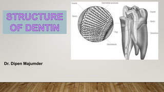
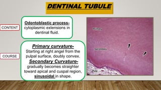
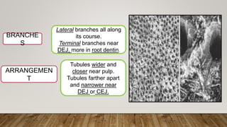
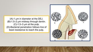
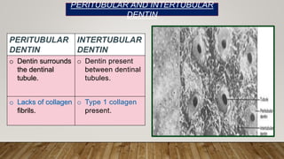
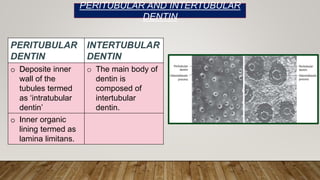
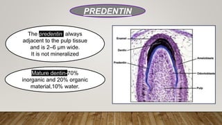
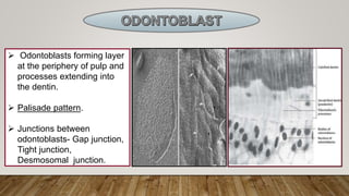
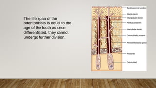
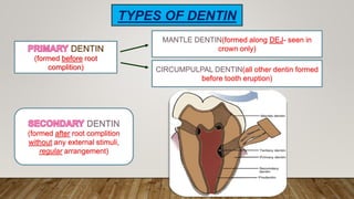
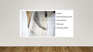
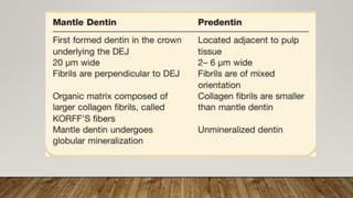
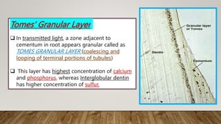
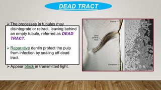
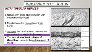
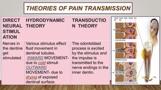
Dr. Dipen Majumder
DENTINAL TUBULE
CONTENT
Odontoblastic process-
cytoplasmic extensions in
dentinal fluid.
COURSE
Primary curvature-
Starting at right angel from the
pulpal surface, doubly convex.
Secondary Curvature-
gradually becomes straighter
toward apical and cuspal region,
sinusoidal in shape.
ARRANGEMEN
T
Tubules wider and
closer near pulp.
Tubules farther apart
and narrower near
DEJ or CEJ.
BRANCHE
S
Lateral branches all along
its course.
Terminal branches near
DEJ, more in root dentin.
(A)-1 μm in diameter at the DEJ.
(B)-1.5–2 μm midway through dentin.
(C)-1.5–3 μm at the pulp.
(D)-Bacterial penetration follows line of
least resistance to reach the pulp.
PERITUBULAR
DENTIN
INTERTUBULAR
DENTIN
o Dentin surrounds
the dentinal
tubule.
o Dentin present
between dentinal
tubules.
o Lacks of collagen
fibrils.
o Type 1 collagen
present.
PERITUBULAR AND INTERTUBULAR
DENTIN
PERITUBULAR
DENTIN
INTERTUBULAR
DENTIN
o Deposite inner
wall of the
tubules termed
as ‘intratubular
dentin’
o The main body of
dentin is
composed of
intertubular
dentin.
o Inner organic
lining termed as
lamina limitans.
PERITUBULAR AND INTERTUBULAR
DENTIN
PREDENTIN
The predentin always
adjacent to the pulp tissue
and is 2–6 µm wide.
It is not mineralized
Mature dentin-70%
inorganic and 20% organic
material,10% water.
 Odontoblasts forming layer
at the periphery of pulp and
processes extending into
the dentin.
 Palisade pattern.
 Junctions between
odontoblasts- Gap junction,
Tight junction,
Desmosomal junction.
The life span of the
odontoblasts is equal to the
age of the tooth as once
differentiated, they cannot
undergo further division.
DENTIN
(formed before root
complition)
MANTLE DENTIN(formed along DEJ- seen in
crown only)
CIRCUMPULPAL DENTIN(all other dentin formed
before tooth eruption)
DENTIN
(formed after root complition
without any external stimuli,
regular arrangement)
TYPES OF DENTIN
DENTIN
(formed after tooth eruption
due to external stimuli,
irregular in arrangement)
REPARATIVE DENTIN
SCLEROTIC DENTIN
REACTIONARY DENTIN
HISTOLOGY OF
DENTIN
 Thicker dentin seen in boys than girls of
similar age group and this increases during
puberty.
 The buccal surfaces of the tooth showed
maximum thickness, followed by lingual.
CLINICAL CONTENTS
Dentinal tubules branches, anastomoses to
form canalicular system enhancing carious
process.
INCREMENTAL GROWTH LINES
OF VON EBNER
 These lines reflect the daily
rhythmic, recurrent deposition of
dentin matrix as well as a hesitation
in the daily formative process.
 Theses lines situated about 20µm
apart.
 Run at right angle to the tubule.
 Deposit at 4 µm daily and
mineralize in a 12-hour cycle.
The contour lines formed from secondary
curvatures between neighbouring dentinal
tubules due to hypomineralisation known
as CONTOUR LINES OF OWEN
Prenatal and postnatal dentin are
separated by an accentuated contour line
known as NEONATAL LINE
 Between the
odontoblastic process and
the peritubular dentin,
periodontoblastic space
is present.
 This space contains
dentinal fluid which is
outward from the pulp,
having higher K+ and
lower Na+ content.
PERIODONTOBLASTIC
SPACE
• Hypomineralised dentin wherein globules
of dentin fail to coalesce into a
homogenous mass, appears dark in
transmitted light.
INTERGLOBULAR
DENTIN
• Prevalent with vitamin D deficiency or
exposure to high levels of fluoride at the
time of dentin formation.
Tomes’ Granular Layer
 In transmitted light, a zone adjacent to
cementum in root appears granular called as
TOMES GRANULAR LAYER (coalescing and
looping of terminal portions of tubules)
 This layer has highest concentration of calcium
and phosphorus, whereas Interglobular dentin
has higher concentration of sulfur.
HYALINE HOPEWELL-
SMITH LAYER
 Hyaline layer present outside
the granular layer(15-20 µm
wide)
 Nontubular and structureless.
The processes in tubules may
disintegrate or retract, leaving behind
an empty tubule, referred as DEAD
TRACT.
Reparative dentin protect the pulp
from infection by sealing off dead
tract.
Appear black in transmitted light.
DEAD TRACT
INNERVATION OF DENTIN
INTRATUBULAR NERVES-
 Nerves with close approximation with
odontoblastic process.
 Mostly located in coronal zone(pulp
horn).
 Synapse like relation seen between the
nerve and the odontoblastic process.
 The subodontoblastic plexus of
Raschkow seen in the cell-free zone of
Weil.
THEORIES OF PAIN TRANSMISSION
DIRECT
NEURAL
STIMUL
ATION
HYDRODYNAMIC
THEORY
TRANSDUCTIO
N THEORY
Nerves in
the dentine
get
stimulated.
Various stimulus effect
fluid movement in
dentinal tubules.
INWARD MOVEMENT-
due to cold stimuli
OUTWARD
MOVEMENT- due to
drying of exposed
dentinal surface.
The odontoblast
process is excited
by the stimulus and
the impulse is
transmitted to the
nerve endings in the
inner dentin.
DENTIN seminar  DENTIN seminar  DENTIN seminar

DENTIN seminar DENTIN seminar DENTIN seminar

  • 2.
  • 3.
    DENTINAL TUBULE CONTENT Odontoblastic process- cytoplasmicextensions in dentinal fluid. COURSE Primary curvature- Starting at right angel from the pulpal surface, doubly convex. Secondary Curvature- gradually becomes straighter toward apical and cuspal region, sinusoidal in shape.
  • 4.
    ARRANGEMEN T Tubules wider and closernear pulp. Tubules farther apart and narrower near DEJ or CEJ. BRANCHE S Lateral branches all along its course. Terminal branches near DEJ, more in root dentin.
  • 5.
    (A)-1 μm indiameter at the DEJ. (B)-1.5–2 μm midway through dentin. (C)-1.5–3 μm at the pulp. (D)-Bacterial penetration follows line of least resistance to reach the pulp.
  • 6.
    PERITUBULAR DENTIN INTERTUBULAR DENTIN o Dentin surrounds thedentinal tubule. o Dentin present between dentinal tubules. o Lacks of collagen fibrils. o Type 1 collagen present. PERITUBULAR AND INTERTUBULAR DENTIN
  • 7.
    PERITUBULAR DENTIN INTERTUBULAR DENTIN o Deposite inner wallof the tubules termed as ‘intratubular dentin’ o The main body of dentin is composed of intertubular dentin. o Inner organic lining termed as lamina limitans. PERITUBULAR AND INTERTUBULAR DENTIN
  • 8.
    PREDENTIN The predentin always adjacentto the pulp tissue and is 2–6 µm wide. It is not mineralized Mature dentin-70% inorganic and 20% organic material,10% water.
  • 9.
     Odontoblasts forminglayer at the periphery of pulp and processes extending into the dentin.  Palisade pattern.  Junctions between odontoblasts- Gap junction, Tight junction, Desmosomal junction.
  • 10.
    The life spanof the odontoblasts is equal to the age of the tooth as once differentiated, they cannot undergo further division.
  • 11.
    DENTIN (formed before root complition) MANTLEDENTIN(formed along DEJ- seen in crown only) CIRCUMPULPAL DENTIN(all other dentin formed before tooth eruption) DENTIN (formed after root complition without any external stimuli, regular arrangement) TYPES OF DENTIN
  • 14.
    DENTIN (formed after tootheruption due to external stimuli, irregular in arrangement) REPARATIVE DENTIN SCLEROTIC DENTIN REACTIONARY DENTIN
  • 15.
  • 16.
     Thicker dentinseen in boys than girls of similar age group and this increases during puberty.  The buccal surfaces of the tooth showed maximum thickness, followed by lingual. CLINICAL CONTENTS Dentinal tubules branches, anastomoses to form canalicular system enhancing carious process.
  • 17.
    INCREMENTAL GROWTH LINES OFVON EBNER  These lines reflect the daily rhythmic, recurrent deposition of dentin matrix as well as a hesitation in the daily formative process.  Theses lines situated about 20µm apart.  Run at right angle to the tubule.  Deposit at 4 µm daily and mineralize in a 12-hour cycle.
  • 19.
    The contour linesformed from secondary curvatures between neighbouring dentinal tubules due to hypomineralisation known as CONTOUR LINES OF OWEN Prenatal and postnatal dentin are separated by an accentuated contour line known as NEONATAL LINE
  • 20.
     Between the odontoblasticprocess and the peritubular dentin, periodontoblastic space is present.  This space contains dentinal fluid which is outward from the pulp, having higher K+ and lower Na+ content. PERIODONTOBLASTIC SPACE
  • 21.
    • Hypomineralised dentinwherein globules of dentin fail to coalesce into a homogenous mass, appears dark in transmitted light. INTERGLOBULAR DENTIN • Prevalent with vitamin D deficiency or exposure to high levels of fluoride at the time of dentin formation.
  • 22.
    Tomes’ Granular Layer In transmitted light, a zone adjacent to cementum in root appears granular called as TOMES GRANULAR LAYER (coalescing and looping of terminal portions of tubules)  This layer has highest concentration of calcium and phosphorus, whereas Interglobular dentin has higher concentration of sulfur.
  • 23.
    HYALINE HOPEWELL- SMITH LAYER Hyaline layer present outside the granular layer(15-20 µm wide)  Nontubular and structureless.
  • 24.
    The processes intubules may disintegrate or retract, leaving behind an empty tubule, referred as DEAD TRACT. Reparative dentin protect the pulp from infection by sealing off dead tract. Appear black in transmitted light. DEAD TRACT
  • 25.
    INNERVATION OF DENTIN INTRATUBULARNERVES-  Nerves with close approximation with odontoblastic process.  Mostly located in coronal zone(pulp horn).  Synapse like relation seen between the nerve and the odontoblastic process.  The subodontoblastic plexus of Raschkow seen in the cell-free zone of Weil.
  • 26.
    THEORIES OF PAINTRANSMISSION DIRECT NEURAL STIMUL ATION HYDRODYNAMIC THEORY TRANSDUCTIO N THEORY Nerves in the dentine get stimulated. Various stimulus effect fluid movement in dentinal tubules. INWARD MOVEMENT- due to cold stimuli OUTWARD MOVEMENT- due to drying of exposed dentinal surface. The odontoblast process is excited by the stimulus and the impulse is transmitted to the nerve endings in the inner dentin.