Download to read offline
![http://astonjournals.com/csj
1Chemical Sciences Journal, Vol. 2011: CSJ-46
Determination of Selenium in Human Blood Serum by
Electrothermal Atomic Absorption Spectrometry
F Čuparigova, T Stafilov*
Institute of Chemistry, Faculty of Science, Sts. Cyril and Methodius University, POB 162,
1000 Skopje, Macedonia.
*Correspondence to: Trajče Stafilov, trajcest@pmf.ukim.mk
Accepted: August 8, 2011; Published: November 11, 2011
Abstract
A method for direct selenium determination in human blood serum by electrothermal atomic absorption spectrometry
(ETAAS) was developed. Total selenium was measured by ETAAS employing 10 g of palladium as matrix modifier in a
graphite atomizer with pyrolytically coated tubes and Zeeman background correction. Blood serum was diluted 1+2 with
0.1 % v/v nitric acid and 0.1 % Triton X-100. Pyrolysis and atomization temperatures for palladium modifier are 1100 ºC and
2500 ºC, respectively. The measurements were confirmed by the analyses of standard reference material and by the
method of standard additions. Reference serum material Seronorm, Trace elements, Serum level 1 (lot JL 4409) was
analyzed and the results are in agreement with the certified value. The limit of detection of the direct ETAAS based on 3σ
of the blank signal is 0.60 g L
-1
Se in blood serum samples. The precision of the method ranges from 2.06 % to 5.95 %. The
obtained data from the selenium concentrations in serum samples from 83 patients show that the content of selenium is
relatively low, ranging from 43.91 ± 4.80 µg L
-1
for female to 45.21 ± 5.60 µg L
-1
for male.
Keywords: Selenium; serum; determination; ETAAS.
1. Introduction
Selenium is an essential micronutrient at low concentration but toxic at high concentration whit a relatively
small difference between these levels. Human body uses selenium to produce glutathione peroxidase [1],
which works with vitamin E [2] to protect cell membranes from damage caused by dangerous, naturally
occurring substances known as free radicals produced by oxidative metabolism [3]. Selenium is taking center
stage as a potential anticancer agent [4, 5] by promoting formation of white blood cells which destroys the
cancer cells and is an essential component of more than 10 selenoproteins with multiple biochemical
functions. Moreover, it boosts the immune system [6] by increasing the activity and number of white blood
cells and prevents premature aging, degenerative diseases, cardiovascular diseases, inflammatory diseases,
stroke, cataracts, and rheumatoid arthritis. It is also necessary for normal thyroid functions [7] and protection
of heavy metal toxicity. Deficiency of the element can cause Keshan disease, characterized by an enlarged
heart and poor heart function [8] or to be a factor for essential hypertension [9]. This disorder is endemic in
some of most selenium-poor soils in the world. High blood levels of selenium can result in selenosis, which is
associated with gastrointestinal upsets, hair loss, white blotchy nails and mild nerve damage [10]. The
optimum daily dietary intake of selenium is 55 µg/day for women and 70 µg/day for men [11].
The trace elements concentration in blood, serum, urine and tissue is used as an indicator of the trace
elements status of the human body. Namely, determination of metals in vivo in organs is not possible;
therefore indirect determination of metals in accessible tissue can point at the presence of toxic elements.
Because of that, it is very important to introduce accurate and precise methods for trace metal determination
(and their chemical forms, as well) in different biological fluids and tissue. There are a number of methods for
selenium determination in different biological materials, mainly by atomic absorption spectrometry (AAS),
both hydride generation AAS (HG-AAS) [12, 13] and electrothermal AAS (ETAAS) [14-17]. Atomic absorption
spectrometry is one of the most used techniques for trace element determination.
Direct electrothermal atomic absorption spectrometry (ETAAS) shows sensitivity and accuracy adequate for
selenium determination in biological fluids and requires little sample preparation, but is very sensitive to
matrix interference and reliable methods have been validated only for blood plasma and serum [18, 19]. In
direct ETAAS assay on diluted biological fluids, the main concern is reliability of background correction and](https://image.slidesharecdn.com/csj-46vol2011-150220001242-conversion-gate01/85/Csj-46-vol2011-1-320.jpg)
![http://astonjournals.com/csj
3Chemical Sciences Journal, Vol. 2011: CSJ-46
The following certified reference materials were used for the validation purposes: Seronorm, Trace elements,
Serum level 1 (lot JL 4409). All disposable devices were rigorously clean before use by brief immersion in hot
concentrated nitric acid and rinsing twice whit distilled water.
Serum samples were obtained from 83 presumable healthy volunteers. All patients signed agreement for
selenium testing of their serum during systematic medical checkup. Clinical experiments were performed
according to the Ethics Committee provisions of the Institute of Preventive Medical Care and Toxicology at the
Clinical Health Institution Center (Skopje, Macedonia). The serum samples were collected whit plastic iv
cannula No. 24 (TIK, Slovenia) whit an injection valve. The samples were kept frozen (-18˚C) until analysis.
2.3. Procedures
Blood serum samples (500 L) were diluted 1+2 with 0.1% V/V nitric acid and 0.1% Triton X-100 and 10 µL
were introduced into graphite furnace whit appropriate volume of palladium modifier (1.2 g; 2 g; 2.4 g; 5
g and 10 g) and iridium modifier (0.1 g; 0.5 g and 1 g) . The calibration curves (4–60 µg mL-1
Se) were
prepared using human serum in nitric acid-Triton X-100 mixture spiked with known amount of selenium
standard solution and palladium or iridium solution as matrix modifier.
3. Results and Discussion
3.1. Selection of chemical modifier
Several preliminary experiments were carried out to select an efficient chemical modifier for the measurement
of selenium in serum. The modifiers tested are: palladium (10 g L
-1
) and iridium (1 g L
-1
) modifier. Typically,
relatively high pyrolysis temperatures could be used for selenium determination, so the action of the modifier
is to assist and ensure complete matrix mineralization and removal during the ashing step. The matrix
modifiers used in this work were Pd and Ir with optimal concentration of 500 µg mL
-1
for Pd modifier and 100
µg mL
-1
for Ir modifier. Different masses of modifiers were tested, for palladium (1.2 g; 2 g; 2.4 g; 5 g and
10 g) and for iridium (0.1 g; 0.5 g and 1 g). Best results yielded 10 g or palladium and 1 g of iridium.
From this point of view, palladium is very useful to its high efficiency at relatively low masses and good
performance in presence of organic mater. It was observed that in presence of palladium the nonspecific
absorbance signals were remarkably lower, than iridium; therefore this modifier was selected for all further
investigations.
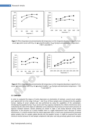
3.2. GFAAS program optimization
Optimization of temperature program for Se determination by electrothermal atomic absorption spectrometry
in human blood serum samples was performed. For the optimization of pretreatment and atomization
temperatures, pyrolysis–atomization curves were constructed from serum sample previously spiked with 10 g
L-1
Se in the presence of palladium (10 g) and iridium (1 g) as most widely used modifiers for thermal
stabilization of selenium in ETAAS. The modifiers were applied through the autosampler directly into the
graphite furnace with a volume of 10 µL for 10 µL serum sample. Parameters of the drying step were carefully
optimized so as to ensure complete matrix decomposition and removal during this step. Ashing temperatures
(from 800 to 1200
o
C) and atomization temperatures (from 2000
o
C to 2700
o
C) were assayed by using wall
atomization (pyrolytically graphite coated graphite tubes). Effects of pyrolysis and atomization temperatures
and times on integrated absorbance for serum samples with Pd modifier are given in Fig. 1 and with iridium
modifier are given in Fig. 2. As it can be seen, the optimal pyrolysis temperature with Pd and Ir modifiers was
found to be 1100
o
C, wile the optimal atomization temperature was 2500
o
C using Pd modifier and 2600
o
C
with Ir modifier. The optimal pyrolysis ramp time was established to be 5 s and hold time was 32 s (the last 2 s
without argon flow). The optimal atomization ramp time was 1 s and hold time 3 s. The behavior of Pd and Ir
for thermal stabilization of selenium proved identical. Both modifiers ensured loss-free ashing up to 1100 o
C is
serum samples diluted with 0.1% V/V nitric acid and 0.1% Triton X-100. Lower atomization temperatures are
preferable in this case because of lower background absorption signals, as well as better shaped absorbance-
time profiles for selenium. Therefore palladium is recommended as efficient modifier for ETAAS determination
of total Se in serum samples [20-23].](https://image.slidesharecdn.com/csj-46vol2011-150220001242-conversion-gate01/85/Csj-46-vol2011-3-320.jpg)
![http://astonjournals.com/csj
6 Research Article
between women 43.91 ± 4.80 (16.88-89.93) µg L
-1
and men 45.21 ± 5.60 (12.25-93.02) µg L
-1
[9]. Our data
reveal that serum selenium levels of healthy people in Macedonia are among the lowest in Europe. These
values are comparable with those in serum of adults in Bulgaria (66.5±15.5 µg L-1
) [14], in Czech Republic
(46±14 µg L
-1
) [24], in Montenegro (51±26 µg L
-1
) [25] in Greece (63±14 µg L
-1
) [26], in Hungary (50±11 µg L
-1
)
[27], in Poland (57±8 µg L
-1
) [28], in Bosnia-Herzegovina (64±19 µg L
-1
) [25], in Croatia (69±17 µg L
-1
) [29] and in
Serbia (41±20 µg L-1
) [25].
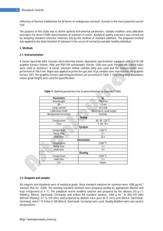
Table 4: Content of Se in blood serum samples for total, male and female.
N Sex
Average age
(from-to) Range/µg L
-1
Median/µg L
-1
(Mean ± CL*)/µg L
-1
47 f 46 (15-84) 16.88 - 89.93 42.35 43.91 ± 4.80
36 m 46 (18-75) 12.25 – 93.02 52.63 45.21 ± 5.60
83 f + m 45 (15-84) 12.25 – 93.02 42.90 44.47 ± 5.13
N – Number of patients; f – female; m – male; *CL - Confidence level
4. Conclusion
The method for total selenium determination in human blood serum by ETAAS was optimized. It was found
that Pd modifier should be applied with the optimal pyrolysis temperature of 1100 ºC and optimal atomizing
temperature of 2500 ºC. It was also established that the serum samples should be diluted 1+2 with a mixture
of 0.1% v/v nitric acid and 0.1% Triton X-100. This method was applied for selenium determination in 83
samples of human serum collected during systematic medical checkup from patients of Clinical Centre “Bit
Pazar” in Skopje. The obtained data show that the content of selenium is relatively low, ranging from 16.88 to
89.93 µg L
-1
for female and from 12.25 to 93.02 µg L
-1
for male, which indicate that dietary intake of Se in the
Republic of Macedonia is low [19, 20]. The highest value for Se was found to be 93.02±1.52 µg L
-1
and the
lowest value was 12.25±0.57 µg L
-1
. These levels are in agreement with previous reports for this
biogeochemical region by Maksimović [25]: 35±7 µg L
-1
for selenium in blood serum. In comparison to other
European countries these selenium levels are among the lowest.
Competing Interests
The authors declare that they have no competing interests.
Authors’ Contributions
TS supervised the experimental work, preparation of manuscript and communicated to the journal; FČ did the
actual work and prepared the manuscript.
References
1. Hatanaka N, Nakaden H, Yamamoto Y, et al., 2000. Selenium kinetics and changes in glutathione peroxidase
activities in patients receiving long-therm parenteral nutrition and effects of supplementation with
selenite. Nutrition, 16: 22-26.
2. Douillet C, Bost M, Accominotti M, et al., 1998. Effect of selenium and vitamin E supplementation on lipid
abnormalities in plasma, aorta and adipose tissue of Zucker rats. Trace Elements Research, 65: 221-236.
3. Clausen J, Nielsen SA, Kristensen M, 1989. Biochemical and clinical effects of an antioxidant
supplementation of geriatric patients. Biological Trace Element Research, 20: 135-151.
4. Clark LC, Combs GF, Turnbull BW, et al., 1996. The nutritional prevention of cancer with selenium. 1983-
1993: a randomized clinical trial. Journal of the American Medical Association, 276: 1957-1963.
5. Combs GF, Clark LC, 1999. Selenium and cancer. In: Nutritional Oncology: Academic Press, San Diego.](https://image.slidesharecdn.com/csj-46vol2011-150220001242-conversion-gate01/85/Csj-46-vol2011-6-320.jpg)

This document describes a method for determining selenium levels in human blood serum using electrothermal atomic absorption spectrometry (ETAAS). Palladium was used as a matrix modifier to minimize interference. Blood serum samples were diluted and spiked with known amounts of selenium. Optimal pyrolysis and atomization temperatures were determined to be 1100°C and 2500°C, respectively. Calibration curves were prepared using the standard additions method to account for matrix effects. The method was validated using certified reference materials, with recoveries ranging from 94.6-100.4%. The method achieved a detection limit of 0.60 μg/L and was applied to determine selenium levels in 83 patient serum samples.


