craniofacial anomalies
•Download as PPTX, PDF•
33 likes•3,795 views

craniifacial anomalies

Recommended
Recommended
More Related Content
What's hot
What's hot (20)
Similar to craniofacial anomalies
Similar to craniofacial anomalies (20)
Recently uploaded
Recently uploaded (20)
craniofacial anomalies
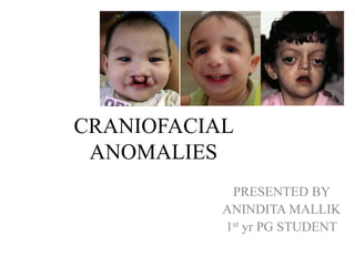
- 1. CRANIOFACIAL ANOMALIES PRESENTED BY ANINDITA MALLIK 1st yr PG STUDENT
- 2. • A craniofacial malformation is an anomaly of embryonic development - results in a serious impairment of the normal anatomy of skull, jaws and adjacent soft tissues. • Most of the malformations diagnosed at birth fall in the category “craniofacial” • Craniofacial anomalies (CFA) are a diverse group of deformities in the growth of the head and facial bones
- 5. Facial syndromes and congenital defects Post natal growth disturbances Fetal alcohol syndrome Trauma – maxilla - Mandible Hemifacial microsomia Acromegaly & hemimandibular hypertrophy Mandibulofacial dysostosis Facial clefting syndromes Achondroplasia Premature fusion of cranial & facial sutures – craniosynostosis syndromes : crouson & aspert Fetal molding & birth injuries – pierre robin syndrome -trauma to mandible
- 6. • Classification of craniofacial malformation (F Van der Meulen et al 1983). • CEREBROCRANIAL DYSPLASIA • CERERBOFACIAL DYSPLASIA • CRANIOFACIAL DYSPLASIA • CRANIOFACIAL DYSPLASIAS WITH OTHER ORIGIN
- 7. CEREBROCRANIAL DYSPLASIA • Anencephaly • Microcephaly CEREBROFACIAL DYSPLASIA • Rhineencepahlic dysplasia • Oculo-orbital dysplasia
- 8. CRANIOFACIAL DYSPLASIA a) With clefting • Latero-nasomaxilary cleft • Medio-nasomaxillary • Intermaxillary clefting • Maxillo-mandibular cleft b) With dysostosis • Sphenoidal • Spheno-frontal • Frontal • Fronto-frontal • Fronto-nasoethmoidal • Internasal
- 9. • Nasal • Premaxillo maxillary and intermaxillo-palatine • Naso maxillary and maxillary • Maxillo zygomatic • Zygomatic • Zygo-auromandibular • Temporo-aural • Temporo-auromandibular • Mandibular • Intermandibular
- 10. c) With synostosis • Cranio synostosis – Parieto-occipital – Interparietal – Interfrontal • Craniofacio synostosis – Spheno-frontopareital – Fronto-parietal – Fronto interpareital Faciosynostosis •Fronto-malar Vomero premaxillary (Binder) •Perimaxillary (posterior) (clefting) •Perimaxillary (anterior) (pseudocrouzon) •Perimaxillary (total) (crouzon)
- 11. d) With dysostosis and synostosis • Crouzon • Acro-cephalosyndactaly (Apert) • Triphyllocephaly (clover leaf skull) e) With dyschondrosis • Achondroplasia
- 12. a) Osseous • Osteopetrosis • Cranio tubular dysplasia • Fibrous dysplasia b) Cutaneous • ectodermal dysplasia c) Neurocutaneous d) Neuromuscular Robin syndrome Mobius syndrome e) Muscular Glossoschizis f) Vascular Haemangioma Haemolymphangioma Lymphangioma CRANIOFACIAL DYSPLASIAS WITH OTHER ORIGIN
- 13. Anencephaly: • Due to - absent closure of the neural tube. • Characterized by absence of – - vault of the skull. - The anterior brain structures - replaced by a spongy vascular mass called psedocephaly
- 14. Fetal alcohol syndrome • caused by consumption of alcohol by mother during pregnancy • characterised by retardation of mental development and of physical growth particularly of skull and face of the infant
- 15. Microcephaly: • Brain is reduced in size and enclosed in a small skull, cerebellum is normal in size • Primary (heredity) or secondary (rubella or toxoplasmosis)
- 16. Rhinencephalic dysplasia: • characterized by forebrain malformations and agenesis of the midline structures of the face.
- 17. • Cyclopia: • Forebrain fails to divide into cerebral hemispheres. • Lateral ventricles are fused. • 1 optic canal, Eyes are fused into a simple orbit. • All the midline structures are absent
- 18. Cebocephaly: • Absence of the falx, corpus callosum, olfactory bulbs and tracts. • Facial dysmorphism -severe hypotelorism and hypodevelopment of the midline structures. • Nose-rudimentary and flat, encompasses a unique nostril simplified into a blind pit.
- 19. Pre-maxillary aplasia or hypoplasia: Type 1 – • cerebral anomalies involves semilobar holoprosencephaly; absence of olfactory bulbs and tracts. The optic foramina lies in a common bony canal. • Hypotelorism and wide palatal clefting is observed. • Nose - flat , columella and philltrum are absent.
- 20. Type 2 – • Brain development may be normal. • Hypotelorism is less severe, the nose is flat and palatal clefting is seen. • Often associated with cardiovascular malformations.
- 21. • True or primary clefts are caused by the persistence of epithelium between the borders of the facial processes, due to deficient epithelial cell degeneration. Their existence is therefore restricted to – – latero-nasomaxillary clefting (naso-ocular clefts) – medio-nasomaxillary clefting (cleft lip) – intermaxillary clefting (cleft palate) – maxillo-mandibular clefting (macrostomia) CRANIOFACIAL DISPLASIAS WITH CLEFTING
- 22. Sites of primary (true) clefts: A latero-nasomaxillary; B medio-nasomaxillary; C maxillomandibular; D intermaxillary
- 23. CLEFT LIPAND PALATE • They are one of the most common congenital anomalies occurring in about 1.97 to 1.23 /1000 in Indians and 2/1000 in mongoloids • In 2/3rd of the cases cleft palate is on the left than the right side • CL(P) is seen more in male and CP alone more in females
- 24. • Cleft lip results from failure of fusion of the median nasal, lateral nasal and the maxillary processes on either or both sides. – Reasons • Hypoplasia of the facial processes • Altered facial geometry • Defective ability of surface epithelia to participate in the fusion process • Excessive cell depth in the fusing palatal seams, mesenchymal deficiency and post fusion rupture
- 25. • Thus they can be unilateral or bilateral clefting ; complete or incomplete , of the lip and/or primary palate till the incisive foramen
- 26. • Embryogenesis of the palate - movement of the initially vertical palatal shelves lateral to the tongue into a horizontal supralingual position with fusion beginning anteriorly and later in the soft palate.
- 27. • Reasons- – Hypoplasia of the palatal shelves – Failure of the palatal shelf elevation at the correct time due to diminished intrinsic force; increased resistance mainly by the tongue position being high – Excessive head width causing failure of normal sized palatal shelves to meet
- 28. Dentofacial relationships in unoperated cases • Unilateral cleft - nasal septum and columella is deviated to the non cleft side of facial midline whereas incisors deviate towards the cleft • In UCLP and BCLP - tendency for the mandible to be retruded and for the mandibular plane to be steep with a relatively shorter posterior facial height and a longer anterior facial height
- 29. • Mandibular incisors- labially proclined in UCLA while lingually inclined in CLP • In BCLP -maxillary intercanine dimension were much smaller than UCLP and UCLA • In maxillary arch the non cleft segment has a tendency to rotate forwards hence increasing the overjet while the cleft side rotates medially hence edge to edge bite of the canines. Teeth also tend to roll superiorly hence an openbite on that side due to infraocclusion
- 30. Presurgical orthopedics 1 to 4weeks Repositioning palatal segments can facilitate lip repair Lip closure 8 to 12 weeks May be preceded by primary lip adhesion as an alteration to presurgical orthopedics Palatal closure 18 to 24 months Closing only the soft plate initially is an alternative but one stage closure of hard and soft palate possible Speech therapy 6 to 11 years Articulation errors develop after a child tries to compensate for the cleft Early orthodontics 7 –8years Usually anterior alignment and maxilla transverse expansion Alveolar grafting 6 to 10 years Needed before the permanent canines erupt; being determined by stage and sequence of eruption Pharyngeal flap surgery 9 to 19 years Occurrence of nasal air leakage Orthognathic surgery 17 to 19 years Maxillary advancement and mandibular setback Fixed orthodontics 17 to 19 years Replacement of missing lateral incisors
- 31. Maxillo-mandibular clefting • It is not formed between the maxillary and mandibular bone but between the facial processes with the same names. • It is essentially a soft tissue defect affecting skin, muscle and mucosa, is usually called macrostomia. It may be unilateral or rarely bilateral. Its range of malformations varies from minor elongation of the oral angle to a wide cleft extending towards the tragal area.
- 32. • In the majority of cases it is associated with preauricular appendages or fistulae that may be found anywhere between the angle of the mouth and the tragus occasionally also with temporoaural and/or mandibular abnormalities.
- 33. CRANIOFACIAL DYSPLASIAS WITH DYSOSTOSIS • MEDIAN CLEFT FACE SYNDROME/ fronto-nasal syndrome/Internasal dysplasia
- 34. • At one end is bifidity of the nasal tip or dorsum, sometimes associated with a median cleft lip and with duplication of the labial frenulum. • Grooves and folds along the dorsum nasi are also occasionally observed.
- 35. • At the other end widely separated nasal halves and extreme orbital hypertelorism, including other anomalies caused by frontonasoethmoidal dysplasia
- 36. • Premaxilla may be retarded in development and bifid, maxilla may show a keel-shaped deformity, with the incisors rotated upward in each half of the alveolar process. • Sometimes a medial cleft of the palate is also found and this may extend upwards to the cribriform plate as an inverted V
- 37. • NASAL APLASIA – • complete absence of one nasal half. The nasal cavity is missing and pneumatiziation of the maxillary ethmoidal and frontal sinuses has failed . • There is no nasolacrimal duct. The affected half of the maxilla is hypoplastic and the palatal vault is high and acutely arched.
- 38. • NASAL DUPLICATION- • ranges from a supernumerary nostril in an otherwise normal nose to duplication of the upper face (diprosopia). The supernumerary nostril is usually the medial one. It may end blindly, be stenotic or open into a nasal cavity.
- 39. • In the milder cases there may be one continuous midline septum, while in the more severe cases duplication of the anterior part of the septum or full duplication may be observed
- 40. MANDIBULOFACIAL DYSOSTOSIS/ Treacher Collins' syndrome /Zygomatic dysplasia • Caused by a change in a single gene & Treacher Collin gene is located on chromosome 5 • Inherited as an autosomal dominant gene with complete penetrance but variable expressivity.
- 41. Features - • Malar & zygomatic hypoplasia • Anti mongoloid slant of the palpebral fissures • Coloboma in the outer third of the lower eyelid(75%) • Deficiency of eyelashes in the medial third of the eyelids
- 42. • Hair extending down & forward from the temporal region on to the cheek. • Unusual tongue shape (25% cases) • Flattening of the cheeks • Body of the mandible is frequently hypoplastic and the chin severely retruded.
- 43. • Radiographs show antigonial notch in the lower border of the mandible along with hypoplasia of coronoid & condylar processes. • Cleft palate is found in approximately 30% of the cases.
- 44. • Posterior maxillary height is decreased and anterior height is increased resulting in a steep anteroinferior cant. • Open bite is related to shortening of the mandibular rami and premature posterior teeth contact
- 45. • Deformed external ear, ear tags & pre-auricular pits, absence of external auditory meatus frequently accompanied by malformations of the middle ear
- 46. Miller syndrome / Postaxial acrofacial dysostosis • Has resemblance to mandibulofacial dysostosis but there is postaxial limb deficiency. • Malar bones are hypoplastic with downslanting palpebral fissures. • Eyelids may exhibit coloboma • Cleft lip and/or cleft palate are common
- 47. • Pinnae tend to be cup-shaped. The external auditory canals and middle ears are often malformed. • Various congenital heart defects have been documented
- 48. • Postaxial agenesis of a digit of the hands and feet is seen • Abnormal thumbs occur in about 50%. The radius and ulna tend to be short and, in some cases, there is radioulnar synostosis
- 49. Nager syndrome / Preaxial acrofacial dysostosis • Similar to mandibulofacial dysostosis. • The zygomatic hypoplasia results in downslanting palpebral fissures. • The lower eyelids exhibit colobomas • reduced numbers of eyelashes
- 50. • External ear defects and cleft palate are common • Velopharyngeal insufficiency • Micrognathia is usually more marked • mild mental retardation
- 51. • Thumb is hypoplastic or aplastic and the anomalies are usually asymmetric • Unilateral radial hypoplasia seen in 50% cases
- 52. HEMIFACIAL MICROSOMIA / Temporo auromandibular dysplasia / Goldenhars syndrome Facial asymmetry with deviation of the chin towards the affected side and ear anomalies are the 'hallmarks' of this entity.
- 53. • Ear - anotia to an ill-defined mass of tissue that is displaced anteriorly and inferiorly, to a mildly dysmorphic ear - 65%. Preauricular tags of skin and cartilage are extremely common, and maybe unilateral or bilateral.
- 54. • Both the horizontal and ascending ramus of the mandible may have macrostomia. • Malformations are most severe in the condylar region and less near the middle sector, with flattening of the gonial angle and accentuation of the antigonial notch.
- 55. • Hypoplasia of the maxilla on the affected side is shown by obliquity of the occlusal plane
- 56. • A depression and recession of the inferiolateral angle of the orbit indicates involvement of the malar bone. • Orbital dystopia may be observed • Temporalis, masseter and lateral pterygoid may be differentially hypoplastic.
- 57. • Aplasia of the levator veli palatini, resulting in abnormal elevation of the soft palate towards the unaffected side • Parotid gland may be absent, producing a preauricular concavity.
- 58. • Maxillary, temporal, and malar bones on the involved side are reduced in size and flattened • Narrow external auditory canals are found in more mild cases; atretic canals are seen in more severe cases.
- 59. • Epibulbar tumors - 35% - solid yellowish or pinkish white ovoid masses. • Blepharoptosis or narrowing of the palpebral fissure occurs on the affected side in about 10%
- 60. • Unilateral or bilateral cleft lip and/or cleft palate occurs in 7-15% of patients • Tooth development tends to be delayed and missing on the affected side • 35% have velopharyngeal insufficiency
- 61. CRANIOSYNOSTOSIS – Conditions in which one or more sutures closes too early causing problems with normal brain & skull growth – 1 in 2000 live births – 2M>F – Can be inherited as: • Autosomal recessive • Autosomal dominant
- 62. Synostotic posterior plagiocephaly / Parieto-occipital • Premature closure of the lambdoid sutures • found isolated, associated with synostosis of the sagittal suture or as part of multiple synostoses. • It causes hypoplasia and flattening of the occiput, with slight compensatory development of the ipsilateral anterior cranial region.
- 63. Scaphocephaly/ interparietal • Early fusion of the interparietal sagittal suture • Elongated narrow shape of the skull, resembling the hull of a ship
- 64. • From front, the skull is high and narrow • From side - skull is elongated • From front to back with posterior occipital protrusion and excessive bulging of the frontal bones anteriorly.
- 65. Trigonocephaly / interfrontal • Premature closure of the frontal suture. The frontal area becomes triangular. • Results in a prominent ridge running down the forehead • Extent of skull malformation depends on how early the synostosis takes place; this usually occurs during intra-uterine life.
- 66. Plagiocephaly/spheno-frontoparietal • Asymmetric malformation secondary to fusion of one half of the coronal suture • Produces flattening of forehead & the brow on the affected side with forehead excessively prominent on the opposite side • Eye on the affected side may also have a different shape
- 67. Brachycephaly / frontoparietal • refers to craniofacial dysmorphism secondary to premature bilateral coronal stenosis • the skull is shortened in the sagittal plane and compensatory lateral development occurs in breadth or in height.
- 68. Binder's syndrome / Maxillo-nasal dysostosis • Nasomaxillary deformity which mainly affects the lower part of the nose and the premaxilla • It is due to an alteration of the inferior mesenchymal portion of the medial strut formed by the vomer pushing the premaxilla forward.
- 69. • Nasofrontal angle is absent and the nose is hypoplastic with flattened ala with nostrils being half moon shaped • Frontal sinuses are hypoplastic
- 70. • Philtrum is poorly developed • Premaxilla is hypoplastic with shortening of the dental arch • All patients have relative mandibular prognathism with anterior crossbite
- 71. Crouzon syndrome • The developmental arrest affects the Maxilla, the Orbit and the Vault • It is an autosomal dominant condition. • Two genes known to be associated are FGFR2 and FGFR3.
- 72. • Cranium – craniosynostosis at birth in which several sutures are always involved
- 73. • Eyes – – Exopthalmos, the cardinal sign is constant – eyes give the patient a ‘ toad like ’ appearance. This appearance is due to hypoplasia of the maxilla, of the malar bone and of the orbital roof, resulting in the reduction in the size of the orbital cavities – Divergent strabismus or defective convergence is frequent – Hypertelorism may be present
- 74. • Face – – Flattened and sometimes concave. – Parrot beak appearance of nose - maxillary retrusion. – Dental malpositioning- supernumerary or abnormal ‘peg- shaped’ teeth. – Palate is high arched, narrow & pointed – Nasal root is flat, the dorsum and the nostrils are wide.
- 75. • Vision – Lack of skeletal protection may result in exposure keratitis or even dislocation of the globe. • Respiration – Constriction of airway may result in chronic or intermittent respiratory problems.
- 76. • Five clinical forms seen – – Maxillary Crouzon – – minor exorbitism is seen with severe maxillary retrusion
- 77. – Pseudo-Crouzon – – it is based on the combination of moderate exorbitism and inferior orbital retrusion. – prominent forehead and marked digital impressions are seen – Occlusion is normal.
- 78. – Facial Crouzon- – Retromaxilla with /without exorbitism in the absence of cranial abnormalities or with discreet frontal flattening. – These malformations are due to fusion of the posterior part of the perimaxillary sutural system. With Exorbitism Without Exorbitism
- 79. – Cranial Crouzon – – a sphenoidal dysostosis with variable facial involvement – occlusion is mostly normal
- 80. – Craniofacial Crouzon - – Disproportion b/w minor degree of facial retrusion & severity of cranial involvement
- 81. Apert's syndrome • Inherited in an autosomal dominant manner. • The gene involved is FGFR2 (fibroblast growth factor receptor 2) located on chromosome 10 • Having craniosynostosis - 4-5% have Apert’s syndrome
- 82. • In infancy- • Midline calvarial defect from the nose to the posterior fontanelle. • The defect is widely patent during infancy and only gradually fills in completely during the third year of life. • Bony islands form within the calvarial defect
- 83. • Down slanting palpebral fissures, strabismus, orbital hypertelorism. • ears may appear low set and Otitis media is common • Midface deficiency (maxillary hypoplasia). • Class III malocclusion is present, with anterior open bite and anterior and posterior crossbite • Delayed dental eruption
- 84. • symmetric syndactyly of hands and feet involving 2nd , 3rd and 4th digits. • fusion of some bones in the neck and differences in the arms that can be seen on X-rays. • Thumb and big toe may be broader than normal and deviates radially
- 85. • palate is high arched; constricted, and has a median furrow. Lateral palatal swellings (Hyaluronic acid) are present, which increase in size with age. • Maxillary dental arch is V-shaped with severely crowded teeth and bulging alveolar ridge
- 86. • Unique- Growth pattern – Length and weight at birth tend to be increased and head circumference is approximately normal. – in infancy and childhood consists of a gradual decrease in height
- 87. Pfeiffer syndrome • Autosomal dominant transmission • Main features- craniosynostosis, broad thumbs & great toes, and soft tissue syndactyly of the hands • skull is usually turribrachycephalic. • Craniofacial asymmetry may be present • Maxillary hypoplasia
- 88. • Hypertelorism, downslanting palpebral fissures, ocular proptosis, and strabismus are common • palate is highly arched, alveolar ridges are broad, and teeth are crowded • thumbs and great toes are broad • Mild soft tissue syndactyly
- 89. Saethre-Chotzen syndrome • Craniosynostosisis a facultative feature • Brachycephaly or acrocephaly with coronal sutural synostosis is seen, producing plagiocephaly and facial asymmetry • Frontal bossing, parietal bossing, and flattened occiput with late- closing fontanels are seen
- 90. • Low-set frontal hairline is commonly observed. • Ptosis of eyelids, hypertelorism, and strabismus are common • ears may be low set, small, posteriorly angulated • nasofrontal angle may be flattened • Maxillary hypoplasia
- 91. • Oral anomalies include narrow or highly arched palate, cleft palate • supernumerary teeth, enamel hypoplasia • Some degree of brachydactyly and partial cutaneous syndactyly is present
- 92. Cloverleaf anomaly, Triphyllocephaly • Characterized by hydrocephalus and a trilobular skull with synostosis of the lambdoidal and coronal and metopic sutures, with bulging of the cerebrum through the open sagittal sutures and a widely patent anterior fontanelle.
- 93. • Characteristics – Hydrocephaly – Retrusion of orbital roof – Exorbitism – Maxillary retrusion – Severe downward displacement of ears and zygomatic arches
- 94. – nasal flattening and an arched palate – Macrostomia; macroglossia; oblique facial clefting – Iris colobomas and blindness – Obstructed nasolacrimal ducts – Absent external auditory canals
- 95. CRANIOFACIAL DYSPLASIAS WITH DYSCHONDROSIS • Achondrodysplasia – deficient formation of enchondral bone – transmitted as an autosomal dominant trait. – Height is usually under 1.4 m. – Short, thick muscular extremities.
- 96. – The skull is voluminous with a prominent occiput and a bulging forehead overhanging a small impacted nose. – Legs are bowed, hands small, fingers stubby – Cranial base is shortened. – The alterations predominantly affect the ethmoidal part and the cribriform plate
- 97. – middle third of the face is short. – lower third- long and protruding. – The upper lip is shortened and labial incompetence is associated with buccal respiration. – Class III malocclusion is seen.
- 98. Ectodermal dysplasia • affect series of ectodermal derivatives including the teeth the sweat glands and the of the adnexa the skin derivatives(nails, hairs). • hypohydrosis, hypotrichosis, hypodontia are the main characteristics • sex-linked recessive trait - males
- 99. • Main features – Thin and/or small nails – Person cannot perspire and consequently suffers from hyperpyrexia & inability to endure warm temp – the midface is retruded due to deficient alveolar growth. Jaw and facial development are normal – forehead is prominent and the nose flattened
- 100. – the skin is thin and dry with multiple ridges – hairs are scarce and underdeveloped. – complete or partial absence of teeth & when present teeth may be truncated or cone shaped. – Palatal arch is frequently high and a cleft palate may be present.
- 101. – Forehead is prominent and nose flattened – Xerostomia may be present. – Hypoplasia of the nasal & pharyngeal mucous glands which leads to chronic rhinitis &/or pharyngitis, sometimes associated with dysphagia & hoarseness.
- 102. Neurofibromatosis • Characterized by neurofibromas or other neural tumours and by focal cutaneous hyperpigmentation (cafe- aulait spots) caused by aggregation of melanoblasts in the basal layer of the epidermis. • derivatives from the neural crest, are primarily affected.
- 103. Skeletal malformations – – macrocranium & interosseous cysts and perforating defects – expansion of the middle cranial fossa – hypoplasia of the sphenoid resulting in wide areas of communication between the cranial cavity and the orbit – downward displacement of the zygoma, maxilla and the mandible on the affected side.
- 104. Pierre Robin syndrome • It’s a combination of problems that begins with Micrognathia . • Causing not enough room for the tongue to lie flat in the mouth, so it rests at the back of the mouth (Glossoptosis) • Glossoptosis prevents palate from closing resulting in Cleft palate
- 105. • It is a disturbance of muscular maturation of nervous origin which affects the masticatory muscles, the tongue and the pharyngeal slings
- 106. Stickler syndrome • It is a connective tissue disorder caused by a change in one of the 3 genes for connective tissue.
- 107. • Features : – Cleft palate and a small lower jaw. Of those with stickler syndrome , 60% have pierre robbin syndrome – Eyes - near sightedness. – increased risk of cataracts & retinal detachment.
- 108. • Hearing loss of some degree affects around 80% patients. • Joints may be enlarged and hyperextensible. • About 50% of the affected people have Mitral Valve Prolapse (MVP).
- 109. Mobius syndrome • involves paralysis of certain facial nerves (unilateral or bilateral). • Mainly the intra-cerebral nuclear part of the 6th & 7th nerves are affected. • face is motionless with a characteristic nasiolabial grin.
- 110. • No side to side eye movements, but they will be able to move them up & down. • Blinking action may be difficult • hypoglossia & microstomia may be seen • skeletal involvement include clubfoot, missing or webbed fingers
- 111. Cleidocranial dysplasia • autosomal dominant inheritance • individuals are usually short • skull is brachycephalic, with pronounced frontal and parietal bossing. • maxilla and zygomas are hypoplastic.
- 112. • skull is large and short • Closure of the anterior fontanel and sagittal and metopic sutures is delayed • Secondary centers of ossification appear in the suture lines, and many Wormian bones are formed
- 113. • Delayed union at the mandibular symphysisis. • nose is broad at the base, with the bridge depressed. • neck appears long, and the shoulders are narrow and droop markedly
- 114. • Clavicles are absent unilaterally or bilaterally • variations in size, origin, and insertion of muscles related to the clavicles, especially the sternocleidomastoid, trapezius, deltoid, and pectoralis major
- 115. • palate is highly arched. • Submucous cleft of palate and complete cleft of the hard and soft palates is seen • Development of the premaxilla is poor with relative prognathism
- 116. • multiple supernumerary teeth • Multiple crown and root abnormalities, crypt formation around impacted teeth, ectopic location of teeth, and lack of tooth eruption
- 117. Acromegaly • Cause – anterior pituitary tumor that secrete growth hormone • Thickening of the facial skin • Mandibular overgrowth • Macroglossia • Anterior open bite • Class III malocclusion
- 118. CFA TEAM • It is agreed worldwide that management of patients with CFAs is best provided by a multidisciplinary team of specialists. – Plastic /craniofacial surgeon – Neurosurgeon – Pediatrician – Orthodontist – Pedodontist – Speech & language specialist – Otolaryngologist – Audiologist
- 119. – Opthalmologist – Genetic councellor – Nurse team coordinator – Social worker – Psychiatrist
- 120. • The surgeon and the orthodontist plan at the very beginning for diagnosis and treatment planning . • A detailed treatment plan should be written, including a specific definition of what orthodontic teeth movement is to be done prior to surgery; how the orthodontic appliance will be used for surgical fixation; and what orthodontic tooth movement will be required to finish the case following surgery.
- 121. • The efficacy of orthodontic and orthopedic treatment in case of craniofacial anomalies depend on the type of deformity, taking mainly into consideration the growth potential.
- 122. Presurgical orthodontic treatment • The main objective of this stage is to arrange the teeth so that they will approximately fit when the arches are surgically moved • Continuous arch wire technique • Segmented arch technique
- 123. • Continuous arch wire technique – Used for total maxillary surgical procedures. – progressively the size of the arch wires is increased to achieve final stability in the post-surgical occlusion. – If .018 slot is used, the minimum size of arch wire for a total maxillary surgical splint is .016x.022 without palatal splinting and .016x.016 if acrylic or metal palatal splinting
- 124. • Segmented arch technique- – used in preparation for a segmented surgical procedure. – orthodontic treatment time is shortened because alignment of each segment is done without being concerned about the relationship of the segments to each other.
- 125. • Disadvantage- when surgical suspension wires are used inadequate fixation will allow the crowns of the segments to be buccally torqued, causing posterior buccal overjet and open bite
- 126. Post surgical orthodontic treatment • Involves various final adjustments in the occlusal relationships and the final tooth alignment. • This final phase usually lasts form 3 to 4 months
- 127. Transverse Maxillary Deficiency • 3 main factors should be considered- – amount of arch length discrepancy- • In moderate to minimal space deficiency, RME will increase arch circumference sufficiently to permit alignment of the crowded anteriors without the necessity of extraction of premolars
- 128. – Arch morphology- • Cases in which a transverse deficiency exists will exhibit a narrow, tapering arch form. • The discrepancy will be most pronounced in the canine region. • If nonextraction orthodontic therapy is decided- lateral maxillary osteotomies and rapid maxillary expansion is the treatment of choice to achieve proper arch morphology
- 129. • Cases which do not exhibit severe constriction in the anterior region, a two-piece maxillary procedure with a midline osteotomy and resultant diastema may be done • consideration to wound healing after creation of an interincisal space should be done. When excessive the gingiva may detach and interproximal bone may be exposed with a possibility of devascularization and osteonecrosis of the underlying bone
- 130. – vertical dimension- • In cases exhibiting an anterior open-bite with a severely accentuated maxillary curve of spee ; orthodontic treatment by extrusion of incisors and/or intrusion of posterior teeth may compromise the postsurgical stability. • Segmentalized orthodontic therapy with a three-piece or four-piece maxillary surgical procedure is indicated
- 131. True Unilateral Transverse Maxillary Deficiency • should be treated by maxillary segmental surgery with the osteotomy mesial to the most anterior tooth in palatal cross-bite. • Orthodontic management of such patients will depend upon the necessity of extractions for alignment of crowded anterior teeth.
- 132. • In some cases the apparent maxillary deficiency may be due to the ectopic eruption of one or two posterior teeth in one quadrant and be treated by orthodontic means
- 133. Transverse Maxillary Excess • Seen mostly in cases with skeletal class II • The aim of presurgical orthodontics in these cases is to position the malaligned teeth over their bases so that the maxilla can be surgically positioned into a satisfactory overbite-overjet relationship
- 134. • Many technical modifications of the Le Fort I osteotomy are feasible to facilitate simultaneous anteroposterior, vertical, or horizontal movements of the anterior and posterior segments of the maxilla.
- 135. Hemifacial Microsomia • Harvold advocates the use of activators to guide eruption of teeth and prevent midline shift until the time of surgery. • This approach may have a stimulator effect on muscle development and serves to prevent canting of the occlusal plane. • conventional orthodontic tooth movement is of little value
- 136. • In a cephalometric study by Bachmayer, Ross and Munro (AJO 1986) on maxillary growth following Le Fort III osteotomy in children with Crouzon-Apert, Pfeiffer (CAP) syndromes it was found that the maxillary growth after surgery is negligible. • Vertical maxillary growth following surgery is identical to that in unoperated CAP and normal children, amounting to 1.3 mm/yr.
- 137. • Graysun et al ( AJO 1983) in a study on unilateral craniofacial microsomia • the lateral ceph analysis of patients with unilateral craniofacial microsomia confirmed the clinical impression of an increased gonial angle and decreased ramal height and body length on the affected side.
- 138. • The ramal height on the unaffected side was also decreased. • The mandibular plane angle was greater than normal on both affected and unaffected sides. • They conclude that the unaffected side too is characterized by abnormalities in the skeletal anatomy.
- 139. • Schudy ( JCO 1986) described the surgical correction of Crouzon's and Apert's syndromes by Dr. Paul Tessier. The orthodontic treatment involves no special procedures and is performed in the usual manner. Good arch forms were established for the prospect of good future occlusion before the surgery was performed. After the surgery was done brackets remained on for a further 24 months to improve the occlusion
- 140. Skeletal Mandibular Deficiency. • 3 types of dentoalveolar problems that require orthodontic treatment often accompany it – – Malalignment of the teeth ie: crowding or protrusion. Most of these are dental compensation for the skeletal deficiency – crossbite tendency appears as the mandible is advanced.
- 141. – Deep bite, with an accentuated curve of Spee due either to elongation of the mandibular incisors or due to vertical under development of the premolar segment of the arch.
- 142. Distraction ostegenesis. • specially effective in cases of unilateral mandibular deficiency • involves the deliberate fracturing of the bone side and holding it in close but not exact approximation by means of a complex system of extra oral positioners
- 143. • Principle- osteogenesis takes place in the intervening space. • As soon the bone formation is complete the set up is adjusted so that the bone segments move a bit away from each other. • The bone segments are held in that place till new bone is formed and so forth, till the bone achieves the required length.
- 144. • General principles of treatment- – Orthodontic intrusion of teeth must be done prior to surgery. – Extrusion of teeth can be done following surgery. – tooth movement in the transverse or crossbite plane of space can be deferred until after surgery.
- 145. • Tooth movement that occurs immediately after surgery, while the patient is in IMF but before bone healing occurs should also be considered. • Orthodontic tooth movement takes place to maintain the dental relationship. The mandibular dentition slips forward on the mandible (2mm) increasing the prominence of the lower incisors. The maxillary dentition is retracted, decreasing the prominence of the maxillary incisors.
- 146. Orthodontic Procedures To Be Avoided Prior To Surgery For Mandibular Deficiency. • use of Class II intermaxillary elastics to reduce overjet- – produces forward positioning of the lower incisors. – will cause vertical extrusion of the anterior maxillary segment, tending to extrude teeth
- 147. Mandibular excess • characterized by a prominent lower third of the face. • orthodontic treatment modalities- – Chin-cap therapy – Activator appliances – Fully banded orthodontic appliances.
- 148. Chin-cap therapy – the pressure against the chin would be transmitted to the growing areas of the mandible and the growth would be impeded or at least directed more favorably.
- 149. • two approaches- – impede mandibular growth by applying heavy pressure in the vicinity of the growing condyle of the mandible. The force is applied upward and backward, opposite to the vector of downward and forward mandibular growth. – redirect the growth of the mandible. It is based on the principle that when the mandible is rotated downward it rotates backward.
- 150. Activator appliances: • effective in the treatment of class III malocclusion using a class III activator causing a downward and backward displacement of the mandible. • It may be trimmed to allow posterior teeth to erupt so that the vertical dimension is maintained
- 151. Fully banded orthodontic appliances • can only be carried out satisfactorily without surgery only when the problem is minor, because it is very difficult to position mandibular teeth so as to camouflage the mandibular prominence.
- 152. – Conclusion • Orthodontic management for patients with craniofacial anomalies tends to be more complex, takes more time and clinical resources and should be based on a precise coordination with multiple dental, surgical and medical providers to achieve the best long-term esthetic and functional results.
- 153. • As the orthodontic management is commonly needed prior to most surgical procedures associated with craniofacial anomalies, management protocols should be based on a precise understanding of the exact nature of the anomalies as certain mechanics may be provided efficiently, safely and with acceptable durability, while at the same time, other techniques might be not effectively applied with some complications.
- 154. THANK YOU
Editor's Notes
- CC IS
- The optic foramina lie close together in a common bony canal