Download to read offline

![The VLU was evaluated by the investigating physician
every 2 weeks until week 8, even if the tested dressing had
been discontinued for whatever reason (except consent with-
drawal or healing). At each visit, the wound evaluations were
repeated (clinical assessment, acetate tracing, and wound
photo). Investigators were required to notify any unexpected
local adverse events whether dressing related or not. In the
case of complete wound closure, an acetate tracing and
a photo documenting complete epithelialization were to be
taken.
Between the biweekly investigator’s assessments, all local
procedures were recorded by health-care professionals,
including detailed information on dressing removal and appli-
cation and the compression system applied. The parameters
(ease of dressing application and removal, pain at removal,
and between dressing changes and periwound maceration)
were subjectively assessed using a four-level scale (from
“very easy” to “extremely difficult” or “none” to “very impor-
tant” depending on the parameter).
The EuroQol Quality of Life Questionnaire was docu-
mented by the patient at baseline and at week 8 (or before if a
treatment discontinuation occurred), including five items rep-
resenting the five following dimensions: mobility, autonomy,
activity, pain–discomfort, and anxiety–depression, each one
being assessed using a three-level scale.Avisual analogic scale
(VAS) was also associated to this patients’ self-completed
questionnaire.
Outcomes
Primary study outcome was relative Wound Area Reduction
(WAR) calculated as [Arealast – Areat0)/Areat0] ¥ 100 and
expressed as a percentage (%). Arealast was the last available
wound tracing value. All acetate tracings were blinded and
centrally measured by two nonparticipating clinicians using
digital software (DeskTop Ruler™). Wound area and wound
perimeter were recorded as the mean of both measurements.
Neither of the measurements deviated by more than 5%. In
addition, 10% of the tracings were randomly selected for a
controlled evaluation using Scion Image™ program.
Secondary outcomes were absolute WAR (Arealast – Areat0,
expressed in square centimeter) and wound edge progression
according to Gilman’s formula. Wound edge progression is
calculated as 2 ¥ (Arealast – Areat0)/(Pto + Plast), where P repre-
sents the wound perimeter. This formula is expected to give a
wound area evolution independent of the baseline surface area
value.
Other efficacy outcomes were the wound healing rate
[(Arealast – Areat0)/(tlast – t0)], expressed in square millimeter
per day of treatment, the percentage of wounds with a relative
WAR Ն40% (WAR40%) and Ն60% (WAR60%) at last avail-
able tracing, and the mean time to reach the WAR Ն40% goal.
Other end points included local tolerance (occurrence of
local adverse events documented by the investigating physi-
cian) and the acceptability of the tested dressings (assessed by
the nursing staff) at each dressing change during the 8-week
follow-up.
Ethical considerations
This study was conducted according to European Good Clini-
cal Pratices recommendations, the principles of the Declara-
tion of Helsinki (1975), and current French regulations. Study
protocol and documentation were submitted to the Comité de
Protection des Personnes (French Ethics Committee) of Paris
VIII (Ambroise Paré University Hospital), which gave its
approval on January 21, 2009. The study was also approved
by the French Competent Authority (Agence Française de
Sécurité Sanitaire des Produits de Santé [AFSSAPS]) on
January 13, 2009 (AFSSAPS Registration Number 2008-
A1573-52).
At baseline, before being included in the trial, all patients
received full information on the study objectives, potential
harm, and benefits both verbally and in writing and gave their
written consent to participate in the trial.
Statistics
Sample size was calculated to document the superiority of
the TLC-NOSF dressing compared with the control TLC
dressing, after 8-week follow-up.
A difference of 15% between the two groups had to be
detected, on relative WAR, if standard deviation (SD) of the
parameter is 34% maximum, on the basis of the previous trial,
with a power of 80%. The alpha risk (bilateral) was fixed at
5%. Accordingly, 82 patients per group were required and it
was decided to include a minimum of 180 patients in case of
dropouts.
Analyses were conducted using SPSS 18.0 software on an
unblinded database (allocated dressings were identified as A
or B and treatment disclosure was performed after the final
statistical report had been written).
Baseline comparability of the two groups was verified using
adapted tests (Student’s t-test, nonparametric Mann–Whitney
test, and chi-square test), dependent on the distribution and the
nature of the variables.
Knowing the large deviation of wound regression variable
distributions from normal and the difficulties in normalizing
these distributions, only nonparametric Mann–Whitney tests
were used to compare the allocated dressing effects on
primary and secondary efficacy outcomes.
For the other outcomes, chi-square tests were used and
odds ratio (OR) was calculated with 95% confidence intervals
(CIs). Time to reach relative WAR40% was evaluated using a
Kaplan–Meier approach followed by log-rank test. Addition-
ally, for WAR40%, a binary logistic regression was conducted
with wound surface area at baseline, wound duration, and
whether the ulcer was recurrent including in the model.
Local tolerance analyses were described and the percentage
of patients suffering from at least one adverse event was
compared by using chi-square test.
All analyses were conducted on an “intent-to-treat” (ITT)
population, defined as all randomized patients who received
the allocated dressing at least once.
All tests were bilateral and a p-value <0.05 was considered
to be indicative of statistical significance.
Post hoc subgroup evaluations were performed to appreci-
ate the magnitude of treatment differences according to base-
line wound area (10 cm2
cutoff), recurrent ulcer status, ulcer
duration (one cutoff), and the nature of the compression
system used during the study period (monolayer or multilayer
compression therapy).
Scale variables are presented by their mean Ϯ SD, their
median, and range. Median differences are given with 95%
CIs according to the method proposed by Bonett and Price.19
Randomized, double-blinded trial with dressing in VLU Meaume et al.
Wound Rep Reg (2012) 20 500–511 © 2012 by the Wound Healing Society502](https://image.slidesharecdn.com/contentserver10-150114215057-conversion-gate02/85/Content-server-10-A-randomized-controlled-double-blind-prospective-trial-with-a-Lipido-Colloid-Technology-Nano-OligoSaccharide-Factor-wound-dressing-in-the-local-management-of-venous-leg-ulcers-3-320.jpg)
![Mean ulcer area was 16.8 Ϯ 15.7 cm2
(median: 11.2 cm2
;
range: 2.3–86.9 cm2
; wound area >10 cm2
in 102 ulcers outof
187, 54.5%), without any difference between the two groups.
Efficacy outcomes
Patient compliance with the compression therapy prescribed
at inclusion was very good with 98.9% of the patients seen at
week 2, 96.6% at week 4, and 96.4% at week 6 still wearing
compression. Between these visits, compression wearing was
also checked by nurses during all the documented local pro-
cedures.
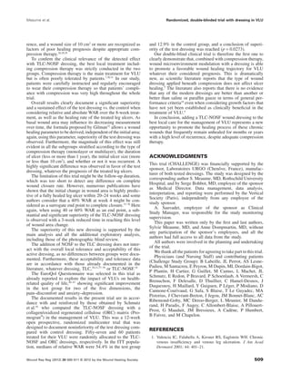
Concerning the primary outcome, the median relative WAR
(Table 3) decreased by -58.3% in the test group and by
-31.6% in the control group (difference: -26.7%; 95% CI for
median difference: -38.3 to -15.1%; p = 0.002), as presented
in Figure 2. Cumulative distributions of relative WAR are
presented in Figure 3, showing a left shift of distribution for
the test group compared with the control group.
By using the process of last-observed value carried forward
to compensate for missing data, the profiles of relative WAR
between-group differences over the 8-week period are pre-
sented in Figure 4; a superior effect of the test dressing was
observed after only 2 weeks and increased steadily thereafter.
A highly statistically significant difference was observed in
favor of the test dressing for absolute WAR (-6.1 cm2
in the
test group and -3.2 cm2
in the control group) and healing rate
(-10.81 mm2
/day in the test group and -5.15 mm2
/day in the
control group) (Table 3).
Moreover, when considering the Gilman’s formula (wound
edge progression), which allows a calculation of the wound
area independent of the baseline wound area value, the supe-
riority of the test dressing can be confirmed (p = 0.001).
The WAR of more than 40% from baseline value (WAR
>40%) was noted in 65.6% of patients receiving test dressing
Table 1. Distribution at baseline of the patient’s characteris-
tics for the treatment groups (n = 187)
TLC-NOSF TLC
n = 93 n = 94
Females (n, %) 62 (66.7%) 60 (63.8%)
Age (year)
(mean Ϯ SD)
72.6 Ϯ 13.0 74.4 Ϯ 12.1
BMI (kg/m2
)
(mean Ϯ SD)
30.5 Ϯ 8.7 30.1 Ϯ 6.9
BMI >30 kg/m2
(n, %) 40 (43.0%) 40 (42.6%)
Diabetes (n, %) 13 (14.0%) 17 (18.1%)
Smoking (n, %) 10 (10.8%) 14 (14.9%)
History of deep
venous
thrombosis
(n, %)
40 (43.0%) 32 (34.0%)
History of venous
surgery (n, %)
32 (34.4%) 37 (39.4%)
History of VLU (n, %) 67 (72.0%) 69 (73.4%)
ABPI
(mean Ϯ SD) 1.05 Ϯ 0.14 1.03 Ϯ 0.12
Median [range] 1.00 [0.8; 1.5] 1.00 [0.8; 1.3]
Patient status (n, %)
• Outpatient 75 (80.6%) 77 (81.9%)
• Hospitalized 18 (19.4%) 17 (18.1%)
Ankle mobility (n, %)
• Fully mobile 65 (69.9 %) 56 (59.6 %)
• Limited mobility 25 (26.9 %) 35 (37.2 %)
• Immobile 3 (3.2 %) 3 (3.2 %)
Autonomy of the
patient (n, %)
• Can easily walk 53 (57.0 %) 45 (47.9 %)
• Can walk with
difficulty
39 (41.9 %) 48 (51.1 %)
• Confined to bed 1 (1.1 %) 1 (1.1 %)
ABPI, Ankle Brachial Pressure Index; BMI, body mass index;
TLC-NOSF, Lipido-Colloid Technology-Nano-OligoSaccharide
Factor; VLU, venous leg ulcer.
Table 2. Distribution at baseline of the VLU characteristics for
the treatment groups (n = 187)
TLC-NOSF TLC
n = 93 n = 94
Duration of VLU (month) 15.6 Ϯ 9.1 15.1 Ϯ 8.7
Median [range] 12 [3; 35] 12 [6; 36]
Duration >1 year
(n; %)
54 (58.1%) 49 (52.7%)
Recurrent ulcer (n; %) 51 (54.8%) 49 (52.1%)
Healthy periwound
skin (n; %)
35 (37.6%) 43 (45.7%)
Erythematous
periwound skin
(n; %)
34 (36.6%) 37 (39.4%)
Periwound eczema
(n; %)
23 (24.7%) 15 (16.0%)
Wound bed aspect*
% granulation 71.4 Ϯ 17.9 72.8 Ϯ 17.0
Median [range] 70 [30; 100] 72 [30; 100]
% slough 28.6 Ϯ 17.9 27.0 Ϯ 16.8
Median [range] 30 [0; 70] 27.5 [0; 70]
Wound size
Wound area (cm2
) 17.0 Ϯ 15.6 16.6 Ϯ 15.8
Median [range] 12.9 [2.3; 86.9] 10.5 [2.7; 85.3]
Wound perimeter (cm) 19.3 Ϯ 9.4 19.8 Ϯ 10.9
Median [range] 17.2 [6.5; 54.2] 16.7 [7.7; 70.4]
Area >10 cm2
(n; %) 54 (58.1%) 48 (51.1%)
*Percentage of wound area covered by granulation tissue or
sloughy tissue (colorimetric scale).
TLC-NOSF, Lipido-Colloid Technology-Nano-OligoSaccharide
Factor; VLU, venous leg ulcer.
Randomized, double-blinded trial with dressing in VLU Meaume et al.
Wound Rep Reg (2012) 20 500–511 © 2012 by the Wound Healing Society504](https://image.slidesharecdn.com/contentserver10-150114215057-conversion-gate02/85/Content-server-10-A-randomized-controlled-double-blind-prospective-trial-with-a-Lipido-Colloid-Technology-Nano-OligoSaccharide-Factor-wound-dressing-in-the-local-management-of-venous-leg-ulcers-5-320.jpg)
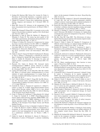
![compared with 39.4% in the control group (OR: 2.9; 95% CI:
1.6; 5.3; p = 0.0003; Table 3) and the median time to reach
WAR >40% was 43 days (95% CI: 37.2–48.8 days) in
the test group and 63 days (95% CI: 57.8–68.1 days) in the
control group, showing a statistically significant difference
(p = 0.002; log-rank test).
When a more stringent criterion is used (relative WAR
Ն60%), the OR is 2.2 (95% CI: 1.2; 4.0; p = 0.013). Figure 5
shows the percentages of patients presenting WAR Ն40% and
WAR Ն60% in the TLC-NOSF group (A) and TLC group
(B) at each investigator’s evaluation (weeks 2, 4, 6, and 8;
Table 4).
By week 8, six and seven wounds had healed (with 100%
reepithelialization and no further need of a primary dressing)
in the test and control groups, respectively.
Complementary analyses were undertaken to document the
relative WAR when considering parameters of poor healing
prognosis such as the recurrence of the leg ulcer, the duration
of the ulcer of more than 1 year, or an initial surface area
greater than 10 cm2
(Table 5). Whichever subgroup is consid-
ered, the superiority of the TLC-NOSF dressing is docu-
mented as having a very homogeneous effect, more marked
when ulcers are of poor prognosis.
Finally, by using a binary logistic regression method that
includes basal wound area (10 cm2
cutoff), ulcer recurrence
(yes/no), and duration (<1, 1–2, and >2 years) in the model,
OR of WAR >40% is 3.3 (95% CI: 1.8–6.1; p < 0.001) in
favor of the test dressing. Only the basal area was significant
in this model (OR area <10 cm2
vs. Ն10 cm2
: 2.1; 95% CI:
1.1–4.0; p = 0.020).
At the end of the trial, the blind review, based on the
photographs considered valuable by the two independent cli-
nicians, was undertaken on 86 patients in the test group and 82
patients in the control group; 81.4% (70/86) ulcers were con-
sidered improved in the test group compared with 65.9%
(54/82) in the control group, which is significantly different
(p = 0.022). Thus, the blind review fully corroborates the
evaluations made by the investigators during the course of
the trial.
Dressing acceptability, adverse events,
and local tolerance
Over the study period, a total of 3,547 local procedures were
documented by the participating nurses (1,804 in the test group
Table 3. Efficacy outcomes in patients randomized to TLC-NOSF (n = 93) and TLC dressing (n = 94) on an ITT basis
TLC-NOSF TLC
p*n = 93 n = 94
Last area (cm2
) Mean Ϯ SD 10.1 Ϯ 15.4 14.0 Ϯ 17.6
Median (range) 5.0 [0.0; 101.1] 6.1 [0.0; 87.4]
Last perimeter (cm) Mean Ϯ SD 14.4 Ϯ 10.3 17.9 Ϯ 13.8
Median (range) 11.8 [0.0; 53.1] 13.2 [0.0; 75.8]
Relative WAR (%) Mean Ϯ SD –45.2 Ϯ 47.9 –21.4 Ϯ 81.0 0.002
Median (range) –58.3 [–100.0; 173.0] –31.6 [–100.0; 571.0]
Absolute WAR (cm2
) Mean Ϯ SD –6.9 Ϯ 11.4 –2.5 Ϯ 11.9 0.003
Median (range) –6.1 [–55.5; 31.7] –3.2 [–33.1; 74.4]
Wound edge progression (mm)
(Gilman)
Mean Ϯ SD –1.15 Ϯ 1.20 –0.56 Ϯ 1.19 0.001
Median (range) –1.15 [–3.96; 2.20] –0.56 [–3.43; 6.97]
Healing rate (mm2
/day) Mean Ϯ SD –13.32 Ϯ 24.56 –4.54 Ϯ 23.20 0.005
Median (range) –10.83 [–158.32; 57.59] –5.15 [–60.19; 132.87]
*Mann–Whitney test.
ITT, intent to treat; SD, standard deviation; TLC-NOSF, Lipido-Colloid Technology-Nano-OligoSaccharide Factor; WAR, Wound Area
Reduction.
Figure 2. Relative Wound Area Reduction (WAR) calculated
as [Arealast - Areat0)/Areat0] ¥ 100 (Areat0 is the baseline wound
tracing value and Arealast is the last available wound tracing
value) and expressed as a percentage over the 8-week
follow-up in patients randomized to TLC-NOSF (test) (n = 93)
and TLC dressing (control) (n = 94) on an ITT basis. Values
indicate median. ITT, intent to treat; TLC-NOSF, Lipido-Colloid
Technology-Nano-OligoSaccharide Factor.
Meaume et al. Randomized, double-blinded trial with dressing in VLU
Wound Rep Reg (2012) 20 500–511 © 2012 by the Wound Healing Society 505](https://image.slidesharecdn.com/contentserver10-150114215057-conversion-gate02/85/Content-server-10-A-randomized-controlled-double-blind-prospective-trial-with-a-Lipido-Colloid-Technology-Nano-OligoSaccharide-Factor-wound-dressing-in-the-local-management-of-venous-leg-ulcers-6-320.jpg)
![NOSF and TLC groups. So, no difference between the two
groups has been noted whatever acceptability parameter that
was considered and documented by the nursing staff during
the trial.
At least one local adverse event (emergent or already
known at baseline) was reported in 29 patients allocated to the
test dressing (31.2%; 95% CI: 22.0–41.6%) and in 27 allo-
cated to the control dressing (28.7%; 95% CI: 19.9–38.0%).
A total of 66 local adverse events were noted (Table 6),
with no obvious differences in event prevalence between the
two groups. Periwound eczema was the most frequently
reported problem (23 of the 187 patients, 12.3%), but this was
already present in these patients at the time of randomization.
Of the 66 local adverse events documented, 23 (10 in the test
group and 13 in the control group) were considered by the
investigators as most probably dressing related.
For 11 patients in the test group and 12 patients in the
control group, the local adverse event was the reason justify-
ing the discontinuation of the dressing before week 8.
Figure 5. Percentages of patients presenting Wound Area
Reduction (WAR) Ն40% and WAR Ն60% in the TLC-NOSF
group (A) and TLC group (B) at each evaluation (weeks
2, 4, 6, and 8). TLC-NOSF, Lipido-Colloid Technology-Nano-
OligoSaccharide Factor.
Table 4. Percentage of wounds with a relative Wound Area
Reduction Ն40% (WAR Ն40%) and Ն60% (WAR Ն60%) at
last available tracing on an ITT basis
Relative WAR
Ն40%
Relative WAR
Ն60%
TLC-NOSF (n, %) 61/93 (65.6%) 42/93 (45.2%)
TLC (n, %) 37/94 (39.4%) 26/94 (27.7%)
Odds ratio (95% CI) 2.9 [1.6; 5.3] 2.2 [1.2; 4.0]
p 0.0003 0.013
CI, confidence interval; ITT, intent to treat; TLC-NOSF,
Lipido-Colloid Technology-Nano-OligoSaccharide Factor.
Table5.RelativeWoundAreaReduction(WAR).Subgroupanalysis.Calculatedwithrespectofthecompressionbandaging(monolayerormultilayerbandages),the
ulcerduration(Յ1yearor>1year),therecurrence,andthebaselinewoundarea(Յ10or>10cm2
).Expressedasapercentageoverthe8-weekfollow-upinpatients
randomizedtoTLC-NOSF(test;n=93)andTLCdressing(control;n=94)onanITTbasis
TLC-NOSFTLC
Mediandifference95%CIofmediandiffnMeanϮSDMediannMeanϮSDMedian
Compression
bandaging*
Monolayer57–40.4Ϯ54.9–58.361–16Ϯ91.8–31.7–26.6[–42.3;-11.0]
Multilayer27–51.8Ϯ33.3–58.226–27.5Ϯ58.7–32.7–25.5[–51.9;0.9]
UlcerdurationՅ1year39–55.9Ϯ37.5–63.344–34.2Ϯ49.8–38.1–25.3[–49.5;-1.0]
>1year54–37.4Ϯ53.1–55.250–10Ϯ100.0–23.1–32.0[–48.7;-15.4]
RecurrentulcerYes51–40.8Ϯ54.6–58.249–16.9Ϯ61.2–26.1–32.1[–49.8;-14.5]
No42–50.5Ϯ38.1–61.845–26.2Ϯ98.7–39.3–22.5[–40.5;-4.6]
BaselinewoundareaՅ10cm2
39–48.7Ϯ54.0–61.746–38.1Ϯ50.7–40.6–21.1[–39.8;-2.3]
>10cm2
54–42.6Ϯ43.3–55.248–5.3Ϯ100.0–19.2–35.9[–54.5;-17.3]
*Patientswithfullconcordancetoprescribedcompressiontherapy(84intestand87incontrolgroup).
CI,confidenceinterval;ITT,intenttotreat;SD,standarddeviation;TLC-NOSF,Lipido-ColloidTechnology-Nano-OligoSaccharideFactor.
Meaume et al. Randomized, double-blinded trial with dressing in VLU
Wound Rep Reg (2012) 20 500–511 © 2012 by the Wound Healing Society 507](https://image.slidesharecdn.com/contentserver10-150114215057-conversion-gate02/85/Content-server-10-A-randomized-controlled-double-blind-prospective-trial-with-a-Lipido-Colloid-Technology-Nano-OligoSaccharide-Factor-wound-dressing-in-the-local-management-of-venous-leg-ulcers-8-320.jpg)


This study evaluates the efficacy of a new lipido-colloid technology-nano-oligosaccharide factor (TLC-NOSF) dressing for managing venous leg ulcers (VLUs) in a randomized, controlled, double-blind trial with 187 participants. Results showed a median wound area reduction of 58.3% in the TLC-NOSF group compared to 31.6% in the control group, indicating superior healing with the new dressing. The findings suggest the TLC-NOSF dressing significantly enhances the healing process of VLUs as compared to traditional dressings.


![Espondilodiscite[1]](https://cdn.slidesharecdn.com/ss_thumbnails/espondilodiscite1-190205175546-thumbnail.jpg?width=640&height=640&fit=bounds)


![PERI-PROSTHETIC FRACTURE NAIL-PLATE CONSTRUCT [NPC].pptx](https://cdn.slidesharecdn.com/ss_thumbnails/drarunkumardrmohamedashrafperiprostheticfrasturenail-plateconstructnpc-260209164459-7e9d15a1-thumbnail.jpg?width=640&height=640&fit=bounds)

