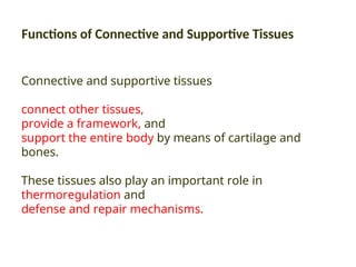
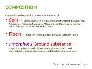
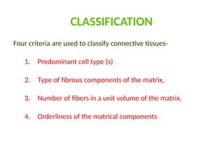
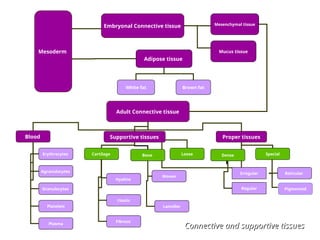
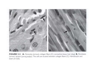
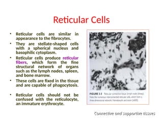
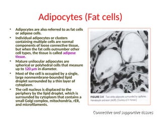
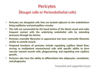
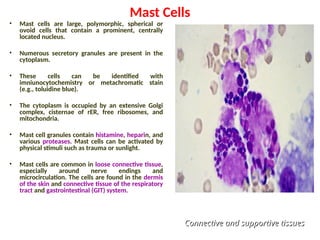
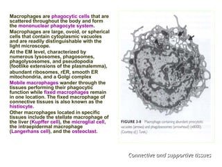
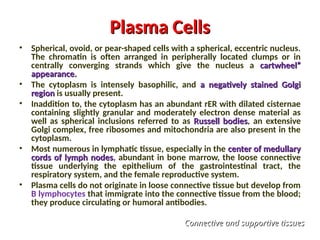
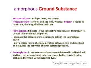
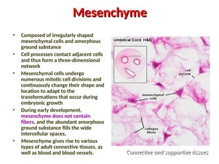
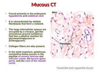
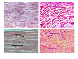
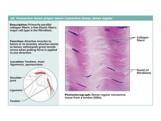
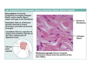
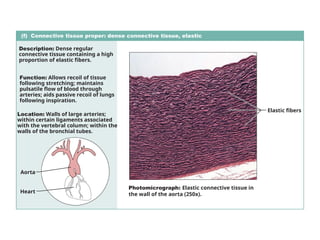
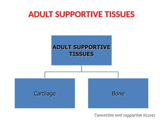
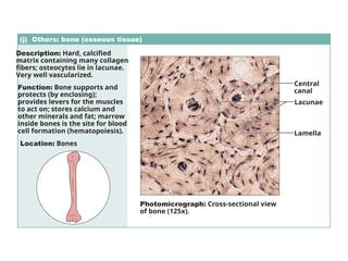
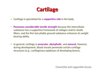
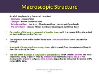
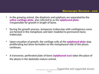
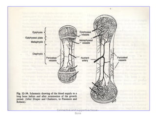
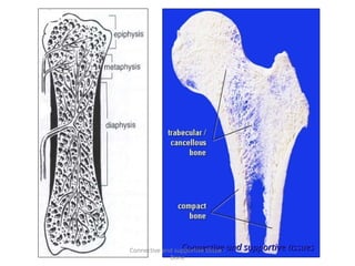
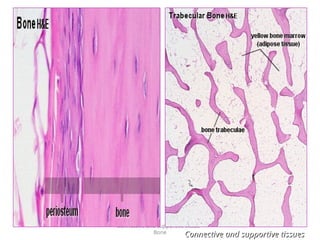
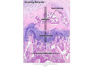
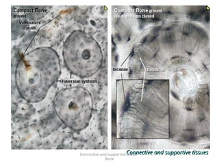
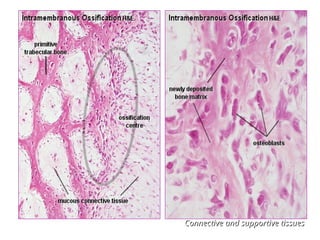
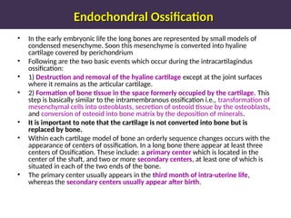
CONNECTIVE AND SUPPORTIVE TISSUES
Dr. Shah Nawaz Sial
Functions of Connective and Supportive Tissues
Connective and supportive tissues
connect other tissues,
provide a framework, and
support the entire body by means of cartilage and
bones.
These tissues also play an important role in
thermoregulation and
defense and repair mechanisms.
COMPOSITION
Connective and supportive tissues are composed of
• Cells – Mesenchymal cells, Fibrocytes and fibroblast, Reticular cells,
Adipocytes, Pericytes, Mast cells, Macrophages, Plasma cells, pigment
cells, other cells of loose connective tissue.
• Fibers – Collagen Fibers, Elastic Fibers and Reticular Fibers
• amorphous Ground substance –
a hydrated gel composed of glycosaminoglycans (GAGs), and
proteoglycans, plasma constituents, metabolites, water, and ions.
Connective and supportive tissues
CLASSIFICATION
Four criteria are used to classify connective tissues-
1. Predominant cell type (s)
2. Type of fibrous components of the matrix,
3. Number of fibers in a unit volume of the matrix,
4. Orderliness of the matrical components
Adult Connective tissue
Blood Supportive tissues Proper tissues
Erythrocytes
Agranulocytes
Granulocytes
Platelets
Plasma
Cartilage Bone
Hyaline
Elastic
Fibrous
Woven
Lameller
Loose Dense Special
Irregular
Regular
Reticular
Pigmented
Mesenchymal tissue
Embryonal Connective tissue
Mucus tissue
Mesoderm
Adipose tissue
Brown fat
White fat
Connective and supportive tissues
CONNECTIVE TISSUE CELLS
- Fixed Cells –
(NATIVE TO THE TISSUE IN WHICH THEY ARE FOUND)
Mesenchymal cell
Fibroblsts /Fibrocytes
Reticular cells
Adipocytes
Pericytes
Mesenchymal Cells
• Mesenchymal cells are irregularly
shaped with multiple processes.
• They are smaller than fibroblasts
and have fewer cytoplasmic
organelles.
• The large, oval nucleus has a
prominent nucleolus and fine
chromatin.
• The mesenchymal cell population
serves as a reservoir of
pluripotent cells that can
differentiate into other types of
connective tissue cells as needed.
Connective and supportive tissues
Fibrocytes and Fibroblasts
Fibrocytes
• The most common cell of connective tissue is the fibrocyte.
• Fibrocytes are generally elongated and spindle-shaped, with processes that connect
adjacent cells and fibers.
• Their heterochromacic nucleus is surrounded by a scant amount of pale cytoplasm.
• Secretory vesicles in the cytoplasm discharge their contents (e.g., procollagen,
proteoglycans, proelastin) into the surrounding microenvironment.
Fibroblasts
• The fibroblast has a larger, more euchromatic nucleus and more abundant,
basophilic cytoplasm than the fibrocyte.
• At the EM level, abundant rER and a prominent Golgi complex are present in the
cytoplasm.
• In certain situations, fibroblasts may differentiate into adipose cells, chondroblasts,
or osteoblasts.
Myofibroblasts
• are fibroblasts that contain actin filaments associated with dense bodies; hence,
they resemble smooth muscle cells.
• It is believed that myofibroblasts play a role in contraction of the wound during
healing.
Reticular Cells
• Reticular cells are similar in
appearance to the fibrocytes.
• They are stellate-shaped cells
with a spherical nucleus and
basophilic cytoplasm.
• Reticular cells produce reticular
fibers, which form the fine
structural network of organs
such as the lymph nodes, spleen,
and bone marrow.
• These cells are fixed in the tissue
and are capable of phagocytosis.
• Reticular cells should not be
confused with the reticulocyte,
an immature erythrocyte.
Connective and supportive tissues
Adipocytes (Fat cells)
• Adipocytes are also referred to as fat cells
or adipose cells.
• Individual adipocytes or clusters
containing multiple cells are normal
components of loose connective tissue,
but when the fat cells outnumber other
cell types, the tissue is called adipose
tissue.
• Mature unilocular adipocytes are
spherical or polyhedral cells that measure
up to 120 µm in diameter.
• Most of the cell is occupied by a single,
large nonmembrane-bounded lipid
droplet surrounded by a thin layer of
cytoplasm.
• The cell nucleus is displaced to the
periphery by the lipid droplet, which is
surrounded by cytoplasm that contains a
small Golgi complex, mitochondria, rER,
and microfilaments.
Connective and supportive tissues
Pericytes
(Rouget cells or Periendothelial cells)
• Pericytes are elongated cells that are located adjacent to the endothelium
lining capillaries and postcapillary venules.
• The cells are surrounded by the basal lamina of the blood vessel and make
frequent contact with the underlying endothelial cells by extending
processes through the lamina.
• Pericytes resemble fibrocytes in appearance but have contractile filaments
similar to smooth muscle.
• Proposed functions of pericytes include regulating capillary blood flow;
serving as multipotent mesenchymal cells with specific ability to form
vascular smooch muscle cells; phagocytosing; and regulating new capillary
growth,
• Pericyres also have the ability to differentiate into adipocytes, osreoblasts,
and phagocytes.
Connective and supportive tissues
Connective Tissue Cells
- Wandering cells -
Mast cells
Macrophages
Plasma cells,
Pigment cells
Other Blood derived CT cells
Mast Cells
• Mast cells are large, polymorphic, spherical or
ovoid cells that contain a prominent, centrally
located nucleus.
• Numerous secretory granules are present in the
cytoplasm.
• These cells can be identified with
imniunocytochemistry or metachromatic stain
(e.g., toluidine blue).
• The cytoplasm is occupied by an extensive Golgi
complex, cisternae of rER, free ribosomes, and
mitochondria.
• Mast cell granules contain histamine, heparin, and
various proteases. Mast cells can be activated by
physical stimuli such as trauma or sunlight.
• Mast cells are common in loose connective tissue,
especially around nerve endings and
microcirculation. The cells are found in the dermis
of the skin and connective tissue of the respiratory
tract and gastrointestinal (GIT) system.
Connective and supportive tissues
Macrophages
Macrophages are phagocytic cells that are
scattered throughout the body and form
the mononuclear phagocyte system.
Macrophages are large, ovoid, or spherical
cells that contain cytoplasmic vacuoles
and are readily distinguishable with the
light microscope.
At the EM level, characterized by
numerous lysosomes, phagosomes,
phagolysosomes, and pseudopodia
(footlike extensions of the plasmalemma),
abundant ribosomes, rER, smooth ER
mitochondria, and a Golgi complex
Mobile macrophages wander through the
tissues performing their phagocytic
function while fixed macrophages remain
in one location. The fixed macrophage of
connective tissues is also known as the
histiocyte.
Other macrophages located in specific
tissues include the stellate macrophage of
the liver (Kupffer cell), the microglial cell,
the intraepidermal macrophage
(Langerhans cell), and the osteoclast.
Connective and supportive tissues
Plasma Cells
Plasma Cells
• Spherical, ovoid, or pear-shaped cells with a spherical, eccentric nucleus.
The chromatin is often arranged in peripherally located clumps or in
centrally converging strands which give the nucleus a cartwheel”
cartwheel”
appearance.
appearance.
• The cytoplasm is intensely basophilic, and a negatively stained Golgi
a negatively stained Golgi
region
region is usually present.
• Inaddition to, the cytoplasm has an abundant rER with dilated cisternae
containing slightly granular and moderately electron dense material as
well as spherical inclusions referred to as Russell bodies
Russell bodies, an extensive
Golgi complex, free ribosomes and mitochondria are also present in the
cytoplasm.
• Most numerous in lymphatic tissue, especially in the center of medullary
center of medullary
cords of lymph nodes
cords of lymph nodes, abundant in bone marrow, the loose connective
tissue underlying the epithelium of the gastrointestinal tract, the
respiratory system, and the female reproductive system.
• Plasma cells do not originate in loose connective tissue but develop from
B lymphocytes that immigrate into the connective tissue from the blood;
they produce circulating or humoral antibodies.
Connective and supportive tissues
Pigment Cells
• Cells in connective tissue may
contain pigments, including
melanin in domestic animals or
pteridines and purines in fish
and amphibians. When present
in large numbers, the cells
impart color to the connective
tissue.
• Occur in various locations such
as the dermis, uterine caruncles
of sheep, meninges, choroid, and
iris.
Connective and supportive tissues
CONNECTIVE TISSUE FIBERS
Connective and supportive tissues
Collagen fibers
Collagen fibers
• Collagen is the principal fiber type in mature connective tissue.
• Fresh collagen fibers are white, and in histologic preparations they stain
with acid dyes, Thus, they are red to pink in H&E stained sections, red
with van Gieson’s method, and blue in Mallory and Masson’s triple
stains (green when light green stain is used).
• The fibers are flexible and can adapt to the movements and changes in
size of the organs with which they are associated.
• Collagen fibers are characterized by a high tensile strength and a poor
shear strength, and stretch is limited to approximately 5% of their initial
length. Consequently, they are found wherever high tensile strength is
required, such as in tendons, ligaments, and organ capsules.
Connective and supportive tissues
Elastic Fibers
Elastic Fibers
• Elastic fibers and/or sheets (laminae) are present in organs in which normal
function requires elasticity in addition to tensile strength.
• Elastic fibers can be stretched as much as two and one-half times their original
length, to which they return when released.
• Found in the pinna of the ear, vocal cords epiglottis, lungs, ligamentum nuchae,
dermis, aorta, and muscular arteries, elastic fibers are one of the most resilient
connective tissue fibers, with standing chemical maceration and auroclaving.
• Elastic fibers usually occur as individual, branching, and anastomosing fibers.
Their diameters vary within a wide range, from 0.2 to 5.0 µm in loose connective
tissue to as large as 12µm in elastic ligaments, such as the ligamenturn nuchae in
the neck.
• In H&E-stained histologic sections, the larger elastic fibers in elastic ligaments are
readily distinguished as highly retractile, amorphous, light pink strands; they are
stainable by certain selective dyes, such as orcein and resorcin-fuchsin.
Connective and supportive tissues
Reticular Fibers
Reticular Fibers
• In routine histologic preparations, reticular fibers cannot be distinguished from
other small collagen fibers, These fibers can be identified only with certain silver
impregnations (thus the term argyrophilic or argenraffin fibers) or with the
periodic acid Schiff (PAS) reagent.
• Reticular fibers are actually individual collagen fibrils (type Ill collagen) coated by
proteoglycans and glycoproteins. This coating increases the affinity of the fibers
for silver salts. When individual reticular fibers are bundled to form collagen
fibers, the coating is supposedly displaced and the argyrophilia decreases.
• Reticular fibers form delicate, flexible networks around capillaries, muscle fibers,
nerves, adipose cells, and heparocytes and serve as a scaffllding to support cells
or cell groups of endocrine, lymphatic, and blood-forming organs. They are an
integral part of basement membranes.
Connective and supportive tissues
Amorphous Ground Substance
• The cells and fibers of connective tissue are embedded in an amorphous
ground substance composed of glycosaminoglycans (GAGs),
proteoglyrans, plasma constituents, metabolites, water, and ions.
• The ground substance forms a hydrated gel that, by virtue of its high
water content, has unique properties of resiliency.
• Seven major types of GAGS can be distinguished, namely,
Hyaluronan (hyaluronic arid) vitreous humor of the eye, synovial fluid;
umbilical cord, loose connective tissue, skin, and cartilage.
Chondroitin-4-sulfate and chondroitin 6 are abundant in cartilage
arteries, skin, and cornea. A smaller amount is found in bone.
Dermatan sulfate - skin, tendon, ligamentum nuchae, sclera, and lung.
Continued…
Connective and supportive tissues
amorphous Ground Substance
Keratan sulfate - cartilage, bone, and cornea.
Heparan sulfate - arteries and the lung, whereas heparin is found in
mast cells, the lung, the liver, and skin.
– Proteoglycans fill space in the connective tissue matrix and impart its
unique biomechanical properties,
- regulate the passage of molecules and cells in the intercellular
space,
- play a major role in chemical signaling between cells and may bind
and regulate the activities of other secreted proteins.
– Proteoglycans in low concentrations are not deterred in H&E-stained
sections, but when present in higher concentrations, as in hyaline
cartilage, they stain with basophilic dyes.
Connective and supportive tissues
EMBRYONIC
CONNECTIVE TISSUES
Mesenchyme
Mesenchyme
• Composed of irregularly shaped
mesenchymal cells and amorphous
ground substance
• Cell processes contact adjacent cells
and thus form a three-dimensional
network
• Mesenchymal cells undergo
numerous mitotic cell divisions and
continuously change their shape and
location to adapt to the
transformations that occur during
embryonic growth
• During early development,
mesenchyme does not contain
fibers, and the abundant amorphous
ground substance fills the wide
intercellular spaces.
• Mesenchyme gives rise to various
types of adult connective tissues, as
well as blood and blood vessels. Connective and supportive tissues
Connective and supportive tissues
Mucous CT
Mucous CT
• Found primarily in the embryonic
hypodermis and umbilical cord
• It is characterized by stellate
fibroblasts that form a network.
• The large intercellular spaces are
occupied by a viscous, gel-like
amorphous ground substance
that has a positive reaction for
glycosaminoglycans or
proteoglycans.
• Collagen fibers are also present.
• In the adult organism, gelatinous
connective tissue occurs in the
papillae of omasal laminae and
reticular crests, the bovine glans
penis, and the core of the rooster
comb.
Connective and supportive tissues
ADULT
CONNECTIVE TISSUES
Connective and supportive tissues
Loose (Fibrous) Connective Tissue
• Most widely distributed type of connective tissue in the adult animal.
• The cells and fibers are widely separated by ground substance.
• Compared to other types of connective tissue, the cells are more abundant and
include both fixed and mobile populations
• Contain all three fiber types (reticular, collagen, and elastic).
• The relative abundance and orientation of fibers vary widely, depend primarily on
the location and specific function of the tissue.
• Early connective tissue is highly cellular with fine reticular fibers; later
connective tissue has predominantly thick collagen fibers
Occurrence:
• Beneath many epithelia, forms the interstitial tissue in most organs, around nerve
and skeletal muscle bundles.
• The pia mater and arachnoid of the brain and spinal cord are also composed of
loose connective tissue.
• Functions range from the mechanical support to repair and defense activities
(inflammation).
Connective and supportive tissues
(a) Connective tissue proper: loose connective tissue, areolar
Description: Gel-like matrix with all
three fiber types; cells: fibroblasts,
macrophages, mast cells, and some
white blood cells.
Function: Wraps and cushions
organs; its macrophages phagocytize
bacteria; plays important role in
inflammation; holds and conveys
tissue fluid.
Location: Widely distributed under
epithelia of body, e.g., forms lamina
propria of mucous membranes;
packages organs; surrounds
capillaries.
Photomicrograph: Areolar connective tissue, a
soft packaging tissue of the body (300x).
Epithelium
Lamina
propria
Fibroblast
nuclei
Elastic
fibers
Collagen
fibers
Dense Fibrous Connective Tissue
Dense regular Connective Tissue + Dense Irregular Connective Tissue
• Fibrocytes are the predominant cell population
• Collagen fibers are generally arranged in bundles that cross each other at varying
angles.
• In thin aponeuroses or muscle fasciae, fiber bundles are located in a single layer. In
heavier aponeuroses, organ capsules, or dermis, the bundles are superimposed in
several layers and interlace with one another in multiple planes.
• Irregular configuration allows adaptation to changes in the size of an organ or the
diameter of a muscle, and stretching forces can be withstood in any direction.
• Continuation of surface connective tissue into the organ or muscle enhances
strength.
• Elastic networks facilitate a rapid return to resting conditions.
• Special functional and morphologic features are described with the various organ
systems.
• Found in a variety of locations, such as the lamina propria of the initial portions of
the digestive system, the capsule of the lung (visceral pleura) and other organs
(spleen, liver, kidney, testis), fasciae, aponeuroses, joint capsules, pericardium, and
dermis.
(d) Connective tissue proper: dense connective tissue, dense regular
Description: Primarily parallel
collagen fibers; a few elastic fibers;
major cell type is the fibroblast.
Function: Attaches muscles to
bones or to muscles; attaches bones
to bones; withstands great tensile
stress when pulling force is applied
in one direction.
Location: Tendons, most
ligaments, aponeuroses.
Photomicrograph: Dense regular connective
tissue from a tendon (500x).
Shoulder
joint
Ligament
Tendon
Collagen
fibers
Nuclei of
fibroblasts
(e) Connective tissue proper: dense connective tissue, dense irregular
Description: Primarily
irregularly arranged collagen
fibers; some elastic fibers;
major cell type is the fibroblast.
Function: Able to withstand
tension exerted in many
directions; provides structural
strength.
Location: Fibrous capsules of
organs and of joints; dermis of
the skin; submucosa of
digestive tract.
Photomicrograph: Dense irregular
connective tissue from the dermis of the
skin (400x).
Collagen
fibers
Nuclei of
fibroblasts
Fibrous
joint
capsule
(f) Connective tissue proper: dense connective tissue, elastic
Description: Dense regular
connective tissue containing a high
proportion of elastic fibers.
Function: Allows recoil of tissue
following stretching; maintains
pulsatile flow of blood through
arteries; aids passive recoil of lungs
following inspiration.
Location: Walls of large arteries;
within certain ligaments associated
with the vertebral column; within the
walls of the bronchial tubes.
Elastic fibers
Aorta
Heart
Photomicrograph: Elastic connective tissue in
the wall of the aorta (250x).
ADULT SUPPORTIVE TISSUES
Connective and supportive tissues
(j) Others: bone (osseous tissue)
Description: Hard, calcified
matrix containing many collagen
fibers; osteocytes lie in lacunae.
Very well vascularized.
Function: Bone supports and
protects (by enclosing);
provides levers for the muscles
to act on; stores calcium and
other minerals and fat; marrow
inside bones is the site for blood
cell formation (hematopoiesis).
Location: Bones
Photomicrograph: Cross-sectional view
of bone (125x).
Lacunae
Lamella
Central
canal
Cartilage
Cartilage
– Cartilage is specialized for a supportive role in the body.
– Possesses considerable tensile strength because the intercellular
substance has a supportive framework of collagen and/or elastic
fibers, and the firm but pliable ground substance enhances its weight
bearing ability.
– In general, cartilage is avascular, alymphatic, and aneural, however,
during development, blood vessels penetrate certain cartilage
structures (e.g., cartilaginous epiphyses of developing bones).
Connective and supportive tissues
Chondroblast
• Found in growing cartilage.
• Oval-shaped with a spherical nucleus and
a prominent Golgi apparatus.
• The cytoplasm is basophilic as a result of
large quantities of rER.
• The chondroblast actively forms the
matrix of cartilage that surrounds the
perimeter of the cell.
Cartilage Cells: Chondroblast, Chondrocyte
Cartilage Cells: Chondroblast, Chondrocyte.
.
Connective and supportive tissues
Chondrocyte
• Elongate to spherical in shape, depending
on the location within the cartilage.
• Each chondrocyte is located within a
lacuna, a cavity within the semirigid
cartilage matrix. In living cartilage or at
the fine-structural level, the cell fills the
lacuna.
• Short cytoplasmic processes extend into
the intercellular substance, in most light
microscopic preparations, the cell surface
appears separated from the lacunar walls
because of shrinkage. The chondrocyte
has a spherical nucleus with one or more
nucleoli and abundant rER and prominent
Golgi complex.
• Glycogen and lipid accumulate in the
cytoplasm of old chondrocytes.
• Chondrocyte is responsible for continual
ongoing maintenance of the surrounding
matrix.
Connective and supportive tissues
Cartilage Matrix
Cartilage Matrix
• Composed of fibers and ground substance
• Collagen forms the framework of the matrix.
• The ground substance contains the GAGs chondroitin sulfate, keratan
sulfate, and hyaluronic acid, all of which play an important role in
transporting water and electrolytes as well as in binding water to give
hyaline cartilage its resiliency.
• Overall, the matrix is slightly basophilic when stained with H&E, reacts
positively with PAS.
Connective and supportive tissues
Hyaline cartilage
Hyaline cartilage
• Chondrocytes in mature hyaline cartilage vary in size - small with elliptic
lacunae near the surface, larger and more polyhedral within deep cartilage
• Some lacunae contain only one cell; others contain two, four, or sometimes
six cells. Multicellular lacunae are called cell nests - isogenous cell groups.
• The amorphous ground substance of hyaline cartilage is a firm gel
containing a network of collagen fibrils. Fibrils have the same refractive
index as the amorphous ground substance.
• Surrounding each chondrocyte is a thin layer of pericellular matrix that has
proteoglycans but lacks collagen.
• The territorial matrix surrounds pericellular matrix - a network of collagen
fibrils and ground substance.
Connective and supportive tissues
Hyaline cartilage
Hyaline cartilage
• The interterritorial matrix lies outside the territorial matrix arid fills the
remaining matrix space. This matrix region contains large collagen fibrils
and abun dant proteoglycans. Differences in collagen and proteoglycan
content account for the staining differences between regions as observed
with the light microscope.
• Except on articular surfaces, hyaline cartilage is surrounded by vascular
connective tissue called the perichondrium, composed of two distinct
layers: cellular or chondrogenic layer and the outer fibrous layer.
• Found on the articulating surfaces of bones in synovial joints, nose, larynx.
trachea, and bronchi. It forms most of the entire appendicular and axial
skeleton in the embryo
Connective and supportive tissues
(g) Cartilage: hyaline
Description: Amorphous but firm
matrix; collagen fibers form an
imperceptible network; chondroblasts
produce the matrix and when mature
(chondrocytes) lie in lacunae.
Function: Supports and reinforces;
has resilient cushioning properties;
resists compressive stress.
Location: Forms most of the
embryonic skeleton; covers the ends
of long bones in joint cavities; forms
costal cartilages of the ribs; cartilages
of the nose, trachea, and larynx.
Photomicrograph: Hyaline cartilage from the
trachea (750x).
Costal
cartilages
Chondrocyte
in lacuna
Matrix
Connective and supportive tissues
Elastic cartilage
Elastic cartilage
• Elastic cartilage occurs where elasticity, as well as some rigidity, is needed,
such as in the epiglottis and external auditory canal. It is also part of the
corniculate and cuneiform processes of the larynx.
• In addition to all of the structural components of hyaline cartilage, elastic
cartilage possesses a dense network of elastic fibers that are visible in
ordinary H&E preparations
• The elastic fibers are few in number near the perichondrium but form a
dense network within the cartilaginous mass.
• Chondrocytes located away from the surface of elastic cartilage contain
many fat vacuoles. Later in adults, these fat-containing cells often become
adipose tissue.
Connective and supportive tissues
(h) Cartilage: elastic
Description: Similar to hyaline
cartilage, but more elastic fibers
in matrix.
Function: Maintains the shape
of a structure while allowing
great flexibility.
Location: Supports the external
ear (pinna); epiglottis.
Photomicrograph: Elastic cartilage from
the human ear pinna; forms the flexible
skeleton of the ear (800x).
Chondrocyte
in lacuna
Matrix
Fibrocartilage
Fibrocartilage
• Of the three cartilage types, fibrocartilage occur least frequently. It often
interposed between other tissues and hyaline cartilage, tendons, or ligaments.
• Found in the intervertebral disks and forms the menisci of the stifle joint. In dogs,
fibrocartilage is found in the cardiac skeleton, which joins the atrial and ventricular
heart muscles.
• The most striking characteristic of fibrocartilage is the presence of prominent type
I collagen fibers in the matrix.
• The microscopic appearance of fibrocartilage may vary with location
• The amorphous ground substance is most abundant in the vicinity of the cells,
whereas the remainder of the matrix contains primarily collagen fiber bundles.
• Fibrocartitage lacks a distinct perichondrium; the cartilage is surrounded by
collagen fibers in some locations, but a cellular layer is absent.
Connective and supportive tissues
(i) Cartilage: fibrocartilage
Description: Matrix similar to
but less firm than that in hyaline
cartilage; thick collagen fibers
predominate.
Function: Tensile strength
with the ability to absorb
compressive shock.
Location: Intervertebral discs;
pubic symphysis; discs of knee
joint.
Photomicrograph: Fibrocartilage of an
intervertebral disc (125x). Special staining
produced the blue color seen.
Intervertebral
discs
Chondrocytes
in lacunae
Collagen
fiber
Connective and supportive tissues
Nutrition of Cartilage
• Unlike other connective tissues, most cartilage is avascular.
Therefore, the chondrocytes must depend on diffusion of
nutrients through the gelled matrix. These nutrients diffuse
from nearby capillaries within the perichondrium or from
synovial fluid bathing the cartilage surface.
• When the intercellular matrix becomes calcified, diffusion is
no longer possible, and the chondrocytes die. This
phenomenon occurs in aging and is natural in endochondral
bone development.
Connective and supportive tissues
BONE
BONE
Connective tissue with cells and fibers embedded
in a hard, mineralized substance that is well suited
for supportive and protective functions.
Connective and supportive tissue -
Bone
Connective and Supportive Tissues
Functions
Functions
– Provides internal support for the entire body as well as
attachment sites for the muscles and tendons necessary for
movement.
– Protects the brain and organs in the thoracic cavity and
contains the bone marrow within its medullary space.
– Functions metabolically by providing a source of calcium to
maintain proper blood calcium levels and various growth
factors (e.g. transforming growth factor- beta) that play a role
in remodeling.
– Bone is a dynamic tissue that is renewed and remodeled
throughout the life of all mammal.
– Its construction is unique because it provides the greatest
tensile strength with the least amount of weight of any tissue.
Connective and supportive tissue -
Bone
1.
1. Osteoblast –bone forming cells
Osteoblast –bone forming cells
• Responsible for active formation and mineralization of bone matrix.
• Columnar to squamous in shape, located on surfaces of bone where new
bone deposits.
• The nucleus is located in the basal region of the intensely basophilic
cytoplasm. The Golgi apparatus and rER are prominent between the
nucleus and the secretory surface of the osteoblast.
• The cell deposits osteoid (type-1collagen (90%) and proteoglycans), the
unmineralized matrix of bone.
• Ostecblasts originate from pluripotent mesenchymal stem cells
(osteoproginator cells) that also give rise to chondroblasts, fibroblasts, and
other cell types.
Connective and supportive tissue -
Bone
Connective and supportive tissues
Bone cells
1.
1. Osteoblast –bone forming cells
Osteoblast –bone forming cells
• Flattened, resting osteoblasts are known as bone lining cells - found on the
surfaces of adult trabeculae and compact bone, capable of becoming
active osteoblasts when appropriately stimulated.
• Osteoblasts have receptors for parathyroid hormone on their surface.
When PTH binds to the osteoblast, the cell releases factors that stimulate
osteoclastic activity.
Connective and supportive tissue -
Bone
Connective and supportive tissue -
Bone
2.
2. Osteocytes – true bone cells
Osteocytes – true bone cells
• The osteocyte is the principal cell in mature bone and resides in a lacuna
surrounded by calcified interstitial matrix.
• Numerous long, slender processes extend from the cell body into canaliculi within
the matrix to contact adjacent osteocytes. Gap junctions are present at the contact
points and provide communication between osteocytes.
• The long cellular processes of the osteocyte are able to shorten and lengthen. This
activity may serve as a ‘pump” to move fluid through lacunae and canaliculi to
transfer metabolites from the surface of the bone.
• The organelles of young osteocytes resemble osteoblasts, but as they mature, the
Golgi complex and rER are less prominent and lysosomes increase in number.
• Osteocytes are essential in presenting bone structure because, upon their death,
osteoclasts immediately move to the area and resorb the bone. Therefore, signals
from apoptotic osteocytes may be part of a signaling pathway to initiate bone
remodeling.
Connective and supportive tissue -
Bone
Bone cells
Connective and supportive tissues
Osteocytes
Osteocytes
Connective and supportive tissue -
Bone
3.
3. Osteoclast –bone resorbing cells
Osteoclast –bone resorbing cells
• The osteoclast is a large, multinucleared cell located on the surface of bone (15 to
30 nuclei per cell, 40 to 100 µm in diameter). Occasional mono-nuclear
osteoclasts are not easily recognized.
• The cytoplasm is acidophilic and contains a small amount of rER, ribosomes,
numerous smooth vesicles, and mitochondria.
• The activated osteoclast has a ruffled border created by extensive infoldings of the
cell membrane that sweep across the bony surface. The cell secretes acid and
lysosomal enzymes into this region. The cell membrane immediately adjacent to
the ruffled border adheres tightly to the bony surface, thereby scaling the area of
active bone resorption.
• Osteoclasts are derived from pluripotent stem cells of the bone marrow that also
give rise to monocytes and macrophages. Final differentiation to an osteoclast
from a circulating monocyte occurs after the cells are recruited to bone resorption
sites. At the end of their cell life span, osteoclasts undergo apoptosis.
Connective and supportive tissue -
Bone
Connective and supportive tissues
Bone cells
Osteoclasts
Connective and supportive tissue -
Bone
Connective and supportive tissue -
Bone
Osteoclast
Osteoblast
Osteocyte in
Lacuna
Bone Matrix
Connective and supportive tissue -
Bone
Bone Matrix
Bone Matrix
• The matrix of bone is composed of osteoid produced by the osteoblasts.
• Mineralization of the osteoid occurs as hydrocrystals are deposited in the osteoid
framework.
• The organic intercellular substance of bone contains sulfated glycosaminoglycans,
glycoproteins, and collagen- Glycoproteins of bone include alkaline phosphatase,
osteonectin, osteopontin, and sialoprotein. These glycoproteins are thought to
play various roles in bone mineralization.
• The inorganic component of bone consists of submicroscopic hydroxyapatite
crystals deposited as slender needles within the collagen fibril network. Such an
efficient arrangement enhances the tensile strength.
• The principal ions in bone salt are Ca, CO3, P04 and OH, and the amounts of Na,
Mg, and Fe are substantial.
• Bone, thus, is a major storehouse for calcium and phosphorus, which are
mobilized whenever they are needed.
Connective and supportive tissue -
Bone
Connective and supportive tissues
Structural and Functional Characteristics
• Adult bone is distinguished from cartilage by the presence of both a
canalicular system and a direct vascular supply.
• The growth process of bone also differs from that of cartilage, bone has a
unique lacunar-canalicular system for supplying the bone cells with
metabolites in a mineralized matrix in which diffusion is not an option.
• The canalicular system provides a conduit system for nourishment of the
mature osteocytes located deep in the bone, and the extensive capillary
supply of bone further enhances the efficiency of the canalicular system.
• Unlike cartilage, bone grows by apposition only, because the intercellular
substance mineralizes so rapidly. Interstitial growth of bone is not
possible.
Connective and supportive tissue -
Bone
Connective and supportive tissues
Macroscopic Structure
• An adult long bone (e.g., humerus) consists of
- Epiphyses - enlarged ends
- Diaphysis - hollow cylindrical shaft
- Articular cartilage - thin layer of hyaline cartilage covering epiphyseal ends
- The periosteum - vascular fibrous membrane covering ext. surface of bone
• Each region of the bone is composed of lamellar bone
Each region of the bone is composed of lamellar bone, but it is arranged differently to best
perform its biomechanical function.
• The epiphyses have a thin shell of dense bone (subchondral bone) under the articular
cartilage.
• A network of trabeculae forms spongy bone
A network of trabeculae forms spongy bone, which extends from the subchondral bone to
form the center of the bone.
• The wall of the diaphysis is composed of compact bone
The wall of the diaphysis is composed of compact bone,
, which contains osteons. The inner
(medullary) cavity of bone is lined by endosteum and contains adipose tissue or red
(hemopoietic) or yellow (adipose) bone marrow, depending on the age of the animal or the
region of the bone.
Connective and supportive tissue -
Bone
• In the growing animal, the diaphysis and epiphysis are separated by the
yaline cartilage plate, also referred to as the epiphyseal plate,
hresponsible for growth in length of bone.
• During the growth process, temporary trabeculae with cartilaginous cores
are formed in the metaphysis and later modeled to permanent bony
trabeculae.
• Upon cessation of growth, the cartilage cells of the epiphyseal plate stop
proliferating but bone formation on the metaphyseal side of the physis
continues.
• A transverse, perforated plate of bone (epiphyseal scar) takes the place of
the physis in the skeletally mature animal.
Connective and supportive tissue -
Bone
Connective and supportive tissues
Macroscopic Structure – cont.
Connective and supportive tissue -
Bone
Connective and supportive tissues
Connective and supportive tissue -
Bone
Microscopic structure
• The outermost layers of the shaft of a long bone consist of compact bone
arranged as outer circumferential lamellae (2 to 8 thick).
•
• Deep to the outer circumferential lamellae are osteons (Haversian systems)
formed by concentric Iamellae surrounding longitudinally oriented vascular
channels (central canals).
• Internal surfaces of compact bone from adult animals are composed of
inner circumferential lamellae encircling the medullary cavity.
• Lacunae are located between each lamella of the compact bone. Radiating
from the lacunae are the branching canaliculi that penetrate and join
canaliculi of adjacent lamelIae.
• The lacunae and canaliculi form an extensive system of interconnecting
passageways for the transport of nutrients.
Connective and supportive tissue -
Bone
• The central canal (Haversian canal) of each osteon contains capillaries,
lymphatic vessels, and nonmyelinated nerve fibers, all supported by reticular
connective tissue.
• Central canals are connected with each other and with the free surfrce by
transverse or horizontal channels called perforating canal (Volkmann’s
canals).
• Most bones are invested with a tough connective tissue layer, the
periosteum having two layers:
an inner osteogenic layer that provides cells necessary to form bone, an
outer fibrous layer - irregularly arranged collagen fibers and blood vessels.
• The vessels branch and enter the perforating canals and ultimately reach the
central canal of the osteons. The cellular layer is more evident in young
animals than in adults. The periosteum is attached firmly to the bone by
bundles of coarse collagen fibers that have been incorporated into the outer
circumferential lamellae of the bone. These fibers are called perforating
(Sharpey’s) fibers.
• A periosteum is absent over surface of hyaline articular cartilage and at sites
where tendons and ligaments insert on bones.
Connective and supportive tissue -
Bone
Microscopic structure - conti
Microscopic structure - conti.
Connective and supportive tissue -
Bone
Connective and supportive tissue -
Bone
Connective and supportive tissues
Connective and supportive tissue -
Bone
Connective and supportive tissues
Connective and supportive tissue -
Bone
Connective and supportive tissue -
Bone
Connective and supportive tissues
OSTEOGENESIS
OSTEOGENESIS
• The process of bone formation is called as Ossification.
• Ossification starts in the early embryonic life and continues in the
adulthood
• The bony tissue like all other types of CT, derived from the
mesenchyme
Connective and supportive tissue - Bone
Bones develop from mesenchyme by two methods:
1) Intramembranous Ossification
Mesenchyme is directly converted into bone without any intervening
stage of cartilage formation – may be called as direct method of bone
formation
Flat bones of skull develop by intramembranous method of bone
formation. In addition, maxilla, mandible and clavical bones
develop by this method.
2) Endochondral Ossification
Mesenchyme is first converted into cartilage which serves as a
temporary framework – Indirect method of bone formation
Replacement of the cartilage is a slow process which is not achieved
until the bone has reached its full size and the growth has ceased.
Most of the bones of the body develop by this method of ossification
Connective and supportive tissue - Bone
Intramembranous Ossification
• The process begins in the 2nd
month of intrauterine life
• In the areas where bone is to be formed, the mesechyme becomes condensed in the form of
sheet or membrane
• The mesenchymal membrane becomes highly vascularized at several regions by the ingrowth
of capillaries which give rise to profuse networks.
• The mesenchymal cells in the center of the vascularized region gradually differentiate into
osteoblasts, such regions are known as centers of ossification
• The osteeoblasts secrete in each center of ossification a special type of intercellular
substance called as OSTEOID
• OSTEOID IS A SOFT AND PLIABLE HYALINE MATERIAL WHICH CONSISTS OF COLLAGEN FIBERS
EMBEDDED IN AMORPHOUS GROUND SUBSTANCE
• The osteoid represents the organic component of bone matrix
• The osteoid is converted into mature bone matrix ny the deposition of minerals in close
association with collagen fibers
• These minerals mainly Calcium and Phosphorus, are deposited as crystals of calcium
phosphorus which occurs in the form of hydroxyapatite.
• This process which converts osteoid into bone matrix is known as Calcification
Connective and supportive tissue - Bone
• It occurs under the influence of the enzyme alkaline phosphatas
• As the bone formation progresses at several foci, a number of needle
like bone spicules are formed which progressively radiate from each of
such foci
• Initially, bone mass is spongy in nature consisting of spicules and
trabeculae of bone tissue
• Later, spongy bone is replaced by the compact bone as the spaces
between the trabeculae become filled with lamellar bone. Thus, inner
and outer tables of the flat bones are formed.
• Between the tables, the spongy bone remains as diploe, and the spaces
within it form primary marrow cavities which become occupied by bone
marrow
• The entire primordium of the developing bone becomes surrounded by
dense mesenchyme which gives rise to a layer of fibrous CT – THE
PERICHONDRIUM.
Conti….
Intramembranous Ossification
Connective and supportive tissues
Endochondral Ossification
• In the early embryonic life the long bones are represented by small models of
condensed mesenchyme. Soon this mesenchyme is converted into hyaline
cartilage covered by perichondrium
• Following are the two basic events which occur during the intracartilagindus
ossification:
• 1) Destruction and removal of the hyaline cartilage except at the joint surfaces
where it remains as the articular cartilage.
• 2) Formation of bone tissue in the space formerly occupied by the cartilage. This
step is basically similar to the intramembranous ossification i.e., transformation of
mesenchymal cells into osteoblasts, secretion of osteoid tissue by the osteoblasts,
and conversion of osteoid into bone matrix by the deposition of minerals.
• It is important to note that the cartilage is not converted into bone but is
replaced by bone.
• Within each cartilage model of bone an orderly sequence changes occurs with the
appearance of centers of ossification. In a long bone there appear at least three
centers of Ossification. These include: a primary center which is located in the
center of the shaft, and two or more secondary centers, at least one of which is
situated in each of the two ends of the bone.
• The primary center usually appears in the third month of intra-uterine life,
whereas the secondary centers usually appear after birth.
Connective and supportive tissue - Bone
Course of Events in the intracartilaginous ossification
Primary Centers of Ossification
• A vascular bud invades the central region of the shaft of the
cartilaginous model of bone. In this region the chondrocytes, along with
their lacunae, undergo an enlargement in size.
• As the process of lacunar enlargement continues, the enlarging
chondrocytes undergo progressive degeneration and they ultimately die,
leaving their enlarged lacunae vacant.
• The cartilage matrix forming the intervening walls of these lacunae
becomes calcified. Just at the time when these changes are occurring in
the cartilage, the perichondrium around the primary center of
ossification changes its character and becomes periosteum, i.e., the cells
in its inner (cellular) layer transform into osteoblasts.
• These periosteal osteoblasts lay down a peripheral collar of bone in the
same way as in intramembranous ossification (i.e., secretion of osteoid
and its conversion into bone matrix by deposition of minerals).
• This bony collar surrounds the primary center of ossification.
Connective and supportive tissue - Bone
• They bony collar surrounding the walls of empty and
enlarged lacunae of the calcified hyaline cartilage is invaded
by vascular sprouts from the periosteum.
• These sprouts consist of blood capillaries which are
accompanied by mesenchymal cells. These cells continually
divide and transform into osteoclasts.
• The osteoclasts exacavate passages through the bony collar
to pass into the underlying calcified cartilage in the primary
center of ossification.
• These osteoclasts also erode the calcified cartilage matrix
and produce an irregular system of intercommunicating
spaces, known as primary marrow spaces.
• These spaces become filled with embryonic bone marrow.
The delicate walls of the primary marrow spaces (which are
formed of calcified cartilage) become covered with a layer of
osteoblasts.
• These cells lay down osteoid which is soon converted into
bone matrix by the deposition of mineral salts.
Connective and supportive tissue - Bone
• Formation of bone by osteoblasts in the center of ossification is
known as interstitial growth, whereas the deposition of periosteal
layers of the bone is called appositional growth of the bone.
• With cotinuous deposition of subperiosteal bone, formation of
bone on the walls of the centrally placed lacunae stops and a
process of erosion begins.
• This results in the removal of early spicules of bone and
enlargement and confluence of the primary marrow spaces until a
primitive medullary cavity is produced in the center of the
growing bone.
• The process of ossification gradually spreads from the primary
center of ossification toward the ends of the bone.
• Progressively, the regions adjoining the primary center of
ossification also undergo the above described sequence of
changes and the none formation extends above and below the
primary center.
• That part of long bone which develops from the primary center of
ossification (i.e., the shaft) is known as diaphysis.
Connective and supportive tissue - Bone
Connective and supportive tissues
cartilaginous growth
plate
Secondary Centers of Ossification
• These appear in the cartilaginous ends of the developing bone
and, here also, the cartilage is replaced by bone through the
same sequence of events as described for th primary center
of ossificataion. Those parts of long bone which develop from
secondary centers of ossification (i.e., the ends) are known as
epiphyses.
• The plate of cartilage intervening between the diaphysis and
epiphysis is known as epiphyseal cartilage or growth plate.
This plate. This plate is responsible for he longitudinal growth
of the bone during childhood and early adult age. Sg from
epiphyseal side, following five successive zones can be
recognized in the epiphyseal cartilage:
• Zone of Reserve Cartilage. This is the zone of hyaline cartilage in which the
chondrocytes are arranged as groups.
• Zone of Cell Multiplication. In this zone the cartilage cells divide mitotically
and become aligned in longitudinal columns. It is the continuous mitotic
division of cartilage cells in this zone which is responsible for the
longitudinal growth of the bone.
• Zone of Lacunar Enlargtement and Cellular Hypertrophy. In this zone the
cartilage cells increase in size which corresponding increase in the size of
the lacunae. Later on, the chondrocytes die leaving the enlarged lacunae
vacant.
• Zone of Cartilage Calcification. In this zone the cartilage matrix, forming
the walls of enlarged lacunae, becomes calcified.
• Zone of Cartilage Removal and Bone Deposition. In this zone the vascular
buds from the diaphyseal side approach the epiphyseal cartilage. With
them, they bring osteoblasts and osteoclasts.
• The osteoclasts remove the calcified cartilage and produce bigger spaces.
On the walls of these spaces the osteoblasts align themselves and deposit
osteoid tissue which is converted into bone by deposition of minerals. This
zone, which is the most active region of bone growth, is also known as
metaphysis.
Connective and supportive tissues
Connective and supportive tissue -
Bone
Zone of reserve cartilage
Zone of multiplication
Zone of hypertrophy
Zone of calcification
Osteocytes
Osteoblast
Connective and supportive tissue -
Bone
Zone of multiplication
Zone of hypertrophy
Zone of Calcification
Zone of ossification
Osteoclast
• When adequate size of the bone has been achieved and no
more increase in length is required, the chondrocytes of the
epiphyseal cartilage cease to divide mitotically and very
soon the whole growth plate becomes replaced by bone and
the diaphysis joins the epiphysis. This junction is indicated in
later life by a faint ridge (the epiphyseal line) on the outer
surface of the bone.
Connective and supportive tissues
Connective and supportive tissue -
Bone
BLOOD – A Fluid Connective Tissue
Consists of specialized cells derived from the bone marrow suspended in
a liquid called plasma.
In an adult animal, the blood volume is about 8 to 10% of the b.w
or 40ml of blood for each pound of b.w. ; A 1000-lb horse contains 40 L;
A 10-lb dog or cat contains 400ml; Laboratory animals ie mice may be as
low as 6% of b.w.
Plasma component 55% of the blood volume, formed or cellular elements
making up the remaining 45 percent.
Plasma is colorless to lightly yellow depending on the animal species,
Slightly alkaline fluid consisting of approx. 92% water and 8% solids.
Cellular Elements of Blood
Erythrocytes
Erythrocytes
Leukocytes
Leukocytes
Granulocytes
Granulocytes
 Neutrophils
 Eosinophils
 Basophils
Agranulocytes
Agranulocytes
 Monocytes
 Lymphocytes
Platelets
Platelets
Functions of Blood
Functions of Blood
1. The blood has several important functions:
2. First the hemoglobin contained within red blood cells carries oxygen
to the tissues and collects carbon dioxide to facilitate its removal,
3. Blood also conveys nutrients such as amino acids, sugars, and
minerals to the tissues, and it is a conduit for byproducts and toxic
substances that may be removed by the liver and kidney.
4. Hormones, enzymes, and vitamins make their way to tissue targets
by means of the blood.
5. As a result of the phagocytic activity of leukocytes, the killing potential
of their granules, and the humoral and cell- mediated immune
responses mounted by lymphocytes,
6. The blood provides a defense system for the animal.
7. Finally, the platelets are tiny cellular elements that play a major rote in
hemostasis, preventing the entire blood volume from being lost during
hemorrhage.
Erythrocytes
Erythrocytes
Red Blood Cells
Red Blood Cells
 Red blood cells (also called erythrocytes) are shaped like slightly
Red blood cells (also called erythrocytes) are shaped like slightly
indented, flattened disks.
 RBCs contain the iron-rich protein hemoglobin.
RBCs contain the iron-rich protein hemoglobin.
 Blood gets its bright red color when hemoglobin picks up
Blood gets its bright red color when hemoglobin picks up
oxygen in the lungs.
 As the blood travels through the body, the hemoglobin releases
As the blood travels through the body, the hemoglobin releases
oxygen to the tissues.
 The body contains more RBCs than any other type of cell, and
The body contains more RBCs than any other type of cell, and
each has a life span of about 4 months.
 Each day, the body produces new RBCs to replace those that die
Each day, the body produces new RBCs to replace those that die
or are lost from the body.
Leukocytes
Leukocytes
Neutrophils
Neutrophils (Heterophils)
(Heterophils)
 Approx. 12 to 15 µm in diameter
 Distinguished by a segmented
nucleus, often comprised of three
to four lobes containing clumped or
heterochromatic chromatin
 Granules are small and neutral-
staining in most mammalian
neutrophils
 In most species, neurrophils are
the most numerous of the
circulating white cells, accounting
for 40 to 80% of the total white cell
numbers
 Function as the body’s first line of
defense against microbial
infections
 Approx. the same size as the neutrophil
 Bright reddish granules in their cytoplasm
 The granules of the eosinophil have an
affinity for eosin, a red acidophilic dye found
in Wright’s stain
 Nucleus has rarely more than two lobes
 Golgi complex elaborates many primary
granules (azurophilic granules)
 Eosinophils usually account for only 0 to 8%
of the total leukocyte count, giving absolute
numbers of 0 to 500 eosinophils/pl of blood
 The intravascular lifespan of the eosinophil
is extremely short, estimated at less than 1
hour in the dog.
 The eosinophil plays an important role in
acute inflammatory, allergic, and
anaphylactic reactions, and in controlling
infestations by helminthic parasites.
Eosinophils
Eosinophils
Basophils
Basophils
 The basophil measures 10 to 15
pm and has a segmented nucleus.
 Characteristic deep purple
granules often fill the cytoplasm
and obscure the nucleus
 The basophil is the least numerous
granulocyte in the peripheral blood,
rarely accounting for more than 0
to 1.5% of the total leukocyte count
or 0 to 200 basophils/µL
 Evidence supports the role of the
basophil in allergic conditions,
including urticaria, allergic rhinitis,
allergic conjunctivitis, asthma,
allergic gastroenteritis, and
anaphylaxis caused by drug
reactions or in sect stings.
Lymphocytes
Lymphocytes
 Lymphocytes are variable in size.
The smaller cells are 6 to 9 pm in
diameter, which is only slightly
larger than a red blood cell, while
larger lymphocytes measure up to
15 pm in diameter
 The number of lymphocytes in the
peripheral circulation varies
among the species. These cells
account for 20 to 40% of the total
leukocyte count in dogs, cats, and
horses, but may be 50 to 60% of
the leukocyte differential in cows,
mice, and pigs.
 Lymphocytes are key components
of the adaptive immune response.
Monocytes
Monocytes
 Monocytes are the largest leukocyres in the
blood
 They are 12 to 18 µm in diameter and have
a pleomorphic nucleus, which may appear
elongated, folded, indented, horse shoe-
shaped, and even lobed.
 The nuclear chromarin of the monocyte is
lacy or reticular with some areas of
condensation, and nucleoli are
inconspicuous.
 The cytoplasm is abundant and grayish blue
in color, often containing a few discrete
vacuoles and/or fine azurophilic granules
 Monocytes account for 3 to 8% of the total
leukocyte count.
 Upon leaving the blood, monocytes
differentiate into long- lived macrophages
Circulating monocyres and tissue
macrophages comprise the mononuclear
phagocyte system (MPS). Phagocytosis and
digestion of cellular debris, microorganisms,
and particulate matter are major functions of
the macrophage
Platelets
Platelets
 Platelets vary in size from 5 to 7 µm in
length and 1.3 to 4.7 µm in width
among the animal species.
 A slight variation in platelet size is
present in most species, but is greatly
accentuated in the cat.
 In stained blood smears, platelets are
discoid, oval, or elongated fragments
of cytoplasm that lack a nucleus and
have fine, reddish-purple granules.
 The term platelet and thrombocyte are
often used interchangeably; however,
when describing the nucleated platelet
in fish, reptiles, and birds, the term
thrombocyte is preferred
 The number of platelets in circulation
ranges from about 200,000 to
400,000/µL of blood.
 In general, the lifespan of circulating
platelets in domestic animal species is
about 8 to 1 2 days.
 A key role of the platelet is
maintenance of primary (platelet plug
formation) and secondary
(coagulation) hemostasis.

Connecive Tissue Lecture (t) by shah.ppt

  • 1.
    CONNECTIVE AND SUPPORTIVETISSUES CONNECTIVE AND SUPPORTIVE TISSUES Dr. Shah Nawaz Sial
  • 2.
    Functions of Connectiveand Supportive Tissues Connective and supportive tissues connect other tissues, provide a framework, and support the entire body by means of cartilage and bones. These tissues also play an important role in thermoregulation and defense and repair mechanisms.
  • 3.
    COMPOSITION Connective and supportivetissues are composed of • Cells – Mesenchymal cells, Fibrocytes and fibroblast, Reticular cells, Adipocytes, Pericytes, Mast cells, Macrophages, Plasma cells, pigment cells, other cells of loose connective tissue. • Fibers – Collagen Fibers, Elastic Fibers and Reticular Fibers • amorphous Ground substance – a hydrated gel composed of glycosaminoglycans (GAGs), and proteoglycans, plasma constituents, metabolites, water, and ions. Connective and supportive tissues Connective and supportive tissues
  • 4.
    CLASSIFICATION Four criteria areused to classify connective tissues- 1. Predominant cell type (s) 2. Type of fibrous components of the matrix, 3. Number of fibers in a unit volume of the matrix, 4. Orderliness of the matrical components
  • 5.
    Adult Connective tissue BloodSupportive tissues Proper tissues Erythrocytes Agranulocytes Granulocytes Platelets Plasma Cartilage Bone Hyaline Elastic Fibrous Woven Lameller Loose Dense Special Irregular Regular Reticular Pigmented Mesenchymal tissue Embryonal Connective tissue Mucus tissue Mesoderm Adipose tissue Brown fat White fat Connective and supportive tissues Connective and supportive tissues
  • 7.
    CONNECTIVE TISSUE CELLS -Fixed Cells – (NATIVE TO THE TISSUE IN WHICH THEY ARE FOUND) Mesenchymal cell Fibroblsts /Fibrocytes Reticular cells Adipocytes Pericytes
  • 8.
    Mesenchymal Cells • Mesenchymalcells are irregularly shaped with multiple processes. • They are smaller than fibroblasts and have fewer cytoplasmic organelles. • The large, oval nucleus has a prominent nucleolus and fine chromatin. • The mesenchymal cell population serves as a reservoir of pluripotent cells that can differentiate into other types of connective tissue cells as needed. Connective and supportive tissues Connective and supportive tissues
  • 9.
    Fibrocytes and Fibroblasts Fibrocytes •The most common cell of connective tissue is the fibrocyte. • Fibrocytes are generally elongated and spindle-shaped, with processes that connect adjacent cells and fibers. • Their heterochromacic nucleus is surrounded by a scant amount of pale cytoplasm. • Secretory vesicles in the cytoplasm discharge their contents (e.g., procollagen, proteoglycans, proelastin) into the surrounding microenvironment. Fibroblasts • The fibroblast has a larger, more euchromatic nucleus and more abundant, basophilic cytoplasm than the fibrocyte. • At the EM level, abundant rER and a prominent Golgi complex are present in the cytoplasm. • In certain situations, fibroblasts may differentiate into adipose cells, chondroblasts, or osteoblasts. Myofibroblasts • are fibroblasts that contain actin filaments associated with dense bodies; hence, they resemble smooth muscle cells. • It is believed that myofibroblasts play a role in contraction of the wound during healing.
  • 11.
    Reticular Cells • Reticularcells are similar in appearance to the fibrocytes. • They are stellate-shaped cells with a spherical nucleus and basophilic cytoplasm. • Reticular cells produce reticular fibers, which form the fine structural network of organs such as the lymph nodes, spleen, and bone marrow. • These cells are fixed in the tissue and are capable of phagocytosis. • Reticular cells should not be confused with the reticulocyte, an immature erythrocyte. Connective and supportive tissues Connective and supportive tissues
  • 12.
    Adipocytes (Fat cells) •Adipocytes are also referred to as fat cells or adipose cells. • Individual adipocytes or clusters containing multiple cells are normal components of loose connective tissue, but when the fat cells outnumber other cell types, the tissue is called adipose tissue. • Mature unilocular adipocytes are spherical or polyhedral cells that measure up to 120 µm in diameter. • Most of the cell is occupied by a single, large nonmembrane-bounded lipid droplet surrounded by a thin layer of cytoplasm. • The cell nucleus is displaced to the periphery by the lipid droplet, which is surrounded by cytoplasm that contains a small Golgi complex, mitochondria, rER, and microfilaments. Connective and supportive tissues Connective and supportive tissues
  • 13.
    Pericytes (Rouget cells orPeriendothelial cells) • Pericytes are elongated cells that are located adjacent to the endothelium lining capillaries and postcapillary venules. • The cells are surrounded by the basal lamina of the blood vessel and make frequent contact with the underlying endothelial cells by extending processes through the lamina. • Pericytes resemble fibrocytes in appearance but have contractile filaments similar to smooth muscle. • Proposed functions of pericytes include regulating capillary blood flow; serving as multipotent mesenchymal cells with specific ability to form vascular smooch muscle cells; phagocytosing; and regulating new capillary growth, • Pericyres also have the ability to differentiate into adipocytes, osreoblasts, and phagocytes. Connective and supportive tissues Connective and supportive tissues
  • 14.
    Connective Tissue Cells ConnectiveTissue Cells - Wandering cells - Mast cells Macrophages Plasma cells, Pigment cells Other Blood derived CT cells
  • 15.
    Mast Cells • Mastcells are large, polymorphic, spherical or ovoid cells that contain a prominent, centrally located nucleus. • Numerous secretory granules are present in the cytoplasm. • These cells can be identified with imniunocytochemistry or metachromatic stain (e.g., toluidine blue). • The cytoplasm is occupied by an extensive Golgi complex, cisternae of rER, free ribosomes, and mitochondria. • Mast cell granules contain histamine, heparin, and various proteases. Mast cells can be activated by physical stimuli such as trauma or sunlight. • Mast cells are common in loose connective tissue, especially around nerve endings and microcirculation. The cells are found in the dermis of the skin and connective tissue of the respiratory tract and gastrointestinal (GIT) system. Connective and supportive tissues Connective and supportive tissues
  • 16.
    Macrophages Macrophages are phagocyticcells that are scattered throughout the body and form the mononuclear phagocyte system. Macrophages are large, ovoid, or spherical cells that contain cytoplasmic vacuoles and are readily distinguishable with the light microscope. At the EM level, characterized by numerous lysosomes, phagosomes, phagolysosomes, and pseudopodia (footlike extensions of the plasmalemma), abundant ribosomes, rER, smooth ER mitochondria, and a Golgi complex Mobile macrophages wander through the tissues performing their phagocytic function while fixed macrophages remain in one location. The fixed macrophage of connective tissues is also known as the histiocyte. Other macrophages located in specific tissues include the stellate macrophage of the liver (Kupffer cell), the microglial cell, the intraepidermal macrophage (Langerhans cell), and the osteoclast. Connective and supportive tissues Connective and supportive tissues
  • 17.
    Plasma Cells Plasma Cells •Spherical, ovoid, or pear-shaped cells with a spherical, eccentric nucleus. The chromatin is often arranged in peripherally located clumps or in centrally converging strands which give the nucleus a cartwheel” cartwheel” appearance. appearance. • The cytoplasm is intensely basophilic, and a negatively stained Golgi a negatively stained Golgi region region is usually present. • Inaddition to, the cytoplasm has an abundant rER with dilated cisternae containing slightly granular and moderately electron dense material as well as spherical inclusions referred to as Russell bodies Russell bodies, an extensive Golgi complex, free ribosomes and mitochondria are also present in the cytoplasm. • Most numerous in lymphatic tissue, especially in the center of medullary center of medullary cords of lymph nodes cords of lymph nodes, abundant in bone marrow, the loose connective tissue underlying the epithelium of the gastrointestinal tract, the respiratory system, and the female reproductive system. • Plasma cells do not originate in loose connective tissue but develop from B lymphocytes that immigrate into the connective tissue from the blood; they produce circulating or humoral antibodies. Connective and supportive tissues Connective and supportive tissues
  • 19.
    Connective and supportivetissues Connective and supportive tissues
  • 20.
    Pigment Cells • Cellsin connective tissue may contain pigments, including melanin in domestic animals or pteridines and purines in fish and amphibians. When present in large numbers, the cells impart color to the connective tissue. • Occur in various locations such as the dermis, uterine caruncles of sheep, meninges, choroid, and iris. Connective and supportive tissues Connective and supportive tissues
  • 21.
    CONNECTIVE TISSUE FIBERS Connectiveand supportive tissues Connective and supportive tissues
  • 22.
    Collagen fibers Collagen fibers •Collagen is the principal fiber type in mature connective tissue. • Fresh collagen fibers are white, and in histologic preparations they stain with acid dyes, Thus, they are red to pink in H&E stained sections, red with van Gieson’s method, and blue in Mallory and Masson’s triple stains (green when light green stain is used). • The fibers are flexible and can adapt to the movements and changes in size of the organs with which they are associated. • Collagen fibers are characterized by a high tensile strength and a poor shear strength, and stretch is limited to approximately 5% of their initial length. Consequently, they are found wherever high tensile strength is required, such as in tendons, ligaments, and organ capsules. Connective and supportive tissues Connective and supportive tissues
  • 23.
    Elastic Fibers Elastic Fibers •Elastic fibers and/or sheets (laminae) are present in organs in which normal function requires elasticity in addition to tensile strength. • Elastic fibers can be stretched as much as two and one-half times their original length, to which they return when released. • Found in the pinna of the ear, vocal cords epiglottis, lungs, ligamentum nuchae, dermis, aorta, and muscular arteries, elastic fibers are one of the most resilient connective tissue fibers, with standing chemical maceration and auroclaving. • Elastic fibers usually occur as individual, branching, and anastomosing fibers. Their diameters vary within a wide range, from 0.2 to 5.0 µm in loose connective tissue to as large as 12µm in elastic ligaments, such as the ligamenturn nuchae in the neck. • In H&E-stained histologic sections, the larger elastic fibers in elastic ligaments are readily distinguished as highly retractile, amorphous, light pink strands; they are stainable by certain selective dyes, such as orcein and resorcin-fuchsin. Connective and supportive tissues Connective and supportive tissues
  • 24.
    Reticular Fibers Reticular Fibers •In routine histologic preparations, reticular fibers cannot be distinguished from other small collagen fibers, These fibers can be identified only with certain silver impregnations (thus the term argyrophilic or argenraffin fibers) or with the periodic acid Schiff (PAS) reagent. • Reticular fibers are actually individual collagen fibrils (type Ill collagen) coated by proteoglycans and glycoproteins. This coating increases the affinity of the fibers for silver salts. When individual reticular fibers are bundled to form collagen fibers, the coating is supposedly displaced and the argyrophilia decreases. • Reticular fibers form delicate, flexible networks around capillaries, muscle fibers, nerves, adipose cells, and heparocytes and serve as a scaffllding to support cells or cell groups of endocrine, lymphatic, and blood-forming organs. They are an integral part of basement membranes. Connective and supportive tissues Connective and supportive tissues
  • 25.
    Amorphous Ground Substance •The cells and fibers of connective tissue are embedded in an amorphous ground substance composed of glycosaminoglycans (GAGs), proteoglyrans, plasma constituents, metabolites, water, and ions. • The ground substance forms a hydrated gel that, by virtue of its high water content, has unique properties of resiliency. • Seven major types of GAGS can be distinguished, namely, Hyaluronan (hyaluronic arid) vitreous humor of the eye, synovial fluid; umbilical cord, loose connective tissue, skin, and cartilage. Chondroitin-4-sulfate and chondroitin 6 are abundant in cartilage arteries, skin, and cornea. A smaller amount is found in bone. Dermatan sulfate - skin, tendon, ligamentum nuchae, sclera, and lung. Continued… Connective and supportive tissues Connective and supportive tissues
  • 26.
    amorphous Ground Substance Keratansulfate - cartilage, bone, and cornea. Heparan sulfate - arteries and the lung, whereas heparin is found in mast cells, the lung, the liver, and skin. – Proteoglycans fill space in the connective tissue matrix and impart its unique biomechanical properties, - regulate the passage of molecules and cells in the intercellular space, - play a major role in chemical signaling between cells and may bind and regulate the activities of other secreted proteins. – Proteoglycans in low concentrations are not deterred in H&E-stained sections, but when present in higher concentrations, as in hyaline cartilage, they stain with basophilic dyes. Connective and supportive tissues Connective and supportive tissues
  • 27.
  • 28.
    Mesenchyme Mesenchyme • Composed ofirregularly shaped mesenchymal cells and amorphous ground substance • Cell processes contact adjacent cells and thus form a three-dimensional network • Mesenchymal cells undergo numerous mitotic cell divisions and continuously change their shape and location to adapt to the transformations that occur during embryonic growth • During early development, mesenchyme does not contain fibers, and the abundant amorphous ground substance fills the wide intercellular spaces. • Mesenchyme gives rise to various types of adult connective tissues, as well as blood and blood vessels. Connective and supportive tissues Connective and supportive tissues
  • 29.
    Mucous CT Mucous CT •Found primarily in the embryonic hypodermis and umbilical cord • It is characterized by stellate fibroblasts that form a network. • The large intercellular spaces are occupied by a viscous, gel-like amorphous ground substance that has a positive reaction for glycosaminoglycans or proteoglycans. • Collagen fibers are also present. • In the adult organism, gelatinous connective tissue occurs in the papillae of omasal laminae and reticular crests, the bovine glans penis, and the core of the rooster comb. Connective and supportive tissues Connective and supportive tissues
  • 30.
    ADULT CONNECTIVE TISSUES Connective andsupportive tissues Connective and supportive tissues
  • 31.
    Loose (Fibrous) ConnectiveTissue Loose (Fibrous) Connective Tissue • Most widely distributed type of connective tissue in the adult animal. • The cells and fibers are widely separated by ground substance. • Compared to other types of connective tissue, the cells are more abundant and include both fixed and mobile populations • Contain all three fiber types (reticular, collagen, and elastic). • The relative abundance and orientation of fibers vary widely, depend primarily on the location and specific function of the tissue. • Early connective tissue is highly cellular with fine reticular fibers; later connective tissue has predominantly thick collagen fibers Occurrence: • Beneath many epithelia, forms the interstitial tissue in most organs, around nerve and skeletal muscle bundles. • The pia mater and arachnoid of the brain and spinal cord are also composed of loose connective tissue. • Functions range from the mechanical support to repair and defense activities (inflammation). Connective and supportive tissues Connective and supportive tissues
  • 32.
    (a) Connective tissueproper: loose connective tissue, areolar Description: Gel-like matrix with all three fiber types; cells: fibroblasts, macrophages, mast cells, and some white blood cells. Function: Wraps and cushions organs; its macrophages phagocytize bacteria; plays important role in inflammation; holds and conveys tissue fluid. Location: Widely distributed under epithelia of body, e.g., forms lamina propria of mucous membranes; packages organs; surrounds capillaries. Photomicrograph: Areolar connective tissue, a soft packaging tissue of the body (300x). Epithelium Lamina propria Fibroblast nuclei Elastic fibers Collagen fibers
  • 34.
    Dense Fibrous ConnectiveTissue Dense Fibrous Connective Tissue Dense regular Connective Tissue + Dense Irregular Connective Tissue • Fibrocytes are the predominant cell population • Collagen fibers are generally arranged in bundles that cross each other at varying angles. • In thin aponeuroses or muscle fasciae, fiber bundles are located in a single layer. In heavier aponeuroses, organ capsules, or dermis, the bundles are superimposed in several layers and interlace with one another in multiple planes. • Irregular configuration allows adaptation to changes in the size of an organ or the diameter of a muscle, and stretching forces can be withstood in any direction. • Continuation of surface connective tissue into the organ or muscle enhances strength. • Elastic networks facilitate a rapid return to resting conditions. • Special functional and morphologic features are described with the various organ systems. • Found in a variety of locations, such as the lamina propria of the initial portions of the digestive system, the capsule of the lung (visceral pleura) and other organs (spleen, liver, kidney, testis), fasciae, aponeuroses, joint capsules, pericardium, and dermis.
  • 36.
    (d) Connective tissueproper: dense connective tissue, dense regular Description: Primarily parallel collagen fibers; a few elastic fibers; major cell type is the fibroblast. Function: Attaches muscles to bones or to muscles; attaches bones to bones; withstands great tensile stress when pulling force is applied in one direction. Location: Tendons, most ligaments, aponeuroses. Photomicrograph: Dense regular connective tissue from a tendon (500x). Shoulder joint Ligament Tendon Collagen fibers Nuclei of fibroblasts
  • 37.
    (e) Connective tissueproper: dense connective tissue, dense irregular Description: Primarily irregularly arranged collagen fibers; some elastic fibers; major cell type is the fibroblast. Function: Able to withstand tension exerted in many directions; provides structural strength. Location: Fibrous capsules of organs and of joints; dermis of the skin; submucosa of digestive tract. Photomicrograph: Dense irregular connective tissue from the dermis of the skin (400x). Collagen fibers Nuclei of fibroblasts Fibrous joint capsule
  • 38.
    (f) Connective tissueproper: dense connective tissue, elastic Description: Dense regular connective tissue containing a high proportion of elastic fibers. Function: Allows recoil of tissue following stretching; maintains pulsatile flow of blood through arteries; aids passive recoil of lungs following inspiration. Location: Walls of large arteries; within certain ligaments associated with the vertebral column; within the walls of the bronchial tubes. Elastic fibers Aorta Heart Photomicrograph: Elastic connective tissue in the wall of the aorta (250x).
  • 39.
    ADULT SUPPORTIVE TISSUES Connectiveand supportive tissues Connective and supportive tissues
  • 40.
    (j) Others: bone(osseous tissue) Description: Hard, calcified matrix containing many collagen fibers; osteocytes lie in lacunae. Very well vascularized. Function: Bone supports and protects (by enclosing); provides levers for the muscles to act on; stores calcium and other minerals and fat; marrow inside bones is the site for blood cell formation (hematopoiesis). Location: Bones Photomicrograph: Cross-sectional view of bone (125x). Lacunae Lamella Central canal
  • 41.
    Cartilage Cartilage – Cartilage isspecialized for a supportive role in the body. – Possesses considerable tensile strength because the intercellular substance has a supportive framework of collagen and/or elastic fibers, and the firm but pliable ground substance enhances its weight bearing ability. – In general, cartilage is avascular, alymphatic, and aneural, however, during development, blood vessels penetrate certain cartilage structures (e.g., cartilaginous epiphyses of developing bones). Connective and supportive tissues Connective and supportive tissues
  • 42.
    Chondroblast • Found ingrowing cartilage. • Oval-shaped with a spherical nucleus and a prominent Golgi apparatus. • The cytoplasm is basophilic as a result of large quantities of rER. • The chondroblast actively forms the matrix of cartilage that surrounds the perimeter of the cell. Cartilage Cells: Chondroblast, Chondrocyte Cartilage Cells: Chondroblast, Chondrocyte. . Connective and supportive tissues Connective and supportive tissues
  • 43.
    Chondrocyte • Elongate tospherical in shape, depending on the location within the cartilage. • Each chondrocyte is located within a lacuna, a cavity within the semirigid cartilage matrix. In living cartilage or at the fine-structural level, the cell fills the lacuna. • Short cytoplasmic processes extend into the intercellular substance, in most light microscopic preparations, the cell surface appears separated from the lacunar walls because of shrinkage. The chondrocyte has a spherical nucleus with one or more nucleoli and abundant rER and prominent Golgi complex. • Glycogen and lipid accumulate in the cytoplasm of old chondrocytes. • Chondrocyte is responsible for continual ongoing maintenance of the surrounding matrix. Connective and supportive tissues Connective and supportive tissues
  • 44.
    Cartilage Matrix Cartilage Matrix •Composed of fibers and ground substance • Collagen forms the framework of the matrix. • The ground substance contains the GAGs chondroitin sulfate, keratan sulfate, and hyaluronic acid, all of which play an important role in transporting water and electrolytes as well as in binding water to give hyaline cartilage its resiliency. • Overall, the matrix is slightly basophilic when stained with H&E, reacts positively with PAS. Connective and supportive tissues Connective and supportive tissues
  • 45.
    Hyaline cartilage Hyaline cartilage •Chondrocytes in mature hyaline cartilage vary in size - small with elliptic lacunae near the surface, larger and more polyhedral within deep cartilage • Some lacunae contain only one cell; others contain two, four, or sometimes six cells. Multicellular lacunae are called cell nests - isogenous cell groups. • The amorphous ground substance of hyaline cartilage is a firm gel containing a network of collagen fibrils. Fibrils have the same refractive index as the amorphous ground substance. • Surrounding each chondrocyte is a thin layer of pericellular matrix that has proteoglycans but lacks collagen. • The territorial matrix surrounds pericellular matrix - a network of collagen fibrils and ground substance. Connective and supportive tissues Connective and supportive tissues
  • 46.
    Hyaline cartilage Hyaline cartilage •The interterritorial matrix lies outside the territorial matrix arid fills the remaining matrix space. This matrix region contains large collagen fibrils and abun dant proteoglycans. Differences in collagen and proteoglycan content account for the staining differences between regions as observed with the light microscope. • Except on articular surfaces, hyaline cartilage is surrounded by vascular connective tissue called the perichondrium, composed of two distinct layers: cellular or chondrogenic layer and the outer fibrous layer. • Found on the articulating surfaces of bones in synovial joints, nose, larynx. trachea, and bronchi. It forms most of the entire appendicular and axial skeleton in the embryo Connective and supportive tissues Connective and supportive tissues
  • 47.
    (g) Cartilage: hyaline Description:Amorphous but firm matrix; collagen fibers form an imperceptible network; chondroblasts produce the matrix and when mature (chondrocytes) lie in lacunae. Function: Supports and reinforces; has resilient cushioning properties; resists compressive stress. Location: Forms most of the embryonic skeleton; covers the ends of long bones in joint cavities; forms costal cartilages of the ribs; cartilages of the nose, trachea, and larynx. Photomicrograph: Hyaline cartilage from the trachea (750x). Costal cartilages Chondrocyte in lacuna Matrix
  • 48.
    Connective and supportivetissues Connective and supportive tissues
  • 49.
    Connective and supportivetissues Connective and supportive tissues
  • 50.
    Elastic cartilage Elastic cartilage •Elastic cartilage occurs where elasticity, as well as some rigidity, is needed, such as in the epiglottis and external auditory canal. It is also part of the corniculate and cuneiform processes of the larynx. • In addition to all of the structural components of hyaline cartilage, elastic cartilage possesses a dense network of elastic fibers that are visible in ordinary H&E preparations • The elastic fibers are few in number near the perichondrium but form a dense network within the cartilaginous mass. • Chondrocytes located away from the surface of elastic cartilage contain many fat vacuoles. Later in adults, these fat-containing cells often become adipose tissue.
  • 51.
    Connective and supportivetissues Connective and supportive tissues
  • 52.
    (h) Cartilage: elastic Description:Similar to hyaline cartilage, but more elastic fibers in matrix. Function: Maintains the shape of a structure while allowing great flexibility. Location: Supports the external ear (pinna); epiglottis. Photomicrograph: Elastic cartilage from the human ear pinna; forms the flexible skeleton of the ear (800x). Chondrocyte in lacuna Matrix
  • 53.
    Fibrocartilage Fibrocartilage • Of thethree cartilage types, fibrocartilage occur least frequently. It often interposed between other tissues and hyaline cartilage, tendons, or ligaments. • Found in the intervertebral disks and forms the menisci of the stifle joint. In dogs, fibrocartilage is found in the cardiac skeleton, which joins the atrial and ventricular heart muscles. • The most striking characteristic of fibrocartilage is the presence of prominent type I collagen fibers in the matrix. • The microscopic appearance of fibrocartilage may vary with location • The amorphous ground substance is most abundant in the vicinity of the cells, whereas the remainder of the matrix contains primarily collagen fiber bundles. • Fibrocartitage lacks a distinct perichondrium; the cartilage is surrounded by collagen fibers in some locations, but a cellular layer is absent. Connective and supportive tissues Connective and supportive tissues
  • 54.
    (i) Cartilage: fibrocartilage Description:Matrix similar to but less firm than that in hyaline cartilage; thick collagen fibers predominate. Function: Tensile strength with the ability to absorb compressive shock. Location: Intervertebral discs; pubic symphysis; discs of knee joint. Photomicrograph: Fibrocartilage of an intervertebral disc (125x). Special staining produced the blue color seen. Intervertebral discs Chondrocytes in lacunae Collagen fiber
  • 55.
    Connective and supportivetissues Connective and supportive tissues
  • 56.
    Nutrition of Cartilage Nutritionof Cartilage • Unlike other connective tissues, most cartilage is avascular. Therefore, the chondrocytes must depend on diffusion of nutrients through the gelled matrix. These nutrients diffuse from nearby capillaries within the perichondrium or from synovial fluid bathing the cartilage surface. • When the intercellular matrix becomes calcified, diffusion is no longer possible, and the chondrocytes die. This phenomenon occurs in aging and is natural in endochondral bone development. Connective and supportive tissues Connective and supportive tissues
  • 57.
    BONE BONE Connective tissue withcells and fibers embedded in a hard, mineralized substance that is well suited for supportive and protective functions. Connective and supportive tissue - Bone Connective and Supportive Tissues Connective and Supportive Tissues
  • 58.
    Functions Functions – Provides internalsupport for the entire body as well as attachment sites for the muscles and tendons necessary for movement. – Protects the brain and organs in the thoracic cavity and contains the bone marrow within its medullary space. – Functions metabolically by providing a source of calcium to maintain proper blood calcium levels and various growth factors (e.g. transforming growth factor- beta) that play a role in remodeling. – Bone is a dynamic tissue that is renewed and remodeled throughout the life of all mammal. – Its construction is unique because it provides the greatest tensile strength with the least amount of weight of any tissue. Connective and supportive tissue - Bone
  • 59.
    1. 1. Osteoblast –boneforming cells Osteoblast –bone forming cells • Responsible for active formation and mineralization of bone matrix. • Columnar to squamous in shape, located on surfaces of bone where new bone deposits. • The nucleus is located in the basal region of the intensely basophilic cytoplasm. The Golgi apparatus and rER are prominent between the nucleus and the secretory surface of the osteoblast. • The cell deposits osteoid (type-1collagen (90%) and proteoglycans), the unmineralized matrix of bone. • Ostecblasts originate from pluripotent mesenchymal stem cells (osteoproginator cells) that also give rise to chondroblasts, fibroblasts, and other cell types. Connective and supportive tissue - Bone Connective and supportive tissues Connective and supportive tissues Bone cells
  • 60.
    1. 1. Osteoblast –boneforming cells Osteoblast –bone forming cells • Flattened, resting osteoblasts are known as bone lining cells - found on the surfaces of adult trabeculae and compact bone, capable of becoming active osteoblasts when appropriately stimulated. • Osteoblasts have receptors for parathyroid hormone on their surface. When PTH binds to the osteoblast, the cell releases factors that stimulate osteoclastic activity. Connective and supportive tissue - Bone
  • 61.
  • 62.
    2. 2. Osteocytes –true bone cells Osteocytes – true bone cells • The osteocyte is the principal cell in mature bone and resides in a lacuna surrounded by calcified interstitial matrix. • Numerous long, slender processes extend from the cell body into canaliculi within the matrix to contact adjacent osteocytes. Gap junctions are present at the contact points and provide communication between osteocytes. • The long cellular processes of the osteocyte are able to shorten and lengthen. This activity may serve as a ‘pump” to move fluid through lacunae and canaliculi to transfer metabolites from the surface of the bone. • The organelles of young osteocytes resemble osteoblasts, but as they mature, the Golgi complex and rER are less prominent and lysosomes increase in number. • Osteocytes are essential in presenting bone structure because, upon their death, osteoclasts immediately move to the area and resorb the bone. Therefore, signals from apoptotic osteocytes may be part of a signaling pathway to initiate bone remodeling. Connective and supportive tissue - Bone Bone cells
  • 63.
    Connective and supportivetissues Connective and supportive tissues Osteocytes Osteocytes Connective and supportive tissue - Bone
  • 64.
    3. 3. Osteoclast –boneresorbing cells Osteoclast –bone resorbing cells • The osteoclast is a large, multinucleared cell located on the surface of bone (15 to 30 nuclei per cell, 40 to 100 µm in diameter). Occasional mono-nuclear osteoclasts are not easily recognized. • The cytoplasm is acidophilic and contains a small amount of rER, ribosomes, numerous smooth vesicles, and mitochondria. • The activated osteoclast has a ruffled border created by extensive infoldings of the cell membrane that sweep across the bony surface. The cell secretes acid and lysosomal enzymes into this region. The cell membrane immediately adjacent to the ruffled border adheres tightly to the bony surface, thereby scaling the area of active bone resorption. • Osteoclasts are derived from pluripotent stem cells of the bone marrow that also give rise to monocytes and macrophages. Final differentiation to an osteoclast from a circulating monocyte occurs after the cells are recruited to bone resorption sites. At the end of their cell life span, osteoclasts undergo apoptosis. Connective and supportive tissue - Bone Connective and supportive tissues Connective and supportive tissues Bone cells
  • 65.
  • 66.
  • 67.
  • 68.
    Bone Matrix Bone Matrix •The matrix of bone is composed of osteoid produced by the osteoblasts. • Mineralization of the osteoid occurs as hydrocrystals are deposited in the osteoid framework. • The organic intercellular substance of bone contains sulfated glycosaminoglycans, glycoproteins, and collagen- Glycoproteins of bone include alkaline phosphatase, osteonectin, osteopontin, and sialoprotein. These glycoproteins are thought to play various roles in bone mineralization. • The inorganic component of bone consists of submicroscopic hydroxyapatite crystals deposited as slender needles within the collagen fibril network. Such an efficient arrangement enhances the tensile strength. • The principal ions in bone salt are Ca, CO3, P04 and OH, and the amounts of Na, Mg, and Fe are substantial. • Bone, thus, is a major storehouse for calcium and phosphorus, which are mobilized whenever they are needed. Connective and supportive tissue - Bone Connective and supportive tissues Connective and supportive tissues
  • 69.
    Structural and FunctionalCharacteristics Structural and Functional Characteristics • Adult bone is distinguished from cartilage by the presence of both a canalicular system and a direct vascular supply. • The growth process of bone also differs from that of cartilage, bone has a unique lacunar-canalicular system for supplying the bone cells with metabolites in a mineralized matrix in which diffusion is not an option. • The canalicular system provides a conduit system for nourishment of the mature osteocytes located deep in the bone, and the extensive capillary supply of bone further enhances the efficiency of the canalicular system. • Unlike cartilage, bone grows by apposition only, because the intercellular substance mineralizes so rapidly. Interstitial growth of bone is not possible. Connective and supportive tissue - Bone Connective and supportive tissues Connective and supportive tissues
  • 70.
    Macroscopic Structure Macroscopic Structure •An adult long bone (e.g., humerus) consists of - Epiphyses - enlarged ends - Diaphysis - hollow cylindrical shaft - Articular cartilage - thin layer of hyaline cartilage covering epiphyseal ends - The periosteum - vascular fibrous membrane covering ext. surface of bone • Each region of the bone is composed of lamellar bone Each region of the bone is composed of lamellar bone, but it is arranged differently to best perform its biomechanical function. • The epiphyses have a thin shell of dense bone (subchondral bone) under the articular cartilage. • A network of trabeculae forms spongy bone A network of trabeculae forms spongy bone, which extends from the subchondral bone to form the center of the bone. • The wall of the diaphysis is composed of compact bone The wall of the diaphysis is composed of compact bone, , which contains osteons. The inner (medullary) cavity of bone is lined by endosteum and contains adipose tissue or red (hemopoietic) or yellow (adipose) bone marrow, depending on the age of the animal or the region of the bone. Connective and supportive tissue - Bone
  • 71.
    • In thegrowing animal, the diaphysis and epiphysis are separated by the yaline cartilage plate, also referred to as the epiphyseal plate, hresponsible for growth in length of bone. • During the growth process, temporary trabeculae with cartilaginous cores are formed in the metaphysis and later modeled to permanent bony trabeculae. • Upon cessation of growth, the cartilage cells of the epiphyseal plate stop proliferating but bone formation on the metaphyseal side of the physis continues. • A transverse, perforated plate of bone (epiphyseal scar) takes the place of the physis in the skeletally mature animal. Connective and supportive tissue - Bone Connective and supportive tissues Connective and supportive tissues Macroscopic Structure – cont. Macroscopic Structure – cont.
  • 72.
  • 73.
    Connective and supportivetissues Connective and supportive tissues Connective and supportive tissue - Bone
  • 74.
    Microscopic structure Microscopic structure •The outermost layers of the shaft of a long bone consist of compact bone arranged as outer circumferential lamellae (2 to 8 thick). • • Deep to the outer circumferential lamellae are osteons (Haversian systems) formed by concentric Iamellae surrounding longitudinally oriented vascular channels (central canals). • Internal surfaces of compact bone from adult animals are composed of inner circumferential lamellae encircling the medullary cavity. • Lacunae are located between each lamella of the compact bone. Radiating from the lacunae are the branching canaliculi that penetrate and join canaliculi of adjacent lamelIae. • The lacunae and canaliculi form an extensive system of interconnecting passageways for the transport of nutrients. Connective and supportive tissue - Bone
  • 75.
    • The centralcanal (Haversian canal) of each osteon contains capillaries, lymphatic vessels, and nonmyelinated nerve fibers, all supported by reticular connective tissue. • Central canals are connected with each other and with the free surfrce by transverse or horizontal channels called perforating canal (Volkmann’s canals). • Most bones are invested with a tough connective tissue layer, the periosteum having two layers: an inner osteogenic layer that provides cells necessary to form bone, an outer fibrous layer - irregularly arranged collagen fibers and blood vessels. • The vessels branch and enter the perforating canals and ultimately reach the central canal of the osteons. The cellular layer is more evident in young animals than in adults. The periosteum is attached firmly to the bone by bundles of coarse collagen fibers that have been incorporated into the outer circumferential lamellae of the bone. These fibers are called perforating (Sharpey’s) fibers. • A periosteum is absent over surface of hyaline articular cartilage and at sites where tendons and ligaments insert on bones. Connective and supportive tissue - Bone Microscopic structure - conti Microscopic structure - conti.
  • 76.
  • 77.
    Connective and supportivetissue - Bone Connective and supportive tissues Connective and supportive tissues
  • 78.
    Connective and supportivetissue - Bone Connective and supportive tissues Connective and supportive tissues
  • 79.
  • 80.
    Connective and supportivetissue - Bone Connective and supportive tissues Connective and supportive tissues
  • 81.
    OSTEOGENESIS OSTEOGENESIS • The processof bone formation is called as Ossification. • Ossification starts in the early embryonic life and continues in the adulthood • The bony tissue like all other types of CT, derived from the mesenchyme Connective and supportive tissue - Bone
  • 82.
    Bones develop frommesenchyme by two methods: 1) Intramembranous Ossification Mesenchyme is directly converted into bone without any intervening stage of cartilage formation – may be called as direct method of bone formation Flat bones of skull develop by intramembranous method of bone formation. In addition, maxilla, mandible and clavical bones develop by this method. 2) Endochondral Ossification Mesenchyme is first converted into cartilage which serves as a temporary framework – Indirect method of bone formation Replacement of the cartilage is a slow process which is not achieved until the bone has reached its full size and the growth has ceased. Most of the bones of the body develop by this method of ossification Connective and supportive tissue - Bone
  • 83.
    Intramembranous Ossification Intramembranous Ossification •The process begins in the 2nd month of intrauterine life • In the areas where bone is to be formed, the mesechyme becomes condensed in the form of sheet or membrane • The mesenchymal membrane becomes highly vascularized at several regions by the ingrowth of capillaries which give rise to profuse networks. • The mesenchymal cells in the center of the vascularized region gradually differentiate into osteoblasts, such regions are known as centers of ossification • The osteeoblasts secrete in each center of ossification a special type of intercellular substance called as OSTEOID • OSTEOID IS A SOFT AND PLIABLE HYALINE MATERIAL WHICH CONSISTS OF COLLAGEN FIBERS EMBEDDED IN AMORPHOUS GROUND SUBSTANCE • The osteoid represents the organic component of bone matrix • The osteoid is converted into mature bone matrix ny the deposition of minerals in close association with collagen fibers • These minerals mainly Calcium and Phosphorus, are deposited as crystals of calcium phosphorus which occurs in the form of hydroxyapatite. • This process which converts osteoid into bone matrix is known as Calcification Connective and supportive tissue - Bone
  • 84.
    • It occursunder the influence of the enzyme alkaline phosphatas • As the bone formation progresses at several foci, a number of needle like bone spicules are formed which progressively radiate from each of such foci • Initially, bone mass is spongy in nature consisting of spicules and trabeculae of bone tissue • Later, spongy bone is replaced by the compact bone as the spaces between the trabeculae become filled with lamellar bone. Thus, inner and outer tables of the flat bones are formed. • Between the tables, the spongy bone remains as diploe, and the spaces within it form primary marrow cavities which become occupied by bone marrow • The entire primordium of the developing bone becomes surrounded by dense mesenchyme which gives rise to a layer of fibrous CT – THE PERICHONDRIUM. Conti…. Intramembranous Ossification
  • 86.
    Connective and supportivetissues Connective and supportive tissues
  • 87.
    Endochondral Ossification Endochondral Ossification •In the early embryonic life the long bones are represented by small models of condensed mesenchyme. Soon this mesenchyme is converted into hyaline cartilage covered by perichondrium • Following are the two basic events which occur during the intracartilagindus ossification: • 1) Destruction and removal of the hyaline cartilage except at the joint surfaces where it remains as the articular cartilage. • 2) Formation of bone tissue in the space formerly occupied by the cartilage. This step is basically similar to the intramembranous ossification i.e., transformation of mesenchymal cells into osteoblasts, secretion of osteoid tissue by the osteoblasts, and conversion of osteoid into bone matrix by the deposition of minerals. • It is important to note that the cartilage is not converted into bone but is replaced by bone. • Within each cartilage model of bone an orderly sequence changes occurs with the appearance of centers of ossification. In a long bone there appear at least three centers of Ossification. These include: a primary center which is located in the center of the shaft, and two or more secondary centers, at least one of which is situated in each of the two ends of the bone. • The primary center usually appears in the third month of intra-uterine life, whereas the secondary centers usually appear after birth. Connective and supportive tissue - Bone
  • 89.
    Course of Eventsin the intracartilaginous ossification Primary Centers of Ossification • A vascular bud invades the central region of the shaft of the cartilaginous model of bone. In this region the chondrocytes, along with their lacunae, undergo an enlargement in size. • As the process of lacunar enlargement continues, the enlarging chondrocytes undergo progressive degeneration and they ultimately die, leaving their enlarged lacunae vacant. • The cartilage matrix forming the intervening walls of these lacunae becomes calcified. Just at the time when these changes are occurring in the cartilage, the perichondrium around the primary center of ossification changes its character and becomes periosteum, i.e., the cells in its inner (cellular) layer transform into osteoblasts. • These periosteal osteoblasts lay down a peripheral collar of bone in the same way as in intramembranous ossification (i.e., secretion of osteoid and its conversion into bone matrix by deposition of minerals). • This bony collar surrounds the primary center of ossification. Connective and supportive tissue - Bone
  • 91.
    • They bonycollar surrounding the walls of empty and enlarged lacunae of the calcified hyaline cartilage is invaded by vascular sprouts from the periosteum. • These sprouts consist of blood capillaries which are accompanied by mesenchymal cells. These cells continually divide and transform into osteoclasts. • The osteoclasts exacavate passages through the bony collar to pass into the underlying calcified cartilage in the primary center of ossification. • These osteoclasts also erode the calcified cartilage matrix and produce an irregular system of intercommunicating spaces, known as primary marrow spaces. • These spaces become filled with embryonic bone marrow. The delicate walls of the primary marrow spaces (which are formed of calcified cartilage) become covered with a layer of osteoblasts. • These cells lay down osteoid which is soon converted into bone matrix by the deposition of mineral salts. Connective and supportive tissue - Bone
  • 92.
    • Formation ofbone by osteoblasts in the center of ossification is known as interstitial growth, whereas the deposition of periosteal layers of the bone is called appositional growth of the bone. • With cotinuous deposition of subperiosteal bone, formation of bone on the walls of the centrally placed lacunae stops and a process of erosion begins. • This results in the removal of early spicules of bone and enlargement and confluence of the primary marrow spaces until a primitive medullary cavity is produced in the center of the growing bone. • The process of ossification gradually spreads from the primary center of ossification toward the ends of the bone. • Progressively, the regions adjoining the primary center of ossification also undergo the above described sequence of changes and the none formation extends above and below the primary center. • That part of long bone which develops from the primary center of ossification (i.e., the shaft) is known as diaphysis. Connective and supportive tissue - Bone
  • 93.
    Connective and supportivetissues Connective and supportive tissues
  • 94.
  • 95.
    Secondary Centers ofOssification • These appear in the cartilaginous ends of the developing bone and, here also, the cartilage is replaced by bone through the same sequence of events as described for th primary center of ossificataion. Those parts of long bone which develop from secondary centers of ossification (i.e., the ends) are known as epiphyses. • The plate of cartilage intervening between the diaphysis and epiphysis is known as epiphyseal cartilage or growth plate. This plate. This plate is responsible for he longitudinal growth of the bone during childhood and early adult age. Sg from epiphyseal side, following five successive zones can be recognized in the epiphyseal cartilage:
  • 96.
    • Zone ofReserve Cartilage. This is the zone of hyaline cartilage in which the chondrocytes are arranged as groups. • Zone of Cell Multiplication. In this zone the cartilage cells divide mitotically and become aligned in longitudinal columns. It is the continuous mitotic division of cartilage cells in this zone which is responsible for the longitudinal growth of the bone. • Zone of Lacunar Enlargtement and Cellular Hypertrophy. In this zone the cartilage cells increase in size which corresponding increase in the size of the lacunae. Later on, the chondrocytes die leaving the enlarged lacunae vacant. • Zone of Cartilage Calcification. In this zone the cartilage matrix, forming the walls of enlarged lacunae, becomes calcified. • Zone of Cartilage Removal and Bone Deposition. In this zone the vascular buds from the diaphyseal side approach the epiphyseal cartilage. With them, they bring osteoblasts and osteoclasts. • The osteoclasts remove the calcified cartilage and produce bigger spaces. On the walls of these spaces the osteoblasts align themselves and deposit osteoid tissue which is converted into bone by deposition of minerals. This zone, which is the most active region of bone growth, is also known as metaphysis.
  • 97.
    Connective and supportivetissues Connective and supportive tissues
  • 98.
    Connective and supportivetissue - Bone Zone of reserve cartilage Zone of multiplication Zone of hypertrophy Zone of calcification Osteocytes Osteoblast
  • 99.
    Connective and supportivetissue - Bone Zone of multiplication Zone of hypertrophy Zone of Calcification Zone of ossification Osteoclast
  • 100.
    • When adequatesize of the bone has been achieved and no more increase in length is required, the chondrocytes of the epiphyseal cartilage cease to divide mitotically and very soon the whole growth plate becomes replaced by bone and the diaphysis joins the epiphysis. This junction is indicated in later life by a faint ridge (the epiphyseal line) on the outer surface of the bone.
  • 101.
    Connective and supportivetissues Connective and supportive tissues
  • 102.
  • 103.
    BLOOD – AFluid Connective Tissue Consists of specialized cells derived from the bone marrow suspended in a liquid called plasma. In an adult animal, the blood volume is about 8 to 10% of the b.w or 40ml of blood for each pound of b.w. ; A 1000-lb horse contains 40 L; A 10-lb dog or cat contains 400ml; Laboratory animals ie mice may be as low as 6% of b.w. Plasma component 55% of the blood volume, formed or cellular elements making up the remaining 45 percent. Plasma is colorless to lightly yellow depending on the animal species, Slightly alkaline fluid consisting of approx. 92% water and 8% solids.
  • 104.
    Cellular Elements ofBlood Cellular Elements of Blood Erythrocytes Erythrocytes Leukocytes Leukocytes Granulocytes Granulocytes  Neutrophils  Eosinophils  Basophils Agranulocytes Agranulocytes  Monocytes  Lymphocytes Platelets Platelets
  • 105.
    Functions of Blood Functionsof Blood 1. The blood has several important functions: 2. First the hemoglobin contained within red blood cells carries oxygen to the tissues and collects carbon dioxide to facilitate its removal, 3. Blood also conveys nutrients such as amino acids, sugars, and minerals to the tissues, and it is a conduit for byproducts and toxic substances that may be removed by the liver and kidney. 4. Hormones, enzymes, and vitamins make their way to tissue targets by means of the blood. 5. As a result of the phagocytic activity of leukocytes, the killing potential of their granules, and the humoral and cell- mediated immune responses mounted by lymphocytes, 6. The blood provides a defense system for the animal. 7. Finally, the platelets are tiny cellular elements that play a major rote in hemostasis, preventing the entire blood volume from being lost during hemorrhage.
  • 107.
  • 108.
    Red Blood Cells RedBlood Cells  Red blood cells (also called erythrocytes) are shaped like slightly Red blood cells (also called erythrocytes) are shaped like slightly indented, flattened disks. indented, flattened disks.  RBCs contain the iron-rich protein hemoglobin. RBCs contain the iron-rich protein hemoglobin.  Blood gets its bright red color when hemoglobin picks up Blood gets its bright red color when hemoglobin picks up oxygen in the lungs. oxygen in the lungs.  As the blood travels through the body, the hemoglobin releases As the blood travels through the body, the hemoglobin releases oxygen to the tissues. oxygen to the tissues.  The body contains more RBCs than any other type of cell, and The body contains more RBCs than any other type of cell, and each has a life span of about 4 months. each has a life span of about 4 months.  Each day, the body produces new RBCs to replace those that die Each day, the body produces new RBCs to replace those that die or are lost from the body. or are lost from the body.
  • 110.
  • 111.
    Neutrophils Neutrophils (Heterophils) (Heterophils)  Approx.12 to 15 µm in diameter  Distinguished by a segmented nucleus, often comprised of three to four lobes containing clumped or heterochromatic chromatin  Granules are small and neutral- staining in most mammalian neutrophils  In most species, neurrophils are the most numerous of the circulating white cells, accounting for 40 to 80% of the total white cell numbers  Function as the body’s first line of defense against microbial infections
  • 112.
     Approx. thesame size as the neutrophil  Bright reddish granules in their cytoplasm  The granules of the eosinophil have an affinity for eosin, a red acidophilic dye found in Wright’s stain  Nucleus has rarely more than two lobes  Golgi complex elaborates many primary granules (azurophilic granules)  Eosinophils usually account for only 0 to 8% of the total leukocyte count, giving absolute numbers of 0 to 500 eosinophils/pl of blood  The intravascular lifespan of the eosinophil is extremely short, estimated at less than 1 hour in the dog.  The eosinophil plays an important role in acute inflammatory, allergic, and anaphylactic reactions, and in controlling infestations by helminthic parasites. Eosinophils Eosinophils
  • 113.
    Basophils Basophils  The basophilmeasures 10 to 15 pm and has a segmented nucleus.  Characteristic deep purple granules often fill the cytoplasm and obscure the nucleus  The basophil is the least numerous granulocyte in the peripheral blood, rarely accounting for more than 0 to 1.5% of the total leukocyte count or 0 to 200 basophils/µL  Evidence supports the role of the basophil in allergic conditions, including urticaria, allergic rhinitis, allergic conjunctivitis, asthma, allergic gastroenteritis, and anaphylaxis caused by drug reactions or in sect stings.
  • 114.
    Lymphocytes Lymphocytes  Lymphocytes arevariable in size. The smaller cells are 6 to 9 pm in diameter, which is only slightly larger than a red blood cell, while larger lymphocytes measure up to 15 pm in diameter  The number of lymphocytes in the peripheral circulation varies among the species. These cells account for 20 to 40% of the total leukocyte count in dogs, cats, and horses, but may be 50 to 60% of the leukocyte differential in cows, mice, and pigs.  Lymphocytes are key components of the adaptive immune response.
  • 115.
    Monocytes Monocytes  Monocytes arethe largest leukocyres in the blood  They are 12 to 18 µm in diameter and have a pleomorphic nucleus, which may appear elongated, folded, indented, horse shoe- shaped, and even lobed.  The nuclear chromarin of the monocyte is lacy or reticular with some areas of condensation, and nucleoli are inconspicuous.  The cytoplasm is abundant and grayish blue in color, often containing a few discrete vacuoles and/or fine azurophilic granules  Monocytes account for 3 to 8% of the total leukocyte count.  Upon leaving the blood, monocytes differentiate into long- lived macrophages Circulating monocyres and tissue macrophages comprise the mononuclear phagocyte system (MPS). Phagocytosis and digestion of cellular debris, microorganisms, and particulate matter are major functions of the macrophage
  • 116.
    Platelets Platelets  Platelets varyin size from 5 to 7 µm in length and 1.3 to 4.7 µm in width among the animal species.  A slight variation in platelet size is present in most species, but is greatly accentuated in the cat.  In stained blood smears, platelets are discoid, oval, or elongated fragments of cytoplasm that lack a nucleus and have fine, reddish-purple granules.  The term platelet and thrombocyte are often used interchangeably; however, when describing the nucleated platelet in fish, reptiles, and birds, the term thrombocyte is preferred  The number of platelets in circulation ranges from about 200,000 to 400,000/µL of blood.  In general, the lifespan of circulating platelets in domestic animal species is about 8 to 1 2 days.  A key role of the platelet is maintenance of primary (platelet plug formation) and secondary (coagulation) hemostasis.