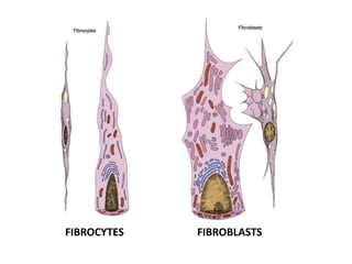
connective tissue contains cells, fibers and ground substance. cells include fibroblasts, undifferentiated mesenchymal cells, adipocytes and pigment cells also wandering cells. fibers include collagen, elastic, and reticular fibers.
3. INTRODUCTION
• Connective tissue are defined as those
composed predominantly of intercellular
material that is extracellular matrix which is
secreted by the connective tissue cells.
• The cells are therefore usually widely
seperated by their matrix, which is composed
of fibrous protein & a relatively amorphous
ground substance.
4. • The term connective is applied to a tissue that
fills the interstices between more specialised
elements.
• It is found in almost every part of the body
• It is conspicuous in some regions & scanty in
others
6. FUNCTIONS
• Connect, Bind, Support, Strengthen the
tissues
• Protects and insulates internal organs
• Major transport system
• Energy reserve and insulation against heat loss
• Repair as in wound healing
• Regeneration of tissues
• Defense of the body
10. 1.FIBROBLASTS
• Principal cell of connective tissue
• Synthesis of collagen, reticular & elastic fibers
& the complex carbohydrates of the ground
substance
• Reside in close proximity to collagen fibers so
called fixed cells
16. RETICULOCYTE/ RETICULAR CELLS
• Fibroblasts secreting reticular fibers
• Reticular tissue: Fibrocellular network of
fibroblasts & delicate collagenous fibers
• Seen in liver, kidney, spleen & most lymphoid
tissues
17. Myofibroblasts
• H&E: bundles of actin filament with actin
motor proteins- nonmuscle myosin
• Expression of α- smooth muscle actin is
regulated by TGF-β1
• Fibronexus
• Mechanotransduction system
23. • As more specialised cells are formed they lose
their ability to transform into other types.
Eg: fibroblasts
24. 3. ADIPOCYTES
(Lipocytes/ Fat cells)
• Differentiated from mesenchymal stem cells
• Central large fat globule(unilocular): Glycerol
esters of oleic, palmitic & stearic acids
• Store neutral fat, produce a variety of
hormones, inflammatory mediators & growth
factors
• Store chemical energy
26. FUNCTIONS:
• Control Energy balance & lipid homeostasis
• Endocrine role: Adipokine secretion
• Mobilization of fat is under nervous &
hormonal control
• No new adipose tissue is thought to form after
the immediate postnatal period
27. Brown fat:
• Multilocular adipose tissue
• Metabolize fatty acids & generate heat
• Uncouple cellular respiration via the
mitochondrial uncoupling protein (UCP1)
• PET scan: supraclavicular, in the neck & along
the spine
• Beige fat cell: Distinct type of thermogenic fat
cell, thought to be derived from precursor
cells in white fat
28.
29. 4. PIGMENT CELLS
• Cells containing a brown pigment (usually
melanin) in their cytoplasm
• Sites:
→iris & choroid of eyeball
→epidermis of skin
• Cells of 2 types:
1. Melanocytes
2. Melanophores/ Chromatophores
30. MELANOCYTE
• Synthesize melanin
• Neural crest origin
MELANOPHORE/CHROMATOPHORE
• Engulf pigment released by
other cells
• Modified fibroblasts
31. • The fibroblasts & blood vessels within healing
wounds develop from mesenchymal stem cells
associated with the tunica adventitia of
venules
37. M2 macrophages
(Alternatively activated macrophages)
• Activated by IL-4,5,10,13
• Anti- inflammatory, proliferative & angiogenic
• Resolution of inflammation & promote wound
repair
• Efficient at combating parasitic infections:
Schistosomiasis
• Involved in pathogenesis of allergy & asthma
38. Mononuclear phagocyte system
(MPS)
• Primarily derived from monocytes
• Phagocytose Trypan blue & India ink
• Cells become fixed in specific tissues & may
adopt a variety of morphologic appearances.
• Functions: phagocytosis, secretion of
lymphokines, antigen processing & antigen
presentation
• Exception: Microglia, Osteoclasts & Fibroblasts
39. MAST CELLS
• Develop in bone marrow & differentiate in
connective tissue
• 20-30µm in diameter
• Gluteraldehyde fixation toluidine blue
Metachromatic staining of granules (heparin)
• The mast cell is related to the basophil that it
contain similar granules
• Cell surface: high affinity Fc receptors (Fc£RI)
41. METACHROMASIA
• When components of a cell or tissue stain in a
colour different from that of the dye used, the
staining is said to be metachromatic.
43. MCTC MAST CELLS
• In the vicinity of small blood vessels, hair
follicles, sebaceous glands & sweat glands & in
the capsules of organs
• Exception: central nervous system
• Exception: Spleen
44. BASOPHILS
• Develop & differentiate in bone marrow
• Share many features with mast cells:
basophilic secretory granules, similar
mediators & high affinity Fc receptors
• Don’t produce PGD2 & IL-5
46. PLASMA CELLS
• Antibody producing cells derived from B
lymphocytes
• Prominent consituent in loose connective
tissue: GI & respiratory tracts
• short life span: 10-30 days
• H&E: cartwheel / analog clock face
47.
48.
49. •Produce large amounts
of only one type of
protein- a specific
antibody
•Produced antibodies are
discharged locally or
stored within the cell
•Antibodies stored within
the cell form inclusons
called Russell’s bodies
52. CONNECTIVE TISSUE FIBERS
• The most conspicuous components of
connective tissue are the fibers within it
• They provide strength & support to the
connective tissue
• 3 types:
Collagen
Reticular
Elastic
53. 1. COLLAGEN FIBERS
• Most abundant structural components of the
connective tissue
• 28 different types
• Seen in bundles
• Bundles may be straight or wavy
• Bundles often branch or anastomose with
adjacent bundles
54. PHYSICAL PROPERTIES
• Flexible & have high tensile
strength
• Pliable & can bend easily
• Birefringence
• Swell & become soft when
treated with weak acid/ alkali
• On boiling converted to
gelatin
55. COLLAGEN FIBRILS
• 8-12nm diameter
• Osmium staining:
65nm- banding pattern
• Collagen molecule/
Tropocollagen is the
subunit
AFM
56. Collagen molecule/ Tropocollagen
• 300nm long & 1.5nm thick
• Head to tail alignment in overlapping rows
• Covalent bonds between the collagen
molecules of adjacent rows gives tensile
strength
• Triple helix of 3 intertwined polypeptides
known as α chains
57. α chains
• Vary in size from 600-3000 aminoacids
• 42 types of α chains
• Encoded by different genes
• 29 types of collagen: Roman numerals I – XXIX
• Collagen molecule can be Homotrimeric /
Heterotrimeric
58. TYPE IV COLLAGEN
TYPE IV COLLAGEN
• Short filaments that form
sheets
• seen in basal lamina,
kidney glomeruli & lens
capsule
• Collagenopathy: Alport’s
syndrome
59. RETICULAR FIBERS
• Supporting framework-
liver, kidneys, bone marrow,
spleen & other lymphoid
organs
• Consist of collagen fibrils-
type III
• 65nm banding pattern
• 20nm diameter
• Exhibit branching pattern
• Do not form bundles
61. ELASTIC FIBERS
• Run singly, branch &
anastomose with each other
• Thinner than collagen
• But thick bunbles in
ligamentum flava &
ligamentum nuchae
• Interwoven with collagen
fibers
62. Staining properties
• H & E
• Orecin / Resorcin-fuchsin
• Aldehyde fuchsin
• Verhoeff’s method
63. • Produced by: fibroblasts, smooth muscle
cells,…
• Elastic property is related to its unusual
polypeptide backbone, which causes random
coiling
• Composed of
Central core of elastin
Fibrillin microfibrils
64. ELASTIN
• 70KDa
• Hydrophobic
• Desmosine &
Isodesmosine
• Encoded by one of the
largest genes:
28kilobases, less than
10% encodes elastin