Subjective classification and objective analysis of the mandibular dental arc...EdwardHAngle
Our objective was to evaluate the relationship between subjective classification of dental-arch shape, objective analyses via arch-width measurements, and the fitting with the fourth-order polynomial equation.
Clinical study of impacted maxillary canine in the Arab population in IsraelAbu-Hussein Muhamad
The objective of the present study was to determine the prevalence of impacted maxillary canine in patients in Arabs
Community in Israel (ARAB48,Israel) visiting our Center For Dentistry,Research & Aesthetics,Jatt,Almothalath,Israel,
4250 patients . This study comprises data from patients who attended the O.P.D.2200 patients between Jun. 2006 to Dec
2013. Patients were examined in order to detect the impacted maxillary canines by intraoral examination, palpation, dental
records and followed by radiographs. It was found that the prevalence of canine impaction was 0,8 % (N=4250), 1,6
(N=2200), 43,9 (N-82) in males and 1,1% (N=4250), 2,1 (N=2200), 56,1 (N-82) in females suggesting that prevalence of
impacted maxillary canines is more in females than males and it is statistically significant. The overall prevalence for
maxillary impacted canines was found to be 3,7 % (N=2200) which suggested that it is much higher than previous studies.
The results of this study were slightly different than other studies, while the dissimilarities may be attributed to the sample
selection, method of the study and area of patient selection, which suggest racial and genetic differences.
Digital workflow in full mouth rehabilitation using CBCTApurva Thampi
This is a journal club presentation on the digital workflow of a full mouth rehabilitation using implants and as CBCT as a guide.
The presentation and all the materials collected is available on request. Mail me at apurvathampi@gmail.com
Craniofacial growth in untreated skeletal class i subjects with low, average,...EdwardHAngle
The dental measurements showed few changes with growth in all groups. In terms of skeletal measurements from ages 9 to 18, similar growth changes were found between the sexes in most angular measurements, but males had larger values in linear measurements than females.
Malposition of unerupted mandibular second premolar in children with cleft li...EdwardHAngle
Objective: To determine whether distoangular malposition of the unerupted mandibular second
premolar (MnP2) is more frequent in children with unilateral clefts of the lip and palate.
Materials and Methods: This retrospective study examined panoramic radiographs from 45 patients
with unilateral clefts of the lip and/or palate who had no previous orthodontics. A control
sample consisted of age- and sex-matched patients. The distal angle formed between the long
axis of MnP2 and the tangent to the inferior border was measured. The mean, standard deviation,
and range were calculated for the angles measured in the cleft and the control groups. The
significance of the differences between the means was evaluated by the paired t-test. The angles
of the cleft and noncleft sides were also measured and compared.
Results: The mean inclination of the MnP2 on the cleft side was 73.6°, compared with 84.6° in
the control group. This difference was highly significant statistically (P < .0001). The difference
in angles from the cleft and noncleft sides was 0.7°, not statistically significant. A significant association
was found between clefting and distoangular malposition of the developing MnP2, suggesting
a shared genetic etiology. This association is independent of the clefting side, ruling out
possible local mechanical effects.
Conclusion: Clinicians should be aware of the potential for anomalous development of MnP2 in
children with clefts.
Long term clinical and bacterial effetcx of xylitol on paitnets with fixed or...EdwardHAngle
Background: The objective of this study was to evaluate long-term clinical and bacterial effects of using 6 g of
xylitol per day for 3 months on patients with full fixed orthodontic appliances.
Methods: The study was a pilot clinical trial that included 41 subjects who were undergoing orthodontic
treatment. The subjects were randomly divided into three groups. Group A received xylitol chewing gum, group B
received xylitol dissolvable chewable tablets, and Group C served as the control group and did not receive xylitol
gums or tablets. Clinical examination and the collection of plaque and saliva samples were carried out at baseline
and 3, 6, and 12 months. All three groups were given oral hygiene instruction and were put on a 6-month cleaning
and topical fluoride schedule. Plaque scores and bacterial counts were used to evaluate the effectiveness of the
different approaches at reducing the caries risk.
Results: Xylitol groups did not experience any more reduction in plaque score, plaque MS counts, or salivary MS
counts than the control group nor did they have lower values at any of the time points. Chewing gum did not
significantly increase the incidence of debonded brackets over the other groups.
Conclusions: Xylitol does not have a clinical or bacterial benefit in patients with fixed orthodontic appliances. Oral
hygiene instructions and 6-month topical fluoride application were effective at reducing plaque scores and bacterial
counts in patients with full fixed appliances regardless of whether or not xylitol was used.
Fixed and removable orthodontic appliance application for class III malocclus...iosrjce
IOSR Journal of Dental and Medical Sciences is one of the speciality Journal in Dental Science and Medical Science published by International Organization of Scientific Research (IOSR). The Journal publishes papers of the highest scientific merit and widest possible scope work in all areas related to medical and dental science. The Journal welcome review articles, leading medical and clinical research articles, technical notes, case reports and others.
Transverse growth of the maxilla and mandible in untreated girls with low, av...EdwardHAngle
The purpose of this study was to investigate maxillary and mandibular transverse growth in
untreated female subjects with low, average, and high mandibular plane angles longitudinally from ages 6 to 18.
ABSTRACT
Objective: To evaluate the role of age as a moderator of bone regeneration patterns and
symphysis remodeling after genioplasty.
Method: Fifty-four patients who underwent genioplasty at the end of their orthodontic treatment
were divided into three age groups: younger than 15 years at the time of surgery (group 1), 15 to
19 years (group 2), and 20 years or older (group 3). Twenty-three patients who did not accept
genioplasty and had a follow-up radiograph 2 years after the end of their orthodontic treatment
were used as a control group. Patients were evaluated at three time points: immediate preoperative
(T1), immediate postoperative (T2,) and 2 years postsurgery (T3).
Results: The mean genial advancement at surgery was similar for the three age groups, but the
extent of remodeling around the repositioned chin was greater in group 1, less in group 2, and still
less in group 3. Symphysis thickness increased significantly during the 2-year postsurgery interval
for the three groups, and this increase was significantly greater in group 1 than in group 3.
Remodeling above and behind the repositioned chin also was greater in the younger patients. This
was related to greater vertical growth of the dentoalveolar process in the younger patients. There
was no evidence of a deleterious effect on mandibular growth.
Conclusion: The outcomes of forward-upward genioplasty include increased symphysis
thickness, bone apposition above B point, and remodeling at the inferior border. When indications
for this type of genioplasty are recognized, early surgical correction (before age 15) produces a
better outcome in terms of bone remodeling. (Angle Orthod. 0000;00:000–000.)
Dr. Rahul VC Tiwari - Fellowship In Orthognathic Surgery - Jubilee Mission Medical College Hospital and Research Center, Thrissur, Kerala - 30th publicationJAMDSR 6TH name
Effects of rapid palatal expansion on the sagittal and vertical dimensions of...EdwardHAngle
The purpose of this study was to use cone-beam computed tomography imaging to examine the skeletal and dental changes in the sagittal and vertical dimensions after rapid palatal expansion.
Mandibular arch form the relationship between dental and basal anatomyEdwardHAngle
We investigated mandibular dental arch form at the levels of both the clinically relevant application points of the orthodontic bracket and the underlying anatomic structure of the apical base. The correlation of both forms was evaluated and examined to determine whether the basal arch could be used to derive a standardized clinical arch form.
Clinical study of impacted maxillary canine in the Arab population in IsraelAbu-Hussein Muhamad
The objective of the present study was to determine the prevalence of impacted maxillary canine in patients in Arabs Community in Israel (ARAB48,Israel) visiting our Center For Dentistry,Research & Aesthetics,Jatt,Almothalath,Israel, 4250 patients . This study comprises data from patients who attended the O.P.D.2200 patients between Jun. 2006 to Dec 2013. Patients were examined in order to detect the impacted maxillary canines by intraoral examination, palpation, dental records and followed by radiographs. It was found that the prevalence of canine impaction was 0,8 % (N=4250), 1,6 (N=2200), 43,9 (N-82) in males and 1,1% (N=4250), 2,1 (N=2200), 56,1 (N-82) in females suggesting that prevalence of impacted maxillary canines is more in females than males and it is statistically significant. The overall prevalence for maxillary impacted canines was found to be 3,7 % (N=2200) which suggested that it is much higher than previous studies. The results of this study were slightly different than other studies, while the dissimilarities may be attributed to the sample selection, method of the study and area of patient selection, which suggest racial and genetic differences.
Tooth Contact Sounds-Can It Evaluate Occlusion?QUESTJOURNAL
Purpose: To study the relevance of tooth contact sounds in a wave form in evaluating the quality of occlusion and to check if these sounds can be used to identify occlusal events. Methods: 42 subjects having 28 or 32 permanent teeth with Angle’s class I molar relashionship were selected . None of the teeth had any restorations . Subjects were asked to make tooth contacts in maximum intercuspation position and eccentric movements in right lateral, left lateral and protrusive positions . Tooth contact sounds were converted into wave forms by a polygraph machine .The components of the wave pattern were studied as ‘impacts’ and ‘slides’. One way ANOVA test was done to evaluate the statistical significance. Results : The wave patterns analyzed were classified into four types. Number of waves and duration of waves in the four different occlusal positions were calculated. Mean value of slides in maximum intercuspation position differed in four types and was found to be statistically significant (p<0.5).The statistically significant difference in the mean number of slides in the maximum intercuspation position in different types signified that tooth contact sounds could be used to study different qualities of occlusion. Conclusion: The study of wave pattern of ‘impacts‘and ‘slides’ in occlusal contact could help in correcting the occlusion to a stable position and this could be used as a ready reckoner to assess the quality of occlusion and also as a guide for future reference.
Report Back from SGO 2024: What’s the Latest in Cervical Cancer?bkling
Are you curious about what’s new in cervical cancer research or unsure what the findings mean? Join Dr. Emily Ko, a gynecologic oncologist at Penn Medicine, to learn about the latest updates from the Society of Gynecologic Oncology (SGO) 2024 Annual Meeting on Women’s Cancer. Dr. Ko will discuss what the research presented at the conference means for you and answer your questions about the new developments.
Tom Selleck Health: A Comprehensive Look at the Iconic Actor’s Wellness Journeygreendigital
Tom Selleck, an enduring figure in Hollywood. has captivated audiences for decades with his rugged charm, iconic moustache. and memorable roles in television and film. From his breakout role as Thomas Magnum in Magnum P.I. to his current portrayal of Frank Reagan in Blue Bloods. Selleck's career has spanned over 50 years. But beyond his professional achievements. fans have often been curious about Tom Selleck Health. especially as he has aged in the public eye.
Follow us on: Pinterest
Introduction
Many have been interested in Tom Selleck health. not only because of his enduring presence on screen but also because of the challenges. and lifestyle choices he has faced and made over the years. This article delves into the various aspects of Tom Selleck health. exploring his fitness regimen, diet, mental health. and the challenges he has encountered as he ages. We'll look at how he maintains his well-being. the health issues he has faced, and his approach to ageing .
Early Life and Career
Childhood and Athletic Beginnings
Tom Selleck was born on January 29, 1945, in Detroit, Michigan, and grew up in Sherman Oaks, California. From an early age, he was involved in sports, particularly basketball. which played a significant role in his physical development. His athletic pursuits continued into college. where he attended the University of Southern California (USC) on a basketball scholarship. This early involvement in sports laid a strong foundation for his physical health and disciplined lifestyle.
Transition to Acting
Selleck's transition from an athlete to an actor came with its physical demands. His first significant role in "Magnum P.I." required him to perform various stunts and maintain a fit appearance. This role, which he played from 1980 to 1988. necessitated a rigorous fitness routine to meet the show's demands. setting the stage for his long-term commitment to health and wellness.
Fitness Regimen
Workout Routine
Tom Selleck health and fitness regimen has evolved. adapting to his changing roles and age. During his "Magnum, P.I." days. Selleck's workouts were intense and focused on building and maintaining muscle mass. His routine included weightlifting, cardiovascular exercises. and specific training for the stunts he performed on the show.
Selleck adjusted his fitness routine as he aged to suit his body's needs. Today, his workouts focus on maintaining flexibility, strength, and cardiovascular health. He incorporates low-impact exercises such as swimming, walking, and light weightlifting. This balanced approach helps him stay fit without putting undue strain on his joints and muscles.
Importance of Flexibility and Mobility
In recent years, Selleck has emphasized the importance of flexibility and mobility in his fitness regimen. Understanding the natural decline in muscle mass and joint flexibility with age. he includes stretching and yoga in his routine. These practices help prevent injuries, improve posture, and maintain mobilit
Knee anatomy and clinical tests 2024.pdfvimalpl1234
This includes all relevant anatomy and clinical tests compiled from standard textbooks, Campbell,netter etc..It is comprehensive and best suited for orthopaedicians and orthopaedic residents.
Title: Sense of Taste
Presenter: Dr. Faiza, Assistant Professor of Physiology
Qualifications:
MBBS (Best Graduate, AIMC Lahore)
FCPS Physiology
ICMT, CHPE, DHPE (STMU)
MPH (GC University, Faisalabad)
MBA (Virtual University of Pakistan)
Learning Objectives:
Describe the structure and function of taste buds.
Describe the relationship between the taste threshold and taste index of common substances.
Explain the chemical basis and signal transduction of taste perception for each type of primary taste sensation.
Recognize different abnormalities of taste perception and their causes.
Key Topics:
Significance of Taste Sensation:
Differentiation between pleasant and harmful food
Influence on behavior
Selection of food based on metabolic needs
Receptors of Taste:
Taste buds on the tongue
Influence of sense of smell, texture of food, and pain stimulation (e.g., by pepper)
Primary and Secondary Taste Sensations:
Primary taste sensations: Sweet, Sour, Salty, Bitter, Umami
Chemical basis and signal transduction mechanisms for each taste
Taste Threshold and Index:
Taste threshold values for Sweet (sucrose), Salty (NaCl), Sour (HCl), and Bitter (Quinine)
Taste index relationship: Inversely proportional to taste threshold
Taste Blindness:
Inability to taste certain substances, particularly thiourea compounds
Example: Phenylthiocarbamide
Structure and Function of Taste Buds:
Composition: Epithelial cells, Sustentacular/Supporting cells, Taste cells, Basal cells
Features: Taste pores, Taste hairs/microvilli, and Taste nerve fibers
Location of Taste Buds:
Found in papillae of the tongue (Fungiform, Circumvallate, Foliate)
Also present on the palate, tonsillar pillars, epiglottis, and proximal esophagus
Mechanism of Taste Stimulation:
Interaction of taste substances with receptors on microvilli
Signal transduction pathways for Umami, Sweet, Bitter, Sour, and Salty tastes
Taste Sensitivity and Adaptation:
Decrease in sensitivity with age
Rapid adaptation of taste sensation
Role of Saliva in Taste:
Dissolution of tastants to reach receptors
Washing away the stimulus
Taste Preferences and Aversions:
Mechanisms behind taste preference and aversion
Influence of receptors and neural pathways
Impact of Sensory Nerve Damage:
Degeneration of taste buds if the sensory nerve fiber is cut
Abnormalities of Taste Detection:
Conditions: Ageusia, Hypogeusia, Dysgeusia (parageusia)
Causes: Nerve damage, neurological disorders, infections, poor oral hygiene, adverse drug effects, deficiencies, aging, tobacco use, altered neurotransmitter levels
Neurotransmitters and Taste Threshold:
Effects of serotonin (5-HT) and norepinephrine (NE) on taste sensitivity
Supertasters:
25% of the population with heightened sensitivity to taste, especially bitterness
Increased number of fungiform papillae
Lung Cancer: Artificial Intelligence, Synergetics, Complex System Analysis, S...Oleg Kshivets
RESULTS: Overall life span (LS) was 2252.1±1742.5 days and cumulative 5-year survival (5YS) reached 73.2%, 10 years – 64.8%, 20 years – 42.5%. 513 LCP lived more than 5 years (LS=3124.6±1525.6 days), 148 LCP – more than 10 years (LS=5054.4±1504.1 days).199 LCP died because of LC (LS=562.7±374.5 days). 5YS of LCP after bi/lobectomies was significantly superior in comparison with LCP after pneumonectomies (78.1% vs.63.7%, P=0.00001 by log-rank test). AT significantly improved 5YS (66.3% vs. 34.8%) (P=0.00000 by log-rank test) only for LCP with N1-2. Cox modeling displayed that 5YS of LCP significantly depended on: phase transition (PT) early-invasive LC in terms of synergetics, PT N0—N12, cell ratio factors (ratio between cancer cells- CC and blood cells subpopulations), G1-3, histology, glucose, AT, blood cell circuit, prothrombin index, heparin tolerance, recalcification time (P=0.000-0.038). Neural networks, genetic algorithm selection and bootstrap simulation revealed relationships between 5YS and PT early-invasive LC (rank=1), PT N0—N12 (rank=2), thrombocytes/CC (3), erythrocytes/CC (4), eosinophils/CC (5), healthy cells/CC (6), lymphocytes/CC (7), segmented neutrophils/CC (8), stick neutrophils/CC (9), monocytes/CC (10); leucocytes/CC (11). Correct prediction of 5YS was 100% by neural networks computing (area under ROC curve=1.0; error=0.0).
CONCLUSIONS: 5YS of LCP after radical procedures significantly depended on: 1) PT early-invasive cancer; 2) PT N0--N12; 3) cell ratio factors; 4) blood cell circuit; 5) biochemical factors; 6) hemostasis system; 7) AT; 8) LC characteristics; 9) LC cell dynamics; 10) surgery type: lobectomy/pneumonectomy; 11) anthropometric data. Optimal diagnosis and treatment strategies for LC are: 1) screening and early detection of LC; 2) availability of experienced thoracic surgeons because of complexity of radical procedures; 3) aggressive en block surgery and adequate lymph node dissection for completeness; 4) precise prediction; 5) adjuvant chemoimmunoradiotherapy for LCP with unfavorable prognosis.
These simplified slides by Dr. Sidra Arshad present an overview of the non-respiratory functions of the respiratory tract.
Learning objectives:
1. Enlist the non-respiratory functions of the respiratory tract
2. Briefly explain how these functions are carried out
3. Discuss the significance of dead space
4. Differentiate between minute ventilation and alveolar ventilation
5. Describe the cough and sneeze reflexes
Study Resources:
1. Chapter 39, Guyton and Hall Textbook of Medical Physiology, 14th edition
2. Chapter 34, Ganong’s Review of Medical Physiology, 26th edition
3. Chapter 17, Human Physiology by Lauralee Sherwood, 9th edition
4. Non-respiratory functions of the lungs https://academic.oup.com/bjaed/article/13/3/98/278874
Couples presenting to the infertility clinic- Do they really have infertility...Sujoy Dasgupta
Dr Sujoy Dasgupta presented the study on "Couples presenting to the infertility clinic- Do they really have infertility? – The unexplored stories of non-consummation" in the 13th Congress of the Asia Pacific Initiative on Reproduction (ASPIRE 2024) at Manila on 24 May, 2024.
ARTIFICIAL INTELLIGENCE IN HEALTHCARE.pdfAnujkumaranit
Artificial intelligence (AI) refers to the simulation of human intelligence processes by machines, especially computer systems. It encompasses tasks such as learning, reasoning, problem-solving, perception, and language understanding. AI technologies are revolutionizing various fields, from healthcare to finance, by enabling machines to perform tasks that typically require human intelligence.
HOT NEW PRODUCT! BIG SALES FAST SHIPPING NOW FROM CHINA!! EU KU DB BK substit...GL Anaacs
Contact us if you are interested:
Email / Skype : kefaya1771@gmail.com
Threema: PXHY5PDH
New BATCH Ku !!! MUCH IN DEMAND FAST SALE EVERY BATCH HAPPY GOOD EFFECT BIG BATCH !
Contact me on Threema or skype to start big business!!
Hot-sale products:
NEW HOT EUTYLONE WHITE CRYSTAL!!
5cl-adba precursor (semi finished )
5cl-adba raw materials
ADBB precursor (semi finished )
ADBB raw materials
APVP powder
5fadb/4f-adb
Jwh018 / Jwh210
Eutylone crystal
Protonitazene (hydrochloride) CAS: 119276-01-6
Flubrotizolam CAS: 57801-95-3
Metonitazene CAS: 14680-51-4
Payment terms: Western Union,MoneyGram,Bitcoin or USDT.
Deliver Time: Usually 7-15days
Shipping method: FedEx, TNT, DHL,UPS etc.Our deliveries are 100% safe, fast, reliable and discreet.
Samples will be sent for your evaluation!If you are interested in, please contact me, let's talk details.
We specializes in exporting high quality Research chemical, medical intermediate, Pharmaceutical chemicals and so on. Products are exported to USA, Canada, France, Korea, Japan,Russia, Southeast Asia and other countries.
New Drug Discovery and Development .....NEHA GUPTA
The "New Drug Discovery and Development" process involves the identification, design, testing, and manufacturing of novel pharmaceutical compounds with the aim of introducing new and improved treatments for various medical conditions. This comprehensive endeavor encompasses various stages, including target identification, preclinical studies, clinical trials, regulatory approval, and post-market surveillance. It involves multidisciplinary collaboration among scientists, researchers, clinicians, regulatory experts, and pharmaceutical companies to bring innovative therapies to market and address unmet medical needs.
Ethanol (CH3CH2OH), or beverage alcohol, is a two-carbon alcohol
that is rapidly distributed in the body and brain. Ethanol alters many
neurochemical systems and has rewarding and addictive properties. It
is the oldest recreational drug and likely contributes to more morbidity,
mortality, and public health costs than all illicit drugs combined. The
5th edition of the Diagnostic and Statistical Manual of Mental Disorders
(DSM-5) integrates alcohol abuse and alcohol dependence into a single
disorder called alcohol use disorder (AUD), with mild, moderate,
and severe subclassifications (American Psychiatric Association, 2013).
In the DSM-5, all types of substance abuse and dependence have been
combined into a single substance use disorder (SUD) on a continuum
from mild to severe. A diagnosis of AUD requires that at least two of
the 11 DSM-5 behaviors be present within a 12-month period (mild
AUD: 2–3 criteria; moderate AUD: 4–5 criteria; severe AUD: 6–11 criteria).
The four main behavioral effects of AUD are impaired control over
drinking, negative social consequences, risky use, and altered physiological
effects (tolerance, withdrawal). This chapter presents an overview
of the prevalence and harmful consequences of AUD in the U.S.,
the systemic nature of the disease, neurocircuitry and stages of AUD,
comorbidities, fetal alcohol spectrum disorders, genetic risk factors, and
pharmacotherapies for AUD.
1. Condylar position assessed by magnetic
resonance imaging after various bite
position registrations
Sanjivan Kandasamy,a
Rudolf Boeddinghaus,b
and Estie Krugerc
Nedlands and Subiaco, Western Australia, Australia, and St Louis, Mo
Introduction: In this study, we evaluated the reliability and validity of 3 bite registrations in relation to condylar
position in the glenoid fossae using magnetic resonance imaging in a symptom-free population. Methods: Nine-
teen subjects, 14 men and 5 women (ages, 20-39 years) without temporomandibular disorders were examined.
Three bite registrations were taken and evaluated on each subject: centric occlusion, centric relation, and Roth
power centric relation. The differences in condyle position among the 3 bite registrations were determined for the
left and right condyles: centric occlusion-centric relation, centric occlusion-Roth power centric relation, and
centric relation-Roth power centric relation for each plane of space. Results: The results indicated that (1) all
measurements collected had large standard deviations and ranges with no statistical significance, and (2) of
the 19 subjects and 38 condyles assessed, 33 condyles (87%) were concentric in an anteroposterior plane.
In the transverse anatomic plane, all condyles were concentric. Conclusions: The clinical concept of positioning
the condyles in specific positions in the fossae with various bite registrations as a preventive measure for tempo-
romandibular disorders and as a diagnosis and treatment planning tool is not supported by this study. (Am J
Orthod Dentofacial Orthop 2013;144:512-7)
T
he relationship among condylar position, occlu-
sion, and temporomandibular disorders (TMD)
has been the subject of much debate in dentistry
for more than a century. A significant part of the debate
involves establishing coincidence between a specific
definition of centric relation and centric occlusion. It is
believed by many gnathologists and the like that failure
to achieve this position will predispose patients to devel-
oping TMD in the future.1
Despite the lack of data available to support this
assertion, many dentists continue to persist in marrying
a particular definition of centric relation with centric oc-
clusion as a main goal of treatment. With the current
emphasis in dentistry on evidence-based decision-mak-
ing, the routine establishment of centric occlusion with
centric relation in all patients is subject to question.
Centric relation is defined as a position of the con-
dyles in the glenoid fossae, irrespective of the occlusion
or tooth contact. Centric occlusion is an interocclusal
dental position of the maxillary teeth relative to the
mandibular teeth. Over the past half century, the defini-
tion of centric relation has evolved from a posterior po-
sition of the condyle in relation to the glenoid fossa to a
posterosuperior position to eventually an anterior and
superior position.2-6
Before 1968, centric relation was
considered to be a retruded (posterior) condylar
position. The latest edition of the Glossary of
Prosthodontic Terms defines centric relation as “a
maxillomandibular relationship in which the condyles
articulate with the thinnest avascular portion of their
respective disks with the complex in the anterosuperior
position against the slopes of the articular eminences.”6
Contemporary orthodontic gnathologists believe in
attaining an anterosuperior condyle position at the
a
Clinical associate professor, Department of Orthodontics, School of Dentistry,
University of Western Australia, Nedlands, Western Australia, Australia; adjunct
assistant professor, Center for Advanced Dental Education, Saint Louis Univer-
sity, St Louis, Mo.
b
Clinical senior lecturer, School of Surgery, University of Western Australia, Ned-
lands; and Perth Radiological Clinic, Subiaco, Western Australia, Australia.
c
Associate professor, Dental Public Health, Faculty of Medicine, Dentistry and
Health Sciences, University of Western Australia, Nedlands, Western Australia,
Australia.
All authors have completed and submitted the ICMJE Form for Disclosure of Po-
tential Conflicts of Interest, and none were reported.
Funded by the Australian Society of Orthodontists Foundation for Research and
Education.
Reprint requests to: Sanjivan Kandasamy, Department of Orthodontics, School of
Dentistry, University of Western Australia, 17 Monash Ave, Nedlands, 6009, WA,
Australia; e-mail, sanj@kandasamy.com.au.
Submitted, May 2013; revised and accepted, June 2013.
0889-5406/$36.00
Copyright Ó 2013 by the American Association of Orthodontists.
http://dx.doi.org/10.1016/j.ajodo.2013.06.014
512
ORIGINAL ARTICLE
2. same time the teeth are in centric occlusion (centric rela-
tion equals centric occlusion); however, there is little or
no scientific evidence to support this view.7
Dentists who
believe in establishing a coincidence of a specific defini-
tion of centric relation with centric occlusion
unnecessarily subject their patients to procedures that
might include orthodontics and irreversible bite-
altering procedures, leading to significant financial
and biological costs to their patients. In fact, the evi-
dence supports the contrary notion. According to Alex-
ander et al,8
magnetic resonance imaging (MRI) data
of the temporomandibular joint showed that condyles
are not located in the assumed positions as advocated
by certain centric bite registrations such as retruded
centric relation and leaf-gauge generated centric rela-
tion registrations. Centric bite registrations attempting
to locate retruded (posterosuperior) centric relation
and contemporary anterosuperior centric relation do
not correspond to the condyle positions of people
without TMD. The location of the condyles in the gle-
noid fossa, irrespective of position, has not been demon-
strated to be consequential to the presence or absence of
TMD symptoms.9-12
The Roth power centric relation is perhaps the most
commonly used contemporary gnathologic record aimed
at attaining an anterosuperior centric relation position.
To date, MRI has not verified the actual anatomic posi-
tion of the condyles when this bite registration is taken.
The condylar positions in centric occlusion and Roth
power centric relation have only been studied on articu-
lators with mandibular or condylar position indicators.
The validity of this approach has been questioned and
is not supported by MRI data, presumably the most ac-
curate ionizing radiation-free means of assessing 3-
dimensional condyle-disc-fossa relationships.13
In this
study, condylar position was assessed using MRI after
the use of 3 common bite registrations: centric occlu-
sion, retruded centric relation, and the so-called Roth
power centric relation.14-16
MATERIAL AND METHODS
Ethics approval for this study was obtained from the
Human Research Ethics Committee at the University of
Western Australia. Guided by a power analysis, a sample
of 19 subjects was studied.
The subjects were 14 men and 5 women (ages, 20-39
years) who met the following criteria: (1) permanent
dentition, (2) no congenital craniofacial anomaly or syn-
drome, (3) no skeletal asymmetry or premature tooth
contacts leading to any functional mandibular displace-
ments, (4) no history of trauma to the face or jaws, (5)
positive overjet and overbite, (6) no TMD signs or symp-
toms (all subjects underwent a TMD examination based
on the Research Diagnostic Criteria for TMD; although
clicking without other symptoms does not indicate pa-
thosis or dysfunction, subjects with clicking were
excluded from the study17
), and (8) completed an MRI
safety screening check.
Each participant was also informed of the risk of
finding any incidental pathology after the MRI scan,
but no significant incidental pathology was found.
To standardize the bite registration technique, all
records were taken by the same investigator (S.K.). Three
bite positions were assessed. The first was centric occlu-
sion, with the subject biting together in maximum inter-
cuspation.
The remaining 2 bite registrations were taken in wax
(blue wax; Delar, Lake Oswego, Ore).
Retruded centric relation was taken by applying
distal pressure to the chin and taking a wax record at
the first point of light occlusal contact. All subjects ex-
hibited a discrepancy between centric relation and
centric occlusion. By taking a record at the first point
of light occlusal contact, we were able to establish a
reference point to base our repeated measurements.
This proved to be a highly reproducible registration
technique in this study.
Roth power centric relation was taken with a 2-
piece wax registration consisting of anterior and poste-
rior sections.14-16
The anterior section was first
constructed at a vertical where the posterior teeth are
at least 2 mm apart. This piece of wax was chilled
and allowed to harden. This wax was then placed
back into the mouth, a softened posterior section was
placed, and the patient was instructed to bite. The
mandibular anterior teeth were guided into the
hardened anterior section of wax without a slide into
the indentations. As the patient closed into the
hardened anterior section, he or she was instructed to
“close firmly and hold.” The posterior section was
chilled with air. When the posterior section had
hardened sufficiently to prevent distortion, both wax
sections were removed and chilled.
Once the hard wax bite registrations were obtained,
they were used to position the subjects' mandibles in
the various occlusal positions as the MRI data were ob-
tained. Originally, acrylic records of the bite positions
were fabricated after the articulation of plaster, with
the assumption that they would be more accurate. We
found, however, that these acrylic records distorted
significantly during the fabrication process and provided
an inaccurate reflection of the bite records. Since the
changes in the bite positions were small, this would
have significantly affected the results. We then decided
to use the hard wax records, which were stable and
more accurate.
Kandasamy, Boeddinghaus, and Kruger 513
American Journal of Orthodontics and Dentofacial Orthopedics October 2013 Vol 144 Issue 4
3. MRI scans of each subject were acquired as the sub-
jects occluded at each of the 3 bite relationships. All
scans were performed on the same MRI scanner (1.5 T,
Signa Excite; General Electric, Fairfield, Conn). Scans
were performed with dedicated phased array surface
temporomandibular joint coils. An initial low-resolution
T1-weighted (repetition time [TR]: 340 ms; echo time
[TE], 8 ms) axial localizing scan was followed by a
high-resolution T2-weighted (TR 2800 ms, TE 72 ms)
sagittal oblique scan acquired perpendicular to the
long axis of each condyle (Fig 1). The MRI scans were in-
terpreted by a head and neck radiologist (R.B.) with 8
years of experience reading MRIs of the temporoman-
dibular joint. He was blinded as to which bite registration
he was assessing in each subject, and the order of scan-
ning was randomized by the MRI technician. On each
side and in each position, measurements of the antero-
posterior and superoinferior positions of the condyle
with respect to the temporal bone were measured using
the following standard cortical bony landmarks: for the
anteroposterior position, the anterior margin of the
condyle and the summit of the articular eminence; for
the superoinferior position, the highest point of the
condyle and the deepest concavity of the glenoid fossa
(Fig 2).
From the sagittal and transverse anatomic plane MRI
views, the radiologist evaluated the concentricity of the
left and right condyles in the glenoid fossa in the centric
occlusion position. This was done by dividing the
condyle into thirds and determining which third was
within the central point of the glenoid fossa.
Guided by a power analysis, the 19 subjects were
studied to produce 80% power to detect mean differ-
ences of at least 1 mm at the 0.05% level of signifi-
cance.
To assess the variability and reproducibility associ-
ated with taking the centric occlusion, centric relation,
and Roth power centric relation bite registrations, 3
sets of each were recorded on 3 separate occasions on
2 subjects. These readings were analyzed by a 1-way
repeated-measures analysis of variance (ANOVA), which
showed no significant (P0.05) difference between the
repeated positions, an indication that this method of
determining centric occlusion, centric relation, and
Roth power centric relation is reliable.
One observer (the radiologist, R.B.) made all mea-
surements, 3 times on 2 randomly chosen subjects
with a 1-week interval between measurements. Intra-
class correlation coefficients (ICC) were used to deter-
mine the intraobserver reliability, and 95% prediction
limits for the errors in measurement are provided. A
mean ICC of 0.992 was determined, with an upper limit
of 0.995 and a lower limit of 0.985.
Statistical analysis
For both the left and right condyles, the differences in
condyle position among the 3 bite registrations were
determined: centric occlusion-centric relation, centric
occlusion-Roth power centric relation, and centric
relation-Roth power centric relation for each plane of
space. The data were analyzed by 1-way ANOVA and
by randomized block 1-way ANOVA with Tukey
follow-ups.
RESULTS
For both the left and right condyles, the differences
in condyle position between the different bite registra-
tions were determined as follows: centric occlusion-
centric relation, centric occlusion-Roth power centric
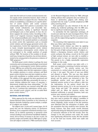
Fig 1. Axial low-resolution T1-weighted planning scan
(A), showing the plane for planning the oblique sagittal
scans (white lines) perpendicular to the long axis of
each condyle (C). Note that there is frequently some
asymmetry of the axes of the condyles, as in this
case. The resultant high-resolution T2-weighted sagittal
oblique image (B), showing the articular disc (between
arrows), the mandibular condyle (C), and the articular
eminence (AE) of the temporal bone.
514 Kandasamy, Boeddinghaus, and Kruger
October 2013 Vol 144 Issue 4 American Journal of Orthodontics and Dentofacial Orthopedics
4. relation, and centric relation-Roth power centric rela-
tion for each plane. The results are presented in the
Table.
All measurements collected had no statistical signif-
icance given the large associated standard deviations
and ranges. Interestingly, asymmetric and opposite
changes were noted between the left and right condyles
between the bite registrations. Qualitatively, the only
measurement with the only noticeable and similar sym-
metric difference at both condyles was the difference be-
tween centric occlusion and Roth power centric relation
bite registrations. Although it was statistically not signif-
icant, both condyles were located more superiorly in the
Roth power centric relation bite registrations by 0.28
mm on average.
Of the 19 subjects and the 38 condyles assessed, 33
condyles (87%) were concentric in an anteroposterior
plane. The remaining 5 were positioned posteriorly.
No pair of condyles had been positioned posteriorly.
In the transverse anatomic plane, all condyles were
concentric.
DISCUSSION
Based on our findings, the differences between the 3
bite positions were small and, more importantly, highly
variable. Since 87% and 100% of the condyles were
concentric in the glenoid fossa in centric occlusion in
the anteroposterior and transverse planes, respectively,
we can safely infer that any significant positioning
that would occur from the centric relation and the
Roth power bite registrations would be detectable. If
the condyles were not concentric in centric occlusion,
this would lead to much ambiguity in terms of condylar
positioning that would occur with the bite registrations.
Our findings show that the posterior condylar posi-
tioning and the anterosuperior condylar positioning
associated with centric relation and Roth power centric
relation bite registrations, respectively, do not occur.
Variability in the findings between the bite registrations
appear to reflect the lack of accuracy and predictability
in these bite registration processes, especially in terms
of determining and positioning the condyles in certain
locations of the glenoid fossae. Based on the findings
that we are not positioning the condyles in specific po-
sitions in the fossae with various bite registrations, the
clinical significance followed by the routine practice of
condylar positioning must be questioned. Hence, previ-
ous studies reporting any changes as a result of these
centric relation bite registrations14,15,18-21
and studies
using these centric relation bite registrations to assess
and report any differences between nongnathologically
treated and gnathologically treated orthodontic
patients would appear to be invalid.22-24
The variability in the findings might be related to the
asymmetry in the long axes of each pair of condyles in
each subject. This is a normal finding,25
and this asymme-
try prevents each pair of condyles from functioning like a
hinge axis as originally proposed by the flawed terminal
hinge axis theory of Posselt.26
The presence of 2 asym-
metrically angulated condylar long axes would naturally
lead to condyles translating and rotating simultaneously
as soon as mandibular opening and closing is initiated.27
The Roth power bite registration is taken when there
is at least 2 mm of separation at the posterior teeth. This
bite registration is then transferred onto an articulator,
with its known inaccuracies, to facilitate mounting of
the upper and lower study casts.28,29
After removal of
the bite registration after the mounting, the upper and
lower casts close down to occlude, reducing the
vertical dimension. Since the condyles rotate and
Fig 2. Reference points for measurement: this is a
sagittal oblique scan from the center of the condyle. The
anteroposterior measurement (A, thick white line) is
made from the anterior cortical margin of the condyle to
the summit of the articular eminence. The superoinferior
measurement (B, thick white line) is made from the high-
est point of the condyle to the highest point (deepest con-
cavity) of the glenoid fossa.
Kandasamy, Boeddinghaus, and Kruger 515
American Journal of Orthodontics and Dentofacial Orthopedics October 2013 Vol 144 Issue 4
5. translate on mandibular opening and closing, this
further reduces the validity of determining or
establishing any position of the condyles in the
glenoid fossae from centric relation to centric
occlusion. Furthermore, if the Roth power bite
registration is taken when there is at least 2 mm of
separation at the posterior teeth, then this is the
position of the condyles at that particular vertical
dimension. This would lead to the assumption that
every patient's vertical dimension is planned to be
opened to at least 2 mm during orthodontic treatment
to establish this particular Roth power centric relation.
We must ask then ourselves, “What happens if further
bite opening or closing occurs during treatment?”
It would appear from the data that if we cannot accu-
rately predict and position condyles in certain locations
in the fossae, then the original position of the condyles
in centric occlusion is a reasonable physiologic guide
on which to base treatment. These findings appear to
support the logical and more compelling notion that
any procedure that deviates or positions the condyles
away from a position they naturally occupy is not only
unphysiologic but perhaps also potentially harmful to
the patient in the long term.
When centric relation bite registration procedures are
carried out in children, it appears that little consideration
is given to any growth-related changes during treatment
in terms of trying to establish and then maintaining this
particular centric relation. Not only are there cranial
base, maxillary, and mandibular growth changes, but
also condylar growth changes and glenoid fossae remodel-
ing.30
This would mean that centric relation bite registra-
tions need to be taken regularly during treatment to
accommodate any growth-related changes; this is not
done. This further reduces the validity of establishing any
particular centric relation before, during, and after ortho-
dontic treatment.
Because the practice of positioning condyles in spe-
cific positions in the fossae routinely to mitigate or pre-
vent TMD has been followed for the last several decades,
it would appear that condyles might have been placed
unpredictably in the glenoid fossae, while clinicians
have assumed otherwise.31-35
According to our
findings, any observed reduction in TMD after
treatment involving condylar positioning with different
bite registrations would most likely be related to
observer bias, the biopsychosocial medical model, the
cyclical nature of TMD, and the placebo effect, rather
than to some specific positioning of the condyles in
the fossae that was actually random.36,37
Because of
the small and unpredictable nature of condylar
positioning associated with centric relation and Roth
power bite registrations, advocating this modality
routinely in clinical practice as a prophylactic measure
for TMD is invalid and unjustified.
According to our findings and the available evidence-
based literature, treatment philosophies based on clin-
ical assumptions purporting an improved long-term
functional superiority of one centric relation to another
is unsupported and clinically insignificant.
CONCLUSIONS
Our data fail to support the claim that certain bite
registrations can accurately and predictably position
condyles into specific locations in the glenoid fossae.
These findings question the physiologic and clinical rele-
vance of certain treatment philosophies that aim to
orthodontically establish occlusions around a particular
centric relation position.
Table. Means, ranges, and standard deviations (mm) for the left and right condyles of the differences in condyle po-
sition between the bite registrations for each plane
Plane Reference Side Mean (SD) Minimum Maximum
Anteroposterior CO-CR Right 0.16 (0.90) À1.20 2.90
Left À0.16 (0.58) À1.00 1.20
CO-Roth CR Right À0.03 (0.78) À1.20 1.30
Left À0.14 (0.80) À1.50 1.70
CR-Roth CR Right 0.19 (0.89) À2.40 0.90
Left 0.01 (0.57) À0.90 1.30
Superoinferior CO-CR Right 0.26 (0.43) À0.70 1.30
Left À0.04 (0.43) À0.80 0.60
CO-Roth CR Right 0.21 (0.53) À0.70 1.40
Left 0.36 (0.43) À0.90 0.80
CR-Roth CR Right À0.06 (0.43) À0.90 0.80
Left 0.07 (0.28) À0.60 0.50
CO, Centric occlusion; CR, centric relation.
516 Kandasamy, Boeddinghaus, and Kruger
October 2013 Vol 144 Issue 4 American Journal of Orthodontics and Dentofacial Orthopedics
6. ACKNOWLEDGMENTS
We thank the Perth Radiological Clinic for subsidiz-
ing the costs of the MRI scans, Jodie Panagopoulos at
Perth Radiological Clinic, Dr Kandasamy's clinical staff
for their involvement, and Marc Tennant for facilitating
the ethics approval of this study.
REFERENCES
1. Rinchuse DJ, Kandasamy S. Centric relation: a historical and
contemporary orthodontic perspective. J Am Dent Assoc 2006;
137:494-501.
2. Glossary of prosthodontic terms. J Prosthet Dent 1987;58:713-62.
3. Academy of Denture Prosthetics. Glossary of prosthodontic terms
(appendix). J Prosthet Dent 1956;6:5-34.
4. The Nomenclature Committee Academy of Denture Prosthetics,
Hickey JC, Boucher CO, Hughes GA. Glossary of prosthodontic
terms. 3rd ed. J Prosthet Dent 1968;20:444-80.
5. The Academy of Prosthodontics. Glossary of prosthodontic terms.
7th ed. J Prosthet Dent 1999;81:39-110.
6. The Academy of Prosthodontics. Glossary of prosthodontic terms.
8th ed. J Prosthet Dent 2005;94:10-92.
7. Rinchuse DJ. Counterpoint: preventing adverse effects on the
temporomandibular joint through orthodontic treatment. Am J
Orthod Dentofacial Orthop 1987;91:500-6.
8. Alexander SR, Moore RN, DuBois LM. Mandibular condyle po-
sition: comparison of articulator mountings and magnetic reso-
nance imaging. Am J Orthod Dentofacial Orthop 1993;104:
230-9.
9. Griffiths RH. Report of the president's conference on the examina-
tion, diagnosis, and management of temporomandibular disor-
ders. J Am Dent Assoc 1983;106:75-7.
10. McNeill C, Mohl ND, Rugh JD, Tanaka TT. Temporomandibular
disorders: diagnosis, management, education, and research. J
Am Dent Assoc 1990;120:253-60.
11. Dixon DC. Diagnostic imaging of the temporomandibular joint.
Dent Clin North Am 1991;35:53-74.
12. Mohl ND, Dixon DC. Current status of diagnostic procedures for
temporomandibular disorders. J Am Dent Assoc 1994;125:
56-64.
13. Petersson A. Imaging of the temporomandibular joint. In:
Manfredini D, editor. Current concepts on temporomandibular
disorders. 1st ed. Berlin, Germany: Quintessence; 2010. p. 207-22.
14. Schmitt ME, Kulbersh R, Freeland T, Bever K, Pink FE. Reproduc-
ibility of the Roth power centric in determining centric relation.
Semin Orthod 2003;9:102-8.
15. Cordray FE. Three-dimensional analysis of models articulated in
the seated condylar position from a deprogrammed asymptomatic
population: a prospective study. Part 1. Am J Orthod Dentofacial
Orthop 2006;129:619-30.
16. Freeland TD. Articulators in orthodontics. Semin Orthod 2012;18:
51-62.
17. Greene CS, Laskin DM. Long-term status of TMD clicking in pa-
tients with myofascial pain and dysfunction. J Am Dent Assoc
1988;117:461-5.
18. Crawford SD. Condylaraxisposition, as determinedbythe occlusion
and measured by the CPI instrument, and signs and symptoms of
temporomandibular dysfunction. Angle Orthod 1999;69:103-10.
19. Utt TW, Meyers CE Jr, Wierzba TF, Hondrum SO. A three-
dimensional comparison of condylar position changes between
centric relation and centric occlusion using the mandibular posi-
tion indicator. Am J Orthod Dentofacial Orthop 1995;107:
298-308.
20. Lavine D, Kulbersh R, Bonner P, Pink FE. Reproducibility of the
condylar position indicator. Semin Orthod 2003;9:96-101.
21. Kulbersh R, Kaczynski R, Freeland T. Orthodontics and gnathol-
ogy: introduction. Semin Orthod 2003;9:93-5.
22. Klar NA, Kulbersh R, Freeland T, Kacynski R. Maximum
intercuspation-centric relation disharmony in 200 consecutively
finished cases in a gnathologically oriented practice. Semin Orthod
2003;9:109-16.
23. Kulbersh R, Dhutia M, Navarro M, Kacynski R. Condylar distraction
effects of standard edgewise therapy versus gnathologically based
edgewise therapy. Semin Orthod 2003;9:117-27.
24. Pangrazio-Kulbersh V, Poggio V, Kulbersh R, Kacynski R. Condylar
distraction effects of two-phase functional appliance/edgewise
therapy versus one-phase gnathologically based edgewise therapy.
Semin Orthod 2003;9:128-39.
25. Kanomi R, Hidaka O, Yamada C, Takada K. Asymmetry in the
condylar long axis and first molar rotation. J Dent Res 2004;83:
109-14.
26. Posselt U. Studies in the mobility of the human mandible. Acta
Odontol Scand 1952;10(Supp):1-160.
27. Lindauer SJ, Sabol G, Isaacson RJ, Davidovitch M. Condylar move-
ment and mandibular rotation during jaw opening. Am J Orthod
Dentofacial Orthop 1995;107:573-7.
28. Rinchuse DJ, Kandasamy S. Articulators in orthodontics: an
evidence-based perspective. Am J Orthod Dentofacial Orthop
2006;129:299-308.
29. Rinchuse DJ, Kandasamy S. Counterpoint: the case against routine
articulator mounting. Am J Orthod Dentofacial Orthop 2012;141:
9-16.
30. Buschang PH, Santos-Pinto A. Condylar growth and glenoid fossa
displacement during childhood and adolescence. Am J Orthod
Dentofacial Orthop 1998;113:437-42.
31. Roth RH. Temporomandibular pain—dysfunction and occlusal re-
lationships. Angle Orthod 1973;43:136-53.
32. Roth RH. The maintenance system and occlusal dynamics. Dent
Clin North Am 1976;20:761-88.
33. Roth RH. Functional occlusion for the orthodontist. J Clin Orthod
1981;15:32-51.
34. Roth RH. Treatment mechanics for the straight-wire appliance. In:
Graber TM, Swain BF, editors. Orthodontics, current principles and
techniques. St Louis: Mosby; 1985. p. 665-716.
35. Roth RH. Functional occlusion for the orthodontist, part III. J Clin
Orthod 1981;15:174-9:182-98.
36. Greene CS, Laskin DM. Temporomandibular disorders: moving
from a dentally based to a medically based model. J Dent Res
2000;79:1736-9.
37. Greene CS, Goddard G, Macaluso GM, Mauro G. Topical review:
placebo responses and therapeutic responses. How are they
related? J Orofac Pain 2009;23:93-107.
Kandasamy, Boeddinghaus, and Kruger 517
American Journal of Orthodontics and Dentofacial Orthopedics October 2013 Vol 144 Issue 4