SlideShare a Scribd company logo
1 of 6
Download to read offline
CONTINUING EDUCATION
Condylar growth and glenoid fossa
during childhood and adolescence
displacement
Peter H. Buschang, PhD, a and Ary Santos-Pinto, DDS, PhD b
Dallas, Texas
This study evaluated age and gender differences in the growth of the mandibular condyle and
displacement of the glenoid fossa. The results pertain to longitudinal samples of untreated French
Canadians, including 118 children and 155 adolescents. Childhood and adolescent growth were
described for girls aged between 6 and 10 years and 9 and 13 years, respectively, and for boys
aged between 8 and 12 years and 11 and 15 years, respectively. Four-year growth changes of the
cephalometric landmarks condylion and articulare were evaluated. Mandibular and cranial/cranial
base structural superimpositions were used to assess condylar growth and fossa displacement,
respectively. The results showed that the condyle grew between 0.8 and 1.3 mm posteriorly and
between 9.0 and 10.7 mm superiorly over the 4-year periods; the articulare landmark showed
significantly more posterior and less superior growth than the condylion landmark. Relative to the
cranial base reference structures, the fossa was displaced between 1.8 and 2.1 mm posteriorly and
between 1.0 and 1.8 mm inferiody. The articulare showed significantly more inferior movement than
the condylion. Boys showed significantly greater superior condylar growth during adolescence than
during childhood. The glenoid fossa demonstrated greater posterior and inferior displacement
during adolescence than during childhood. (Am J Orthod Dentofacial Orthop 1998;113:437-42.)
Orthopedic procedures aimed at improv-
ing the profile by protruding or retruding the mandible
are dependent on mandibular condylar growth and
displacement of the glenoid fossa. The condyle, a well
established mandibular growth site,TM serves as the
primary focus of functional orthopedic therapy to
stimulate5-1~or restrict11-13 mandibular growth. Be-
cause the glenoid fossa determines the posterior/supe-
rior limit of the mandible, it holds important implica-
tions for mandibular displacement. 14-a6Both condylar
growth and fossa displacement must be evaluated to
fully understand mandibular growth changes occurring
with or without therapy.
Although we understand condylar growth better
than fossa displacement, controversies remain and
longitudinal reference data are lacking. Baumrind
and coworkers17 recently suggested that condylar
growth remains relatively constant between 8.5 and
15.5 years for both treated and untreated patients.
In contrast, Bj6rkTM observed condylar growth of 3
mm per year during the childhood period, a slight
From the Department of Orthodontics & Center for Craniofacial Re-
search and Diagnosis, Baylor College of Dentistry,
aAssociate Professor.
bVisiting Professor.
Reprint requests to: Peter H. Buschang, PhD, Department of Orthodon-
tics & Center for Craniofacial Research and Diagnosis, Baylor College of
Dentistry, The Texas A&M University System, 3302 Gaston Ave., Dallas,
TX 75243.
Copyright 9 1998 by the American Association of Orthodontists.
0889-5406/98/$5.00 + 0 8/1/82609
decrease to a prepubertal minimum, followed by an
adolescent spurt peaking at 5.5 mm per year at
approximately 14.5 years of age. In their longitudi-
nal follow-up study of 21 patients, Hfigg and Att-
strom19 observed greater condylar growth before
(11.3 mm/3 years) than after the purbertal peak (9.6
ram/3 yrs). Differences in condylar growth between
untreated subjects with Class I and Class II maloc-
clusion also have been described,a5,2~
Although the glenoid fossa position has been
related to malocclusion,a6'23little is known about its
changes during normal growth and development.
BjOrk24indicated that the distance between the fossa
and nasion increases 7.5 mm between 12 and 20
years of age when the landmark articulate (AR) is
used. Concurrent with the elongation of the poste-
rior cranial base, the fossa and the temporal bone
are displaced inferiorly and posteriorly.15 Such
movements are important because the direction of
fossa displacement in treated patients has been
related to their overall growth patterns. ~4
The available information on condylar growth
and glenoid fossa displacement is limited by the lack
of longitudinal reference data based on valid mea-
sures.19 Sample sizes have typically been small and
the age ranges studied restricted. Moreover, no
systematic evaluation has been made of gender or
age effects, which are essential for refining diag-
noses, formulating treatment plans, and evaluating
treatment outcomes. Finally, the use of AR as a
437
438 Buschang and Santos-Pinto American Journal of Orthodontics and Dentofacial Orthopedics
April 1998
~ o- .......... -4
~. ........... 9
I J J I J J I I
6 7 8 9 10 11 12 13
Age (Yrs)
* } Adolescent
}Childhood
I I
14 15
Fig. 1. Childhood and adolescent age ranges accord-
ing to gender.
Fig. 2. Horizontal (Hod, vertical (Vert), and total (Hyp)
fossa displacement.
surrogate for the condylion landmark (CO) remains
questionable. 25
Considering these factors, the purpose of this
study was to (1) describe the longitudinal growth
changes of CO and AR, on the basis of stable
cranial/cranial base and mandibular references
structures; (2) evaluate age and gender differences
in growth movements of the two landmarks; and (3)
compare the growth changes of AR and CO.
MATERIAL AND METHODS
The data were derived from serial lateral cephalo-
grams collected by the Human Growth Research Center,
University of Montreal. They pertain to French Canadian
children drawn from three school districts representing
the different socioeconomic strata of the larger popula-
tion.26Untreated longitudinal samples of 118 children (54
boys and 64 girls) and 155 adolescents (108 boys and 47
girls) were selected on the basis of available and suitable
serial data.
Childhood and adolescent growth phases were estab-
lished on the basis of the estimated ages of peak adoles-
cent velocity of the samples.27Adolescence was defined as
Table I. Condylar growth (mm/4yrs) based on mandibular
superimposition--age and gender differences
Girl Boy
Gender
Measurement Period Mean SD Mean SD difference
CO horizontal Childhood -1.3 2.3 -1.1 2.3 -0.2
Adolescence -1.3 2.8 -0.8 2.3 -0.5
Age difference 0.3 -0.7
CO vertical Childhood -9.4 1.9 -9.0 2.4 -0.4
Adolescence -9.1 2.8 -10.7 3.0 1.6:~
Age difference -0.8 1.8:~
CO total Childhood 9.8 1.7 9.6 2.0 0.2
Adolescence 9.6 2.6 11.0 3.0 - 1.47
Age difference 0.5 - 1.4:~
AR horizontal Childhood - 1.6 2.0 -1.8 2.4 0.2
Adolescence - 1.7 2.1 - 1.9 2.4 0.2
Age difference 0.5 -0.2
AR vertical Childhood -8.1 1.9 -7.8 2.6 0.3
Adolescence -8.2 3.0 -9.5 3.0 1.3t
Age difference -0.3 1.7:~
AR total Childhood 8.6 1.7 8.5 2.1 0.1
Adolescence 8.7 2.7 10.0 2.8 -1.3t
Age difference 0.4 - 1.55
tP < 0.01; :~p < 0.001.
the period 3 years before and 1 year after peak velocity;
childhood was defined as the period 4 years preceding
adolescence with a 1-year overlap (Fig. 1).
All cephalograms were traced and digitized by the
same technician. The analyses described the growth of CO
and AR. CO was defined as the point tangential to the
most superior aspect of the condyle using a perpendicular
to the ramal plane for orientation; AR was defined as the
intersection of the posterior border of the ramus and the
inferior cranial surface. The average of the right and left
images was used to identify the landmarks. Technical
reliability (method error) has been estimated at 90% (0.7
mm) and 92% (0.6 mm) for the horizontal and vertical
aspects of CO, respectively; the horizontal and vertical
reliabilities (method error) for AR were 97% (0.3 mm)
and 94% (0.5 mm), respectively.
Glenoid fossa displacement was described by the
movements of CO and AR using cranial/cranial base
superimpositions. Condylar growth was evaluated on the
basis of mandibular superimpositions. The serial radio-
graphs of the subjects were superimposed using natural
reference structures. 28 The reliability of the mandibular
superimpositions ranged between 94% and 99%; the
reliability of cranial base superimpositions ranged be-
tween 98% and 99%.29 All measures Were corrected for
radiographic magnification.
Horizontal and vertical growth changes of CO and AR
were evaluated using Cartesian coordinates. A horizontal
reference plane, defined by sella-nasion minus 7~ was
used for orientation with registration on sella (Fig. 2). The
horizontal and vertical reference planes were constructed
on the first tracing of each series and transferred to
American Journal of Orthodontics and Dentofacial Orthopedics Buschang and Santos-Pinto 439
Volume 113, No. 4
Table II. Fossa displacements (ram/4yrs) based on cranial/
cranial base superimposition--age and gender differences
Gender
Measurement Period differenc~
CO horizontal Childhood -0.3
Adolescence 0.0
Age difference
CO vertical Childhood 0.2
Adolescence -0.5
Age difference
CO total Childhood 0.3
Adolescence -0.8~
Age difference
AR horizontal Childhood -0.2
Adolescence 0.2
Age difference
AR vertical Childhood 0,1
Adolescence - 1.0~:
Age difference
AR total Childhood 0.0
Adolescence - 1.0:~
Age difference
Gid B~
MeanlSD Mean SD
-2.1 1.4 -1.8 1,1
-2.0 1.5 -2.1 1,8
-0.2 0.2
12 1.5 1.0 1.9
1.3 1.5 1.8 2,4
-0.1 -0.7
2.9 1.1 2.7 1,3
2.8 1,3 3.8 1.8
0.3 -0.3
-1.9 1.1 -1.8 1.1
-2.3 1.3 -2.5 1.3
0.3 0,65
2.5 1.0 2.4 1.4
2.2 1.1 3.2 1.4
0.4 -0.7f
3.3 1.0 3.3 1.0
3.4 1.3 4.3 1.4
0.3 -0.7~
tp < 0.01; ~:p < 0.001.
subsequent tracing after superimposition. Total growth
changes were computed as the hypotenuse of the horizon-
tal and vertical movements of the landmark.
All the variables were normally distributed. Paired t
tests were used to compare age groups. Gender differ-
ences were evaluated using t tests. Paired t tests also were
used to compare the growth changes at CO and AR.
RESULTS
Condylar Growth (Table I)
The total growth changes over the 4-year periods
ranged between 9.6 and 11.0 mm and between 8.5
and 10.0 mm for CO and AR, respectively. Superior
growth of the condyles was approximately 8 to 9
times greater than its posterior growth. A large
proportion of subjects demonstrated more than 15
mm of superior growth over the 4-year periods.
Approximately 30% to 40% of the sample showed
anterior condylar growth; 20% to 30% showed
anterior movements of AR.
Boys showed significantly (p < 0.01) more total
condylar growth than girls during adolescence, be-
cause of greater superior growth. The AR values
showed the same pattern of gender differences as
the CO values. No gender differences were noted in
the horizontal movements of CO or AR. Age group
differences were found only among the boys, who
displayed significantly more vertical growth (approx-
imately 1.5 mm/4 years) during adolescence than
childhood for both CO and AR.
Co
13yrs
Ar
I
Fe/l?ales.... 6yr~
-4 mm -2
tam,. Co~ ~ lS-m
1sin / L
14 14
12 12
10 t0
$
4
2
0 Ma~-2z~ 0
-4 mm -2 0
Fig. 3. Condylar growth for girls aged 6 to 13 years and
boys aged 8 to 15 years.
Table III. Growthdifferences (CO minus AR) between
condylion and articulate
Measurement Period
Girl Boy
Difference SD Difference SD
Condylar growth
horizontal Childhood -0.3t 0.1 -0.7"~ 0.1
Adolescence -0.5~: 0.1 -1.0t 0.1
vertical Childhood 1.3t 0.1 1.2# 0.2
Adolescence 0.9t 0.2 1.1# 0.2
Total Childhood -1.3t 0.1 -1.1t 0.2
Adolescence -0.9t 0.2 -0.9"~ 0.2
Fossa displacement
horizontal Childhood 0.2 0.1 -0.01 0.1
Adolescence -0.2 0.1 -0.4t 0.1
vertical Childhood 1.4t 0.1 1.3t 0.2
Adolescence 0.9# 0.2 1.3t 0.2
Total Childhood 0.5t 0.1 0.63" 0.1
Adolescence 0.55 0.1 0.93" 0.1
tp < 0.001; :~p < 0.01.
Fossa Displacement (Table II)
The CO and AR were displaced posteriorly and
inferiorly approximately 3 to 4 mm over the 4-year
periods. Posterior movements of the CO were twice
as great as its inferior movements. It was not
uncommon for adolescents to show more than 5 mm
of posterior fossa displacement. Fewer than 10% of
the samples showed anterior fossa displacement and
20% to 30% showed superior displacements.
Total adolescent movements for CO were signif-
icantly (p < 0.001) greater in boys than in girls,
because of greater inferior fossa displacement. No
significant gender differences were noted in either
horizontal or vertical movements of the fossa. AR
440 Buschang and Santos-Pinto AmericanJournalof Orthodontics and DentofacialOrthopedics
April 1998
-5 mm -4 -3 -2 -1 0
; .... ; .... ; .... ; .... I ' ''~
Co
Ar Female$
-5 =am -4 -3 -2 -1
0 I .... ; .... ; .... ; .... ;,,~,
-1 r~
-2
-3 Co
-5 Malesmm
Ar
0
0
-1
-2
-3
-4
-5 mm
Fig. 4. Fossa displacements for girls aged 6 to 13 years and boys aged 8 to 15 years.
showed significantly greater inferior displacement in
boys than in girls during adolescence. With the
exception of the AR values for boys, which showed
consistently greater posterior and inferior move-
ments during adolescence than childhood, no signif-
icant age group differences were noted in fossa
displacement.
Condylion Versus Articulare (Table III)
Descriptions of condylar growth on the basis of
the amount and direction of movement for CO and
AR were significantly different (Fig. 3). Total
growth of CO was significantly (0.9 to 1.3 mm)
greater than the total growth at AR, because of
differences in vertical growth. AR showed signifi-
cantly greater (0.3 to 1.0 mm) posterior movements
than CO (Fig. 3).
Fossa displacements on the basis of the AR and
CO also were significantly different (Fig. 4). Total
movements over the 4-year period were 0.5 to 0.9
mm greater for AR than for CO. The differences
were primarily in the vertical plane, with AR dem-
onstrating 0.9 to 1.4 mm more inferior movement
than the CO (Fig. 4).
DISCUSSION
One of the most significant findings was that
posterior fossa displacement was almost twice as great
as posterior condylar growth (Fig. 5). The predomi-
nance of fossa displacement over condylar growth
occurred in more than 61% of the children and 65% of
the adolescents. If fossa displacement is greater than
condylar growth, posterior displacement of the chin
might be expected. Because this does not normally
occur, it is theoretically possible that true mandibular
rotation~s'3~plays a more fundamental role in deter-
mining the anterior-posterior position of the chin than
does condylar growth (Fig. 5).
The only other study to evaluate both condylar
growth and fossa displacement reported 1.5 mm
posterior condylar growth and 1.2 mm posterior
fossa displacement for 50 control subjects with Class
II malocclusions aged between 8.4 and 10.6 years.15
The untreated subjects with Class II malocclusions
in that study had greater posterior and less vertical
condylar growth than expected from the results of
the current study, which implies that at least some of
the subjects with Class II malocclusions had condy-
lar growth problems associated with larger gonial
angles, more posteriorly directed ramal planes, and
shorter ramal heights.
The results of the current study are particularly
relevant for patients with Class II skeletal malocclu-
sion (Fig. 6), who have been reported to show
greater than expected posterior fossa displace-
ments. 14'16'23mWhen the observed patterns of vari-
ation are considered, approximately 16% of our
adolescent subjects might be expected to show more
than 4 mm posterior fossa displacement. Therefore
when planning skeletal corrections, posterior fossa
displacements must be added to any existing anterior-
posterior discrepancies and future growth deficiencies.
The observed changes in fossa position suggest
that the existing literature may need to be reevalu-
ated. Although animal studies report that glenoid
fossa remodeling and relocation occur as an adap-
tation to chronic anterior repositioning of the man-
dible,32-34the available human studies remain equiv-
ocal,5-8'1~perhaps because the results of the human
studies are confounded by posterior fossa displace-
ments. 9,14,~5The functional appliance literature may
also need to be reinterpreted if posterior fossa
displacement masks the therapeutic effects on the
condyles.
The results also help resolve controversies con-
cerning age changes in condylar growth. For boys,
American Journal of Orthodontics and Dentofacial Orthopedics Buschang and Santos-Pinto 441
Volume 113,No. 4
Childhood
Adolescence
Childhood
Adolescence
-2.5 -2
I I
L__
~ Males
--~ Females
I__
] T
-1.5 -1 -0.5 0
mm / 4 years
[3Condylar Growth 9 Fossa Displacement ]
Fig. 5. Posterior condylar growth and fossa displacement.
the results were consistent with longitudinal studies
reporting greater mandibular growth rates during
adolescence than childhood, as These differences re-
sulted from vertical rather than horizontal growth.
In contrast, no clear age effects were noted for girls.
Baumrind et al.17reported relatively constant rates
of condylar growth between ages 8.5 and 15.5 years
for a sample of 31 subjects, most of whom were girls.
Buschang and coworkers35also showed that rates of
mandibular growth during the preadolescent and
adolescent spurts were comparable for girls.
The vertical condylar growth of our study subjects
was approximately nine times greater than the hori-
zontal condylar growth, as previously suggested.24The
ramus heights showed greater absolute35-37and rela-
tive27 size increases than the corpus length. This im-
plies that previous growth studies38-4~conducted on
the basis of mandibular length, for example, Co-Gn
(gnathion), Co-Pg (pogonion), and Ar-Pg, were pri-
marily describing vertical growth changes at the con-
dyles. Because the efficacy of functional appliance
therapy is commonly assessed on the basis of changes
in mandibular length,71~ the growth vector most im-
portant for a favorable response may not have been
adequately addressed. Assuming a relationship be-
tween growth potential and treatment response,41 a
much greater vertical than horizontal treatment re-
sponse might be expected.
Sexual dimorphism was restricted to vertical
condylar growth. Superior condylar growth and in-
ferior fossa displacement were both greater in ado-
lescent boys than girls. Previous studies describing
condylar growth and fossa displacements have com-
bined boys and girls,5,14-16making comparisons dif-
Fig. 6. Pretreatment and posttreatment tracings of
Class II high-pull headgear patient. Condylar growth
was masked by fossa relocation with minimal anterior
chin displacement.
ficult. Greater vertical facial growth in boys than
girls, however, has been reported37'4a-44and can be
partially explained by the combined effects de-
scribed above.
Finally, the results showed that AR should not
be used to describe either condylar growth or fossa
displacement. On the basis of its proximity, the
condyle serves as a better reference for the glenoid
442 Buschang and Santos-Pinto American Journal of Orthodontics and Dentofacial Orthopedics
April 1998
fossa than does AR. Using AR rather than CO
values will systematically overestimate inferior dis-
placements (Fig. 4). Because of the inclination of
the occipital bone where it intersects with the ramus
to define AR, any posterior movements of the ramus
are necessarily interpreted as posterior and inferior
movements at AR. Similarly, AR underestimates
vertical condylar growth and overestimates posterior
condylar growth (Fig. 3). Such discrepancies will be
most pronounced for individuals showing the great-
est posterior displacement of the glenoid fossa.
Because the glenoid fossa has been shown to be
positioned more posteriorly in patients with Class II
malocclusions than in patients with Class III maloc-
clusions, 16we might expect growth studies using AR
to systematically overestimate condylar growth in
Class III patients and underestimate condylar
growth in Class II patients. Studies using AR rather
than CO to describe condylar growth or fossa dis-
placement may also need to be reevaluated. 45
CONCLUSIONS
1. Posterior glenoid fossa displacement was almost
twice as great as posterior condylar growth.
2. Vertical condylar growth was approximately nine
times greater than posterior condylar growth.
3. Superior condylar growth and inferior fossa dis-
placement were greater in adolescent boys than in
girls.
4. Articulare (AR) systematically overestimates infe-
rior fossa displacements, underestimates superior
condylar growth, and overestimate posterior con-
dylar growth.
REFERENCES
1. Baume LJ. Principle of cephalometric development revealed by experimental
biology. Am J Orthod 1961;47:881-901.
2. Moss ML. Functional analysis of human mandibular growth. J Prosthet Dent
1960;10:1151-9.
3. Moss ML, Rankow RM. The role of the functional matrix in mandibular growth.
Angle Orthod 1968;38:95-103.
4. Koski KL. Cranial growth centers: facts or fallacies? Am J Orthod 1968;54:566-83.
5. Birkebaek L, Melsen B, Terp S. A laminagraphic study of the alterations in the
temporo-mandibular joint followingactivator treatment. Eur J Orthod 1984;6:257-66.
6. Jakobsson SO. Cephalometric evaluation of treatment effect on Class II, division 1
malocclusion. Am J Orthod 1967;53:446-57.
7. Harvold EP, Vargervik K. Morphogenetic response to activator treatment. Am J
Orthod 1971;60:478-90.
8. Mamandras AH, Allen LP. Mandibular response to orthodontic treatment with the
bionator appliance. Am J Orthod Dentofac Orthop 1990;97:113-20.
9. Vargervik K, Harvold EP. Response to activator treatment in Class II malocclu-
sions. Am J Orthod 1985;88:242-51.
10. Weislander L, LagerstrOm L. The effect of activator treatment on Class II
malocclusion. AM J ORTHOD 1979;75:20-6.
11. Graber LW. Chin cup therapy for mandibular prognathism. Am J Orthod
1977;72:23-41.
12. Sakamoto T, Iwase I, Uka A, Nakamura S. A roentgenocephalometric study of
skeletal changes during and after chin cup treatment. Am J Orthod 1984;85:341-50.
13. Thilander B. Chin cup treatment for Angle Class II malocclusion (a longitudinal
study). Trans Europ Orthod Soc 1965:311-27.
14. Agronin KI, Kokich VG. Displacement of the glenoid fossa: a cephalometric
evaluation of growth during treatment. Am J Orthod 1987;91:42-8.
15. Baumrind S, Korn EL, Issacson RJ, West EE, Molthen R. Superimpositional
assessment of treatment-associated changes in the temporomandibular joint and
the mandibular symphysis.Am J Orthod 1983;84:443-65.
16. Droel R, Isaacson RJ. Some relationships between the glenoid fossa position and
various skeletal discrepancies. Am J Orthod 1972;61:64-78.
17. Baumrind S, Ben-Bassat Y, Korn EL, Bravo LA, Curry S. Mandibular remodeling
measured on cephalograms. 1. Osseus changes relative to superimposition on
metallic implants. Am J Orthod Dentofac Orthop 1992;102:134-42.
18. Bj6rk A. Variations in the growth of the human mandible. Longitudinal radio-
graphic study by the implant method. J Dent Res 1963;42(suppl):400-11.
19. H~iggU, Attstrom K. Mandibular growth estimated by four cephalometric mea-
surements. Am J Orthod Dentofae Orthop 1992;102:146-52.
20. Buschang PH, Tanguay R, Demirjian A, LaPalme L, Turkewicz J. Mathematical
models of longitudinal growth for children with normal and untreated Class II
division ] malocclusion. Eur J Orthod 1988;10:227-34.
21. Odegaard J. Mandibular rotation studied with the aid of metal implants. Am J
Orthod 1970;58:448-54.
22. Odegaard J. Growth of the mandible studied with the aid of metal implants. Am J
Orthod 1970;57:145-57.
23. Anderson O, Popovich F. Relation of cranial base flexure to cranial form and
mandibular position. Am 1 Phys Anthropol 1983;61:181-7.
24. Bj6rk A. Cranial base development. Am J Orthod 1955;41:198-225.
25. Stickel A, Pancherz H. Can "articulate" be used in the cephalometric analysis of
mandibular length? A methodologic study. Eur J Orthod 1988;10:362-8.
26. Demirjian A, Brault Dubuc M, Jenicek M. l~tude comparative de la croissance de
l'enfaut canadien d'orige fran~kaish Montr6aL Can J Public Health 1971;62:111-9.
27. Buschang PH, Baume RM, Nass GG. A craniofacial growth maturity gradient for
males and females between 4 and 16 years of age. Am J Phys Anthropol
1983;61:373-81.
28. Bj6rk A, Skieller V. Normal and abnormal growth of the mandible. A synthesis of
longitudinal cephalometric implant studies over a period of 25 years. Eur J Orthod
1983;5:1-46.
29. Buschang PH, LaPalme L, Tanguay R, Demirjian A. The technical reliability of
superimposition on cranial base and mandibular structures. Eur J Orthod 1986;8:
152-6.
30. Bj6rk A. Prediction of mandibular growth rotation. Am J Orthod 1969;55:585-99.
31. Hopkin GB, Houston WJB, James GA. The cranial base as an aetiological factor
in malocclusion. Angle Orthod 1968;38:250-5.
32. Hintou RJ, McNamara Jr JA. Temporal bone adaptations in response to protrusive
function in juvenile and young adult rhesus monkeys (Macaca mulatta). Eur
J Orthod 1984;6:155-74.
33. St6cki PW, Willert HG. Tissue reactions in the temporomandibular joint resulting
from anterior displacement of mandible in monkey. Am J Orthod 1971;60:142-55.
34. Woodside DG, Metaxas A, Altuna G. The influence of functional appliance
therapy on glenoid fossa remodeling. Am J Orthod 1987;92:181-98.
35. Buschang PH, Tanguay R, LaPalme L, Demirjian A, Goldstein H. Sexual dimor-
phism in mandibular growth of French-Canadian children 6-10 years of age. Am J
Phys Anthropol 1986;71:33-7.
36. Nanda RS. The rates of growth of several facial components measured from serial
cephalometric roentgenograms. Am J Orthod 1955;41:658-73.
37. Riolo ML, Moyers RE, McNamara, JA, Hunter WS. An Atlas of Craniofacial
Growth. Craniofacial Growth Series, Vol 2, Center of Human Growth and
Development. Ann Arbor: University of Michigan. 1974.
38. Bishara SE, Jamison JE, Peterson LC, DeKock WH. Longitudinal changes in
standing height and mandibular parameters between the ages of 8 and 17 years.
Am J Orthod 1981;80:115-35.
39. Tracy WE, Savara BS. Norms of size and annual increments of five anatomical
measures of the mandible in girls from 3 to 16 years of age. Arch Oral Biol
1966;11:587-98.
40. Ursi WJS, Trotman C, McNamara JA, Behrents RG. Sexual dimurphism in normal
craniofacial growth. Angle Orthod 1993;63:47-55.
41. Petrovic A, Stutzmann J, Lavergne J. Mechanisms of eraniofacial growth and
modus operandi of functional appliances: a cell-level and cybernetic approach to
orthodontic decision making. In: Carlson DS, ed. Craniufacial growth theory and
orthodontic treatment. Craniofacial Growth Series, Vol 23, Center of Human
Growth and Development. Ann Arbor: University of Michigan, 1990.
42. Coben, SE. The integration of facial skeletal variants. Am J Orthod 1955;41:407-34.
43. Maj G, Luzi C. Longitudinal study of mandibular growth between 9 and 13 years
as a basis of an attempt of its prediction. Angle Orthod 1964;3:220-30.
44. Prahl-Anderson B, Kowalski CJ. Sexual dimorphism in dentofacial dimensions of
Dutch children in the Nymegen growth study. Rev Beige Med Dent 1976;31:371-6.
45. Williams S, Melsen B. Condylar development and mandibular rotation and
displacement during activator treatment. An implant study. Am J Orthod 1982;81:
322-6.

More Related Content

What's hot

Craniofacial growth in untreated skeletal class i subjects with low, average,...
Craniofacial growth in untreated skeletal class i subjects with low, average,...Craniofacial growth in untreated skeletal class i subjects with low, average,...
Craniofacial growth in untreated skeletal class i subjects with low, average,...EdwardHAngle
 
Alexiev Harcke - US Predict Pavlik DDH Residua - JPO 26(1)16-23
Alexiev Harcke - US Predict Pavlik DDH Residua - JPO 26(1)16-23Alexiev Harcke - US Predict Pavlik DDH Residua - JPO 26(1)16-23
Alexiev Harcke - US Predict Pavlik DDH Residua - JPO 26(1)16-23Venelin Alexiev
 
Frequency of Sella Turcica Bridge and Clinoid Enlargement in Lateral Cephalom...
Frequency of Sella Turcica Bridge and Clinoid Enlargement in Lateral Cephalom...Frequency of Sella Turcica Bridge and Clinoid Enlargement in Lateral Cephalom...
Frequency of Sella Turcica Bridge and Clinoid Enlargement in Lateral Cephalom...Iván E Pérez
 
2016 ghassemi-maxillary advancement versus man
2016 ghassemi-maxillary advancement versus man2016 ghassemi-maxillary advancement versus man
2016 ghassemi-maxillary advancement versus manKlinikum Lippe GmbH
 
mandibular condyle position comparison of articulator mountings and magnetic ...
mandibular condyle position comparison of articulator mountings and magnetic ...mandibular condyle position comparison of articulator mountings and magnetic ...
mandibular condyle position comparison of articulator mountings and magnetic ...Dr. Carlos Joel Sequeira.
 
Duration of pubertal peak in skeletal class I /certified fixed orthodontic co...
Duration of pubertal peak in skeletal class I /certified fixed orthodontic co...Duration of pubertal peak in skeletal class I /certified fixed orthodontic co...
Duration of pubertal peak in skeletal class I /certified fixed orthodontic co...Indian dental academy
 
2013 ghassemi-dcia variants-clan
2013 ghassemi-dcia variants-clan2013 ghassemi-dcia variants-clan
2013 ghassemi-dcia variants-clanKlinikum Lippe GmbH
 
Effects of bonded rapid palatal expansion on the transverse dimensions of the...
Effects of bonded rapid palatal expansion on the transverse dimensions of the...Effects of bonded rapid palatal expansion on the transverse dimensions of the...
Effects of bonded rapid palatal expansion on the transverse dimensions of the...EdwardHAngle
 
RadiologyPracticeSkillSetsPhilipHanlineMD
RadiologyPracticeSkillSetsPhilipHanlineMDRadiologyPracticeSkillSetsPhilipHanlineMD
RadiologyPracticeSkillSetsPhilipHanlineMDPhilip Hanline MD
 
Controversies in cephalometrics /certified fixed orthodontic courses by India...
Controversies in cephalometrics /certified fixed orthodontic courses by India...Controversies in cephalometrics /certified fixed orthodontic courses by India...
Controversies in cephalometrics /certified fixed orthodontic courses by India...Indian dental academy
 

What's hot (17)

Bone age assessent
Bone age  assessentBone age  assessent
Bone age assessent
 
Craniofacial growth in untreated skeletal class i subjects with low, average,...
Craniofacial growth in untreated skeletal class i subjects with low, average,...Craniofacial growth in untreated skeletal class i subjects with low, average,...
Craniofacial growth in untreated skeletal class i subjects with low, average,...
 
Publication
PublicationPublication
Publication
 
eidelman2016.pdf
eidelman2016.pdfeidelman2016.pdf
eidelman2016.pdf
 
Alexiev Harcke - US Predict Pavlik DDH Residua - JPO 26(1)16-23
Alexiev Harcke - US Predict Pavlik DDH Residua - JPO 26(1)16-23Alexiev Harcke - US Predict Pavlik DDH Residua - JPO 26(1)16-23
Alexiev Harcke - US Predict Pavlik DDH Residua - JPO 26(1)16-23
 
Growth prediction
Growth prediction Growth prediction
Growth prediction
 
Risk factors in children who had late DDH - البروفيسور فريح ابوحسان – استشا...
  Risk factors in children who had late DDH - البروفيسور فريح ابوحسان – استشا...  Risk factors in children who had late DDH - البروفيسور فريح ابوحسان – استشا...
Risk factors in children who had late DDH - البروفيسور فريح ابوحسان – استشا...
 
Chungara Vol 46.2 - p - 295
Chungara Vol 46.2 - p - 295Chungara Vol 46.2 - p - 295
Chungara Vol 46.2 - p - 295
 
UOG Journal Club: Aortic and carotid arterial wall thickness in term small-fo...
UOG Journal Club: Aortic and carotid arterial wall thickness in term small-fo...UOG Journal Club: Aortic and carotid arterial wall thickness in term small-fo...
UOG Journal Club: Aortic and carotid arterial wall thickness in term small-fo...
 
Frequency of Sella Turcica Bridge and Clinoid Enlargement in Lateral Cephalom...
Frequency of Sella Turcica Bridge and Clinoid Enlargement in Lateral Cephalom...Frequency of Sella Turcica Bridge and Clinoid Enlargement in Lateral Cephalom...
Frequency of Sella Turcica Bridge and Clinoid Enlargement in Lateral Cephalom...
 
2016 ghassemi-maxillary advancement versus man
2016 ghassemi-maxillary advancement versus man2016 ghassemi-maxillary advancement versus man
2016 ghassemi-maxillary advancement versus man
 
mandibular condyle position comparison of articulator mountings and magnetic ...
mandibular condyle position comparison of articulator mountings and magnetic ...mandibular condyle position comparison of articulator mountings and magnetic ...
mandibular condyle position comparison of articulator mountings and magnetic ...
 
Duration of pubertal peak in skeletal class I /certified fixed orthodontic co...
Duration of pubertal peak in skeletal class I /certified fixed orthodontic co...Duration of pubertal peak in skeletal class I /certified fixed orthodontic co...
Duration of pubertal peak in skeletal class I /certified fixed orthodontic co...
 
2013 ghassemi-dcia variants-clan
2013 ghassemi-dcia variants-clan2013 ghassemi-dcia variants-clan
2013 ghassemi-dcia variants-clan
 
Effects of bonded rapid palatal expansion on the transverse dimensions of the...
Effects of bonded rapid palatal expansion on the transverse dimensions of the...Effects of bonded rapid palatal expansion on the transverse dimensions of the...
Effects of bonded rapid palatal expansion on the transverse dimensions of the...
 
RadiologyPracticeSkillSetsPhilipHanlineMD
RadiologyPracticeSkillSetsPhilipHanlineMDRadiologyPracticeSkillSetsPhilipHanlineMD
RadiologyPracticeSkillSetsPhilipHanlineMD
 
Controversies in cephalometrics /certified fixed orthodontic courses by India...
Controversies in cephalometrics /certified fixed orthodontic courses by India...Controversies in cephalometrics /certified fixed orthodontic courses by India...
Controversies in cephalometrics /certified fixed orthodontic courses by India...
 

Similar to Continuing education of condylar growth and glenoid fossa displacement

EVALUATTION OF ANTEGONIAL NOTCH DEPTH FOR GROWTH PREDICTION.pptx
EVALUATTION OF ANTEGONIAL NOTCH DEPTH FOR GROWTH PREDICTION.pptxEVALUATTION OF ANTEGONIAL NOTCH DEPTH FOR GROWTH PREDICTION.pptx
EVALUATTION OF ANTEGONIAL NOTCH DEPTH FOR GROWTH PREDICTION.pptxSadhuAbhijeet
 
Transverse growth of the maxilla and mandible in untreated girls with low, av...
Transverse growth of the maxilla and mandible in untreated girls with low, av...Transverse growth of the maxilla and mandible in untreated girls with low, av...
Transverse growth of the maxilla and mandible in untreated girls with low, av...EdwardHAngle
 
Jc on gonial angle - dr. priyadershini kasture
Jc on gonial angle - dr. priyadershini kastureJc on gonial angle - dr. priyadershini kasture
Jc on gonial angle - dr. priyadershini kasturepriyadershini rangari
 
Croissance et développement partie 4 estimation de la maturité physique
Croissance et développement partie 4 estimation de la maturité physiqueCroissance et développement partie 4 estimation de la maturité physique
Croissance et développement partie 4 estimation de la maturité physiqueDr Sylvain Chamberland
 
Effects of rapid palatal expansion on the sagittal and vertical dimensions of...
Effects of rapid palatal expansion on the sagittal and vertical dimensions of...Effects of rapid palatal expansion on the sagittal and vertical dimensions of...
Effects of rapid palatal expansion on the sagittal and vertical dimensions of...EdwardHAngle
 
Skeletal Age Assessment and Maturity Indicators
Skeletal Age Assessment and Maturity IndicatorsSkeletal Age Assessment and Maturity Indicators
Skeletal Age Assessment and Maturity IndicatorsAIIMS New Delhi
 
Skeletal maturity indicators /certified fixed orthodontic courses by Indian d...
Skeletal maturity indicators /certified fixed orthodontic courses by Indian d...Skeletal maturity indicators /certified fixed orthodontic courses by Indian d...
Skeletal maturity indicators /certified fixed orthodontic courses by Indian d...Indian dental academy
 
Skeletal and dental changes in the sagittal, vertical, and transverse dimensi...
Skeletal and dental changes in the sagittal, vertical, and transverse dimensi...Skeletal and dental changes in the sagittal, vertical, and transverse dimensi...
Skeletal and dental changes in the sagittal, vertical, and transverse dimensi...EdwardHAngle
 
Skeletal maturity index
Skeletal maturity indexSkeletal maturity index
Skeletal maturity indexSk Aziz Ikbal
 
GROWTH - BASIC CONCEPT & ASSESSMENT OF GROWTH
GROWTH - BASIC CONCEPT & ASSESSMENT OF GROWTHGROWTH - BASIC CONCEPT & ASSESSMENT OF GROWTH
GROWTH - BASIC CONCEPT & ASSESSMENT OF GROWTHDrFirdoshRozy
 
Baccetti tx timing_for_twin_block_therapy
Baccetti tx timing_for_twin_block_therapyBaccetti tx timing_for_twin_block_therapy
Baccetti tx timing_for_twin_block_therapyConsultório Particular
 
Comparison Results between Patients with Developmental Hip Dysplasia Treated ...
Comparison Results between Patients with Developmental Hip Dysplasia Treated ...Comparison Results between Patients with Developmental Hip Dysplasia Treated ...
Comparison Results between Patients with Developmental Hip Dysplasia Treated ...CrimsonPublishersOPROJ
 
Relationship between dental arch width and vertical facial morphology in unt...
 Relationship between dental arch width and vertical facial morphology in unt... Relationship between dental arch width and vertical facial morphology in unt...
Relationship between dental arch width and vertical facial morphology in unt...EdwardHAngle
 
C Axis- A growth vector of maxilla
C Axis- A growth vector of maxillaC Axis- A growth vector of maxilla
C Axis- A growth vector of maxillaDeeksha Bhanotia
 
A Comparative Evaluation of Antegonial Notch Depth, Symphysis Morphology, Ram...
A Comparative Evaluation of Antegonial Notch Depth, Symphysis Morphology, Ram...A Comparative Evaluation of Antegonial Notch Depth, Symphysis Morphology, Ram...
A Comparative Evaluation of Antegonial Notch Depth, Symphysis Morphology, Ram...ijtsrd
 

Similar to Continuing education of condylar growth and glenoid fossa displacement (20)

EVALUATTION OF ANTEGONIAL NOTCH DEPTH FOR GROWTH PREDICTION.pptx
EVALUATTION OF ANTEGONIAL NOTCH DEPTH FOR GROWTH PREDICTION.pptxEVALUATTION OF ANTEGONIAL NOTCH DEPTH FOR GROWTH PREDICTION.pptx
EVALUATTION OF ANTEGONIAL NOTCH DEPTH FOR GROWTH PREDICTION.pptx
 
Transverse growth of the maxilla and mandible in untreated girls with low, av...
Transverse growth of the maxilla and mandible in untreated girls with low, av...Transverse growth of the maxilla and mandible in untreated girls with low, av...
Transverse growth of the maxilla and mandible in untreated girls with low, av...
 
Jc on gonial angle - dr. priyadershini kasture
Jc on gonial angle - dr. priyadershini kastureJc on gonial angle - dr. priyadershini kasture
Jc on gonial angle - dr. priyadershini kasture
 
Croissance et développement partie 4 estimation de la maturité physique
Croissance et développement partie 4 estimation de la maturité physiqueCroissance et développement partie 4 estimation de la maturité physique
Croissance et développement partie 4 estimation de la maturité physique
 
Jc on
Jc onJc on
Jc on
 
Effects of rapid palatal expansion on the sagittal and vertical dimensions of...
Effects of rapid palatal expansion on the sagittal and vertical dimensions of...Effects of rapid palatal expansion on the sagittal and vertical dimensions of...
Effects of rapid palatal expansion on the sagittal and vertical dimensions of...
 
Skeletal Age Assessment and Maturity Indicators
Skeletal Age Assessment and Maturity IndicatorsSkeletal Age Assessment and Maturity Indicators
Skeletal Age Assessment and Maturity Indicators
 
Skeletal maturity indicators /certified fixed orthodontic courses by Indian d...
Skeletal maturity indicators /certified fixed orthodontic courses by Indian d...Skeletal maturity indicators /certified fixed orthodontic courses by Indian d...
Skeletal maturity indicators /certified fixed orthodontic courses by Indian d...
 
Skeletal and dental changes in the sagittal, vertical, and transverse dimensi...
Skeletal and dental changes in the sagittal, vertical, and transverse dimensi...Skeletal and dental changes in the sagittal, vertical, and transverse dimensi...
Skeletal and dental changes in the sagittal, vertical, and transverse dimensi...
 
Skeletal maturity index
Skeletal maturity indexSkeletal maturity index
Skeletal maturity index
 
GROWTH - BASIC CONCEPT & ASSESSMENT OF GROWTH
GROWTH - BASIC CONCEPT & ASSESSMENT OF GROWTHGROWTH - BASIC CONCEPT & ASSESSMENT OF GROWTH
GROWTH - BASIC CONCEPT & ASSESSMENT OF GROWTH
 
Baccetti tx timing_for_twin_block_therapy
Baccetti tx timing_for_twin_block_therapyBaccetti tx timing_for_twin_block_therapy
Baccetti tx timing_for_twin_block_therapy
 
Comparison Results between Patients with Developmental Hip Dysplasia Treated ...
Comparison Results between Patients with Developmental Hip Dysplasia Treated ...Comparison Results between Patients with Developmental Hip Dysplasia Treated ...
Comparison Results between Patients with Developmental Hip Dysplasia Treated ...
 
Dissertation presentation
Dissertation presentation   Dissertation presentation
Dissertation presentation
 
Grwoth prediction
Grwoth predictionGrwoth prediction
Grwoth prediction
 
Cvm
CvmCvm
Cvm
 
Relationship between dental arch width and vertical facial morphology in unt...
 Relationship between dental arch width and vertical facial morphology in unt... Relationship between dental arch width and vertical facial morphology in unt...
Relationship between dental arch width and vertical facial morphology in unt...
 
C Axis- A growth vector of maxilla
C Axis- A growth vector of maxillaC Axis- A growth vector of maxilla
C Axis- A growth vector of maxilla
 
A Comparative Evaluation of Antegonial Notch Depth, Symphysis Morphology, Ram...
A Comparative Evaluation of Antegonial Notch Depth, Symphysis Morphology, Ram...A Comparative Evaluation of Antegonial Notch Depth, Symphysis Morphology, Ram...
A Comparative Evaluation of Antegonial Notch Depth, Symphysis Morphology, Ram...
 
Growth markers
Growth markersGrowth markers
Growth markers
 

More from Dr. Carlos Joel Sequeira.

Transverse effects of surgically assisted rapid maxillary expansion.
Transverse effects of surgically assisted rapid maxillary expansion.Transverse effects of surgically assisted rapid maxillary expansion.
Transverse effects of surgically assisted rapid maxillary expansion.Dr. Carlos Joel Sequeira.
 
Immediate effects of rapid maxillary expansion with haas-type and hyrax-type ...
Immediate effects of rapid maxillary expansion with haas-type and hyrax-type ...Immediate effects of rapid maxillary expansion with haas-type and hyrax-type ...
Immediate effects of rapid maxillary expansion with haas-type and hyrax-type ...Dr. Carlos Joel Sequeira.
 
Rapid maxillary expansion in growing patients.
Rapid maxillary expansion in growing patients.Rapid maxillary expansion in growing patients.
Rapid maxillary expansion in growing patients.Dr. Carlos Joel Sequeira.
 
Corticotomía microcirugía ortodóntica en paciente con periodonto reducido...
Corticotomía  microcirugía ortodóntica en paciente con periodonto reducido...Corticotomía  microcirugía ortodóntica en paciente con periodonto reducido...
Corticotomía microcirugía ortodóntica en paciente con periodonto reducido...Dr. Carlos Joel Sequeira.
 
Comparison of corticotomy facilitated vs standard tooth-movement techniques i...
Comparison of corticotomy facilitated vs standard tooth-movement techniques i...Comparison of corticotomy facilitated vs standard tooth-movement techniques i...
Comparison of corticotomy facilitated vs standard tooth-movement techniques i...Dr. Carlos Joel Sequeira.
 
Articulators in orthodontics an evidencebased
Articulators in orthodontics an evidencebasedArticulators in orthodontics an evidencebased
Articulators in orthodontics an evidencebasedDr. Carlos Joel Sequeira.
 
centric relation a historical and contemporary orthodontic perspective
centric relation a historical and contemporary orthodontic perspectivecentric relation a historical and contemporary orthodontic perspective
centric relation a historical and contemporary orthodontic perspectiveDr. Carlos Joel Sequeira.
 
reproducibility of the condylar position indicator
reproducibility of the condylar position indicatorreproducibility of the condylar position indicator
reproducibility of the condylar position indicatorDr. Carlos Joel Sequeira.
 
Reproducibility of the roth power centric in
Reproducibility of the roth power centric inReproducibility of the roth power centric in
Reproducibility of the roth power centric inDr. Carlos Joel Sequeira.
 

More from Dr. Carlos Joel Sequeira. (20)

Transverse effects of surgically assisted rapid maxillary expansion.
Transverse effects of surgically assisted rapid maxillary expansion.Transverse effects of surgically assisted rapid maxillary expansion.
Transverse effects of surgically assisted rapid maxillary expansion.
 
Immediate effects of rapid maxillary expansion with haas-type and hyrax-type ...
Immediate effects of rapid maxillary expansion with haas-type and hyrax-type ...Immediate effects of rapid maxillary expansion with haas-type and hyrax-type ...
Immediate effects of rapid maxillary expansion with haas-type and hyrax-type ...
 
Rapid maxillary expansion in growing patients.
Rapid maxillary expansion in growing patients.Rapid maxillary expansion in growing patients.
Rapid maxillary expansion in growing patients.
 
Oliveira2008
Oliveira2008Oliveira2008
Oliveira2008
 
Wilcko2015
Wilcko2015Wilcko2015
Wilcko2015
 
Lines1975
Lines1975Lines1975
Lines1975
 
Yang2015
Yang2015Yang2015
Yang2015
 
Corticotomia estudio piloto
Corticotomia estudio pilotoCorticotomia estudio piloto
Corticotomia estudio piloto
 
Corticotomía microcirugía ortodóntica en paciente con periodonto reducido...
Corticotomía  microcirugía ortodóntica en paciente con periodonto reducido...Corticotomía  microcirugía ortodóntica en paciente con periodonto reducido...
Corticotomía microcirugía ortodóntica en paciente con periodonto reducido...
 
Corticotomía perspectiva histórica
Corticotomía  perspectiva histórica Corticotomía  perspectiva histórica
Corticotomía perspectiva histórica
 
Comparison of corticotomy facilitated vs standard tooth-movement techniques i...
Comparison of corticotomy facilitated vs standard tooth-movement techniques i...Comparison of corticotomy facilitated vs standard tooth-movement techniques i...
Comparison of corticotomy facilitated vs standard tooth-movement techniques i...
 
Articulators in orthodontics an evidencebased
Articulators in orthodontics an evidencebasedArticulators in orthodontics an evidencebased
Articulators in orthodontics an evidencebased
 
condylar position assessed by magnetic
condylar position assessed by magneticcondylar position assessed by magnetic
condylar position assessed by magnetic
 
orthodontic dental casts the case against
orthodontic dental casts the case againstorthodontic dental casts the case against
orthodontic dental casts the case against
 
temporo mandibular disorders
 temporo mandibular disorders temporo mandibular disorders
temporo mandibular disorders
 
centric relation a historical and contemporary orthodontic perspective
centric relation a historical and contemporary orthodontic perspectivecentric relation a historical and contemporary orthodontic perspective
centric relation a historical and contemporary orthodontic perspective
 
reproducibility of the condylar position indicator
reproducibility of the condylar position indicatorreproducibility of the condylar position indicator
reproducibility of the condylar position indicator
 
Reproducibility of the roth power centric in
Reproducibility of the roth power centric inReproducibility of the roth power centric in
Reproducibility of the roth power centric in
 
articulators in orthodontics
 articulators in orthodontics articulators in orthodontics
articulators in orthodontics
 
Condylar distraction effects_of_standard
Condylar distraction effects_of_standardCondylar distraction effects_of_standard
Condylar distraction effects_of_standard
 

Recently uploaded

Russian Call Girls in Pune Tanvi 9907093804 Short 1500 Night 6000 Best call g...
Russian Call Girls in Pune Tanvi 9907093804 Short 1500 Night 6000 Best call g...Russian Call Girls in Pune Tanvi 9907093804 Short 1500 Night 6000 Best call g...
Russian Call Girls in Pune Tanvi 9907093804 Short 1500 Night 6000 Best call g...Miss joya
 
💎VVIP Kolkata Call Girls Parganas🩱7001035870🩱Independent Girl ( Ac Rooms Avai...
💎VVIP Kolkata Call Girls Parganas🩱7001035870🩱Independent Girl ( Ac Rooms Avai...💎VVIP Kolkata Call Girls Parganas🩱7001035870🩱Independent Girl ( Ac Rooms Avai...
💎VVIP Kolkata Call Girls Parganas🩱7001035870🩱Independent Girl ( Ac Rooms Avai...Taniya Sharma
 
Call Girls Darjeeling Just Call 9907093804 Top Class Call Girl Service Available
Call Girls Darjeeling Just Call 9907093804 Top Class Call Girl Service AvailableCall Girls Darjeeling Just Call 9907093804 Top Class Call Girl Service Available
Call Girls Darjeeling Just Call 9907093804 Top Class Call Girl Service AvailableDipal Arora
 
Vip Call Girls Anna Salai Chennai 👉 8250192130 ❣️💯 Top Class Girls Available
Vip Call Girls Anna Salai Chennai 👉 8250192130 ❣️💯 Top Class Girls AvailableVip Call Girls Anna Salai Chennai 👉 8250192130 ❣️💯 Top Class Girls Available
Vip Call Girls Anna Salai Chennai 👉 8250192130 ❣️💯 Top Class Girls AvailableNehru place Escorts
 
Call Girls Service Pune Vaishnavi 9907093804 Short 1500 Night 6000 Best call ...
Call Girls Service Pune Vaishnavi 9907093804 Short 1500 Night 6000 Best call ...Call Girls Service Pune Vaishnavi 9907093804 Short 1500 Night 6000 Best call ...
Call Girls Service Pune Vaishnavi 9907093804 Short 1500 Night 6000 Best call ...Miss joya
 
College Call Girls Pune Mira 9907093804 Short 1500 Night 6000 Best call girls...
College Call Girls Pune Mira 9907093804 Short 1500 Night 6000 Best call girls...College Call Girls Pune Mira 9907093804 Short 1500 Night 6000 Best call girls...
College Call Girls Pune Mira 9907093804 Short 1500 Night 6000 Best call girls...Miss joya
 
Lucknow Call girls - 8800925952 - 24x7 service with hotel room
Lucknow Call girls - 8800925952 - 24x7 service with hotel roomLucknow Call girls - 8800925952 - 24x7 service with hotel room
Lucknow Call girls - 8800925952 - 24x7 service with hotel roomdiscovermytutordmt
 
Russian Call Girls in Pune Riya 9907093804 Short 1500 Night 6000 Best call gi...
Russian Call Girls in Pune Riya 9907093804 Short 1500 Night 6000 Best call gi...Russian Call Girls in Pune Riya 9907093804 Short 1500 Night 6000 Best call gi...
Russian Call Girls in Pune Riya 9907093804 Short 1500 Night 6000 Best call gi...Miss joya
 
Low Rate Call Girls Pune Esha 9907093804 Short 1500 Night 6000 Best call girl...
Low Rate Call Girls Pune Esha 9907093804 Short 1500 Night 6000 Best call girl...Low Rate Call Girls Pune Esha 9907093804 Short 1500 Night 6000 Best call girl...
Low Rate Call Girls Pune Esha 9907093804 Short 1500 Night 6000 Best call girl...Miss joya
 
VIP Call Girls Indore Kirti 💚😋 9256729539 🚀 Indore Escorts
VIP Call Girls Indore Kirti 💚😋  9256729539 🚀 Indore EscortsVIP Call Girls Indore Kirti 💚😋  9256729539 🚀 Indore Escorts
VIP Call Girls Indore Kirti 💚😋 9256729539 🚀 Indore Escortsaditipandeya
 
Call Girl Number in Panvel Mumbai📲 9833363713 💞 Full Night Enjoy
Call Girl Number in Panvel Mumbai📲 9833363713 💞 Full Night EnjoyCall Girl Number in Panvel Mumbai📲 9833363713 💞 Full Night Enjoy
Call Girl Number in Panvel Mumbai📲 9833363713 💞 Full Night Enjoybabeytanya
 
VIP Call Girls Pune Vani 9907093804 Short 1500 Night 6000 Best call girls Ser...
VIP Call Girls Pune Vani 9907093804 Short 1500 Night 6000 Best call girls Ser...VIP Call Girls Pune Vani 9907093804 Short 1500 Night 6000 Best call girls Ser...
VIP Call Girls Pune Vani 9907093804 Short 1500 Night 6000 Best call girls Ser...Miss joya
 
(👑VVIP ISHAAN ) Russian Call Girls Service Navi Mumbai🖕9920874524🖕Independent...
(👑VVIP ISHAAN ) Russian Call Girls Service Navi Mumbai🖕9920874524🖕Independent...(👑VVIP ISHAAN ) Russian Call Girls Service Navi Mumbai🖕9920874524🖕Independent...
(👑VVIP ISHAAN ) Russian Call Girls Service Navi Mumbai🖕9920874524🖕Independent...Taniya Sharma
 
Call Girls Service Navi Mumbai Samaira 8617697112 Independent Escort Service ...
Call Girls Service Navi Mumbai Samaira 8617697112 Independent Escort Service ...Call Girls Service Navi Mumbai Samaira 8617697112 Independent Escort Service ...
Call Girls Service Navi Mumbai Samaira 8617697112 Independent Escort Service ...Call girls in Ahmedabad High profile
 
(Rocky) Jaipur Call Girl - 9521753030 Escorts Service 50% Off with Cash ON De...
(Rocky) Jaipur Call Girl - 9521753030 Escorts Service 50% Off with Cash ON De...(Rocky) Jaipur Call Girl - 9521753030 Escorts Service 50% Off with Cash ON De...
(Rocky) Jaipur Call Girl - 9521753030 Escorts Service 50% Off with Cash ON De...indiancallgirl4rent
 
Book Paid Powai Call Girls Mumbai 𖠋 9930245274 𖠋Low Budget Full Independent H...
Book Paid Powai Call Girls Mumbai 𖠋 9930245274 𖠋Low Budget Full Independent H...Book Paid Powai Call Girls Mumbai 𖠋 9930245274 𖠋Low Budget Full Independent H...
Book Paid Powai Call Girls Mumbai 𖠋 9930245274 𖠋Low Budget Full Independent H...Call Girls in Nagpur High Profile
 
Low Rate Call Girls Patna Anika 8250192130 Independent Escort Service Patna
Low Rate Call Girls Patna Anika 8250192130 Independent Escort Service PatnaLow Rate Call Girls Patna Anika 8250192130 Independent Escort Service Patna
Low Rate Call Girls Patna Anika 8250192130 Independent Escort Service Patnamakika9823
 
Bangalore Call Girl Whatsapp Number 100% Complete Your Sexual Needs
Bangalore Call Girl Whatsapp Number 100% Complete Your Sexual NeedsBangalore Call Girl Whatsapp Number 100% Complete Your Sexual Needs
Bangalore Call Girl Whatsapp Number 100% Complete Your Sexual NeedsGfnyt
 
VIP Call Girls Tirunelveli Aaradhya 8250192130 Independent Escort Service Tir...
VIP Call Girls Tirunelveli Aaradhya 8250192130 Independent Escort Service Tir...VIP Call Girls Tirunelveli Aaradhya 8250192130 Independent Escort Service Tir...
VIP Call Girls Tirunelveli Aaradhya 8250192130 Independent Escort Service Tir...narwatsonia7
 

Recently uploaded (20)

Russian Call Girls in Pune Tanvi 9907093804 Short 1500 Night 6000 Best call g...
Russian Call Girls in Pune Tanvi 9907093804 Short 1500 Night 6000 Best call g...Russian Call Girls in Pune Tanvi 9907093804 Short 1500 Night 6000 Best call g...
Russian Call Girls in Pune Tanvi 9907093804 Short 1500 Night 6000 Best call g...
 
💎VVIP Kolkata Call Girls Parganas🩱7001035870🩱Independent Girl ( Ac Rooms Avai...
💎VVIP Kolkata Call Girls Parganas🩱7001035870🩱Independent Girl ( Ac Rooms Avai...💎VVIP Kolkata Call Girls Parganas🩱7001035870🩱Independent Girl ( Ac Rooms Avai...
💎VVIP Kolkata Call Girls Parganas🩱7001035870🩱Independent Girl ( Ac Rooms Avai...
 
Call Girls Darjeeling Just Call 9907093804 Top Class Call Girl Service Available
Call Girls Darjeeling Just Call 9907093804 Top Class Call Girl Service AvailableCall Girls Darjeeling Just Call 9907093804 Top Class Call Girl Service Available
Call Girls Darjeeling Just Call 9907093804 Top Class Call Girl Service Available
 
Vip Call Girls Anna Salai Chennai 👉 8250192130 ❣️💯 Top Class Girls Available
Vip Call Girls Anna Salai Chennai 👉 8250192130 ❣️💯 Top Class Girls AvailableVip Call Girls Anna Salai Chennai 👉 8250192130 ❣️💯 Top Class Girls Available
Vip Call Girls Anna Salai Chennai 👉 8250192130 ❣️💯 Top Class Girls Available
 
Call Girls Service Pune Vaishnavi 9907093804 Short 1500 Night 6000 Best call ...
Call Girls Service Pune Vaishnavi 9907093804 Short 1500 Night 6000 Best call ...Call Girls Service Pune Vaishnavi 9907093804 Short 1500 Night 6000 Best call ...
Call Girls Service Pune Vaishnavi 9907093804 Short 1500 Night 6000 Best call ...
 
College Call Girls Pune Mira 9907093804 Short 1500 Night 6000 Best call girls...
College Call Girls Pune Mira 9907093804 Short 1500 Night 6000 Best call girls...College Call Girls Pune Mira 9907093804 Short 1500 Night 6000 Best call girls...
College Call Girls Pune Mira 9907093804 Short 1500 Night 6000 Best call girls...
 
Lucknow Call girls - 8800925952 - 24x7 service with hotel room
Lucknow Call girls - 8800925952 - 24x7 service with hotel roomLucknow Call girls - 8800925952 - 24x7 service with hotel room
Lucknow Call girls - 8800925952 - 24x7 service with hotel room
 
Russian Call Girls in Pune Riya 9907093804 Short 1500 Night 6000 Best call gi...
Russian Call Girls in Pune Riya 9907093804 Short 1500 Night 6000 Best call gi...Russian Call Girls in Pune Riya 9907093804 Short 1500 Night 6000 Best call gi...
Russian Call Girls in Pune Riya 9907093804 Short 1500 Night 6000 Best call gi...
 
Low Rate Call Girls Pune Esha 9907093804 Short 1500 Night 6000 Best call girl...
Low Rate Call Girls Pune Esha 9907093804 Short 1500 Night 6000 Best call girl...Low Rate Call Girls Pune Esha 9907093804 Short 1500 Night 6000 Best call girl...
Low Rate Call Girls Pune Esha 9907093804 Short 1500 Night 6000 Best call girl...
 
VIP Call Girls Indore Kirti 💚😋 9256729539 🚀 Indore Escorts
VIP Call Girls Indore Kirti 💚😋  9256729539 🚀 Indore EscortsVIP Call Girls Indore Kirti 💚😋  9256729539 🚀 Indore Escorts
VIP Call Girls Indore Kirti 💚😋 9256729539 🚀 Indore Escorts
 
Call Girl Number in Panvel Mumbai📲 9833363713 💞 Full Night Enjoy
Call Girl Number in Panvel Mumbai📲 9833363713 💞 Full Night EnjoyCall Girl Number in Panvel Mumbai📲 9833363713 💞 Full Night Enjoy
Call Girl Number in Panvel Mumbai📲 9833363713 💞 Full Night Enjoy
 
VIP Call Girls Pune Vani 9907093804 Short 1500 Night 6000 Best call girls Ser...
VIP Call Girls Pune Vani 9907093804 Short 1500 Night 6000 Best call girls Ser...VIP Call Girls Pune Vani 9907093804 Short 1500 Night 6000 Best call girls Ser...
VIP Call Girls Pune Vani 9907093804 Short 1500 Night 6000 Best call girls Ser...
 
(👑VVIP ISHAAN ) Russian Call Girls Service Navi Mumbai🖕9920874524🖕Independent...
(👑VVIP ISHAAN ) Russian Call Girls Service Navi Mumbai🖕9920874524🖕Independent...(👑VVIP ISHAAN ) Russian Call Girls Service Navi Mumbai🖕9920874524🖕Independent...
(👑VVIP ISHAAN ) Russian Call Girls Service Navi Mumbai🖕9920874524🖕Independent...
 
Call Girls Service Navi Mumbai Samaira 8617697112 Independent Escort Service ...
Call Girls Service Navi Mumbai Samaira 8617697112 Independent Escort Service ...Call Girls Service Navi Mumbai Samaira 8617697112 Independent Escort Service ...
Call Girls Service Navi Mumbai Samaira 8617697112 Independent Escort Service ...
 
(Rocky) Jaipur Call Girl - 9521753030 Escorts Service 50% Off with Cash ON De...
(Rocky) Jaipur Call Girl - 9521753030 Escorts Service 50% Off with Cash ON De...(Rocky) Jaipur Call Girl - 9521753030 Escorts Service 50% Off with Cash ON De...
(Rocky) Jaipur Call Girl - 9521753030 Escorts Service 50% Off with Cash ON De...
 
Book Paid Powai Call Girls Mumbai 𖠋 9930245274 𖠋Low Budget Full Independent H...
Book Paid Powai Call Girls Mumbai 𖠋 9930245274 𖠋Low Budget Full Independent H...Book Paid Powai Call Girls Mumbai 𖠋 9930245274 𖠋Low Budget Full Independent H...
Book Paid Powai Call Girls Mumbai 𖠋 9930245274 𖠋Low Budget Full Independent H...
 
Escort Service Call Girls In Sarita Vihar,, 99530°56974 Delhi NCR
Escort Service Call Girls In Sarita Vihar,, 99530°56974 Delhi NCREscort Service Call Girls In Sarita Vihar,, 99530°56974 Delhi NCR
Escort Service Call Girls In Sarita Vihar,, 99530°56974 Delhi NCR
 
Low Rate Call Girls Patna Anika 8250192130 Independent Escort Service Patna
Low Rate Call Girls Patna Anika 8250192130 Independent Escort Service PatnaLow Rate Call Girls Patna Anika 8250192130 Independent Escort Service Patna
Low Rate Call Girls Patna Anika 8250192130 Independent Escort Service Patna
 
Bangalore Call Girl Whatsapp Number 100% Complete Your Sexual Needs
Bangalore Call Girl Whatsapp Number 100% Complete Your Sexual NeedsBangalore Call Girl Whatsapp Number 100% Complete Your Sexual Needs
Bangalore Call Girl Whatsapp Number 100% Complete Your Sexual Needs
 
VIP Call Girls Tirunelveli Aaradhya 8250192130 Independent Escort Service Tir...
VIP Call Girls Tirunelveli Aaradhya 8250192130 Independent Escort Service Tir...VIP Call Girls Tirunelveli Aaradhya 8250192130 Independent Escort Service Tir...
VIP Call Girls Tirunelveli Aaradhya 8250192130 Independent Escort Service Tir...
 

Continuing education of condylar growth and glenoid fossa displacement

  • 1. CONTINUING EDUCATION Condylar growth and glenoid fossa during childhood and adolescence displacement Peter H. Buschang, PhD, a and Ary Santos-Pinto, DDS, PhD b Dallas, Texas This study evaluated age and gender differences in the growth of the mandibular condyle and displacement of the glenoid fossa. The results pertain to longitudinal samples of untreated French Canadians, including 118 children and 155 adolescents. Childhood and adolescent growth were described for girls aged between 6 and 10 years and 9 and 13 years, respectively, and for boys aged between 8 and 12 years and 11 and 15 years, respectively. Four-year growth changes of the cephalometric landmarks condylion and articulare were evaluated. Mandibular and cranial/cranial base structural superimpositions were used to assess condylar growth and fossa displacement, respectively. The results showed that the condyle grew between 0.8 and 1.3 mm posteriorly and between 9.0 and 10.7 mm superiorly over the 4-year periods; the articulare landmark showed significantly more posterior and less superior growth than the condylion landmark. Relative to the cranial base reference structures, the fossa was displaced between 1.8 and 2.1 mm posteriorly and between 1.0 and 1.8 mm inferiody. The articulare showed significantly more inferior movement than the condylion. Boys showed significantly greater superior condylar growth during adolescence than during childhood. The glenoid fossa demonstrated greater posterior and inferior displacement during adolescence than during childhood. (Am J Orthod Dentofacial Orthop 1998;113:437-42.) Orthopedic procedures aimed at improv- ing the profile by protruding or retruding the mandible are dependent on mandibular condylar growth and displacement of the glenoid fossa. The condyle, a well established mandibular growth site,TM serves as the primary focus of functional orthopedic therapy to stimulate5-1~or restrict11-13 mandibular growth. Be- cause the glenoid fossa determines the posterior/supe- rior limit of the mandible, it holds important implica- tions for mandibular displacement. 14-a6Both condylar growth and fossa displacement must be evaluated to fully understand mandibular growth changes occurring with or without therapy. Although we understand condylar growth better than fossa displacement, controversies remain and longitudinal reference data are lacking. Baumrind and coworkers17 recently suggested that condylar growth remains relatively constant between 8.5 and 15.5 years for both treated and untreated patients. In contrast, Bj6rkTM observed condylar growth of 3 mm per year during the childhood period, a slight From the Department of Orthodontics & Center for Craniofacial Re- search and Diagnosis, Baylor College of Dentistry, aAssociate Professor. bVisiting Professor. Reprint requests to: Peter H. Buschang, PhD, Department of Orthodon- tics & Center for Craniofacial Research and Diagnosis, Baylor College of Dentistry, The Texas A&M University System, 3302 Gaston Ave., Dallas, TX 75243. Copyright 9 1998 by the American Association of Orthodontists. 0889-5406/98/$5.00 + 0 8/1/82609 decrease to a prepubertal minimum, followed by an adolescent spurt peaking at 5.5 mm per year at approximately 14.5 years of age. In their longitudi- nal follow-up study of 21 patients, Hfigg and Att- strom19 observed greater condylar growth before (11.3 mm/3 years) than after the purbertal peak (9.6 ram/3 yrs). Differences in condylar growth between untreated subjects with Class I and Class II maloc- clusion also have been described,a5,2~ Although the glenoid fossa position has been related to malocclusion,a6'23little is known about its changes during normal growth and development. BjOrk24indicated that the distance between the fossa and nasion increases 7.5 mm between 12 and 20 years of age when the landmark articulate (AR) is used. Concurrent with the elongation of the poste- rior cranial base, the fossa and the temporal bone are displaced inferiorly and posteriorly.15 Such movements are important because the direction of fossa displacement in treated patients has been related to their overall growth patterns. ~4 The available information on condylar growth and glenoid fossa displacement is limited by the lack of longitudinal reference data based on valid mea- sures.19 Sample sizes have typically been small and the age ranges studied restricted. Moreover, no systematic evaluation has been made of gender or age effects, which are essential for refining diag- noses, formulating treatment plans, and evaluating treatment outcomes. Finally, the use of AR as a 437
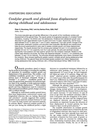
  • 2. 438 Buschang and Santos-Pinto American Journal of Orthodontics and Dentofacial Orthopedics April 1998 ~ o- .......... -4 ~. ........... 9 I J J I J J I I 6 7 8 9 10 11 12 13 Age (Yrs) * } Adolescent }Childhood I I 14 15 Fig. 1. Childhood and adolescent age ranges accord- ing to gender. Fig. 2. Horizontal (Hod, vertical (Vert), and total (Hyp) fossa displacement. surrogate for the condylion landmark (CO) remains questionable. 25 Considering these factors, the purpose of this study was to (1) describe the longitudinal growth changes of CO and AR, on the basis of stable cranial/cranial base and mandibular references structures; (2) evaluate age and gender differences in growth movements of the two landmarks; and (3) compare the growth changes of AR and CO. MATERIAL AND METHODS The data were derived from serial lateral cephalo- grams collected by the Human Growth Research Center, University of Montreal. They pertain to French Canadian children drawn from three school districts representing the different socioeconomic strata of the larger popula- tion.26Untreated longitudinal samples of 118 children (54 boys and 64 girls) and 155 adolescents (108 boys and 47 girls) were selected on the basis of available and suitable serial data. Childhood and adolescent growth phases were estab- lished on the basis of the estimated ages of peak adoles- cent velocity of the samples.27Adolescence was defined as Table I. Condylar growth (mm/4yrs) based on mandibular superimposition--age and gender differences Girl Boy Gender Measurement Period Mean SD Mean SD difference CO horizontal Childhood -1.3 2.3 -1.1 2.3 -0.2 Adolescence -1.3 2.8 -0.8 2.3 -0.5 Age difference 0.3 -0.7 CO vertical Childhood -9.4 1.9 -9.0 2.4 -0.4 Adolescence -9.1 2.8 -10.7 3.0 1.6:~ Age difference -0.8 1.8:~ CO total Childhood 9.8 1.7 9.6 2.0 0.2 Adolescence 9.6 2.6 11.0 3.0 - 1.47 Age difference 0.5 - 1.4:~ AR horizontal Childhood - 1.6 2.0 -1.8 2.4 0.2 Adolescence - 1.7 2.1 - 1.9 2.4 0.2 Age difference 0.5 -0.2 AR vertical Childhood -8.1 1.9 -7.8 2.6 0.3 Adolescence -8.2 3.0 -9.5 3.0 1.3t Age difference -0.3 1.7:~ AR total Childhood 8.6 1.7 8.5 2.1 0.1 Adolescence 8.7 2.7 10.0 2.8 -1.3t Age difference 0.4 - 1.55 tP < 0.01; :~p < 0.001. the period 3 years before and 1 year after peak velocity; childhood was defined as the period 4 years preceding adolescence with a 1-year overlap (Fig. 1). All cephalograms were traced and digitized by the same technician. The analyses described the growth of CO and AR. CO was defined as the point tangential to the most superior aspect of the condyle using a perpendicular to the ramal plane for orientation; AR was defined as the intersection of the posterior border of the ramus and the inferior cranial surface. The average of the right and left images was used to identify the landmarks. Technical reliability (method error) has been estimated at 90% (0.7 mm) and 92% (0.6 mm) for the horizontal and vertical aspects of CO, respectively; the horizontal and vertical reliabilities (method error) for AR were 97% (0.3 mm) and 94% (0.5 mm), respectively. Glenoid fossa displacement was described by the movements of CO and AR using cranial/cranial base superimpositions. Condylar growth was evaluated on the basis of mandibular superimpositions. The serial radio- graphs of the subjects were superimposed using natural reference structures. 28 The reliability of the mandibular superimpositions ranged between 94% and 99%; the reliability of cranial base superimpositions ranged be- tween 98% and 99%.29 All measures Were corrected for radiographic magnification. Horizontal and vertical growth changes of CO and AR were evaluated using Cartesian coordinates. A horizontal reference plane, defined by sella-nasion minus 7~ was used for orientation with registration on sella (Fig. 2). The horizontal and vertical reference planes were constructed on the first tracing of each series and transferred to
  • 3. American Journal of Orthodontics and Dentofacial Orthopedics Buschang and Santos-Pinto 439 Volume 113, No. 4 Table II. Fossa displacements (ram/4yrs) based on cranial/ cranial base superimposition--age and gender differences Gender Measurement Period differenc~ CO horizontal Childhood -0.3 Adolescence 0.0 Age difference CO vertical Childhood 0.2 Adolescence -0.5 Age difference CO total Childhood 0.3 Adolescence -0.8~ Age difference AR horizontal Childhood -0.2 Adolescence 0.2 Age difference AR vertical Childhood 0,1 Adolescence - 1.0~: Age difference AR total Childhood 0.0 Adolescence - 1.0:~ Age difference Gid B~ MeanlSD Mean SD -2.1 1.4 -1.8 1,1 -2.0 1.5 -2.1 1,8 -0.2 0.2 12 1.5 1.0 1.9 1.3 1.5 1.8 2,4 -0.1 -0.7 2.9 1.1 2.7 1,3 2.8 1,3 3.8 1.8 0.3 -0.3 -1.9 1.1 -1.8 1.1 -2.3 1.3 -2.5 1.3 0.3 0,65 2.5 1.0 2.4 1.4 2.2 1.1 3.2 1.4 0.4 -0.7f 3.3 1.0 3.3 1.0 3.4 1.3 4.3 1.4 0.3 -0.7~ tp < 0.01; ~:p < 0.001. subsequent tracing after superimposition. Total growth changes were computed as the hypotenuse of the horizon- tal and vertical movements of the landmark. All the variables were normally distributed. Paired t tests were used to compare age groups. Gender differ- ences were evaluated using t tests. Paired t tests also were used to compare the growth changes at CO and AR. RESULTS Condylar Growth (Table I) The total growth changes over the 4-year periods ranged between 9.6 and 11.0 mm and between 8.5 and 10.0 mm for CO and AR, respectively. Superior growth of the condyles was approximately 8 to 9 times greater than its posterior growth. A large proportion of subjects demonstrated more than 15 mm of superior growth over the 4-year periods. Approximately 30% to 40% of the sample showed anterior condylar growth; 20% to 30% showed anterior movements of AR. Boys showed significantly (p < 0.01) more total condylar growth than girls during adolescence, be- cause of greater superior growth. The AR values showed the same pattern of gender differences as the CO values. No gender differences were noted in the horizontal movements of CO or AR. Age group differences were found only among the boys, who displayed significantly more vertical growth (approx- imately 1.5 mm/4 years) during adolescence than childhood for both CO and AR. Co 13yrs Ar I Fe/l?ales.... 6yr~ -4 mm -2 tam,. Co~ ~ lS-m 1sin / L 14 14 12 12 10 t0 $ 4 2 0 Ma~-2z~ 0 -4 mm -2 0 Fig. 3. Condylar growth for girls aged 6 to 13 years and boys aged 8 to 15 years. Table III. Growthdifferences (CO minus AR) between condylion and articulate Measurement Period Girl Boy Difference SD Difference SD Condylar growth horizontal Childhood -0.3t 0.1 -0.7"~ 0.1 Adolescence -0.5~: 0.1 -1.0t 0.1 vertical Childhood 1.3t 0.1 1.2# 0.2 Adolescence 0.9t 0.2 1.1# 0.2 Total Childhood -1.3t 0.1 -1.1t 0.2 Adolescence -0.9t 0.2 -0.9"~ 0.2 Fossa displacement horizontal Childhood 0.2 0.1 -0.01 0.1 Adolescence -0.2 0.1 -0.4t 0.1 vertical Childhood 1.4t 0.1 1.3t 0.2 Adolescence 0.9# 0.2 1.3t 0.2 Total Childhood 0.5t 0.1 0.63" 0.1 Adolescence 0.55 0.1 0.93" 0.1 tp < 0.001; :~p < 0.01. Fossa Displacement (Table II) The CO and AR were displaced posteriorly and inferiorly approximately 3 to 4 mm over the 4-year periods. Posterior movements of the CO were twice as great as its inferior movements. It was not uncommon for adolescents to show more than 5 mm of posterior fossa displacement. Fewer than 10% of the samples showed anterior fossa displacement and 20% to 30% showed superior displacements. Total adolescent movements for CO were signif- icantly (p < 0.001) greater in boys than in girls, because of greater inferior fossa displacement. No significant gender differences were noted in either horizontal or vertical movements of the fossa. AR
  • 4. 440 Buschang and Santos-Pinto AmericanJournalof Orthodontics and DentofacialOrthopedics April 1998 -5 mm -4 -3 -2 -1 0 ; .... ; .... ; .... ; .... I ' ''~ Co Ar Female$ -5 =am -4 -3 -2 -1 0 I .... ; .... ; .... ; .... ;,,~, -1 r~ -2 -3 Co -5 Malesmm Ar 0 0 -1 -2 -3 -4 -5 mm Fig. 4. Fossa displacements for girls aged 6 to 13 years and boys aged 8 to 15 years. showed significantly greater inferior displacement in boys than in girls during adolescence. With the exception of the AR values for boys, which showed consistently greater posterior and inferior move- ments during adolescence than childhood, no signif- icant age group differences were noted in fossa displacement. Condylion Versus Articulare (Table III) Descriptions of condylar growth on the basis of the amount and direction of movement for CO and AR were significantly different (Fig. 3). Total growth of CO was significantly (0.9 to 1.3 mm) greater than the total growth at AR, because of differences in vertical growth. AR showed signifi- cantly greater (0.3 to 1.0 mm) posterior movements than CO (Fig. 3). Fossa displacements on the basis of the AR and CO also were significantly different (Fig. 4). Total movements over the 4-year period were 0.5 to 0.9 mm greater for AR than for CO. The differences were primarily in the vertical plane, with AR dem- onstrating 0.9 to 1.4 mm more inferior movement than the CO (Fig. 4). DISCUSSION One of the most significant findings was that posterior fossa displacement was almost twice as great as posterior condylar growth (Fig. 5). The predomi- nance of fossa displacement over condylar growth occurred in more than 61% of the children and 65% of the adolescents. If fossa displacement is greater than condylar growth, posterior displacement of the chin might be expected. Because this does not normally occur, it is theoretically possible that true mandibular rotation~s'3~plays a more fundamental role in deter- mining the anterior-posterior position of the chin than does condylar growth (Fig. 5). The only other study to evaluate both condylar growth and fossa displacement reported 1.5 mm posterior condylar growth and 1.2 mm posterior fossa displacement for 50 control subjects with Class II malocclusions aged between 8.4 and 10.6 years.15 The untreated subjects with Class II malocclusions in that study had greater posterior and less vertical condylar growth than expected from the results of the current study, which implies that at least some of the subjects with Class II malocclusions had condy- lar growth problems associated with larger gonial angles, more posteriorly directed ramal planes, and shorter ramal heights. The results of the current study are particularly relevant for patients with Class II skeletal malocclu- sion (Fig. 6), who have been reported to show greater than expected posterior fossa displace- ments. 14'16'23mWhen the observed patterns of vari- ation are considered, approximately 16% of our adolescent subjects might be expected to show more than 4 mm posterior fossa displacement. Therefore when planning skeletal corrections, posterior fossa displacements must be added to any existing anterior- posterior discrepancies and future growth deficiencies. The observed changes in fossa position suggest that the existing literature may need to be reevalu- ated. Although animal studies report that glenoid fossa remodeling and relocation occur as an adap- tation to chronic anterior repositioning of the man- dible,32-34the available human studies remain equiv- ocal,5-8'1~perhaps because the results of the human studies are confounded by posterior fossa displace- ments. 9,14,~5The functional appliance literature may also need to be reinterpreted if posterior fossa displacement masks the therapeutic effects on the condyles. The results also help resolve controversies con- cerning age changes in condylar growth. For boys,
  • 5. American Journal of Orthodontics and Dentofacial Orthopedics Buschang and Santos-Pinto 441 Volume 113,No. 4 Childhood Adolescence Childhood Adolescence -2.5 -2 I I L__ ~ Males --~ Females I__ ] T -1.5 -1 -0.5 0 mm / 4 years [3Condylar Growth 9 Fossa Displacement ] Fig. 5. Posterior condylar growth and fossa displacement. the results were consistent with longitudinal studies reporting greater mandibular growth rates during adolescence than childhood, as These differences re- sulted from vertical rather than horizontal growth. In contrast, no clear age effects were noted for girls. Baumrind et al.17reported relatively constant rates of condylar growth between ages 8.5 and 15.5 years for a sample of 31 subjects, most of whom were girls. Buschang and coworkers35also showed that rates of mandibular growth during the preadolescent and adolescent spurts were comparable for girls. The vertical condylar growth of our study subjects was approximately nine times greater than the hori- zontal condylar growth, as previously suggested.24The ramus heights showed greater absolute35-37and rela- tive27 size increases than the corpus length. This im- plies that previous growth studies38-4~conducted on the basis of mandibular length, for example, Co-Gn (gnathion), Co-Pg (pogonion), and Ar-Pg, were pri- marily describing vertical growth changes at the con- dyles. Because the efficacy of functional appliance therapy is commonly assessed on the basis of changes in mandibular length,71~ the growth vector most im- portant for a favorable response may not have been adequately addressed. Assuming a relationship be- tween growth potential and treatment response,41 a much greater vertical than horizontal treatment re- sponse might be expected. Sexual dimorphism was restricted to vertical condylar growth. Superior condylar growth and in- ferior fossa displacement were both greater in ado- lescent boys than girls. Previous studies describing condylar growth and fossa displacements have com- bined boys and girls,5,14-16making comparisons dif- Fig. 6. Pretreatment and posttreatment tracings of Class II high-pull headgear patient. Condylar growth was masked by fossa relocation with minimal anterior chin displacement. ficult. Greater vertical facial growth in boys than girls, however, has been reported37'4a-44and can be partially explained by the combined effects de- scribed above. Finally, the results showed that AR should not be used to describe either condylar growth or fossa displacement. On the basis of its proximity, the condyle serves as a better reference for the glenoid
  • 6. 442 Buschang and Santos-Pinto American Journal of Orthodontics and Dentofacial Orthopedics April 1998 fossa than does AR. Using AR rather than CO values will systematically overestimate inferior dis- placements (Fig. 4). Because of the inclination of the occipital bone where it intersects with the ramus to define AR, any posterior movements of the ramus are necessarily interpreted as posterior and inferior movements at AR. Similarly, AR underestimates vertical condylar growth and overestimates posterior condylar growth (Fig. 3). Such discrepancies will be most pronounced for individuals showing the great- est posterior displacement of the glenoid fossa. Because the glenoid fossa has been shown to be positioned more posteriorly in patients with Class II malocclusions than in patients with Class III maloc- clusions, 16we might expect growth studies using AR to systematically overestimate condylar growth in Class III patients and underestimate condylar growth in Class II patients. Studies using AR rather than CO to describe condylar growth or fossa dis- placement may also need to be reevaluated. 45 CONCLUSIONS 1. Posterior glenoid fossa displacement was almost twice as great as posterior condylar growth. 2. Vertical condylar growth was approximately nine times greater than posterior condylar growth. 3. Superior condylar growth and inferior fossa dis- placement were greater in adolescent boys than in girls. 4. Articulare (AR) systematically overestimates infe- rior fossa displacements, underestimates superior condylar growth, and overestimate posterior con- dylar growth. REFERENCES 1. Baume LJ. Principle of cephalometric development revealed by experimental biology. Am J Orthod 1961;47:881-901. 2. Moss ML. Functional analysis of human mandibular growth. J Prosthet Dent 1960;10:1151-9. 3. Moss ML, Rankow RM. The role of the functional matrix in mandibular growth. Angle Orthod 1968;38:95-103. 4. Koski KL. Cranial growth centers: facts or fallacies? Am J Orthod 1968;54:566-83. 5. Birkebaek L, Melsen B, Terp S. A laminagraphic study of the alterations in the temporo-mandibular joint followingactivator treatment. Eur J Orthod 1984;6:257-66. 6. Jakobsson SO. Cephalometric evaluation of treatment effect on Class II, division 1 malocclusion. Am J Orthod 1967;53:446-57. 7. Harvold EP, Vargervik K. Morphogenetic response to activator treatment. Am J Orthod 1971;60:478-90. 8. Mamandras AH, Allen LP. Mandibular response to orthodontic treatment with the bionator appliance. Am J Orthod Dentofac Orthop 1990;97:113-20. 9. Vargervik K, Harvold EP. Response to activator treatment in Class II malocclu- sions. Am J Orthod 1985;88:242-51. 10. Weislander L, LagerstrOm L. The effect of activator treatment on Class II malocclusion. AM J ORTHOD 1979;75:20-6. 11. Graber LW. Chin cup therapy for mandibular prognathism. Am J Orthod 1977;72:23-41. 12. Sakamoto T, Iwase I, Uka A, Nakamura S. A roentgenocephalometric study of skeletal changes during and after chin cup treatment. Am J Orthod 1984;85:341-50. 13. Thilander B. Chin cup treatment for Angle Class II malocclusion (a longitudinal study). Trans Europ Orthod Soc 1965:311-27. 14. Agronin KI, Kokich VG. Displacement of the glenoid fossa: a cephalometric evaluation of growth during treatment. Am J Orthod 1987;91:42-8. 15. Baumrind S, Korn EL, Issacson RJ, West EE, Molthen R. Superimpositional assessment of treatment-associated changes in the temporomandibular joint and the mandibular symphysis.Am J Orthod 1983;84:443-65. 16. Droel R, Isaacson RJ. Some relationships between the glenoid fossa position and various skeletal discrepancies. Am J Orthod 1972;61:64-78. 17. Baumrind S, Ben-Bassat Y, Korn EL, Bravo LA, Curry S. Mandibular remodeling measured on cephalograms. 1. Osseus changes relative to superimposition on metallic implants. Am J Orthod Dentofac Orthop 1992;102:134-42. 18. Bj6rk A. Variations in the growth of the human mandible. Longitudinal radio- graphic study by the implant method. J Dent Res 1963;42(suppl):400-11. 19. H~iggU, Attstrom K. Mandibular growth estimated by four cephalometric mea- surements. Am J Orthod Dentofae Orthop 1992;102:146-52. 20. Buschang PH, Tanguay R, Demirjian A, LaPalme L, Turkewicz J. Mathematical models of longitudinal growth for children with normal and untreated Class II division ] malocclusion. Eur J Orthod 1988;10:227-34. 21. Odegaard J. Mandibular rotation studied with the aid of metal implants. Am J Orthod 1970;58:448-54. 22. Odegaard J. Growth of the mandible studied with the aid of metal implants. Am J Orthod 1970;57:145-57. 23. Anderson O, Popovich F. Relation of cranial base flexure to cranial form and mandibular position. Am 1 Phys Anthropol 1983;61:181-7. 24. Bj6rk A. Cranial base development. Am J Orthod 1955;41:198-225. 25. Stickel A, Pancherz H. Can "articulate" be used in the cephalometric analysis of mandibular length? A methodologic study. Eur J Orthod 1988;10:362-8. 26. Demirjian A, Brault Dubuc M, Jenicek M. l~tude comparative de la croissance de l'enfaut canadien d'orige fran~kaish Montr6aL Can J Public Health 1971;62:111-9. 27. Buschang PH, Baume RM, Nass GG. A craniofacial growth maturity gradient for males and females between 4 and 16 years of age. Am J Phys Anthropol 1983;61:373-81. 28. Bj6rk A, Skieller V. Normal and abnormal growth of the mandible. A synthesis of longitudinal cephalometric implant studies over a period of 25 years. Eur J Orthod 1983;5:1-46. 29. Buschang PH, LaPalme L, Tanguay R, Demirjian A. The technical reliability of superimposition on cranial base and mandibular structures. Eur J Orthod 1986;8: 152-6. 30. Bj6rk A. Prediction of mandibular growth rotation. Am J Orthod 1969;55:585-99. 31. Hopkin GB, Houston WJB, James GA. The cranial base as an aetiological factor in malocclusion. Angle Orthod 1968;38:250-5. 32. Hintou RJ, McNamara Jr JA. Temporal bone adaptations in response to protrusive function in juvenile and young adult rhesus monkeys (Macaca mulatta). Eur J Orthod 1984;6:155-74. 33. St6cki PW, Willert HG. Tissue reactions in the temporomandibular joint resulting from anterior displacement of mandible in monkey. Am J Orthod 1971;60:142-55. 34. Woodside DG, Metaxas A, Altuna G. The influence of functional appliance therapy on glenoid fossa remodeling. Am J Orthod 1987;92:181-98. 35. Buschang PH, Tanguay R, LaPalme L, Demirjian A, Goldstein H. Sexual dimor- phism in mandibular growth of French-Canadian children 6-10 years of age. Am J Phys Anthropol 1986;71:33-7. 36. Nanda RS. The rates of growth of several facial components measured from serial cephalometric roentgenograms. Am J Orthod 1955;41:658-73. 37. Riolo ML, Moyers RE, McNamara, JA, Hunter WS. An Atlas of Craniofacial Growth. Craniofacial Growth Series, Vol 2, Center of Human Growth and Development. Ann Arbor: University of Michigan. 1974. 38. Bishara SE, Jamison JE, Peterson LC, DeKock WH. Longitudinal changes in standing height and mandibular parameters between the ages of 8 and 17 years. Am J Orthod 1981;80:115-35. 39. Tracy WE, Savara BS. Norms of size and annual increments of five anatomical measures of the mandible in girls from 3 to 16 years of age. Arch Oral Biol 1966;11:587-98. 40. Ursi WJS, Trotman C, McNamara JA, Behrents RG. Sexual dimurphism in normal craniofacial growth. Angle Orthod 1993;63:47-55. 41. Petrovic A, Stutzmann J, Lavergne J. Mechanisms of eraniofacial growth and modus operandi of functional appliances: a cell-level and cybernetic approach to orthodontic decision making. In: Carlson DS, ed. Craniufacial growth theory and orthodontic treatment. Craniofacial Growth Series, Vol 23, Center of Human Growth and Development. Ann Arbor: University of Michigan, 1990. 42. Coben, SE. The integration of facial skeletal variants. Am J Orthod 1955;41:407-34. 43. Maj G, Luzi C. Longitudinal study of mandibular growth between 9 and 13 years as a basis of an attempt of its prediction. Angle Orthod 1964;3:220-30. 44. Prahl-Anderson B, Kowalski CJ. Sexual dimorphism in dentofacial dimensions of Dutch children in the Nymegen growth study. Rev Beige Med Dent 1976;31:371-6. 45. Williams S, Melsen B. Condylar development and mandibular rotation and displacement during activator treatment. An implant study. Am J Orthod 1982;81: 322-6.