SlideShare a Scribd company logo
1 of 10
Download to read offline
Mandibular condyle position: Comparison
articulator mountings and magnetic
resonance imaging
of
Steven R. Alexander, DDS, MS," Robert N. Moore, DDS, PhD, EdD,band
Linda M. DuBois, DDS, PhDc
Olympia, Wash., and Lincoln, Neb.
This study evaluated the reliability of jaw positions, the existence of distinct jaw positions, and
condyle-disk-fossa relationships in a symptom-free population by using articulator mountings and
magnetic resonance imaging (MRI). The subjects examined included 28 men, 22 to 35 years of age,
all having Angle Class I molar relationships and no discernible TMJ dysfunction. Records taken
included the following: an axiographic face-bow to locate retruded hinge axis position, an
interocclusal registration of retruded position (RE), a series of inter0cclusal registrations for centric
occlusion (CO), a leaf gauge-generated centric relation (CR), a series of interocclusal registrations
for CR, and MRI. The mandibular position indicator of the SAM articulator (Great Lakes
Orthodontics, Ltd., Tonawanda, N.Y.) was used to determine reliability and existence of distinct jaw
positions. Magnetic resonance imaging also evaluated jaw positions and anatomic relationships. The
results indicate: (1) The articulator analysis of CO and CR is statistically replicable. (2) A distinct jaw
position could be demonstrated for CO that was separate from RE and CR. It was not possible to
discriminate between RE and CR. (3) Condylar concentricity was observed in half of the sample and
remained consistent in RE, GO, and CR. (4) Of the sample 13% demonstrated anteriorly displaced
disks that were not influenced by posterior condyle placement. (5) The clinical concept of treating to
CR as a preventive measure to improve disk-to-condyle relationships was not supported by this
study. (AMJ ORTHODDENTOFACORTHOP1993;104:230-9.)
One of the goals of orthodontic treatment is
the establishment of a harmony between occlusal func-
tion and the temporomandibularjoint (TMJ).~ However,
conflicting claims are being made about occlusion and
the role of orthodontics in this relationship. Many in-
vestigators think occlusion plays a primary role,15 and
some evidence suggests that orthodontic correction of
malocclusion can have a positive influence on TMJ
health.6s Conversely, other studies contend that ortho-
dontic therapy has a potential negative influence by
contributing to symptoms of temporomandibular dys-
function (TMD),2.9"13especially if it creates a discrep-
ancy between centric occlusion (CO) and centric rela-
tion (CR). 14"16 Still other findings indicate that
occlusion~7a8 or orthodontics has no correlation with
Fromthe UniversityofNebraskaMedicalCenterCollegeof Dentistry.Lincoln,
Nebraska.
This studywas supportedby theUniversityof NebraskaMedicalCenterOrtho-
dontic DevelopmentFund.
*Private Practice, Olympia, Wash.
bProfessorand Chairman, Departmentof Orthodontics,
CAssociateProfessor,Departmentof Adult RestorativeDentistry.
Copyright 9 1993by the AmericanAssociationof Orthodontists.
0889-5406/93/$1.00 + 0.10 8/1/36228
230
the incidence of TMD. ~921Since TMD most likely has
a multifactorial origin, completely discounting the role
of occlusion may be an inappropriate interpretation of
published data.2224
Condylar position is a major issue in studies of the
TMJ.25"26Occlusal disharmonies from preexisting mal-
occlusions or orthodontic treatment have been reported
to create an unphysiologie mandibular position~6"2728
that results in muscle hyperactivity,29,3~internal de-
rangements,3"27"2s'3~and pain dysfunction.2'~5
Many clinicians consider that an ideal oeclusal re-
lationship should be closely correlated to an ideal con-
dyle-disk-fossa relationship, which they refer to as CR.
To assess both condylar and occlusal relationships,
the use of articulator-mounted models in centric relation
is advocated for diagnosis and treatment plan-
ning.2a4'29"3235 Theoretically, this technique allows
placement of the mandible in a more physiologic po-
sition with fewer occlusal-generated condylar deflec-
tions from centric relation. The purpose of this study
is to determine the reliability of a commonly used CR
articulator mounting techniqiae and then to determine
the placement of the condyle in relation to the fossa
and the disk with magnetic resonance imaging (MRI).
American Journal of Orthodontics and Dentofacial Orthopedics Alexander, Moore, and DuBois 231
Volume 104,No. 3
Although the definition of CR and its location re-
mains controversial, 15'36-38 the definition of CR has
changed from a posterior39to an anterior and superior~~
position in the fossa. This position is considered to be
the most reliable reference point for accurately record-
ing the relationship Of the mandible to the maxilla.4''4z
As CR was accepted into other areas of dentistry, CR--
the reference point became CR--the universal treat-
ment position. However, to be meaningfully defined,
9CR must show uniqueness of position among other con-
dylar relationships. It must be reproducible, and the
imposition of the disk must be determined. 25.3s,4~
To better understand condyle and fossa positional
relationships as reflected on articulators, the visualiza-
tion of the TMJ is necessary. In contrast to transcranial
radiographs and tomography, which are unable to reveal
the relationship of the disk to the condyle and
fossa, 31,37,43,44 MRI can image the osseous, muscular,
and fibrous components of the TMJ wthout ionizing
radiationY
MATERIALS AND METHODS
Subject selection
By Using the following criteria, 28 men between the ages
of 23 to 34 years were selected to participate in this study:
(1) full permanent dentition, except third molars, in an Angle
Class I occlusion; and (2) no signs or symptoms of TMD as
defined by a normal range of motion, no mandibular deviation
on opening or closing, no history of TMD as established by
a TMJ health history, and no pain in the prearticular and
postarticular and temporal areas on palpation. Clicking in the
absence of other symptoms did not exclude a subject from
the study since joint sounds are common and may be caused
by conditions other than internal derangements?"6
Records
To standardize technique, all records were taken by the
same investigator. In addition to intraoral photographs and
diagnostic plaster casts, each subject had another set of casts
mounted on a SAM articulator with an axiograph face-bow.
The hinge-axis position was established by manipulating the
mandible to its most retruded position. The mandibular model
was oriented with a fiberglass framework, zinc oxide and
eugenol interocclusal record. This procedure was followed
by three consecutive interocclusal registrations of CO (ha-
bitual bite or maximum intercuspation) and CR (leaf gauge
technique of Williamson".33) with the fiberglass framework
system. The leaf gauge is reported to allow the musculature
of the subject to seat the condyles more superiorly on the
posterior Slope of the articulator eminence and to eliminate
the operator-induced variability from manipulation.14"25The
series of CO and CR records were repeated two more times
with a 3- to 7~.dayinterval between each appointment.
The mandibular position indicator (MPI) of the SAM
articulator was used to determine the reproducibility of CR.
This instrument compares the position of the maxilla with the
mandible and records any differences in the horizontal (X),
transverse (Y), and vertical (Z) positions of the mandible.
Horizontal and vertical dimensions are read in 0.5 mm in-
crements, whereas transverse displacement of the mandible
is measured in 0.1 nun increments from a gauge on the MPI.
Thus the MPI can measure replicability and positional dif-
ferences of CR and CO within the described accuracy of this
instrument.
Acrylic interocclusal registrations of the most clinically
reproducible positions of CO and CR for each patient were
constructed. A third acrylic registration was constructed for
each patient from the rctruded position (RE). These registra-
tions were used to transfer interocclusal relationships in a
given jaw position from the articulated casts to the subject
for MRI recordings. Since the imaging of the joint must be
done in a supine position, the acrylic registration insured
proper mandibular positioning.
Magnetic resonance imaging of the right and left tem-
poromandibular joints was performed in CO, CR, and hinge
axis positions with a General Electric signa system (GE, Mil-
waukee, Wis.). The subject's head was orientated with cross
laser reference lines and supported with surface coils and
styrofoam packing during the imaging.
To measure the movement between the threejaw positions
in the sagittal plane (X and Z positions), a 0.003-inch matte
acetate superimposition was created by tracing reproducible
structures from the image of the hinge axis that would su-
perimpose on the images of CO and CR. The condyle was
also traced from the hinge axis image by using the external
or internal margins of cortical bone. To establish reference
points for measurements, pin holes were made through the
acetate and the image along the glenoid fossa (three holes)
and the condylar head (four holes). The acetate was then
transferred to the CO and CR images and was superimposed
on the stable structures. The CO and CR images were marked
with pin holes through the established holes of the acetate.
The acetate was then superimposed on the condyle where the
same process was repeated.
Once the reference points were transferred, the acetate
could then be realigned on the stable structures of each image
by matching the pin holes of the acetate and the image along
the glcnoid fossa. The condylar position was then marked on
the acetate by locating the condylar pin holes with a tracing
pen. Three different pen colors were used to allow distinction
between the condylar movement of the three jaw positions.
By using the hinge axis as a reference point, measurements
in the X and Z axes were made. The images were also sub-
jectively evaluated by two independent examiners to analyze
the concentricity of the mandibular condyle to the fossa and
the disk relationship to the condyle. Finally, data from the
MPI articulatoranalysis were correlatedwith the data obtained
from the MRI.
Statistical analysis
The reliability among measurements taken at time 1 (T,)
was calculated with correlation coefficients. The values for
each dependent variable that were recorded at a single sitting
were averaged such that each patient had one value for
232 Alexander, Moore, and DuBois AmericanJournalofOrthodonticsandDentofacialOrthopedics
September1993
Table I. MPI means and standard deviations (millimeters) for retruded, centric occlusion, and centric
relations (n = 28)
Plane Statistic Retruded Tt
Transverse (Y) X 0.70 0.02
SD 1.39 0.80
Left vertical (Z) X - 0.02 0.24
SD 0.37 0.44
Left horizontal X - 0.07 0.26
(X) SD 0.40 0.36
Right vertical X 0.21 0.42
(Z) SD 0.42 0.44
Right horizontal X 0.00 0.10
(X) SD 0.38 0.29
Centric occlusion Centric relation
0.12 0.02 0.19 0.27 0.41
0.73 0.78 0.95 0.95 1.06
0.23 0.14 -0.06 --0.09 --0.13
0.42 0.45 0.49 0.45 0.52
0.29 0.29 0.12 0.14 0.11
0.41 0.36 0.56 0.45 0.60
0.30 0.-30 0.05 0.01 -0.01
0.45 0.44 0.42 0.38 0.47
0.14 0.11 -0.04 -0.02 0.04
0.32 0.28 0.39 0.39 0.44
Transverse plane at Tt: RE > CO (p < 0.05).
Vertical plane at TI: Left condyle, CO > CR and CO > RE 07 < 0.05); fight condyle,
Horizontal plane at T~: Left condyle, CO > RE and CO > CR 07 < 0.05).
07< 0.05).
Table II. Alpha levels of reliability analysis among the time periods
Jaw l Transverse Left vertical l Left horizontal
position (Y) (Z) (X)
CO' 0.87 0.95 " 0.88
CRb 0.93 0.90 0.92
Right vertical ] Right horizontal
(z) ] ix)
0.94 0.93
0.92 0.92
'Centrie occlusion measurement at all three time periods.
bCentrie relation measurement at all three time periods.
each dependent variable and the patient was the unit of mea-
surement.
Descriptive statistics were calculated for continuous vari-
ables and percentages or frequencies were calculated for cat-
egorical variables and ordinal data. Repeated measures anal-
ysis of variance with Tukey foll0w-ui~sWereused to compare
RE, CO, and CR atT~ for MPI andMRI data for each plane
and each side.
The validity of the MPI and the MRI was addressed by
comparing data from these techniques with correlational co-
efficients. The Spearman's correlation coefficient was used
to measure the reliability between evaluators of concentricity
of the Condyles. In the following analysis, only instances in
which agreement occurred were used.
9With the Friedman test, RE, CO,and CR were compared
as to percent of superior condyle positioning, condyle con~
centricity, and anterior disk placement.
RESULTS
The results of the SAM articulator MPI records are
presented in Table I. The means and standard deviations
are listed for the three jaw positions, as recorded in the
transverse (Y), vertical (Z), and horizontal (X) planes
for the left and right TMJs. The Y is established at the
left condyle with larger numbers indicating mandibular
movement to the left. Table II is a reliability analysis
of CO and CR at time 1 (TO, time 2 (T2), and time 3
(T3). Results revealed an alpha level of sufficient con-
sistency between scores to confirm the reliability of T~
and to base all other analyses from the T~ results. Anal-
ysis of RE, CO, and CR at T~ for the Y, Z, and X
planes (Table I)indicated differences between the fol-
lowing jaw positions: in the Y axis RE > CO, in the
Z axis CO > CR and CO > RE for the left condyle
and CO > CR for the fight condyle, and in the X axis
CO > RE and CO > CR for the left condyle.
The means and standard deviations as established
from the MRI for the sagittal planes can be seen in
Figs. 1 and 2. Retruded position was used as a reference
position for CO and CR measurements and therefore
was not included in the graph. Significant differences
were shown between the following jaw positions: in
the Z axis CO > RE for the left condyle (Fig. 1) and
in the X axis CO > CR for the fight condyle (Fig. 2)~
Validity as documented by correlation coefficients
showed a weak relationship between theMRI and the
articulator, with significance only between Z right for
CO (r = 0.37) and X right for CO (r = 0.55) at
p < 0.05 (Table III). The MPI data was adjusted tO RE
for correlation with the MRI data.
The superior-most condylar position can be evalu-
American Journal of Orthodontics and Dentofacial Orthopedics Alexander, Moore, and DuBois 233
Volume 104.No. 3
Retruded Position Used as a Reference Point
Verical Plane (Z)
0.6
0.4
'~ 0.2
'~ (0.21
"~ (0.4)
r=
r / ,-i ,-~,/ ! / zi 9r
Centric Occlusion
Jaw Position
I~ght Side N = 28, Left Side N = 26 [] Left [] Right
Left Condyle CO > RE (P<.05)
Fig. 1. MRI comparison of jaw relationships in vertical plane.
l, I
Centric Relation
Retruded Position Used as a Reference Point
Horizontal Plane (X)
0.6
0.4
0.2
•(0.2)
1o.4)
t~ (0.6) I
Centric Occlusion
1~'9hr Side N -~ 28, Left Side N = 26
Right Condyle CO > CR (P<.05)
Jaw Position
[] Left [] Right
Centric Relation
Fig. 2. MRI comparison of jaw relationships in horizontal plane.
ated by comparing condylar movement in the Z axis.
To confirm the Z axis spacial relationship (Fig. I), the
distance from the most concave portion of the fossa to
the most convex surface of the condyle was measured.
Any of the three jaw positions can share the superior
position if no measurable differences can be found be-
tween their vertical positioning. The percentages of su-
perior condylar positions for right and left TMJs is
234 Alexander, Moore, and DuBois AmericanJournalofOrthodonticsandDentofacialOrthopedics
September1993
.~ 80
o.
,~ 20


~N
~N
~XNNXNNX~
"//y/////~N~
7Y/Y////Z .........
Retruded Centric Occlusion
Jaw Position
[] Left [] Right
Right Side N = 28, Left Side N = 26
Fig. 3. Superior condylar position.
,~,',t
Y////////. .......-.........,
7//////A""
Y////////.
/fffffJf/'~
~////////,Fffjf/'/'/',t'.,
;r162162162162162162162
"I///////Z
Centric Relation
Table III.Validity coefficient between MRI and articulator
I Left vertical l Lefth~176 l Right(X) (Z) Righthorizontal
(x)
CO 0.13 -0.18 0.37* 0.55*
CR 0.15 0.04 0.25 0.28
*p < 0.05.
Table IV. Reliability coefficients between two examiners for concentricity and disk placement~
Left Right
RE CR REI co I CO [ CR
Concentricity 0.93 0.92 0.94 0.86 0.91 0.94
Disk placement 1.00 1.00 1,00 0.89 1.00 0.79
'All values were significant (P < 0.001).
presented in Fig. 3. The Friedman test shows there is
no significantdifference between the threejaw positions
with this evaluation.
Two independent clinicians completed a subjective
evaluation of concentricity and disk placement. The
reliability analysis between the two examiners showed
a very high degree of correlation (Table IV). Figs. 4
and 5 show the distribution of condylar position (an-
terior, positive; posterior, negative) in relation to the
three jaw positions. Fig. 6 shows the percent of anterior
disk placement as it relates to the three jaw positions.
A normal disk-to-condyle relationship is shown in Fig.
7, and an anteriorly displaced disk is shown in Fig. 8.
No significant difference existed between the three jaw
positions for concentricity or anterior disk placement.
DISCUSSION
Reliability of jaw positions
The two important jaw relationships examined for
reliability were CO and CR: Retruded (RE) was in-
cluded in the study as a reference for CR because of
the implication found in the old and the new definitions
Percent
50
40
30
20
10
N= 26
-1.0 -.5 0 +.5
//-/#
F//s
r.//.~
,~/j-#
r//#
F//.e
.//.e
///s
F//#
r//..
xi/#
x.//.~
///j
r/-/#
//#'~
r//.e
I/-/-v
1//s
F//s
i///
i//J
AmericanJournalof OrthodonticsandDentofacialOrthopedics Alexander, Moore, and DuBois 235
Volume104, No. 3
+1.0
Concentricity
[] Retruded [] Centric Occlusion [] Centric Relation
60
Fig. 4. Concentricity of left condyle.
N= 28
5O
40
10
0
-1.0 -.5 0 +.5
Concentricity
[] Retruded [] Centric Occlusion [] Centric Relation
Fig. 5. Concentricityof rightcondyle.
+1.0
of CR that these positions should be different from each
other. The results showed CO and CR to be reliable
among tests.
Positional relationships among RE, CO, and CR
Positional relationships were analyzed by transverse
(Y), vertical (Z), and horizontal (X) planes with the
MPI. The sagittal (X and Z) plane was further analyzed
by MRI. This procedure allowed a correlation of data
between MPI and MRI in the sagittal plane.
The transverse (Y) plane recorded positive numbers
for means showing jaw positions slightly to the left of
the MPI center (Table I). This deviation from zero is
not considered significant since manufacturers state that
measurements of 0.4 mm or less are not accurate. A
greater variation existed in the transverse plane than
236 Alexander,Moore, and DuBois American Journal of Orthodontics and Dentofacial Orthopedics
September1993
20
15
5

~XN~X
N%N%NXN%
NNXN~NN~N%
NNNNN%NNN
NNXNNN%NN
NNRetruded Centric Occlusion
Jaw Postion
[] Left [] Right
Centric Related
Right SJde N = 28, Left Side N = 26
Fig. 6. Percent anterior disk placement of TMJ.
Fig. 7. MRI of TMJ. Condyle (C) placed posteriorly in fossa (F) with normal disk (D) to condyle
relationship.
was found in the sagittal planes. Such variation could
be explained by the osseous asymmetry between right
and left halves of the face and the cranium. This vari-
ation then cannot be directly compared with the vari-
ation found in the sagittal plane. The MPI data estab-
lished that a significant difference existed between the
jaw positions of RE and CO.
The MPI and MRI data were used to evaluate dis-
American Journal of Orthodontics and Dentofacial Orthopedics Alexander, Moore, and DuBois 237
Volume 104, No. 3
Fig. 8. MRI of TMJ. Condyle (C) concentric in fossa (F) withanterior disk (D) to condyle relationship.
tinct jaw positions in the sagittal plane. The baseline
or 0 value for the MPI means and standard deviations
was established from the axiographic location of the
hinge axis position. Therefore it would be expected that
interocclusal registrations of RE should be close to the
0 value and that CO and CR would be further from the
baseline if their positions were significantly different.
The variability, as reflected by the standard deviations,
shows approximately -+0.5 mm or less from the mean
for MPI and also MRI values (Table I, Figs. 1 and 2).
This range is satisfactory since measurements less than
0.5 mm were not discernible. These data suggests the
existence of distinct jaw positions between CO and
either RE or CR.
In the vertical (Z) plane CR is often described as
the most superior position.Z5 Evaluation of the MPI data
revealed that significant differences between jaw po-
sitions only existed between CO as compared with RE
and CR for the left condyle, and CO as compared with
CR for the fight condyle (Table I). In all relationships,
CO was positioned inferiorly to RE and CR. Evaluation
of MRI data confirmed that CO was inferior in position
when compared with RE and CR, but significance was
only found between RE and CO for the left condyle
(Fig. 1). The MRI data also illustrated the relationship
of the condylar position in RE when referenced to the
glenoid fossa (Fig. 3). Collectively considered, the data
support the existence of a distinct jaw position for CO
inferior to RE and CR.
In the horizontal (X) plane, the definitions and ar-
ticles previously mentioned would support a CR posi-
tion anterior to RE.~5.36 Evaluation of the MPI data
revealed CO to be positioned anteriorly to RE and CR
on both the left and right sides. However, statistical
significance was only found on the left side between
CO and RE and between CO and CR (Table I). The
MRI data also revealed that CO was positioned ante-
riorly to RE and CR but statistical significance was only
found on the right side between CO and CR (Fig. 2).
A collective evaluation of the data would suggest that
CO was positioned anteriorly to RE and CR, but the
MPI data failed to correlate statistically with the MRI
data. These differences might be explained by the lack
of a sharp demarcation of cortical bone'on the MRI
image that reduced the accuracy of the measurements.
In addition, small measurements and large variations
238 Alexander, Moore, and DuBois AmericanJournalof Orthodonticsand DentofacialOrthopedics
September1993
potentially create small changes in interpretation of
MPI or MRI data that could greatly change statistical
significance.
Combining vertical and horizontal axes of the sag-
ittal plane allows further correlation of the data. The
data of the present study provide evidence that CO is
a distinct jaw position, separate from RE and CR. This
finding is substantiated by a condylar position inferior
and anterior to RE and CR. The data do not support
distinct condylar positions for RE and CR and do not
suggest that CO and CR are coincident. The latter is
in contrast to what was previously described as the ideal
relationship for a healthy TMJ. 29,32,35Possible reasons
for these observations might be that a separation of RE
and CR fails to exist, and CO and CR are not found
coincidentwithin the normal population. Conversely,
RE and CR could be distinct jaw positions, but the leaf
gauge does not establish CR, thus preventing coinci-
dence with CO in the normal population.
Condyle to fosse relationship
The issue of diagnosis and treatment to different
condylar relationships often focuses on the issue of
concentricity. The present study shows that most per-
sons within this population are concentric (Figs. 4 and
5). The distribution pattern of all three jaw positions
supports the concept that a concentrically placed con-
dyle represents the most common position in defining
what is normal. It is important to note, that although
this entire sample was selected from a normal popu-
lation, approximately half of the condyles were not
defined as being concentric. Further evaluation of the
data revealed that approximately half of the patients did
not change their condylar position in reference to the
fossa for RE, CO, or CR. In other words, if the patient
was concentric, anteriorly or posteriorly placed, they
maintained that same classification for all three jaw
relationships, Therefore diagnosis of the health of the
TMJ on the basis of concentricity is not supported by
the present data. Furthermore, changing the concentric,
anterior, or posterior through manipulation status may
not be possible in many cases.
Disk-to-condyle relationship
One of the most common problems affecting the
TMJ is internal derangement, which includes the ab-
normal relationship of the articular disk relative to the
mandibular condyle, the fossa, and the articular emi-
nence. Of the 54 joints examined with MRI, 7 (13%)
were diagnosed as having anterior disk placement. Al-
though this result is less than the 32% reported by
Kircos,47 it does confirm the presence of anterior disk
placement in a symptom-free population.
In half of the anteriorly displaced disks observed in
the present study, the condyle was posteriorly posi-
tioned. In the other half, the condyle was concentric or
anteriorly positioned. This is in contrast to a previous
report that indicated an anteriorly displaceddisk was
associated with a posterior condylar position (Figs. 7
and 8).4s
Two of the seven subjects with anteriorly displaced
disks had joint sounds, and five subjects with normal
disk relationships had joint sounds. This finding sup-
ports previous reports that, in the absence of other
symptoms, joint sounds cannot be considered sufficient
evidence of dysfunction, n
CONCLUSIONS
1. The articulator analysis of CO and CR is statis-
ticaUy replicable.
2. Condylar concentricity was observed in half of
the sample and remained consistent in RE, CO,
and CR.
3. Of the sample 13% demonstrated anteriorly dis-
placed disks that were not influenced by poste-
rior condyle placement.
4. The clinical concept of treating to CR as a pre-
ventive measure to improve disk-to-condyle re-
lationships was not supported by this study.
REFERENCES
I. Perry HI'. Mandibularfunction:an orthodonticresponsibility.
AMJ ORTHOD1975;67:316-23.
2. RothRH. Tcmporomandibularpain-dysfunctionandocclusalre-
latinnships. AngleOrthod 1973;43:136-53.
3. ThompsonJR. Abnormalfunctionof the temporomandibular
joints and relatedmusculature:orthodonticimplications,partI.
AngleOrthod 1986;56:143-63.
4. MorawaAP, Loos PJ, Easton JW. Temporomandibularjoint
dysfunction in children and adolescents:incidence,diagnosis,
and treatment.QulntessenceInt 1985;11:771-7.
5. McLaughlinRP. Malocclusionandthetemporomandibularjoint:
an historicalperspective.AngleOrthod 1988;58:185-91.
6. Myers DR, Ba.renicJ-l',Bell RA, WilliamsonEH. Condylar
positionin chiIdrenwithfunctionalposteriorcrossbites:before
and aftercrossbitecorrection.PediatrDent 1980;2:190-4.
"7. OwcnAH.The maxillarysagittalappliance:aclinicalstudy. AM
J ORTItODDENTOFACORTHOP1987;91:271-85.
8. SadowskyC, BeGoleEA. Long-termstatusof temporomandib-
ular joint function and functionalocclusion afterorthodontic
treatment. AMJ ORTHOD1980;78:201-12.
9. LoftGH, ReynoldsJM, ZwemerJD, ThompsonWO, Dushku
J. The occurrenceof craniomandibulatsymptomsin healthy
youngadultswithand withoutpriororthodontictreatment.AM
J ORTHODDENTOFACORTHOP1989;96:264-5.
10. ThompsonJR. Abnormalfunctionof the temporomandibular
jointsand relatedmusculature:orthodonticimplications,partII.
AngleOrthod 1986;56:181-95.
11. PullingerA, MonteiroA, Liu S. Etiologicalfactorsassociated
with temporomandibulardisorders. [Abstrctno. 848]. J Dent
Res 1985;64:269.
AmericanJournalof Orthodonticsand DentofacialOrthopedics Alexander, Moore, and DuBois 239
Volume 104,No. 3
12. Pullinger A, Seligman D, Solberg W, Liu S. Relationship of
occlusal morphology to TM disorders in young adults. [Abstract
no. 849]. J Dent Res 1985;64:269.
13. Drace JE. Natural history of minimal anterior displacements of
the temporomandibular joint meniscus. [Abstract]. Radiol Soc
North Am 1988.
14. Williamson EH. Occlusion and TMJ dysfunction. J Clin Orthod
1981;15:333-50.
15. Dawson PE. Centric relation: its effect on occluso-muscle har-
mony. Dent Clin North Am 1979;23:169-80.
16. McNeill C. The optimum temporomandibularjoint condyle po-
sition in clinical practice, lnt J Periodontic Restorative Dent
1985;6:53-76.
17. Egermark-Eriksson I, Carlsson GE, Magnusson T. A long-term
epidemiologie study of the relationship between occlusal factors
and mandibular dysfunction in children and adolescents. J Dent
Res 1987;66:67-71.
18. Parker MW. A dynamic model of etiology in temporomandibular
disorders. J Am Dent Assoc 1990;120:283-90.
19. Sadowsky C, Poison AM. Temporomandibular disorders and
functional occlusion after orthodontic treatment: results of two
long-term studies. AM J ORrr~OD1984;86:386-90.
20. Greene CS. Orthodontics and temporomandibular disorders.
Dent Clin North Am 1988;32:529-38.
21. American Academy of Pediatric Dentistry. Treatment of tem-
poromandibular disorders in children: summary statements and
recommendations. J Am Dent Assoc 1990;120;265-9.
22. Mohlin B. Prevalence of mandibular dysfunction and relation
between malocclusion and mandibular dysfunction in a group of
women in Sweden. Eur J Orthod 1983;4:115-23.
23. Mohlin B, Ingervall B, Thilander B. Relation between maloc-
clusion and mandibular dysfunction in Swedish men. Eur J Or-
thod 1980;2:229-38.
24. Perry HT. Relationofocclusiontotemporomandibularjointdys-
function: the orthodontic viewpoint. J Am Dent Assoc
1969;79: i37-4I.
25. Gilboe DB. Centric relation as the treatment position. J Prosthet
Dent 1983;50:685-9.
26. Mongini F, Schmid W. Assessment of the therapeutic position
for orthodontic diagnosis and treatment. AM J ORTHOD
1982;82:513-8.
27. Reuling N. Comparative study of clinical examination, occlusal
analysis and new radiological imagingprocedures in patients with
functional TMJ disorders. J Oral Rehabil 1987;14:165-74.
28. Wyatt WE. Preventingadverse effects on the temporomandibular
joint through orthodontic treatment. A~,t J ORTHODDENTOFAC
OR'mOP 1987;91:493-9.
29. Williamson EH. The role of craniomandibular dysfunction in
orthodontic diagnosis and treatment planning. Dent Clin North
Am 1983;27:541-60.
30. Kirveskari P, Alanen P, Jamsa T. Association between cranio-
mandibular disorders and occlusal interferences. J Prosthet Dent
1989;62:66-9.
31. Owen AH. Orthodontic~orthopedic therapy for cranioman-
dibular pain dysfunction: Part A. Anterior disk displacement,
review of literature. J Craniomandib Pract 1987;5:357-66.
32. Roth RH. Functional occlusion for the orthodontist, part III.
J Clin Orthod 1981;15:174-98.
33. Williamson EH, Steinke RM, Morse PK, Swift TR. Centric
relation: a comparison of muscle-determined position and op-
erator guidance. AM J ORTHOD1980;77;133-45.
34. Salvicek R. Clinical and instrumental functional analysis for
diagnosis and treatment planning, part IV: instrumental analysis
of mandibular casts using the mandibular position indicator.
J Clin Orthod 1988;22:566-75.
35. Parker WS. Centric relation and centric occlusion--an ortho-
dontic responsibility. Ar,t J ORTHOD1978;74:481-500.
36. Celenza FV. The theory and clinical management of centric po-
sitions: II. Centric relation and centric relation occlusion. Int J
Periodontics Restorative Dent 1984;6:63-86.
37. Roth RH. Functional occlusion for the orthodontist. J Clin Or-
thod 1981;15:32-51.
38. Crawford WA. Centrie relation reappraised. J Craniomandib
Pract 1983-84;2:39-45.
39. Academy of Denture Prosthetics. Glossary of Prosthodontic
Terms. J Prosthet Dent 1960.
40. Academy of Denture Prosthetics. Glossary of Prosthodontic
Terms. J Prosthet Dent 1987;58:717-62.
41. Myers ML. Centric relation records--historical review.
J Prosthet Dent 1982;47:141-5.
42. Okeson JP. Fundamentals of occlusion and temporomandibular
disorders. St. Louis: CV Mosby, 1985.
43. Ricketts RM. Abnormal function of the temporomandibular
joint. AM J ORTHOD1955;41:435-41.
44. Weinberg LA, Chastain JK. New TMJ clinical data and the
implication on diagnosis and treatment. J Am Dent Assoc
1990;120:305-I 1.
45. Schach RT, Sadowsky PL. Clinical experience with magnetic
resonance imaging in internal derangements of the TMJ. Angle
Orthod 1988;58:21-32.
46. Dworkin SF, Huggins KH, LeResche L, et al. Epidemiology of
signs and symptoms in temporomandibular disorders: clinical
signs in cases and controls. J Am Dent Assoc 1990;120:273-81.
47. Kircos LT, Ortendahl DA, Arakawa M, Mark A. Demonstration
of TMJ anterior disc position by MRI in asymptomatie subjects.
[Abstract no. 1842]. J Dent Res 1987;66:337.
48. Farrar WB, McCarty WL. A clinical outline of the tempoi-o-
mandibular joint diagnosis and treatment. Montgomery, Ala-
bama: Walker Printing, 1983.
Reprint requests to:
Dr. Robert N. Moore
Department of Orthodontics
College of Dentistry
University of Nebraska Medical Center
Lincoln, NE 68583-0740

More Related Content

What's hot

BaumannAP_EtAl_2012
BaumannAP_EtAl_2012BaumannAP_EtAl_2012
BaumannAP_EtAl_2012Andy Baumann
 
IS THERE ANY CORRELATION BETWEEN BONE QUALITY AND MANDIBULAR HYPERMOBILITY IN...
IS THERE ANY CORRELATION BETWEEN BONE QUALITY AND MANDIBULAR HYPERMOBILITY IN...IS THERE ANY CORRELATION BETWEEN BONE QUALITY AND MANDIBULAR HYPERMOBILITY IN...
IS THERE ANY CORRELATION BETWEEN BONE QUALITY AND MANDIBULAR HYPERMOBILITY IN...Ibrahim Sevki Bayrakdar
 
Calcaneal Lengthening Using Bone Substitute Graft for Neurological Flatfoot T...
Calcaneal Lengthening Using Bone Substitute Graft for Neurological Flatfoot T...Calcaneal Lengthening Using Bone Substitute Graft for Neurological Flatfoot T...
Calcaneal Lengthening Using Bone Substitute Graft for Neurological Flatfoot T...CrimsonPublishersOPROJ
 
The role of radiation diagnostic methods in pathological changes of the hip j...
The role of radiation diagnostic methods in pathological changes of the hip j...The role of radiation diagnostic methods in pathological changes of the hip j...
The role of radiation diagnostic methods in pathological changes of the hip j...SubmissionResearchpa
 
Anterolateral ligament in pediatric knees a radiographic study
Anterolateral ligament in pediatric knees a radiographic studyAnterolateral ligament in pediatric knees a radiographic study
Anterolateral ligament in pediatric knees a radiographic studyProf. Hesham N. Mustafa
 
2009 ghassemi-comparsion of donor beckjoms
2009 ghassemi-comparsion of donor beckjoms2009 ghassemi-comparsion of donor beckjoms
2009 ghassemi-comparsion of donor beckjomsKlinikum Lippe GmbH
 
Frequency of Sella Turcica Bridge and Clinoid Enlargement in Lateral Cephalom...
Frequency of Sella Turcica Bridge and Clinoid Enlargement in Lateral Cephalom...Frequency of Sella Turcica Bridge and Clinoid Enlargement in Lateral Cephalom...
Frequency of Sella Turcica Bridge and Clinoid Enlargement in Lateral Cephalom...Iván E Pérez
 
Physiognomy in classification of individuals with lateral preference in masti...
Physiognomy in classification of individuals with lateral preference in masti...Physiognomy in classification of individuals with lateral preference in masti...
Physiognomy in classification of individuals with lateral preference in masti...Jinny Shaw
 
Controversies in cephalometrics /certified fixed orthodontic courses by India...
Controversies in cephalometrics /certified fixed orthodontic courses by India...Controversies in cephalometrics /certified fixed orthodontic courses by India...
Controversies in cephalometrics /certified fixed orthodontic courses by India...Indian dental academy
 
Surface landmarks for the junction between the transverse and sigmoid sinuses...
Surface landmarks for the junction between the transverse and sigmoid sinuses...Surface landmarks for the junction between the transverse and sigmoid sinuses...
Surface landmarks for the junction between the transverse and sigmoid sinuses...INUB
 
Arthroscopic Single Row Versus Double-Row Suture Anchor Rotator Cuff Repair |...
Arthroscopic Single Row Versus Double-Row Suture Anchor Rotator Cuff Repair |...Arthroscopic Single Row Versus Double-Row Suture Anchor Rotator Cuff Repair |...
Arthroscopic Single Row Versus Double-Row Suture Anchor Rotator Cuff Repair |...Peter Millett MD
 

What's hot (17)

Three-Dimensional Investigation of the Effects of Ectodermal Dysplasia on the...
Three-Dimensional Investigation of the Effects of Ectodermal Dysplasia on the...Three-Dimensional Investigation of the Effects of Ectodermal Dysplasia on the...
Three-Dimensional Investigation of the Effects of Ectodermal Dysplasia on the...
 
BaumannAP_EtAl_2012
BaumannAP_EtAl_2012BaumannAP_EtAl_2012
BaumannAP_EtAl_2012
 
IS THERE ANY CORRELATION BETWEEN BONE QUALITY AND MANDIBULAR HYPERMOBILITY IN...
IS THERE ANY CORRELATION BETWEEN BONE QUALITY AND MANDIBULAR HYPERMOBILITY IN...IS THERE ANY CORRELATION BETWEEN BONE QUALITY AND MANDIBULAR HYPERMOBILITY IN...
IS THERE ANY CORRELATION BETWEEN BONE QUALITY AND MANDIBULAR HYPERMOBILITY IN...
 
Calcaneal Lengthening Using Bone Substitute Graft for Neurological Flatfoot T...
Calcaneal Lengthening Using Bone Substitute Graft for Neurological Flatfoot T...Calcaneal Lengthening Using Bone Substitute Graft for Neurological Flatfoot T...
Calcaneal Lengthening Using Bone Substitute Graft for Neurological Flatfoot T...
 
The role of radiation diagnostic methods in pathological changes of the hip j...
The role of radiation diagnostic methods in pathological changes of the hip j...The role of radiation diagnostic methods in pathological changes of the hip j...
The role of radiation diagnostic methods in pathological changes of the hip j...
 
Anterolateral ligament in pediatric knees a radiographic study
Anterolateral ligament in pediatric knees a radiographic studyAnterolateral ligament in pediatric knees a radiographic study
Anterolateral ligament in pediatric knees a radiographic study
 
2009 ghassemi-comparsion of donor beckjoms
2009 ghassemi-comparsion of donor beckjoms2009 ghassemi-comparsion of donor beckjoms
2009 ghassemi-comparsion of donor beckjoms
 
Frequency of Sella Turcica Bridge and Clinoid Enlargement in Lateral Cephalom...
Frequency of Sella Turcica Bridge and Clinoid Enlargement in Lateral Cephalom...Frequency of Sella Turcica Bridge and Clinoid Enlargement in Lateral Cephalom...
Frequency of Sella Turcica Bridge and Clinoid Enlargement in Lateral Cephalom...
 
Physiognomy in classification of individuals with lateral preference in masti...
Physiognomy in classification of individuals with lateral preference in masti...Physiognomy in classification of individuals with lateral preference in masti...
Physiognomy in classification of individuals with lateral preference in masti...
 
Dania
DaniaDania
Dania
 
condylar position assessed by magnetic
condylar position assessed by magneticcondylar position assessed by magnetic
condylar position assessed by magnetic
 
Controversies in cephalometrics /certified fixed orthodontic courses by India...
Controversies in cephalometrics /certified fixed orthodontic courses by India...Controversies in cephalometrics /certified fixed orthodontic courses by India...
Controversies in cephalometrics /certified fixed orthodontic courses by India...
 
S k jain fm
S k jain fmS k jain fm
S k jain fm
 
2013 gerressen-micro sug
2013 gerressen-micro sug2013 gerressen-micro sug
2013 gerressen-micro sug
 
almamidou assoumane
almamidou assoumanealmamidou assoumane
almamidou assoumane
 
Surface landmarks for the junction between the transverse and sigmoid sinuses...
Surface landmarks for the junction between the transverse and sigmoid sinuses...Surface landmarks for the junction between the transverse and sigmoid sinuses...
Surface landmarks for the junction between the transverse and sigmoid sinuses...
 
Arthroscopic Single Row Versus Double-Row Suture Anchor Rotator Cuff Repair |...
Arthroscopic Single Row Versus Double-Row Suture Anchor Rotator Cuff Repair |...Arthroscopic Single Row Versus Double-Row Suture Anchor Rotator Cuff Repair |...
Arthroscopic Single Row Versus Double-Row Suture Anchor Rotator Cuff Repair |...
 

Similar to mandibular condyle position comparison of articulator mountings and magnetic resonance imaging

MANDIBULAR CANALPANORAMIC XRAY
MANDIBULAR CANALPANORAMIC XRAYMANDIBULAR CANALPANORAMIC XRAY
MANDIBULAR CANALPANORAMIC XRAYDivine Macaranas
 
horizontal jaw relation in complete denture
 horizontal jaw relation in complete denture horizontal jaw relation in complete denture
horizontal jaw relation in complete denturedipalmawani91
 
dipalhorizontaljawrelation-170709154117.pdf
dipalhorizontaljawrelation-170709154117.pdfdipalhorizontaljawrelation-170709154117.pdf
dipalhorizontaljawrelation-170709154117.pdfSrustishastri
 
A magnetic resonance imaging studyof the temporomandibular joint and the disc...
A magnetic resonance imaging studyof the temporomandibular joint and the disc...A magnetic resonance imaging studyof the temporomandibular joint and the disc...
A magnetic resonance imaging studyof the temporomandibular joint and the disc...Abu-Hussein Muhamad
 
A magnetic resonance imaging studyof the temporomandibular joint and the disc...
A magnetic resonance imaging studyof the temporomandibular joint and the disc...A magnetic resonance imaging studyof the temporomandibular joint and the disc...
A magnetic resonance imaging studyof the temporomandibular joint and the disc...iosrjce
 
A cone-beam computed tomography evaluation of buccal bone thickness following...
A cone-beam computed tomography evaluation of buccal bone thickness following...A cone-beam computed tomography evaluation of buccal bone thickness following...
A cone-beam computed tomography evaluation of buccal bone thickness following...AlyOsman4
 
Relationship between dental arch width and vertical facial morphology in unt...
 Relationship between dental arch width and vertical facial morphology in unt... Relationship between dental arch width and vertical facial morphology in unt...
Relationship between dental arch width and vertical facial morphology in unt...EdwardHAngle
 
Articulators in orthodontics an evidencebased
Articulators in orthodontics an evidencebasedArticulators in orthodontics an evidencebased
Articulators in orthodontics an evidencebasedDr. Carlos Joel Sequeira.
 
condylar position indicator
 condylar position indicator  condylar position indicator
condylar position indicator Prince Denz
 
Reproducibility of the roth power centric in
Reproducibility of the roth power centric inReproducibility of the roth power centric in
Reproducibility of the roth power centric inDr. Carlos Joel Sequeira.
 
Bone scanning & nasal bone graft viability
Bone scanning & nasal bone graft viabilityBone scanning & nasal bone graft viability
Bone scanning & nasal bone graft viabilityLawrence Ho
 
Medial Patellofemoral Ligament Reconstruction with Patellar Tendon
Medial Patellofemoral Ligament Reconstruction with Patellar TendonMedial Patellofemoral Ligament Reconstruction with Patellar Tendon
Medial Patellofemoral Ligament Reconstruction with Patellar TendonDavid Sadigursky
 
Evolution of tunnel placement in ACL reconstruction
Evolution of tunnel placement in ACL reconstructionEvolution of tunnel placement in ACL reconstruction
Evolution of tunnel placement in ACL reconstructionDhananjaya Sabat
 
Ceph write up/fixed orthodontic courses by indian dental academy
Ceph write up/fixed orthodontic courses by indian dental academyCeph write up/fixed orthodontic courses by indian dental academy
Ceph write up/fixed orthodontic courses by indian dental academyIndian dental academy
 

Similar to mandibular condyle position comparison of articulator mountings and magnetic resonance imaging (20)

MANDIBULAR CANALPANORAMIC XRAY
MANDIBULAR CANALPANORAMIC XRAYMANDIBULAR CANALPANORAMIC XRAY
MANDIBULAR CANALPANORAMIC XRAY
 
Burstone
BurstoneBurstone
Burstone
 
horizontal jaw relation in complete denture
 horizontal jaw relation in complete denture horizontal jaw relation in complete denture
horizontal jaw relation in complete denture
 
dipalhorizontaljawrelation-170709154117.pdf
dipalhorizontaljawrelation-170709154117.pdfdipalhorizontaljawrelation-170709154117.pdf
dipalhorizontaljawrelation-170709154117.pdf
 
Tilted implants cruz-2009
Tilted implants cruz-2009Tilted implants cruz-2009
Tilted implants cruz-2009
 
A magnetic resonance imaging studyof the temporomandibular joint and the disc...
A magnetic resonance imaging studyof the temporomandibular joint and the disc...A magnetic resonance imaging studyof the temporomandibular joint and the disc...
A magnetic resonance imaging studyof the temporomandibular joint and the disc...
 
A magnetic resonance imaging studyof the temporomandibular joint and the disc...
A magnetic resonance imaging studyof the temporomandibular joint and the disc...A magnetic resonance imaging studyof the temporomandibular joint and the disc...
A magnetic resonance imaging studyof the temporomandibular joint and the disc...
 
A cone-beam computed tomography evaluation of buccal bone thickness following...
A cone-beam computed tomography evaluation of buccal bone thickness following...A cone-beam computed tomography evaluation of buccal bone thickness following...
A cone-beam computed tomography evaluation of buccal bone thickness following...
 
Relationship between dental arch width and vertical facial morphology in unt...
 Relationship between dental arch width and vertical facial morphology in unt... Relationship between dental arch width and vertical facial morphology in unt...
Relationship between dental arch width and vertical facial morphology in unt...
 
2018 behrens-patient-spezcific
2018 behrens-patient-spezcific2018 behrens-patient-spezcific
2018 behrens-patient-spezcific
 
Articulators in orthodontics an evidencebased
Articulators in orthodontics an evidencebasedArticulators in orthodontics an evidencebased
Articulators in orthodontics an evidencebased
 
condylar position indicator
 condylar position indicator  condylar position indicator
condylar position indicator
 
Reproducibility of the roth power centric in
Reproducibility of the roth power centric inReproducibility of the roth power centric in
Reproducibility of the roth power centric in
 
Bone scanning & nasal bone graft viability
Bone scanning & nasal bone graft viabilityBone scanning & nasal bone graft viability
Bone scanning & nasal bone graft viability
 
Medial Patellofemoral Ligament Reconstruction with Patellar Tendon
Medial Patellofemoral Ligament Reconstruction with Patellar TendonMedial Patellofemoral Ligament Reconstruction with Patellar Tendon
Medial Patellofemoral Ligament Reconstruction with Patellar Tendon
 
Evolution of tunnel placement in ACL reconstruction
Evolution of tunnel placement in ACL reconstructionEvolution of tunnel placement in ACL reconstruction
Evolution of tunnel placement in ACL reconstruction
 
Ceph write up/fixed orthodontic courses by indian dental academy
Ceph write up/fixed orthodontic courses by indian dental academyCeph write up/fixed orthodontic courses by indian dental academy
Ceph write up/fixed orthodontic courses by indian dental academy
 
Cephalometrics
CephalometricsCephalometrics
Cephalometrics
 
Pitchfork Analysis
Pitchfork AnalysisPitchfork Analysis
Pitchfork Analysis
 
1 s2.0-s0378603 x16300201-main
1 s2.0-s0378603 x16300201-main1 s2.0-s0378603 x16300201-main
1 s2.0-s0378603 x16300201-main
 

More from Dr. Carlos Joel Sequeira.

Transverse effects of surgically assisted rapid maxillary expansion.
Transverse effects of surgically assisted rapid maxillary expansion.Transverse effects of surgically assisted rapid maxillary expansion.
Transverse effects of surgically assisted rapid maxillary expansion.Dr. Carlos Joel Sequeira.
 
Immediate effects of rapid maxillary expansion with haas-type and hyrax-type ...
Immediate effects of rapid maxillary expansion with haas-type and hyrax-type ...Immediate effects of rapid maxillary expansion with haas-type and hyrax-type ...
Immediate effects of rapid maxillary expansion with haas-type and hyrax-type ...Dr. Carlos Joel Sequeira.
 
Rapid maxillary expansion in growing patients.
Rapid maxillary expansion in growing patients.Rapid maxillary expansion in growing patients.
Rapid maxillary expansion in growing patients.Dr. Carlos Joel Sequeira.
 
Corticotomía microcirugía ortodóntica en paciente con periodonto reducido...
Corticotomía  microcirugía ortodóntica en paciente con periodonto reducido...Corticotomía  microcirugía ortodóntica en paciente con periodonto reducido...
Corticotomía microcirugía ortodóntica en paciente con periodonto reducido...Dr. Carlos Joel Sequeira.
 
Comparison of corticotomy facilitated vs standard tooth-movement techniques i...
Comparison of corticotomy facilitated vs standard tooth-movement techniques i...Comparison of corticotomy facilitated vs standard tooth-movement techniques i...
Comparison of corticotomy facilitated vs standard tooth-movement techniques i...Dr. Carlos Joel Sequeira.
 
centric relation a historical and contemporary orthodontic perspective
centric relation a historical and contemporary orthodontic perspectivecentric relation a historical and contemporary orthodontic perspective
centric relation a historical and contemporary orthodontic perspectiveDr. Carlos Joel Sequeira.
 
reproducibility of the condylar position indicator
reproducibility of the condylar position indicatorreproducibility of the condylar position indicator
reproducibility of the condylar position indicatorDr. Carlos Joel Sequeira.
 

More from Dr. Carlos Joel Sequeira. (17)

Transverse effects of surgically assisted rapid maxillary expansion.
Transverse effects of surgically assisted rapid maxillary expansion.Transverse effects of surgically assisted rapid maxillary expansion.
Transverse effects of surgically assisted rapid maxillary expansion.
 
Immediate effects of rapid maxillary expansion with haas-type and hyrax-type ...
Immediate effects of rapid maxillary expansion with haas-type and hyrax-type ...Immediate effects of rapid maxillary expansion with haas-type and hyrax-type ...
Immediate effects of rapid maxillary expansion with haas-type and hyrax-type ...
 
Rapid maxillary expansion in growing patients.
Rapid maxillary expansion in growing patients.Rapid maxillary expansion in growing patients.
Rapid maxillary expansion in growing patients.
 
Oliveira2008
Oliveira2008Oliveira2008
Oliveira2008
 
Wilcko2015
Wilcko2015Wilcko2015
Wilcko2015
 
Lines1975
Lines1975Lines1975
Lines1975
 
Yang2015
Yang2015Yang2015
Yang2015
 
Corticotomia estudio piloto
Corticotomia estudio pilotoCorticotomia estudio piloto
Corticotomia estudio piloto
 
Corticotomía microcirugía ortodóntica en paciente con periodonto reducido...
Corticotomía  microcirugía ortodóntica en paciente con periodonto reducido...Corticotomía  microcirugía ortodóntica en paciente con periodonto reducido...
Corticotomía microcirugía ortodóntica en paciente con periodonto reducido...
 
Corticotomía perspectiva histórica
Corticotomía  perspectiva histórica Corticotomía  perspectiva histórica
Corticotomía perspectiva histórica
 
Comparison of corticotomy facilitated vs standard tooth-movement techniques i...
Comparison of corticotomy facilitated vs standard tooth-movement techniques i...Comparison of corticotomy facilitated vs standard tooth-movement techniques i...
Comparison of corticotomy facilitated vs standard tooth-movement techniques i...
 
orthodontic dental casts the case against
orthodontic dental casts the case againstorthodontic dental casts the case against
orthodontic dental casts the case against
 
condylar growth and glenoid fossa
condylar growth and glenoid fossacondylar growth and glenoid fossa
condylar growth and glenoid fossa
 
temporo mandibular disorders
 temporo mandibular disorders temporo mandibular disorders
temporo mandibular disorders
 
centric relation a historical and contemporary orthodontic perspective
centric relation a historical and contemporary orthodontic perspectivecentric relation a historical and contemporary orthodontic perspective
centric relation a historical and contemporary orthodontic perspective
 
reproducibility of the condylar position indicator
reproducibility of the condylar position indicatorreproducibility of the condylar position indicator
reproducibility of the condylar position indicator
 
articulators in orthodontics
 articulators in orthodontics articulators in orthodontics
articulators in orthodontics
 

Recently uploaded

Call Girls Jabalpur Just Call 9907093804 Top Class Call Girl Service Available
Call Girls Jabalpur Just Call 9907093804 Top Class Call Girl Service AvailableCall Girls Jabalpur Just Call 9907093804 Top Class Call Girl Service Available
Call Girls Jabalpur Just Call 9907093804 Top Class Call Girl Service AvailableDipal Arora
 
Call Girls Siliguri Just Call 9907093804 Top Class Call Girl Service Available
Call Girls Siliguri Just Call 9907093804 Top Class Call Girl Service AvailableCall Girls Siliguri Just Call 9907093804 Top Class Call Girl Service Available
Call Girls Siliguri Just Call 9907093804 Top Class Call Girl Service AvailableDipal Arora
 
(Rocky) Jaipur Call Girl - 09521753030 Escorts Service 50% Off with Cash ON D...
(Rocky) Jaipur Call Girl - 09521753030 Escorts Service 50% Off with Cash ON D...(Rocky) Jaipur Call Girl - 09521753030 Escorts Service 50% Off with Cash ON D...
(Rocky) Jaipur Call Girl - 09521753030 Escorts Service 50% Off with Cash ON D...indiancallgirl4rent
 
Bangalore Call Girls Nelamangala Number 9332606886 Meetin With Bangalore Esc...
Bangalore Call Girls Nelamangala Number 9332606886  Meetin With Bangalore Esc...Bangalore Call Girls Nelamangala Number 9332606886  Meetin With Bangalore Esc...
Bangalore Call Girls Nelamangala Number 9332606886 Meetin With Bangalore Esc...narwatsonia7
 
Call Girls Cuttack Just Call 9907093804 Top Class Call Girl Service Available
Call Girls Cuttack Just Call 9907093804 Top Class Call Girl Service AvailableCall Girls Cuttack Just Call 9907093804 Top Class Call Girl Service Available
Call Girls Cuttack Just Call 9907093804 Top Class Call Girl Service AvailableDipal Arora
 
VIP Hyderabad Call Girls Bahadurpally 7877925207 ₹5000 To 25K With AC Room 💚😋
VIP Hyderabad Call Girls Bahadurpally 7877925207 ₹5000 To 25K With AC Room 💚😋VIP Hyderabad Call Girls Bahadurpally 7877925207 ₹5000 To 25K With AC Room 💚😋
VIP Hyderabad Call Girls Bahadurpally 7877925207 ₹5000 To 25K With AC Room 💚😋TANUJA PANDEY
 
College Call Girls in Haridwar 9667172968 Short 4000 Night 10000 Best call gi...
College Call Girls in Haridwar 9667172968 Short 4000 Night 10000 Best call gi...College Call Girls in Haridwar 9667172968 Short 4000 Night 10000 Best call gi...
College Call Girls in Haridwar 9667172968 Short 4000 Night 10000 Best call gi...perfect solution
 
Top Quality Call Girl Service Kalyanpur 6378878445 Available Call Girls Any Time
Top Quality Call Girl Service Kalyanpur 6378878445 Available Call Girls Any TimeTop Quality Call Girl Service Kalyanpur 6378878445 Available Call Girls Any Time
Top Quality Call Girl Service Kalyanpur 6378878445 Available Call Girls Any TimeCall Girls Delhi
 
Call Girls Haridwar Just Call 9907093804 Top Class Call Girl Service Available
Call Girls Haridwar Just Call 9907093804 Top Class Call Girl Service AvailableCall Girls Haridwar Just Call 9907093804 Top Class Call Girl Service Available
Call Girls Haridwar Just Call 9907093804 Top Class Call Girl Service AvailableDipal Arora
 
Premium Call Girls Cottonpet Whatsapp 7001035870 Independent Escort Service
Premium Call Girls Cottonpet Whatsapp 7001035870 Independent Escort ServicePremium Call Girls Cottonpet Whatsapp 7001035870 Independent Escort Service
Premium Call Girls Cottonpet Whatsapp 7001035870 Independent Escort Servicevidya singh
 
♛VVIP Hyderabad Call Girls Chintalkunta🖕7001035870🖕Riya Kappor Top Call Girl ...
♛VVIP Hyderabad Call Girls Chintalkunta🖕7001035870🖕Riya Kappor Top Call Girl ...♛VVIP Hyderabad Call Girls Chintalkunta🖕7001035870🖕Riya Kappor Top Call Girl ...
♛VVIP Hyderabad Call Girls Chintalkunta🖕7001035870🖕Riya Kappor Top Call Girl ...astropune
 
(Low Rate RASHMI ) Rate Of Call Girls Jaipur ❣ 8445551418 ❣ Elite Models & Ce...
(Low Rate RASHMI ) Rate Of Call Girls Jaipur ❣ 8445551418 ❣ Elite Models & Ce...(Low Rate RASHMI ) Rate Of Call Girls Jaipur ❣ 8445551418 ❣ Elite Models & Ce...
(Low Rate RASHMI ) Rate Of Call Girls Jaipur ❣ 8445551418 ❣ Elite Models & Ce...parulsinha
 
(👑VVIP ISHAAN ) Russian Call Girls Service Navi Mumbai🖕9920874524🖕Independent...
(👑VVIP ISHAAN ) Russian Call Girls Service Navi Mumbai🖕9920874524🖕Independent...(👑VVIP ISHAAN ) Russian Call Girls Service Navi Mumbai🖕9920874524🖕Independent...
(👑VVIP ISHAAN ) Russian Call Girls Service Navi Mumbai🖕9920874524🖕Independent...Taniya Sharma
 
Top Rated Bangalore Call Girls Ramamurthy Nagar ⟟ 9332606886 ⟟ Call Me For G...
Top Rated Bangalore Call Girls Ramamurthy Nagar ⟟  9332606886 ⟟ Call Me For G...Top Rated Bangalore Call Girls Ramamurthy Nagar ⟟  9332606886 ⟟ Call Me For G...
Top Rated Bangalore Call Girls Ramamurthy Nagar ⟟ 9332606886 ⟟ Call Me For G...narwatsonia7
 
Lucknow Call girls - 8800925952 - 24x7 service with hotel room
Lucknow Call girls - 8800925952 - 24x7 service with hotel roomLucknow Call girls - 8800925952 - 24x7 service with hotel room
Lucknow Call girls - 8800925952 - 24x7 service with hotel roomdiscovermytutordmt
 
Call Girls Ludhiana Just Call 9907093804 Top Class Call Girl Service Available
Call Girls Ludhiana Just Call 9907093804 Top Class Call Girl Service AvailableCall Girls Ludhiana Just Call 9907093804 Top Class Call Girl Service Available
Call Girls Ludhiana Just Call 9907093804 Top Class Call Girl Service AvailableDipal Arora
 
The Most Attractive Hyderabad Call Girls Kothapet 𖠋 6297143586 𖠋 Will You Mis...
The Most Attractive Hyderabad Call Girls Kothapet 𖠋 6297143586 𖠋 Will You Mis...The Most Attractive Hyderabad Call Girls Kothapet 𖠋 6297143586 𖠋 Will You Mis...
The Most Attractive Hyderabad Call Girls Kothapet 𖠋 6297143586 𖠋 Will You Mis...chandars293
 
Call Girls Bhubaneswar Just Call 9907093804 Top Class Call Girl Service Avail...
Call Girls Bhubaneswar Just Call 9907093804 Top Class Call Girl Service Avail...Call Girls Bhubaneswar Just Call 9907093804 Top Class Call Girl Service Avail...
Call Girls Bhubaneswar Just Call 9907093804 Top Class Call Girl Service Avail...Dipal Arora
 
Call Girls Kochi Just Call 9907093804 Top Class Call Girl Service Available
Call Girls Kochi Just Call 9907093804 Top Class Call Girl Service AvailableCall Girls Kochi Just Call 9907093804 Top Class Call Girl Service Available
Call Girls Kochi Just Call 9907093804 Top Class Call Girl Service AvailableDipal Arora
 
💎VVIP Kolkata Call Girls Parganas🩱7001035870🩱Independent Girl ( Ac Rooms Avai...
💎VVIP Kolkata Call Girls Parganas🩱7001035870🩱Independent Girl ( Ac Rooms Avai...💎VVIP Kolkata Call Girls Parganas🩱7001035870🩱Independent Girl ( Ac Rooms Avai...
💎VVIP Kolkata Call Girls Parganas🩱7001035870🩱Independent Girl ( Ac Rooms Avai...Taniya Sharma
 

Recently uploaded (20)

Call Girls Jabalpur Just Call 9907093804 Top Class Call Girl Service Available
Call Girls Jabalpur Just Call 9907093804 Top Class Call Girl Service AvailableCall Girls Jabalpur Just Call 9907093804 Top Class Call Girl Service Available
Call Girls Jabalpur Just Call 9907093804 Top Class Call Girl Service Available
 
Call Girls Siliguri Just Call 9907093804 Top Class Call Girl Service Available
Call Girls Siliguri Just Call 9907093804 Top Class Call Girl Service AvailableCall Girls Siliguri Just Call 9907093804 Top Class Call Girl Service Available
Call Girls Siliguri Just Call 9907093804 Top Class Call Girl Service Available
 
(Rocky) Jaipur Call Girl - 09521753030 Escorts Service 50% Off with Cash ON D...
(Rocky) Jaipur Call Girl - 09521753030 Escorts Service 50% Off with Cash ON D...(Rocky) Jaipur Call Girl - 09521753030 Escorts Service 50% Off with Cash ON D...
(Rocky) Jaipur Call Girl - 09521753030 Escorts Service 50% Off with Cash ON D...
 
Bangalore Call Girls Nelamangala Number 9332606886 Meetin With Bangalore Esc...
Bangalore Call Girls Nelamangala Number 9332606886  Meetin With Bangalore Esc...Bangalore Call Girls Nelamangala Number 9332606886  Meetin With Bangalore Esc...
Bangalore Call Girls Nelamangala Number 9332606886 Meetin With Bangalore Esc...
 
Call Girls Cuttack Just Call 9907093804 Top Class Call Girl Service Available
Call Girls Cuttack Just Call 9907093804 Top Class Call Girl Service AvailableCall Girls Cuttack Just Call 9907093804 Top Class Call Girl Service Available
Call Girls Cuttack Just Call 9907093804 Top Class Call Girl Service Available
 
VIP Hyderabad Call Girls Bahadurpally 7877925207 ₹5000 To 25K With AC Room 💚😋
VIP Hyderabad Call Girls Bahadurpally 7877925207 ₹5000 To 25K With AC Room 💚😋VIP Hyderabad Call Girls Bahadurpally 7877925207 ₹5000 To 25K With AC Room 💚😋
VIP Hyderabad Call Girls Bahadurpally 7877925207 ₹5000 To 25K With AC Room 💚😋
 
College Call Girls in Haridwar 9667172968 Short 4000 Night 10000 Best call gi...
College Call Girls in Haridwar 9667172968 Short 4000 Night 10000 Best call gi...College Call Girls in Haridwar 9667172968 Short 4000 Night 10000 Best call gi...
College Call Girls in Haridwar 9667172968 Short 4000 Night 10000 Best call gi...
 
Top Quality Call Girl Service Kalyanpur 6378878445 Available Call Girls Any Time
Top Quality Call Girl Service Kalyanpur 6378878445 Available Call Girls Any TimeTop Quality Call Girl Service Kalyanpur 6378878445 Available Call Girls Any Time
Top Quality Call Girl Service Kalyanpur 6378878445 Available Call Girls Any Time
 
Call Girls Haridwar Just Call 9907093804 Top Class Call Girl Service Available
Call Girls Haridwar Just Call 9907093804 Top Class Call Girl Service AvailableCall Girls Haridwar Just Call 9907093804 Top Class Call Girl Service Available
Call Girls Haridwar Just Call 9907093804 Top Class Call Girl Service Available
 
Premium Call Girls Cottonpet Whatsapp 7001035870 Independent Escort Service
Premium Call Girls Cottonpet Whatsapp 7001035870 Independent Escort ServicePremium Call Girls Cottonpet Whatsapp 7001035870 Independent Escort Service
Premium Call Girls Cottonpet Whatsapp 7001035870 Independent Escort Service
 
♛VVIP Hyderabad Call Girls Chintalkunta🖕7001035870🖕Riya Kappor Top Call Girl ...
♛VVIP Hyderabad Call Girls Chintalkunta🖕7001035870🖕Riya Kappor Top Call Girl ...♛VVIP Hyderabad Call Girls Chintalkunta🖕7001035870🖕Riya Kappor Top Call Girl ...
♛VVIP Hyderabad Call Girls Chintalkunta🖕7001035870🖕Riya Kappor Top Call Girl ...
 
(Low Rate RASHMI ) Rate Of Call Girls Jaipur ❣ 8445551418 ❣ Elite Models & Ce...
(Low Rate RASHMI ) Rate Of Call Girls Jaipur ❣ 8445551418 ❣ Elite Models & Ce...(Low Rate RASHMI ) Rate Of Call Girls Jaipur ❣ 8445551418 ❣ Elite Models & Ce...
(Low Rate RASHMI ) Rate Of Call Girls Jaipur ❣ 8445551418 ❣ Elite Models & Ce...
 
(👑VVIP ISHAAN ) Russian Call Girls Service Navi Mumbai🖕9920874524🖕Independent...
(👑VVIP ISHAAN ) Russian Call Girls Service Navi Mumbai🖕9920874524🖕Independent...(👑VVIP ISHAAN ) Russian Call Girls Service Navi Mumbai🖕9920874524🖕Independent...
(👑VVIP ISHAAN ) Russian Call Girls Service Navi Mumbai🖕9920874524🖕Independent...
 
Top Rated Bangalore Call Girls Ramamurthy Nagar ⟟ 9332606886 ⟟ Call Me For G...
Top Rated Bangalore Call Girls Ramamurthy Nagar ⟟  9332606886 ⟟ Call Me For G...Top Rated Bangalore Call Girls Ramamurthy Nagar ⟟  9332606886 ⟟ Call Me For G...
Top Rated Bangalore Call Girls Ramamurthy Nagar ⟟ 9332606886 ⟟ Call Me For G...
 
Lucknow Call girls - 8800925952 - 24x7 service with hotel room
Lucknow Call girls - 8800925952 - 24x7 service with hotel roomLucknow Call girls - 8800925952 - 24x7 service with hotel room
Lucknow Call girls - 8800925952 - 24x7 service with hotel room
 
Call Girls Ludhiana Just Call 9907093804 Top Class Call Girl Service Available
Call Girls Ludhiana Just Call 9907093804 Top Class Call Girl Service AvailableCall Girls Ludhiana Just Call 9907093804 Top Class Call Girl Service Available
Call Girls Ludhiana Just Call 9907093804 Top Class Call Girl Service Available
 
The Most Attractive Hyderabad Call Girls Kothapet 𖠋 6297143586 𖠋 Will You Mis...
The Most Attractive Hyderabad Call Girls Kothapet 𖠋 6297143586 𖠋 Will You Mis...The Most Attractive Hyderabad Call Girls Kothapet 𖠋 6297143586 𖠋 Will You Mis...
The Most Attractive Hyderabad Call Girls Kothapet 𖠋 6297143586 𖠋 Will You Mis...
 
Call Girls Bhubaneswar Just Call 9907093804 Top Class Call Girl Service Avail...
Call Girls Bhubaneswar Just Call 9907093804 Top Class Call Girl Service Avail...Call Girls Bhubaneswar Just Call 9907093804 Top Class Call Girl Service Avail...
Call Girls Bhubaneswar Just Call 9907093804 Top Class Call Girl Service Avail...
 
Call Girls Kochi Just Call 9907093804 Top Class Call Girl Service Available
Call Girls Kochi Just Call 9907093804 Top Class Call Girl Service AvailableCall Girls Kochi Just Call 9907093804 Top Class Call Girl Service Available
Call Girls Kochi Just Call 9907093804 Top Class Call Girl Service Available
 
💎VVIP Kolkata Call Girls Parganas🩱7001035870🩱Independent Girl ( Ac Rooms Avai...
💎VVIP Kolkata Call Girls Parganas🩱7001035870🩱Independent Girl ( Ac Rooms Avai...💎VVIP Kolkata Call Girls Parganas🩱7001035870🩱Independent Girl ( Ac Rooms Avai...
💎VVIP Kolkata Call Girls Parganas🩱7001035870🩱Independent Girl ( Ac Rooms Avai...
 

mandibular condyle position comparison of articulator mountings and magnetic resonance imaging

  • 1. Mandibular condyle position: Comparison articulator mountings and magnetic resonance imaging of Steven R. Alexander, DDS, MS," Robert N. Moore, DDS, PhD, EdD,band Linda M. DuBois, DDS, PhDc Olympia, Wash., and Lincoln, Neb. This study evaluated the reliability of jaw positions, the existence of distinct jaw positions, and condyle-disk-fossa relationships in a symptom-free population by using articulator mountings and magnetic resonance imaging (MRI). The subjects examined included 28 men, 22 to 35 years of age, all having Angle Class I molar relationships and no discernible TMJ dysfunction. Records taken included the following: an axiographic face-bow to locate retruded hinge axis position, an interocclusal registration of retruded position (RE), a series of inter0cclusal registrations for centric occlusion (CO), a leaf gauge-generated centric relation (CR), a series of interocclusal registrations for CR, and MRI. The mandibular position indicator of the SAM articulator (Great Lakes Orthodontics, Ltd., Tonawanda, N.Y.) was used to determine reliability and existence of distinct jaw positions. Magnetic resonance imaging also evaluated jaw positions and anatomic relationships. The results indicate: (1) The articulator analysis of CO and CR is statistically replicable. (2) A distinct jaw position could be demonstrated for CO that was separate from RE and CR. It was not possible to discriminate between RE and CR. (3) Condylar concentricity was observed in half of the sample and remained consistent in RE, GO, and CR. (4) Of the sample 13% demonstrated anteriorly displaced disks that were not influenced by posterior condyle placement. (5) The clinical concept of treating to CR as a preventive measure to improve disk-to-condyle relationships was not supported by this study. (AMJ ORTHODDENTOFACORTHOP1993;104:230-9.) One of the goals of orthodontic treatment is the establishment of a harmony between occlusal func- tion and the temporomandibularjoint (TMJ).~ However, conflicting claims are being made about occlusion and the role of orthodontics in this relationship. Many in- vestigators think occlusion plays a primary role,15 and some evidence suggests that orthodontic correction of malocclusion can have a positive influence on TMJ health.6s Conversely, other studies contend that ortho- dontic therapy has a potential negative influence by contributing to symptoms of temporomandibular dys- function (TMD),2.9"13especially if it creates a discrep- ancy between centric occlusion (CO) and centric rela- tion (CR). 14"16 Still other findings indicate that occlusion~7a8 or orthodontics has no correlation with Fromthe UniversityofNebraskaMedicalCenterCollegeof Dentistry.Lincoln, Nebraska. This studywas supportedby theUniversityof NebraskaMedicalCenterOrtho- dontic DevelopmentFund. *Private Practice, Olympia, Wash. bProfessorand Chairman, Departmentof Orthodontics, CAssociateProfessor,Departmentof Adult RestorativeDentistry. Copyright 9 1993by the AmericanAssociationof Orthodontists. 0889-5406/93/$1.00 + 0.10 8/1/36228 230 the incidence of TMD. ~921Since TMD most likely has a multifactorial origin, completely discounting the role of occlusion may be an inappropriate interpretation of published data.2224 Condylar position is a major issue in studies of the TMJ.25"26Occlusal disharmonies from preexisting mal- occlusions or orthodontic treatment have been reported to create an unphysiologie mandibular position~6"2728 that results in muscle hyperactivity,29,3~internal de- rangements,3"27"2s'3~and pain dysfunction.2'~5 Many clinicians consider that an ideal oeclusal re- lationship should be closely correlated to an ideal con- dyle-disk-fossa relationship, which they refer to as CR. To assess both condylar and occlusal relationships, the use of articulator-mounted models in centric relation is advocated for diagnosis and treatment plan- ning.2a4'29"3235 Theoretically, this technique allows placement of the mandible in a more physiologic po- sition with fewer occlusal-generated condylar deflec- tions from centric relation. The purpose of this study is to determine the reliability of a commonly used CR articulator mounting techniqiae and then to determine the placement of the condyle in relation to the fossa and the disk with magnetic resonance imaging (MRI).
  • 2. American Journal of Orthodontics and Dentofacial Orthopedics Alexander, Moore, and DuBois 231 Volume 104,No. 3 Although the definition of CR and its location re- mains controversial, 15'36-38 the definition of CR has changed from a posterior39to an anterior and superior~~ position in the fossa. This position is considered to be the most reliable reference point for accurately record- ing the relationship Of the mandible to the maxilla.4''4z As CR was accepted into other areas of dentistry, CR-- the reference point became CR--the universal treat- ment position. However, to be meaningfully defined, 9CR must show uniqueness of position among other con- dylar relationships. It must be reproducible, and the imposition of the disk must be determined. 25.3s,4~ To better understand condyle and fossa positional relationships as reflected on articulators, the visualiza- tion of the TMJ is necessary. In contrast to transcranial radiographs and tomography, which are unable to reveal the relationship of the disk to the condyle and fossa, 31,37,43,44 MRI can image the osseous, muscular, and fibrous components of the TMJ wthout ionizing radiationY MATERIALS AND METHODS Subject selection By Using the following criteria, 28 men between the ages of 23 to 34 years were selected to participate in this study: (1) full permanent dentition, except third molars, in an Angle Class I occlusion; and (2) no signs or symptoms of TMD as defined by a normal range of motion, no mandibular deviation on opening or closing, no history of TMD as established by a TMJ health history, and no pain in the prearticular and postarticular and temporal areas on palpation. Clicking in the absence of other symptoms did not exclude a subject from the study since joint sounds are common and may be caused by conditions other than internal derangements?"6 Records To standardize technique, all records were taken by the same investigator. In addition to intraoral photographs and diagnostic plaster casts, each subject had another set of casts mounted on a SAM articulator with an axiograph face-bow. The hinge-axis position was established by manipulating the mandible to its most retruded position. The mandibular model was oriented with a fiberglass framework, zinc oxide and eugenol interocclusal record. This procedure was followed by three consecutive interocclusal registrations of CO (ha- bitual bite or maximum intercuspation) and CR (leaf gauge technique of Williamson".33) with the fiberglass framework system. The leaf gauge is reported to allow the musculature of the subject to seat the condyles more superiorly on the posterior Slope of the articulator eminence and to eliminate the operator-induced variability from manipulation.14"25The series of CO and CR records were repeated two more times with a 3- to 7~.dayinterval between each appointment. The mandibular position indicator (MPI) of the SAM articulator was used to determine the reproducibility of CR. This instrument compares the position of the maxilla with the mandible and records any differences in the horizontal (X), transverse (Y), and vertical (Z) positions of the mandible. Horizontal and vertical dimensions are read in 0.5 mm in- crements, whereas transverse displacement of the mandible is measured in 0.1 nun increments from a gauge on the MPI. Thus the MPI can measure replicability and positional dif- ferences of CR and CO within the described accuracy of this instrument. Acrylic interocclusal registrations of the most clinically reproducible positions of CO and CR for each patient were constructed. A third acrylic registration was constructed for each patient from the rctruded position (RE). These registra- tions were used to transfer interocclusal relationships in a given jaw position from the articulated casts to the subject for MRI recordings. Since the imaging of the joint must be done in a supine position, the acrylic registration insured proper mandibular positioning. Magnetic resonance imaging of the right and left tem- poromandibular joints was performed in CO, CR, and hinge axis positions with a General Electric signa system (GE, Mil- waukee, Wis.). The subject's head was orientated with cross laser reference lines and supported with surface coils and styrofoam packing during the imaging. To measure the movement between the threejaw positions in the sagittal plane (X and Z positions), a 0.003-inch matte acetate superimposition was created by tracing reproducible structures from the image of the hinge axis that would su- perimpose on the images of CO and CR. The condyle was also traced from the hinge axis image by using the external or internal margins of cortical bone. To establish reference points for measurements, pin holes were made through the acetate and the image along the glenoid fossa (three holes) and the condylar head (four holes). The acetate was then transferred to the CO and CR images and was superimposed on the stable structures. The CO and CR images were marked with pin holes through the established holes of the acetate. The acetate was then superimposed on the condyle where the same process was repeated. Once the reference points were transferred, the acetate could then be realigned on the stable structures of each image by matching the pin holes of the acetate and the image along the glcnoid fossa. The condylar position was then marked on the acetate by locating the condylar pin holes with a tracing pen. Three different pen colors were used to allow distinction between the condylar movement of the three jaw positions. By using the hinge axis as a reference point, measurements in the X and Z axes were made. The images were also sub- jectively evaluated by two independent examiners to analyze the concentricity of the mandibular condyle to the fossa and the disk relationship to the condyle. Finally, data from the MPI articulatoranalysis were correlatedwith the data obtained from the MRI. Statistical analysis The reliability among measurements taken at time 1 (T,) was calculated with correlation coefficients. The values for each dependent variable that were recorded at a single sitting were averaged such that each patient had one value for
  • 3. 232 Alexander, Moore, and DuBois AmericanJournalofOrthodonticsandDentofacialOrthopedics September1993 Table I. MPI means and standard deviations (millimeters) for retruded, centric occlusion, and centric relations (n = 28) Plane Statistic Retruded Tt Transverse (Y) X 0.70 0.02 SD 1.39 0.80 Left vertical (Z) X - 0.02 0.24 SD 0.37 0.44 Left horizontal X - 0.07 0.26 (X) SD 0.40 0.36 Right vertical X 0.21 0.42 (Z) SD 0.42 0.44 Right horizontal X 0.00 0.10 (X) SD 0.38 0.29 Centric occlusion Centric relation 0.12 0.02 0.19 0.27 0.41 0.73 0.78 0.95 0.95 1.06 0.23 0.14 -0.06 --0.09 --0.13 0.42 0.45 0.49 0.45 0.52 0.29 0.29 0.12 0.14 0.11 0.41 0.36 0.56 0.45 0.60 0.30 0.-30 0.05 0.01 -0.01 0.45 0.44 0.42 0.38 0.47 0.14 0.11 -0.04 -0.02 0.04 0.32 0.28 0.39 0.39 0.44 Transverse plane at Tt: RE > CO (p < 0.05). Vertical plane at TI: Left condyle, CO > CR and CO > RE 07 < 0.05); fight condyle, Horizontal plane at T~: Left condyle, CO > RE and CO > CR 07 < 0.05). 07< 0.05). Table II. Alpha levels of reliability analysis among the time periods Jaw l Transverse Left vertical l Left horizontal position (Y) (Z) (X) CO' 0.87 0.95 " 0.88 CRb 0.93 0.90 0.92 Right vertical ] Right horizontal (z) ] ix) 0.94 0.93 0.92 0.92 'Centrie occlusion measurement at all three time periods. bCentrie relation measurement at all three time periods. each dependent variable and the patient was the unit of mea- surement. Descriptive statistics were calculated for continuous vari- ables and percentages or frequencies were calculated for cat- egorical variables and ordinal data. Repeated measures anal- ysis of variance with Tukey foll0w-ui~sWereused to compare RE, CO, and CR atT~ for MPI andMRI data for each plane and each side. The validity of the MPI and the MRI was addressed by comparing data from these techniques with correlational co- efficients. The Spearman's correlation coefficient was used to measure the reliability between evaluators of concentricity of the Condyles. In the following analysis, only instances in which agreement occurred were used. 9With the Friedman test, RE, CO,and CR were compared as to percent of superior condyle positioning, condyle con~ centricity, and anterior disk placement. RESULTS The results of the SAM articulator MPI records are presented in Table I. The means and standard deviations are listed for the three jaw positions, as recorded in the transverse (Y), vertical (Z), and horizontal (X) planes for the left and right TMJs. The Y is established at the left condyle with larger numbers indicating mandibular movement to the left. Table II is a reliability analysis of CO and CR at time 1 (TO, time 2 (T2), and time 3 (T3). Results revealed an alpha level of sufficient con- sistency between scores to confirm the reliability of T~ and to base all other analyses from the T~ results. Anal- ysis of RE, CO, and CR at T~ for the Y, Z, and X planes (Table I)indicated differences between the fol- lowing jaw positions: in the Y axis RE > CO, in the Z axis CO > CR and CO > RE for the left condyle and CO > CR for the fight condyle, and in the X axis CO > RE and CO > CR for the left condyle. The means and standard deviations as established from the MRI for the sagittal planes can be seen in Figs. 1 and 2. Retruded position was used as a reference position for CO and CR measurements and therefore was not included in the graph. Significant differences were shown between the following jaw positions: in the Z axis CO > RE for the left condyle (Fig. 1) and in the X axis CO > CR for the fight condyle (Fig. 2)~ Validity as documented by correlation coefficients showed a weak relationship between theMRI and the articulator, with significance only between Z right for CO (r = 0.37) and X right for CO (r = 0.55) at p < 0.05 (Table III). The MPI data was adjusted tO RE for correlation with the MRI data. The superior-most condylar position can be evalu-
  • 4. American Journal of Orthodontics and Dentofacial Orthopedics Alexander, Moore, and DuBois 233 Volume 104.No. 3 Retruded Position Used as a Reference Point Verical Plane (Z) 0.6 0.4 '~ 0.2 '~ (0.21 "~ (0.4) r= r / ,-i ,-~,/ ! / zi 9r Centric Occlusion Jaw Position I~ght Side N = 28, Left Side N = 26 [] Left [] Right Left Condyle CO > RE (P<.05) Fig. 1. MRI comparison of jaw relationships in vertical plane. l, I Centric Relation Retruded Position Used as a Reference Point Horizontal Plane (X) 0.6 0.4 0.2 •(0.2) 1o.4) t~ (0.6) I Centric Occlusion 1~'9hr Side N -~ 28, Left Side N = 26 Right Condyle CO > CR (P<.05) Jaw Position [] Left [] Right Centric Relation Fig. 2. MRI comparison of jaw relationships in horizontal plane. ated by comparing condylar movement in the Z axis. To confirm the Z axis spacial relationship (Fig. I), the distance from the most concave portion of the fossa to the most convex surface of the condyle was measured. Any of the three jaw positions can share the superior position if no measurable differences can be found be- tween their vertical positioning. The percentages of su- perior condylar positions for right and left TMJs is
  • 5. 234 Alexander, Moore, and DuBois AmericanJournalofOrthodonticsandDentofacialOrthopedics September1993 .~ 80 o. ,~ 20 ~N ~N ~XNNXNNX~ "//y/////~N~ 7Y/Y////Z ......... Retruded Centric Occlusion Jaw Position [] Left [] Right Right Side N = 28, Left Side N = 26 Fig. 3. Superior condylar position. ,~,',t Y////////. .......-........., 7//////A"" Y////////. /fffffJf/'~ ~////////,Fffjf/'/'/',t'., ;r162162162162162162162 "I///////Z Centric Relation Table III.Validity coefficient between MRI and articulator I Left vertical l Lefth~176 l Right(X) (Z) Righthorizontal (x) CO 0.13 -0.18 0.37* 0.55* CR 0.15 0.04 0.25 0.28 *p < 0.05. Table IV. Reliability coefficients between two examiners for concentricity and disk placement~ Left Right RE CR REI co I CO [ CR Concentricity 0.93 0.92 0.94 0.86 0.91 0.94 Disk placement 1.00 1.00 1,00 0.89 1.00 0.79 'All values were significant (P < 0.001). presented in Fig. 3. The Friedman test shows there is no significantdifference between the threejaw positions with this evaluation. Two independent clinicians completed a subjective evaluation of concentricity and disk placement. The reliability analysis between the two examiners showed a very high degree of correlation (Table IV). Figs. 4 and 5 show the distribution of condylar position (an- terior, positive; posterior, negative) in relation to the three jaw positions. Fig. 6 shows the percent of anterior disk placement as it relates to the three jaw positions. A normal disk-to-condyle relationship is shown in Fig. 7, and an anteriorly displaced disk is shown in Fig. 8. No significant difference existed between the three jaw positions for concentricity or anterior disk placement. DISCUSSION Reliability of jaw positions The two important jaw relationships examined for reliability were CO and CR: Retruded (RE) was in- cluded in the study as a reference for CR because of the implication found in the old and the new definitions
  • 6. Percent 50 40 30 20 10 N= 26 -1.0 -.5 0 +.5 //-/# F//s r.//.~ ,~/j-# r//# F//.e .//.e ///s F//# r//.. xi/# x.//.~ ///j r/-/# //#'~ r//.e I/-/-v 1//s F//s i/// i//J AmericanJournalof OrthodonticsandDentofacialOrthopedics Alexander, Moore, and DuBois 235 Volume104, No. 3 +1.0 Concentricity [] Retruded [] Centric Occlusion [] Centric Relation 60 Fig. 4. Concentricity of left condyle. N= 28 5O 40 10 0 -1.0 -.5 0 +.5 Concentricity [] Retruded [] Centric Occlusion [] Centric Relation Fig. 5. Concentricityof rightcondyle. +1.0 of CR that these positions should be different from each other. The results showed CO and CR to be reliable among tests. Positional relationships among RE, CO, and CR Positional relationships were analyzed by transverse (Y), vertical (Z), and horizontal (X) planes with the MPI. The sagittal (X and Z) plane was further analyzed by MRI. This procedure allowed a correlation of data between MPI and MRI in the sagittal plane. The transverse (Y) plane recorded positive numbers for means showing jaw positions slightly to the left of the MPI center (Table I). This deviation from zero is not considered significant since manufacturers state that measurements of 0.4 mm or less are not accurate. A greater variation existed in the transverse plane than
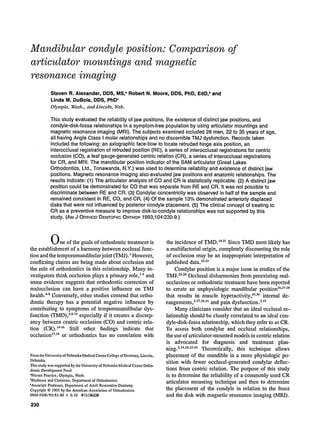
  • 7. 236 Alexander,Moore, and DuBois American Journal of Orthodontics and Dentofacial Orthopedics September1993 20 15 5 ~XN~X N%N%NXN% NNXN~NN~N% NNNNN%NNN NNXNNN%NN NNRetruded Centric Occlusion Jaw Postion [] Left [] Right Centric Related Right SJde N = 28, Left Side N = 26 Fig. 6. Percent anterior disk placement of TMJ. Fig. 7. MRI of TMJ. Condyle (C) placed posteriorly in fossa (F) with normal disk (D) to condyle relationship. was found in the sagittal planes. Such variation could be explained by the osseous asymmetry between right and left halves of the face and the cranium. This vari- ation then cannot be directly compared with the vari- ation found in the sagittal plane. The MPI data estab- lished that a significant difference existed between the jaw positions of RE and CO. The MPI and MRI data were used to evaluate dis-
  • 8. American Journal of Orthodontics and Dentofacial Orthopedics Alexander, Moore, and DuBois 237 Volume 104, No. 3 Fig. 8. MRI of TMJ. Condyle (C) concentric in fossa (F) withanterior disk (D) to condyle relationship. tinct jaw positions in the sagittal plane. The baseline or 0 value for the MPI means and standard deviations was established from the axiographic location of the hinge axis position. Therefore it would be expected that interocclusal registrations of RE should be close to the 0 value and that CO and CR would be further from the baseline if their positions were significantly different. The variability, as reflected by the standard deviations, shows approximately -+0.5 mm or less from the mean for MPI and also MRI values (Table I, Figs. 1 and 2). This range is satisfactory since measurements less than 0.5 mm were not discernible. These data suggests the existence of distinct jaw positions between CO and either RE or CR. In the vertical (Z) plane CR is often described as the most superior position.Z5 Evaluation of the MPI data revealed that significant differences between jaw po- sitions only existed between CO as compared with RE and CR for the left condyle, and CO as compared with CR for the fight condyle (Table I). In all relationships, CO was positioned inferiorly to RE and CR. Evaluation of MRI data confirmed that CO was inferior in position when compared with RE and CR, but significance was only found between RE and CO for the left condyle (Fig. 1). The MRI data also illustrated the relationship of the condylar position in RE when referenced to the glenoid fossa (Fig. 3). Collectively considered, the data support the existence of a distinct jaw position for CO inferior to RE and CR. In the horizontal (X) plane, the definitions and ar- ticles previously mentioned would support a CR posi- tion anterior to RE.~5.36 Evaluation of the MPI data revealed CO to be positioned anteriorly to RE and CR on both the left and right sides. However, statistical significance was only found on the left side between CO and RE and between CO and CR (Table I). The MRI data also revealed that CO was positioned ante- riorly to RE and CR but statistical significance was only found on the right side between CO and CR (Fig. 2). A collective evaluation of the data would suggest that CO was positioned anteriorly to RE and CR, but the MPI data failed to correlate statistically with the MRI data. These differences might be explained by the lack of a sharp demarcation of cortical bone'on the MRI image that reduced the accuracy of the measurements. In addition, small measurements and large variations
  • 9. 238 Alexander, Moore, and DuBois AmericanJournalof Orthodonticsand DentofacialOrthopedics September1993 potentially create small changes in interpretation of MPI or MRI data that could greatly change statistical significance. Combining vertical and horizontal axes of the sag- ittal plane allows further correlation of the data. The data of the present study provide evidence that CO is a distinct jaw position, separate from RE and CR. This finding is substantiated by a condylar position inferior and anterior to RE and CR. The data do not support distinct condylar positions for RE and CR and do not suggest that CO and CR are coincident. The latter is in contrast to what was previously described as the ideal relationship for a healthy TMJ. 29,32,35Possible reasons for these observations might be that a separation of RE and CR fails to exist, and CO and CR are not found coincidentwithin the normal population. Conversely, RE and CR could be distinct jaw positions, but the leaf gauge does not establish CR, thus preventing coinci- dence with CO in the normal population. Condyle to fosse relationship The issue of diagnosis and treatment to different condylar relationships often focuses on the issue of concentricity. The present study shows that most per- sons within this population are concentric (Figs. 4 and 5). The distribution pattern of all three jaw positions supports the concept that a concentrically placed con- dyle represents the most common position in defining what is normal. It is important to note, that although this entire sample was selected from a normal popu- lation, approximately half of the condyles were not defined as being concentric. Further evaluation of the data revealed that approximately half of the patients did not change their condylar position in reference to the fossa for RE, CO, or CR. In other words, if the patient was concentric, anteriorly or posteriorly placed, they maintained that same classification for all three jaw relationships, Therefore diagnosis of the health of the TMJ on the basis of concentricity is not supported by the present data. Furthermore, changing the concentric, anterior, or posterior through manipulation status may not be possible in many cases. Disk-to-condyle relationship One of the most common problems affecting the TMJ is internal derangement, which includes the ab- normal relationship of the articular disk relative to the mandibular condyle, the fossa, and the articular emi- nence. Of the 54 joints examined with MRI, 7 (13%) were diagnosed as having anterior disk placement. Al- though this result is less than the 32% reported by Kircos,47 it does confirm the presence of anterior disk placement in a symptom-free population. In half of the anteriorly displaced disks observed in the present study, the condyle was posteriorly posi- tioned. In the other half, the condyle was concentric or anteriorly positioned. This is in contrast to a previous report that indicated an anteriorly displaceddisk was associated with a posterior condylar position (Figs. 7 and 8).4s Two of the seven subjects with anteriorly displaced disks had joint sounds, and five subjects with normal disk relationships had joint sounds. This finding sup- ports previous reports that, in the absence of other symptoms, joint sounds cannot be considered sufficient evidence of dysfunction, n CONCLUSIONS 1. The articulator analysis of CO and CR is statis- ticaUy replicable. 2. Condylar concentricity was observed in half of the sample and remained consistent in RE, CO, and CR. 3. Of the sample 13% demonstrated anteriorly dis- placed disks that were not influenced by poste- rior condyle placement. 4. The clinical concept of treating to CR as a pre- ventive measure to improve disk-to-condyle re- lationships was not supported by this study. REFERENCES I. Perry HI'. Mandibularfunction:an orthodonticresponsibility. AMJ ORTHOD1975;67:316-23. 2. RothRH. Tcmporomandibularpain-dysfunctionandocclusalre- latinnships. AngleOrthod 1973;43:136-53. 3. ThompsonJR. Abnormalfunctionof the temporomandibular joints and relatedmusculature:orthodonticimplications,partI. AngleOrthod 1986;56:143-63. 4. MorawaAP, Loos PJ, Easton JW. Temporomandibularjoint dysfunction in children and adolescents:incidence,diagnosis, and treatment.QulntessenceInt 1985;11:771-7. 5. McLaughlinRP. Malocclusionandthetemporomandibularjoint: an historicalperspective.AngleOrthod 1988;58:185-91. 6. Myers DR, Ba.renicJ-l',Bell RA, WilliamsonEH. Condylar positionin chiIdrenwithfunctionalposteriorcrossbites:before and aftercrossbitecorrection.PediatrDent 1980;2:190-4. "7. OwcnAH.The maxillarysagittalappliance:aclinicalstudy. AM J ORTItODDENTOFACORTHOP1987;91:271-85. 8. SadowskyC, BeGoleEA. Long-termstatusof temporomandib- ular joint function and functionalocclusion afterorthodontic treatment. AMJ ORTHOD1980;78:201-12. 9. LoftGH, ReynoldsJM, ZwemerJD, ThompsonWO, Dushku J. The occurrenceof craniomandibulatsymptomsin healthy youngadultswithand withoutpriororthodontictreatment.AM J ORTHODDENTOFACORTHOP1989;96:264-5. 10. ThompsonJR. Abnormalfunctionof the temporomandibular jointsand relatedmusculature:orthodonticimplications,partII. AngleOrthod 1986;56:181-95. 11. PullingerA, MonteiroA, Liu S. Etiologicalfactorsassociated with temporomandibulardisorders. [Abstrctno. 848]. J Dent Res 1985;64:269.
  • 10. AmericanJournalof Orthodonticsand DentofacialOrthopedics Alexander, Moore, and DuBois 239 Volume 104,No. 3 12. Pullinger A, Seligman D, Solberg W, Liu S. Relationship of occlusal morphology to TM disorders in young adults. [Abstract no. 849]. J Dent Res 1985;64:269. 13. Drace JE. Natural history of minimal anterior displacements of the temporomandibular joint meniscus. [Abstract]. Radiol Soc North Am 1988. 14. Williamson EH. Occlusion and TMJ dysfunction. J Clin Orthod 1981;15:333-50. 15. Dawson PE. Centric relation: its effect on occluso-muscle har- mony. Dent Clin North Am 1979;23:169-80. 16. McNeill C. The optimum temporomandibularjoint condyle po- sition in clinical practice, lnt J Periodontic Restorative Dent 1985;6:53-76. 17. Egermark-Eriksson I, Carlsson GE, Magnusson T. A long-term epidemiologie study of the relationship between occlusal factors and mandibular dysfunction in children and adolescents. J Dent Res 1987;66:67-71. 18. Parker MW. A dynamic model of etiology in temporomandibular disorders. J Am Dent Assoc 1990;120:283-90. 19. Sadowsky C, Poison AM. Temporomandibular disorders and functional occlusion after orthodontic treatment: results of two long-term studies. AM J ORrr~OD1984;86:386-90. 20. Greene CS. Orthodontics and temporomandibular disorders. Dent Clin North Am 1988;32:529-38. 21. American Academy of Pediatric Dentistry. Treatment of tem- poromandibular disorders in children: summary statements and recommendations. J Am Dent Assoc 1990;120;265-9. 22. Mohlin B. Prevalence of mandibular dysfunction and relation between malocclusion and mandibular dysfunction in a group of women in Sweden. Eur J Orthod 1983;4:115-23. 23. Mohlin B, Ingervall B, Thilander B. Relation between maloc- clusion and mandibular dysfunction in Swedish men. Eur J Or- thod 1980;2:229-38. 24. Perry HT. Relationofocclusiontotemporomandibularjointdys- function: the orthodontic viewpoint. J Am Dent Assoc 1969;79: i37-4I. 25. Gilboe DB. Centric relation as the treatment position. J Prosthet Dent 1983;50:685-9. 26. Mongini F, Schmid W. Assessment of the therapeutic position for orthodontic diagnosis and treatment. AM J ORTHOD 1982;82:513-8. 27. Reuling N. Comparative study of clinical examination, occlusal analysis and new radiological imagingprocedures in patients with functional TMJ disorders. J Oral Rehabil 1987;14:165-74. 28. Wyatt WE. Preventingadverse effects on the temporomandibular joint through orthodontic treatment. A~,t J ORTHODDENTOFAC OR'mOP 1987;91:493-9. 29. Williamson EH. The role of craniomandibular dysfunction in orthodontic diagnosis and treatment planning. Dent Clin North Am 1983;27:541-60. 30. Kirveskari P, Alanen P, Jamsa T. Association between cranio- mandibular disorders and occlusal interferences. J Prosthet Dent 1989;62:66-9. 31. Owen AH. Orthodontic~orthopedic therapy for cranioman- dibular pain dysfunction: Part A. Anterior disk displacement, review of literature. J Craniomandib Pract 1987;5:357-66. 32. Roth RH. Functional occlusion for the orthodontist, part III. J Clin Orthod 1981;15:174-98. 33. Williamson EH, Steinke RM, Morse PK, Swift TR. Centric relation: a comparison of muscle-determined position and op- erator guidance. AM J ORTHOD1980;77;133-45. 34. Salvicek R. Clinical and instrumental functional analysis for diagnosis and treatment planning, part IV: instrumental analysis of mandibular casts using the mandibular position indicator. J Clin Orthod 1988;22:566-75. 35. Parker WS. Centric relation and centric occlusion--an ortho- dontic responsibility. Ar,t J ORTHOD1978;74:481-500. 36. Celenza FV. The theory and clinical management of centric po- sitions: II. Centric relation and centric relation occlusion. Int J Periodontics Restorative Dent 1984;6:63-86. 37. Roth RH. Functional occlusion for the orthodontist. J Clin Or- thod 1981;15:32-51. 38. Crawford WA. Centrie relation reappraised. J Craniomandib Pract 1983-84;2:39-45. 39. Academy of Denture Prosthetics. Glossary of Prosthodontic Terms. J Prosthet Dent 1960. 40. Academy of Denture Prosthetics. Glossary of Prosthodontic Terms. J Prosthet Dent 1987;58:717-62. 41. Myers ML. Centric relation records--historical review. J Prosthet Dent 1982;47:141-5. 42. Okeson JP. Fundamentals of occlusion and temporomandibular disorders. St. Louis: CV Mosby, 1985. 43. Ricketts RM. Abnormal function of the temporomandibular joint. AM J ORTHOD1955;41:435-41. 44. Weinberg LA, Chastain JK. New TMJ clinical data and the implication on diagnosis and treatment. J Am Dent Assoc 1990;120:305-I 1. 45. Schach RT, Sadowsky PL. Clinical experience with magnetic resonance imaging in internal derangements of the TMJ. Angle Orthod 1988;58:21-32. 46. Dworkin SF, Huggins KH, LeResche L, et al. Epidemiology of signs and symptoms in temporomandibular disorders: clinical signs in cases and controls. J Am Dent Assoc 1990;120:273-81. 47. Kircos LT, Ortendahl DA, Arakawa M, Mark A. Demonstration of TMJ anterior disc position by MRI in asymptomatie subjects. [Abstract no. 1842]. J Dent Res 1987;66:337. 48. Farrar WB, McCarty WL. A clinical outline of the tempoi-o- mandibular joint diagnosis and treatment. Montgomery, Ala- bama: Walker Printing, 1983. Reprint requests to: Dr. Robert N. Moore Department of Orthodontics College of Dentistry University of Nebraska Medical Center Lincoln, NE 68583-0740