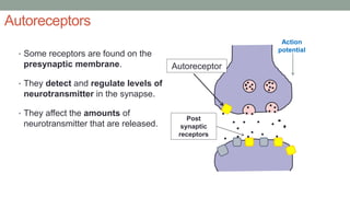
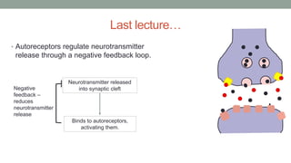
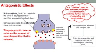
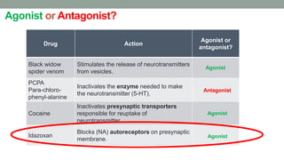
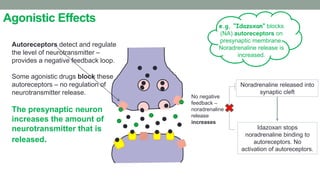
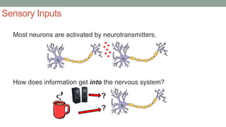
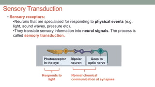
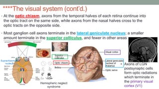
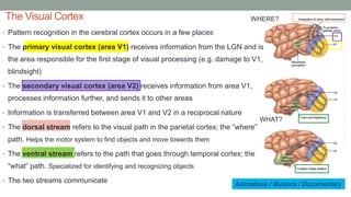
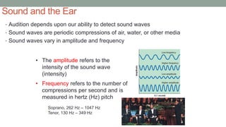
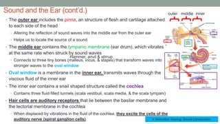
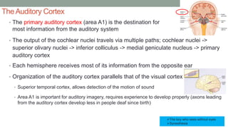
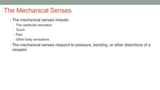
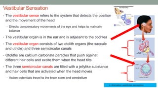
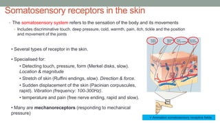
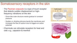
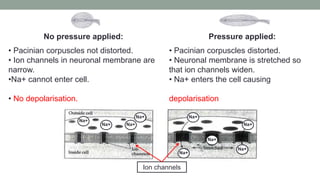
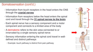
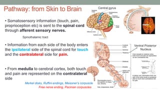
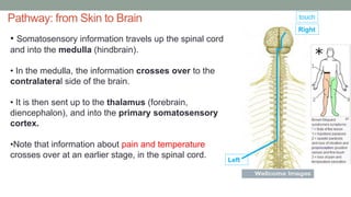
LECTURE 10
SENSORY INPUTS
Dr. Paula Trotter
P.Trotter@mmu.ac.uk
Office hours: Mondays 12:30 – 15:30, BR3.53
Autoreceptors
• Some receptors are found on the
presynaptic membrane.
• They detect and regulate levels of
neurotransmitter in the synapse.
• They affect the amounts of
neurotransmitter that are released.
Action
potential
Post
synaptic
receptors
Autoreceptor
Last lecture…
• Autoreceptors regulate neurotransmitter
release through a negative feedback loop.
Neurotransmitter released
into synaptic cleft
Binds to autoreceptors,
activating them.
Negative
feedback –
reduces
neurotransmitter
release
Antagonistic Effects
Autoreceptors detect and regulate
the level of neurotransmitter –
provides a negative feedback loop.
Some antagonistic drugs stimulate
these autoreceptors.
The presynaptic neuron
reduces the amount of
neurotransmitter that is
released.
e.g. “clonidine” binds to
and activates presynaptic
α2 autoreceptors,
inhibiting further release
of adrenaline and
noradrenaline.
Neurotransmitter released
into synaptic cleft
Both neurotransmitter and
clonidine can bind to
autoreceptors, activating
them.
Negative
feedback is
increased –
reduced
neurotransmitter
release
Agonist or Antagonist?
Drug Action
Agonist or
antagonist?
Black widow
spider venom
Stimulates the release of neurotransmitters
from vesicles.
PCPA
Para-chloro-
phenyl-alanine
Inactivates the enzyme needed to make
the neurotransmitter (5-HT).
Cocaine
Inactivates presynaptic transporters
responsible for reuptake of
neurotransmitter.
Idazoxan
Blocks (NA) autoreceptors on presynaptic
membrane.
Agonist
Antagonist
Agonist
Agonist
Agonistic Effects
Autoreceptors detect and regulate
the level of neurotransmitter –
provides a negative feedback loop.
Some agonistic drugs block these
autoreceptors – no regulation of
neurotransmitter release.
The presynaptic neuron
increases the amount of
neurotransmitter that is
released.
e.g. “Idazoxan” blocks
(NA) autoreceptors on
presynaptic membrane.
Noradrenaline release is
increased.
Noradrenaline released into
synaptic cleft
Idazoxan stops
noradrenaline binding to
autoreceptors. No
activation of autoreceptors.
No negative
feedback –
noradrenaline
release
increases
SENSORY INPUTS
Learning Objectives
• Explain what is meant by the term sensory transduction.
• Describe the mechanisms and processes that underlie
sensory transduction in the visual, auditory, vestibular and
somatosensory systems.
• Describe the pathway that sensory information takes to get
from the periphery to the cortex.
Keywords
• Lateral and Medial Geniculate
Nucleus (LGN & MGN)
• Prosopagnosia
• Visual agnosia
• Dermatome
• Vestibular system
• Mechanoreceptors
• Pacinian corpuscle
• Merkel’s disk
• Ruffini ending
• Free nerve ending
• Synesthesia
• Sensory transduction
• Primary somatosensory cortex
• Propioception
• Bipolar cells
• Ganglion cells
• Amacrine cells
• Horizontal cells
• Photoreceptor
• Rods
• Cones
• Fovea
• Saccade
Our senses
• How many senses do we have?
Touch Smell
Taste Hearing
Sight
FIVE?
General Principles of Perception
• Each of our senses has specialized receptors that are sensitive to a particular
kind of energy
• Receptors “transduce” (convert) energy into electrochemical patterns so that
the brain can perceive sights, sounds, smells, etc.
• Law of specific nerve energies states that activity by a particular nerve always
conveys the same type of information to the brain
• Example: impulses in one neuron indicate light; impulses in another neuron indicate sound
• Which neurons respond, the amount of response, and the timing of response
influence what we perceive
Sensory Inputs
Most neurons are activated by neurotransmitters.
How does information get into the nervous system?
?
?
Sensory Transduction
Photoreceptor
in the eye
Bipolar
neuron
Goes to
optic nerve
• Sensory receptors:
•Neurons that are specialised for responding to physical events (e.g.
light, sound waves, pressure etc).
•They translate sensory information into neural signals. The process is
called sensory transduction.
Responds to
light
Normal chemical
communication at synapses
Photoreceptors in the eye
In the dark
Photoreceptor cells respond to light
Pigment molecules in
receptor membrane
are inactive
Sodium channels are open
– sodium ions enter cell -
neuron is depolarised.
Neurotransmitter
(glutamate) is
released by the cell.
Cell body
Resting potential ~ -40 mV
Axon terminals
Photoreceptors in the eye
In the light
Photoreceptor cells respond to light
Light bleaches
pigment
molecules and
activates them.
Sodium channels
close – sodium ions
cannot enter – cell is
hyperpolarised.
Less
neurotransmitter
(glutamate) is
released by the cell
Cell body Axon terminals
The visual system
• Light enters the eye through an opening in the center of the iris
called the pupil
• Light is focused by the lens and the cornea onto the rear surface
of the eye known as the retina, lined with visual receptors
• Rods: most abundant in the periphery of the eye and
respond to faint light (120 million per retina)
• Cones: most abundant in and around the fovea (6 million)
• Essential for color vision & more useful in bright light
• Light from the left side of the world strikes the right side of the retina and vice versa
• The central portion of the retina is the fovea and allows for acute and detailed vision
• Packed tight with receptors and nearly free of ganglion axons and blood vessels
The visual system (cont’d.)
• Bipolar cells make synapses onto amacrine cells and ganglion
cells
• The axons of ganglion cells join one another to form the optic nerve that travels to
the brain
• Amacrine cells are additional cells that receive information
from bipolar cells and send it to other bipolar, ganglion, or
amacrine cells
• Amacrine cells control the ability of the ganglion cells to
respond to shapes, movements, or other specific aspects of
visual stimuli
• The point at which the optic nerve leaves the back of the eye is
called the blind spot because it contains no receptors
• Visual receptors send messages to neurons called bipolar cells,
located closer to the center of the eye, and horizontal cells.
****The visual system (cont’d.)
• At the optic chiasm, axons from the temporal halves of each retina continue into
the optic tract on the same side, while axons from the nasal halves cross to the
optic tracts on the opposite side.
• Most ganglion cell axons terminate in the lateral geniculate nucleus; a smaller
amount terminate in the superior colliculus, and fewer in other areas
• Axons of LGN
postsynaptic cells
form optic radiations
which terminate in
the primary visual
cortex (V1)
Suprachiasmatic
nucleus
Hemispheric neglect
syndrome
The Visual Cortex
Animations / Illusions / Documentary
• Pattern recognition in the cerebral cortex occurs in a few places
• The primary visual cortex (area V1) receives information from the LGN and is
the area responsible for the first stage of visual processing (e.g. damage to V1,
blindsight)
• The secondary visual cortex (area V2) receives information from area V1,
processes information further, and sends it to other areas
• Information is transferred between area V1 and V2 in a reciprocal nature
• The dorsal stream refers to the visual path in the parietal cortex; the “where”
path. Helps the motor system to find objects and move towards them
• The ventral stream refers to the path that goes through temporal cortex; the
“what” path. Specialized for identifying and recognizing objects
• The two streams communicate
WHERE?
WHAT?
Sound and the Ear
• Audition depends upon our ability to detect sound waves
• Sound waves are periodic compressions of air, water, or other media
• Sound waves vary in amplitude and frequency
• The amplitude refers to the
intensity of the sound wave
(intensity)
• Frequency refers to the number of
compressions per second and is
measured in hertz (Hz) pitch
Soprano, 262 Hz – 1047 Hz
Tenor, 130 Hz – 349 Hz
Sound and the Ear (cont’d.)
• The outer ear includes the pinna, an structure of flesh and cartilage attached
to each side of the head
• Altering the reflection of sound waves into the middle ear from the outer ear
• Helps us to locate the source of a sound
• The middle ear contains the tympanic membrane (ear drum), which vibrates
at the same rate when struck by sound waves
• Connects to three tiny bones (malleus, incus, & stapes) that transform waves into
stronger waves to the oval window
• Oval window is a membrane in the inner ear, transmits waves through the
viscous fluid of the inner ear
• The inner ear contains a snail shaped structure called the cochlea
• Contains three fluid-filled tunnels (scala vestibuli, scala media, & the scala tympani)
• Hair cells are auditory receptors that lie between the basilar membrane and
the tectorial membrane in the cochlea
• When displaced by vibrations in the fluid of the cochlea, they excite the cells of the
auditory nerve (spiral ganglion cells)
outer middle inner
 Animation hearing: Sound transduction
Hammer, anvil & stirrup
The Auditory Cortex
• The primary auditory cortex (area A1) is the destination for
most information from the auditory system
• The output of the cochlear nuclei travels via multiple paths; cochlear nuclei ->
superior olivary nuclei -> inferior colliculus -> medial geniculate nucleus -> primary
auditory cortex
• Each hemisphere receives most of its information from the opposite ear
• Organization of the auditory cortex parallels that of the visual cortex
• Superior temporal cortex, allows detection of the motion of sound
• Area A1 is important for auditory imagery, requires experience to develop properly (axons leading
from the auditory cortex develop less in people deaf since birth)
The boy who sees without eyes
Synesthesia
The Mechanical Senses
• The mechanical senses include:
• The vestibular sensation
• Touch
• Pain
• Other body sensations
• The mechanical senses respond to pressure, bending, or other distortions of a
receptor
Vestibular Sensation
• The vestibular sense refers to the system that detects the position
and the movement of the head
• Directs compensatory movements of the eye and helps to maintain
balance
• The vestibular organ is in the ear and is adjacent to the cochlea
• The vestibular organ consists of two otolith organs (the saccule
and utricle) and three semicircular canals
• Otoliths are calcium carbonate particles that push against
different hair cells and excite them when the head tilts
• The three semicircular canals are filled with a jellylike substance
and hair cells that are activated when the head moves
• Action potentials travel to the brain stem and cerebellum
 Animation vestibular perception
Somatosensory receptors in the skin
• Several types of receptor in the skin.
• Specialised for:
• Detecting touch, pressure, form (Merkel disks, slow).
Location & magnitude
• Stretch of skin (Ruffini endings, slow). Direction & force.
• Sudden displacement of the skin (Pacinian corpuscules,
rapid). Vibration (frequency: 100-300Hz).
• temperature and pain (free nerve ending, rapid and slow).
• Many are mechanoreceptors (responding to mechanical
pressure)
• The somatosensory system refers to the sensation of the body and its movements
• Includes discriminative touch, deep pressure, cold, warmth, pain, itch, tickle and the position
and movement of the joints
 Animation somatosensory receptive fields
Somatosensory receptors in the skin
• The Pacinian corpuscle is a type of touch receptor
that detects sudden displacement or high-
frequency vibrations on the skin
• Onion-like outer structure resists gradual or constant
pressure
• Sudden or vibrating stimulus bends the membrane and
increases the flow of sodium ions to triggers an action
potential
• Chemicals can stimulate receptors for heat and
cold: e.g., capsaicin & menthol
No pressure applied:
• Pacinian corpuscles not distorted.
• Ion channels in neuronal membrane are
narrow.
•Na+ cannot enter cell.
• No depolarisation.
Pressure applied:
• Pacinian corpuscles distorted.
• Neuronal membrane is stretched so
that ion channels widen.
• Na+ enters the cell causing
depolarisation
Ion channels
Na+
Na+
Na+
Na+
Na+
Na+
Na+
Na+
Na+
Somatosensation (cont’d.)
• Information from touch receptors in the head enters the CNS
through the cranial nerves
• Information from receptors below the head enters the spinal
cord and travel through the 31 spinal nerves to the brain
• Each spinal nerve has a sensory component and a motor
component and connects to a limited area of the body
• A dermatome refers to the skin area connected to or
innervated by a single sensory spinal nerve
• Sensory information entering the spinal cord travel in well-
defined and distinct pathways
• Example: touch pathway is distinct from pain pathway
Pathway: from Skin to Brain Central gyrus
Free nerve ending, Pacinian corpuscles
Merkel disks, Ruffini endings, Meissner’s corpuscle
Spinothalamic tract
Ventral Posterior
Nucleus
• Somatosensory information (touch, pain,
proprioception etc) is sent to the spinal cord
through afferent sensory nerves.
• Information from each side of the body enters
the ipsilateral side of the spinal cord for touch
and the contralateral side for pain.
• From medulla to cerebral cortex, both touch
and pain are represented on the contralateral
side
Pathway: from Skin to Brain
• Somatosensory information travels up the spinal cord
and into the medulla (hindbrain).
• In the medulla, the information crosses over to the
contralateral side of the brain.
• It is then sent up to the thalamus (forebrain,
diencephalon), and into the primary somatosensory
cortex.
•Note that information about pain and temperature
crosses over at an earlier stage, in the spinal cord.
Right
Left
touch
Core reading
• Pinel: sections 6.2, and 7.3. (note the textbook is quite
detailed, but might still be of interest to some of you) or
relevant sections in another biopsychology textbook
Recommended Reading
Any questions?

CogBio - Lecture 10 - SensorySystems Student.pptx

  • 1.
    LECTURE 10 SENSORY INPUTS Dr.Paula Trotter P.Trotter@mmu.ac.uk Office hours: Mondays 12:30 – 15:30, BR3.53
  • 2.
    Autoreceptors • Some receptorsare found on the presynaptic membrane. • They detect and regulate levels of neurotransmitter in the synapse. • They affect the amounts of neurotransmitter that are released. Action potential Post synaptic receptors Autoreceptor
  • 3.
    Last lecture… • Autoreceptorsregulate neurotransmitter release through a negative feedback loop. Neurotransmitter released into synaptic cleft Binds to autoreceptors, activating them. Negative feedback – reduces neurotransmitter release
  • 4.
    Antagonistic Effects Autoreceptors detectand regulate the level of neurotransmitter – provides a negative feedback loop. Some antagonistic drugs stimulate these autoreceptors. The presynaptic neuron reduces the amount of neurotransmitter that is released. e.g. “clonidine” binds to and activates presynaptic α2 autoreceptors, inhibiting further release of adrenaline and noradrenaline. Neurotransmitter released into synaptic cleft Both neurotransmitter and clonidine can bind to autoreceptors, activating them. Negative feedback is increased – reduced neurotransmitter release
  • 5.
    Agonist or Antagonist? DrugAction Agonist or antagonist? Black widow spider venom Stimulates the release of neurotransmitters from vesicles. PCPA Para-chloro- phenyl-alanine Inactivates the enzyme needed to make the neurotransmitter (5-HT). Cocaine Inactivates presynaptic transporters responsible for reuptake of neurotransmitter. Idazoxan Blocks (NA) autoreceptors on presynaptic membrane. Agonist Antagonist Agonist Agonist
  • 6.
    Agonistic Effects Autoreceptors detectand regulate the level of neurotransmitter – provides a negative feedback loop. Some agonistic drugs block these autoreceptors – no regulation of neurotransmitter release. The presynaptic neuron increases the amount of neurotransmitter that is released. e.g. “Idazoxan” blocks (NA) autoreceptors on presynaptic membrane. Noradrenaline release is increased. Noradrenaline released into synaptic cleft Idazoxan stops noradrenaline binding to autoreceptors. No activation of autoreceptors. No negative feedback – noradrenaline release increases
  • 7.
  • 8.
    Learning Objectives • Explainwhat is meant by the term sensory transduction. • Describe the mechanisms and processes that underlie sensory transduction in the visual, auditory, vestibular and somatosensory systems. • Describe the pathway that sensory information takes to get from the periphery to the cortex.
  • 9.
    Keywords • Lateral andMedial Geniculate Nucleus (LGN & MGN) • Prosopagnosia • Visual agnosia • Dermatome • Vestibular system • Mechanoreceptors • Pacinian corpuscle • Merkel’s disk • Ruffini ending • Free nerve ending • Synesthesia • Sensory transduction • Primary somatosensory cortex • Propioception • Bipolar cells • Ganglion cells • Amacrine cells • Horizontal cells • Photoreceptor • Rods • Cones • Fovea • Saccade
  • 10.
    Our senses • Howmany senses do we have? Touch Smell Taste Hearing Sight FIVE?
  • 11.
    General Principles ofPerception • Each of our senses has specialized receptors that are sensitive to a particular kind of energy • Receptors “transduce” (convert) energy into electrochemical patterns so that the brain can perceive sights, sounds, smells, etc. • Law of specific nerve energies states that activity by a particular nerve always conveys the same type of information to the brain • Example: impulses in one neuron indicate light; impulses in another neuron indicate sound • Which neurons respond, the amount of response, and the timing of response influence what we perceive
  • 12.
    Sensory Inputs Most neuronsare activated by neurotransmitters. How does information get into the nervous system? ? ?
  • 13.
    Sensory Transduction Photoreceptor in theeye Bipolar neuron Goes to optic nerve • Sensory receptors: •Neurons that are specialised for responding to physical events (e.g. light, sound waves, pressure etc). •They translate sensory information into neural signals. The process is called sensory transduction. Responds to light Normal chemical communication at synapses
  • 14.
    Photoreceptors in theeye In the dark Photoreceptor cells respond to light Pigment molecules in receptor membrane are inactive Sodium channels are open – sodium ions enter cell - neuron is depolarised. Neurotransmitter (glutamate) is released by the cell. Cell body Resting potential ~ -40 mV Axon terminals
  • 15.
    Photoreceptors in theeye In the light Photoreceptor cells respond to light Light bleaches pigment molecules and activates them. Sodium channels close – sodium ions cannot enter – cell is hyperpolarised. Less neurotransmitter (glutamate) is released by the cell Cell body Axon terminals
  • 16.
    The visual system •Light enters the eye through an opening in the center of the iris called the pupil • Light is focused by the lens and the cornea onto the rear surface of the eye known as the retina, lined with visual receptors • Rods: most abundant in the periphery of the eye and respond to faint light (120 million per retina) • Cones: most abundant in and around the fovea (6 million) • Essential for color vision & more useful in bright light • Light from the left side of the world strikes the right side of the retina and vice versa • The central portion of the retina is the fovea and allows for acute and detailed vision • Packed tight with receptors and nearly free of ganglion axons and blood vessels
  • 17.
    The visual system(cont’d.) • Bipolar cells make synapses onto amacrine cells and ganglion cells • The axons of ganglion cells join one another to form the optic nerve that travels to the brain • Amacrine cells are additional cells that receive information from bipolar cells and send it to other bipolar, ganglion, or amacrine cells • Amacrine cells control the ability of the ganglion cells to respond to shapes, movements, or other specific aspects of visual stimuli • The point at which the optic nerve leaves the back of the eye is called the blind spot because it contains no receptors • Visual receptors send messages to neurons called bipolar cells, located closer to the center of the eye, and horizontal cells.
  • 18.
    ****The visual system(cont’d.) • At the optic chiasm, axons from the temporal halves of each retina continue into the optic tract on the same side, while axons from the nasal halves cross to the optic tracts on the opposite side. • Most ganglion cell axons terminate in the lateral geniculate nucleus; a smaller amount terminate in the superior colliculus, and fewer in other areas • Axons of LGN postsynaptic cells form optic radiations which terminate in the primary visual cortex (V1) Suprachiasmatic nucleus Hemispheric neglect syndrome
  • 19.
    The Visual Cortex Animations/ Illusions / Documentary • Pattern recognition in the cerebral cortex occurs in a few places • The primary visual cortex (area V1) receives information from the LGN and is the area responsible for the first stage of visual processing (e.g. damage to V1, blindsight) • The secondary visual cortex (area V2) receives information from area V1, processes information further, and sends it to other areas • Information is transferred between area V1 and V2 in a reciprocal nature • The dorsal stream refers to the visual path in the parietal cortex; the “where” path. Helps the motor system to find objects and move towards them • The ventral stream refers to the path that goes through temporal cortex; the “what” path. Specialized for identifying and recognizing objects • The two streams communicate WHERE? WHAT?
  • 20.
    Sound and theEar • Audition depends upon our ability to detect sound waves • Sound waves are periodic compressions of air, water, or other media • Sound waves vary in amplitude and frequency • The amplitude refers to the intensity of the sound wave (intensity) • Frequency refers to the number of compressions per second and is measured in hertz (Hz) pitch Soprano, 262 Hz – 1047 Hz Tenor, 130 Hz – 349 Hz
  • 21.
    Sound and theEar (cont’d.) • The outer ear includes the pinna, an structure of flesh and cartilage attached to each side of the head • Altering the reflection of sound waves into the middle ear from the outer ear • Helps us to locate the source of a sound • The middle ear contains the tympanic membrane (ear drum), which vibrates at the same rate when struck by sound waves • Connects to three tiny bones (malleus, incus, & stapes) that transform waves into stronger waves to the oval window • Oval window is a membrane in the inner ear, transmits waves through the viscous fluid of the inner ear • The inner ear contains a snail shaped structure called the cochlea • Contains three fluid-filled tunnels (scala vestibuli, scala media, & the scala tympani) • Hair cells are auditory receptors that lie between the basilar membrane and the tectorial membrane in the cochlea • When displaced by vibrations in the fluid of the cochlea, they excite the cells of the auditory nerve (spiral ganglion cells) outer middle inner  Animation hearing: Sound transduction Hammer, anvil & stirrup
  • 22.
    The Auditory Cortex •The primary auditory cortex (area A1) is the destination for most information from the auditory system • The output of the cochlear nuclei travels via multiple paths; cochlear nuclei -> superior olivary nuclei -> inferior colliculus -> medial geniculate nucleus -> primary auditory cortex • Each hemisphere receives most of its information from the opposite ear • Organization of the auditory cortex parallels that of the visual cortex • Superior temporal cortex, allows detection of the motion of sound • Area A1 is important for auditory imagery, requires experience to develop properly (axons leading from the auditory cortex develop less in people deaf since birth) The boy who sees without eyes Synesthesia
  • 23.
    The Mechanical Senses •The mechanical senses include: • The vestibular sensation • Touch • Pain • Other body sensations • The mechanical senses respond to pressure, bending, or other distortions of a receptor
  • 24.
    Vestibular Sensation • Thevestibular sense refers to the system that detects the position and the movement of the head • Directs compensatory movements of the eye and helps to maintain balance • The vestibular organ is in the ear and is adjacent to the cochlea • The vestibular organ consists of two otolith organs (the saccule and utricle) and three semicircular canals • Otoliths are calcium carbonate particles that push against different hair cells and excite them when the head tilts • The three semicircular canals are filled with a jellylike substance and hair cells that are activated when the head moves • Action potentials travel to the brain stem and cerebellum  Animation vestibular perception
  • 25.
    Somatosensory receptors inthe skin • Several types of receptor in the skin. • Specialised for: • Detecting touch, pressure, form (Merkel disks, slow). Location & magnitude • Stretch of skin (Ruffini endings, slow). Direction & force. • Sudden displacement of the skin (Pacinian corpuscules, rapid). Vibration (frequency: 100-300Hz). • temperature and pain (free nerve ending, rapid and slow). • Many are mechanoreceptors (responding to mechanical pressure) • The somatosensory system refers to the sensation of the body and its movements • Includes discriminative touch, deep pressure, cold, warmth, pain, itch, tickle and the position and movement of the joints  Animation somatosensory receptive fields
  • 26.
    Somatosensory receptors inthe skin • The Pacinian corpuscle is a type of touch receptor that detects sudden displacement or high- frequency vibrations on the skin • Onion-like outer structure resists gradual or constant pressure • Sudden or vibrating stimulus bends the membrane and increases the flow of sodium ions to triggers an action potential • Chemicals can stimulate receptors for heat and cold: e.g., capsaicin & menthol
  • 27.
    No pressure applied: •Pacinian corpuscles not distorted. • Ion channels in neuronal membrane are narrow. •Na+ cannot enter cell. • No depolarisation. Pressure applied: • Pacinian corpuscles distorted. • Neuronal membrane is stretched so that ion channels widen. • Na+ enters the cell causing depolarisation Ion channels Na+ Na+ Na+ Na+ Na+ Na+ Na+ Na+ Na+
  • 28.
    Somatosensation (cont’d.) • Informationfrom touch receptors in the head enters the CNS through the cranial nerves • Information from receptors below the head enters the spinal cord and travel through the 31 spinal nerves to the brain • Each spinal nerve has a sensory component and a motor component and connects to a limited area of the body • A dermatome refers to the skin area connected to or innervated by a single sensory spinal nerve • Sensory information entering the spinal cord travel in well- defined and distinct pathways • Example: touch pathway is distinct from pain pathway
  • 29.
    Pathway: from Skinto Brain Central gyrus Free nerve ending, Pacinian corpuscles Merkel disks, Ruffini endings, Meissner’s corpuscle Spinothalamic tract Ventral Posterior Nucleus • Somatosensory information (touch, pain, proprioception etc) is sent to the spinal cord through afferent sensory nerves. • Information from each side of the body enters the ipsilateral side of the spinal cord for touch and the contralateral side for pain. • From medulla to cerebral cortex, both touch and pain are represented on the contralateral side
  • 30.
    Pathway: from Skinto Brain • Somatosensory information travels up the spinal cord and into the medulla (hindbrain). • In the medulla, the information crosses over to the contralateral side of the brain. • It is then sent up to the thalamus (forebrain, diencephalon), and into the primary somatosensory cortex. •Note that information about pain and temperature crosses over at an earlier stage, in the spinal cord. Right Left touch
  • 31.
    Core reading • Pinel:sections 6.2, and 7.3. (note the textbook is quite detailed, but might still be of interest to some of you) or relevant sections in another biopsychology textbook Recommended Reading Any questions?