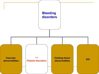
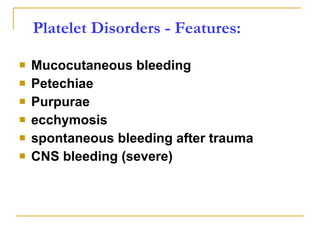
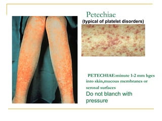
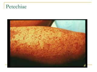
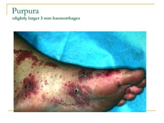
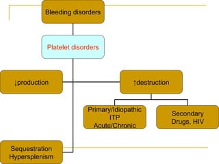
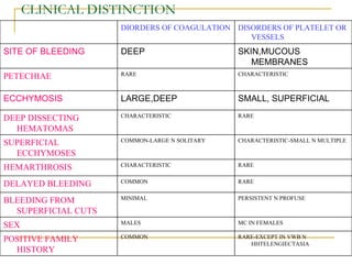
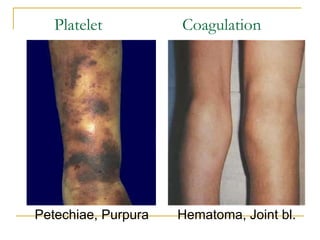
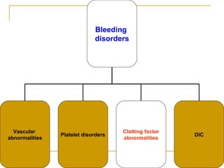
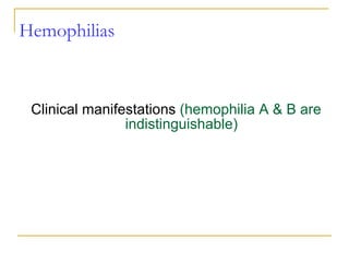
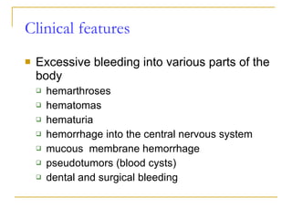
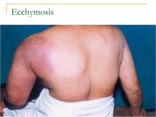
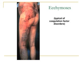
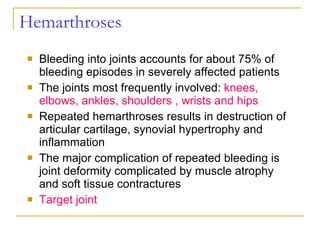
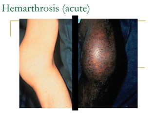
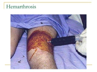
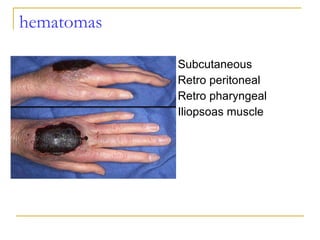
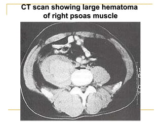
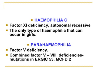
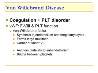
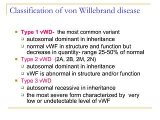
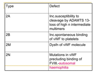
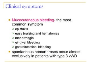
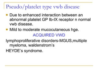
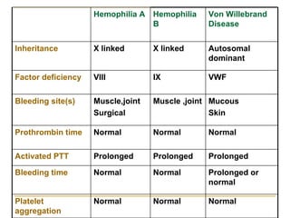
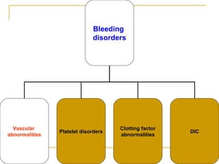
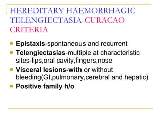
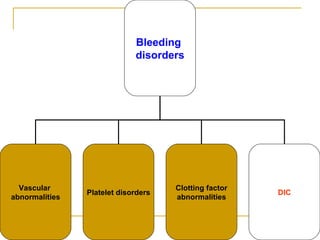
BLEEDING DISORDERS   CLINICAL FEATURES PROF.DR.G.SUNDARAMURTHY’S UNIT M7 BHARGAVI.K
Bleeding  disorders Vascular  abnormalities *** Platelet disorders Clotting factor abnormalities DIC
Platelet Disorders - Features: Mucocutaneous bleeding Petechiae Purpurae ecchymosis spontaneous bleeding after trauma CNS bleeding (severe)
Petechiae   PETECHIAE:minute 1-2 mm hges into skin,mucous membranes or serosal surfaces   Do not blanch with pressure (typical of platelet disorders)
Petechiae
Purpura :slightly larger 3 mm haemorrhages
Bleeding disorders Platelet disorders ↓ production ↑ destruction Sequestration Hypersplenism Primary/Idiopathic ITP Acute/Chronic Secondary Drugs, HIV
Classification of thrombocytopenia PSEUDO-ARTIFACTUAL THROMBOCYTOPENIA Platelet agglutination Platelet satellitism Giant platelets IMPAIRED PLATELET PRODUCTION congenital autosomal dominant
MYH9 RELATED May hegglin,fetchner,epstein,sebastian syndromes Mediterranean macrothrombocytopenia Familial platelet syndrome with predisoposition to AML Thrombocytopenia with linkage to ch 10 Paris-trousseau syndrome AUTOSOMAL RECESSIVE Congenital amegakaryocytic TAR syndrome Bernard soulier syndrome Gray platelet syndrome
X-LINKED WISKOTT-ALDRICH syndrome WITH DYSERYTHROCYTOSIS ACQUIRED Marrow infiltration Infectious disease-HIV,parvo,CMV Radiotherapy n chemotherapy Folic acid and vit b12 deficiency PNH Acquired aplastic anemia Myelodysplastic syndromes
ACCELELERATED PLATELET DESTRUCTION IMMUNE MEDIATED Autoimmune thrombocytopenic purpura Idiopathic secondary-infections,pregnancy Alloimmune neonatal posttransfusion purpura Autoimmune diseases MDS Lymphoproliferative disorders
NONIMMUNE THROMBOCYTOPENIA THROMBOTIC MICROANGIOPATHIES DIC KASSABACHMERIT SYNDROME PLATELET DESTRUCTION BY ARTIFICIAL SURFACES HEMOPHAGOCYTOSIS
ABNORMAL DISTRIBUTION OR POOLING Splenomegaly Hypersplenism Hypothermia Massive transfusion DRUG INDUCED HEPARIN INDUCED OTHERS
Immune Thrombocytopenic Purpura (ITP)   Cause 1-4 weeks following exposure to a common viral infection, small number of children develop an autoantibody directed against platelet surface. Following binding of the antibody to the platelet surface, circulating antibody coated platelet are recognized by receptor on splenic macrophage, ingested  & destroyed. The virus that has been described in association with ITP  including EBV,HIV. Morphology Peripheral Blood thrombocytopenia, abnormally large platelets (megathrombocytes or Giant platelets), Marrow Normal or Increased magakaryocyte
ITP Same Normal or  ↑Megakaryocytes Bone marrow Same Thrombocytopenia & Giant PLTS Peripheral smear IgG against Platelet GP - Pathogenesis >6mnoths <2 months Duration - Viral infection/ vaccine Predisposing Factors Gradual Abrupt Onset Adult/Female Children Age / Sex Chronic Acute Feature
Clinical Manifestation:- 1-the classic presentation of ITP from 1-4 years old who has sudden onset of generalized  petechiae & purpura. 2-often there is  bleeding from gums & mucous membrane. 3- splenomegaly are rare, so also lymphadenopathy or hepatosplenomegaly. 4-70 to 80% of children who present with acute ITP with have spontaneous resolution of their ITP within 6 months. 5-less than 1% of cases develop intracranial hemorrhage.
 
Thrombotic Microangiopathies Thrombotic thrombocytopenic Purpura  (TTP)   Hemolytic-Uremic syndrome  (HUS)
TTP  HUS Exist on a continuum  Diagnosed by a common pentad Microangiopathic Hemolytic Anemia : Schistocytes membranes are sheared passing through  microthrombi Thrombocytopenia :  More sever in TTP Fever Renal Abnormalities : More prominent in HUS: include Renal insufficiency, azotemia, proteinuria, hematuria, and renal failure Neurologic Abnormalities :  hallmark of TTP 1/3 of HUS:  confusion, CN palsies, seizure,coma
Thrombotic Microangiopathies HUS TTP Feature Genetic (vWF metalloprotease-  ADAMTS 13 ) deficiency Cause Infection ( E.coli O157 : H7) Adults  Age  Children  Less prominent Acute Renal Failure Prominent  Prominent  Neurological symptoms Absent
PLATELET FUNCTION DISORDERS Inherited Acquired
Inherited Disorders:  1.GLYCOPROTEIN RECEPTOR ABNORMALITIES Bernard-Soulier disease Glanzmann’s thrombasthenia 2. ABNORMALITIES OF PLATELET GRANULES -D-storage pool deficiency -gray platelet syndrome: a-storage pool deficiency -PARIS TROUSSEAU/JACOBSON syndrome(giant a granule) -QUEBEC platelet factor V disorder 3.PLATELET COAGULANT ACTIVITY ABNORMALITY SCOTT SYNDROME-failure of normal microvesiculation in response to stimuli -not primarily mucocutaneous.
Glanzmann Thromasthenia BSS Inheritance Autosomal Recessive Autosomal Recessive, rarely AD Platelet Count Normal Low Size Normal Giant Glycoprotein Deficiency IIb/IIIa aggregation Ib-IX-V adhesion
Platelet functional  disorders
Bleeding in glanzmann thrombasthenia Menorrhagia Easy bruising,purpura Epistaxis Gingival bleeding GI haemorrhage Hematuria Hemarthrosis Intra cerebral haemorrhage  Visceral hematoma
Bleeding in Bernard soulier Epistaxis Echymoses Menometrorrhagia Gingival haemorrhage GI bleeding Post traumatic bleeding Hematuria Cerebral hge Retinal hge
Drugs affecting platelet function NSAIDS Theinopyridines Gp iib- iii a antagonists Antibiotics Anticoagulants fibrinolytics
Uremia associated abnormal platelet function Renal failure associated anemia Reduced fibrinogen binding Defective aggregation Concurrent medications
Liver Disease and Hemostasis Decreased synthesis of  II, VII, IX, X, XI, and fibrinogen Dietary Vitamin K deficiency (Inadequate intake or malabsortion) Dysfibrinogenemia Enhanced fibrinolysis (Decreased alpha-2-antiplasmin) DIC Thrombocytoepnia and platelet function defects
HIV associated Accelerated destruction because of immune complexes Decreased production Splenic sequestration TTP Medications Concurrent infections
CLINICAL DISTINCTION SMALL, SUPERFICIAL LARGE,DEEP ECCHYMOSIS SKIN,MUCOUS MEMBRANES DEEP SITE OF BLEEDING RARE-EXCEPT IN VWB N HHTELENGIECTASIA COMMON POSITIVE FAMILY HISTORY MC IN FEMALES MALES SEX PERSISTENT N PROFUSE MINIMAL BLEEDING FROM SUPERFICIAL CUTS RARE COMMON DELAYED BLEEDING RARE CHARACTERISTIC HEMARTHROSIS CHARACTERISTIC-SMALL N MULTIPLE COMMON-LARGE N SOLITARY SUPERFICIAL ECCHYMOSES RARE CHARACTERISTIC DEEP DISSECTING HEMATOMAS CHARACTERISTIC RARE PETECHIAE DISORDERS OF PLATELET OR VESSELS DIORDERS OF COAGULATION
Platelet Coagulation Petechiae, Purpura   Hematoma, Joint bl.
Bleeding  disorders Vascular  abnormalities Platelet disorders Clotting factor abnormalities DIC
Clotting factor abnormalities Congenital disorders Factor VIII Deficiency  - Hemophilia A or Classic Type Factor IX Deficiency  – Hemophilia B Acquired disorders Vit. K deficiency =Due to deficient carboxylation of factors II, VII, IX &X or liver disease Accelerated catabolism or thrombolytic therapy Antibody mediated neutralisation Accelerated clearance
1/50,000 1/10,000 males Incidence X-linkedrecessive  X-linkedrecessive  Inheritance  Factor IX  Factor VIII factor deficiency Hemophilia B Hemophilia A
Hge secondary to trauma or surgery Rare spontaneous hge. 6-30% Mild Hge secondary to trauma or surgery Occasional spontaneous hemarthroses 1-5%  Moderate Spontaneous he from early infancy Frequent spontaneous hemarthroses <1% Severe Clinical features Levels Classificat ion
Hemophilias Clinical manifestations  (hemophilia A & B are indistinguishable)
Clinical features Excessive bleeding into various parts of the body hemarthroses hematomas hematuria hemorrhage into the central nervous system mucous  membrane hemorrhage pseudotumors (blood cysts) dental and surgical bleeding
Ecchymosis
Ecchymoses (typical of coagulation factor disorders)
Hemarthroses Bleeding into joints accounts for about 75% of bleeding episodes in severely affected patients The joints most frequently involved:  knees, elbows, ankles, shoulders , wrists and hips Repeated hemarthroses results in destruction of articular cartilage, synovial hypertrophy and inflammation The major complication of repeated bleeding is joint deformity complicated by muscle atrophy and soft tissue contractures Target joint
Hemarthrosis (acute)
Hemarthrosis
hematomas Subcutaneous Retro peritoneal Retro pharyngeal Iliopsoas muscle
CT scan showing large hematoma  of right psoas muscle
Neurologic complications Hemorrhage  into the central nervous system is the most dangerous event in hemophilic patients Intracranial bleeding  may be spontaneous or follows trauma, which may be trivial. SUBDURAL OR EPIDURAL HEMATOMA Hemorrhage into the spinal canal can result in  paraplegia Peripheral nerve compression  is a frequent complication of muscle hematomas, particularly in the extremities
HAEMOPHILIA C Factor XI deficiency, autosomal recessive The only type of haemophilia that can occur in girls. PARAHAEMOPHILIA Factor V deficiency. Combined factor V – VIII  deficiencies-mutations in ERGIC 53, MCFD 2
Von Willebrand Disease Coagulation + PLT disorder vWF: F-VIII & PLT function von Willebrand factor Synthesis in endothelium and megakaryocytes Forms large multimer  Carrier of factor VIII Anchors platelets to subendothelium Bridge between platelets .
 
Classification of  von Willebrand disease Type 1 vWD-   the most common variant autosomal dominant in inheritance normal vWF in structure and function but decrease in quantity- range 25-50% of normal Type 2 vWD  ( 2A, 2B, 2M, 2N) autosomal dominant in inheritance vWF is abnormal in structure and/or function Type 3 vWD autosomal recessive in inheritance the most severe form characterized by  very low or undetectable level of vWF
Mutations in vWF precluding binding of FVIII.- autosomal haemophilia 2N Dysfn of vWF molecule 2M Inc.spontaneous binding of vWF to platelets 2B Inc.susceptibility to cleavage by ADAMTS 13-loss of high n intermediate multimers 2A Defect  Type
Clinical symptoms Mucocutaneous bleeding-  the most common symptom epistaxis  easy bruising and hematomas menorrhagia gingival bleeding gastrointestinal bleeding spontaneous hemarthroses occur almost exclusively in patients with type 3 vWD
Pseudo/platelet type vwb disease Due to enhanced interaction between an abnormal platelet GP Ib-IX receptor n normal vwb disease. Mild to moderate mucocutaneous hge. ACQUIRED VWD lymphoproliferative disorders-MGUS,multiple myeloma, waldenstrom’s HEYDE’s syndrome.
Hemophilia A Hemophilia B Von Willebrand Disease Inheritance X linked X linked Autosomal dominant Factor deficiency VIII IX VWF Bleeding site(s) Muscle,joint Surgical Muscle ,joint Mucous Skin Prothrombin time Normal Normal Normal Activated PTT Prolonged Prolonged Prolonged Bleeding time Normal Normal Prolonged or normal Platelet aggregation Normal Normal Normal
Bleeding  disorders Vascular  abnormalities Platelet disorders Clotting factor abnormalities DIC
Vascular abnormalities Causes Infections Meningococcemia, Rickettsioses , Infective endocarditis Drug reactions   Hereditary hemorrhagic telangiectasia Autosomal dominant  Cushing syndrome Henoch - Schönlein Purpura   systemic hypersensitivity disease of unknown cause  polyarthralgia, and acute Glomerulonephritis  Palpable purpuric rash, colicky abdominal pain  Scurvy and the Ehlers-Danlos syndrome Amyloid infiltration of blood vessels
HEREDITARY HAEMORRHAGIC   TELENGIECTASIA - CURACAO CRITERIA Epistaxis -spontaneous and recurrent Telengiectasias -multiple at characteristic sites-lips,oral cavity,fingers,nose Visceral lesions-with  or without bleeding(GI,pulmonary,cerebral and hepatic) Positive family h/o
Henoch-Sch ő nlein Purpura (Vaculitis) Definition:- It is  hypersensitivity vasculitis  involving the small blood vessels of skin ,joints,gut & kidneys.  Etiology:- in most cases there is history of preceding upper respiratory tract infection. Hypersensitivity vasculitis may be due to virus-antigen/antibody reaction. Age:- It can occur at any age , it is more common in children than in adult. Most commonly between 2-8 years , boys are affected twice as often as girls.
Clinical Manifestation:- 1- Skin rash  (100% of cases) Appear in lower extremities and buttocks. The classic lesion begins as a small maculopapular lesion initially blanch on pressure but later loses this feature & generally become petechial or purpuric rash. There is erythemia multiform & angioedema involving the scalp, eyelid , lips,ears, dorsum of hand & feet. 2- Arthritis( 65% of cases)  Occur in 2/3 of affected children,large joints-particularly the knee & ankles-,joint may be swollen,tender , painful on motion. When present effusion reveal serious fluid ,they are not hemorrhagic.  It resolves after a few days without residual deformity or damage
Henoch-Schonlein purpura
3- Abdominal Manifestation(65 % of cases) There is severe colicky pain ,vomiting is common & may be associated with hematemesis, malena or occult blood in stool may be present,the most important complication in intussusception. 4- Renal Manifestation(20 % of cases) There is hematuria  Nephrotic syndrome ,hypertension, oliguria may occasionally occur. 5-   C.N.S.  (not common convulsion,paresis & coma.
Henoch-Schonlein purpura
Bleeding  disorders Vascular  abnormalities Platelet disorders Clotting factor abnormalities DIC
Disseminated Intravascular Coagulation (DIC) Mechanism Systemic activation of coagulation Intravascular deposition of fibrin Depletion of platelets and coagulation factors Bleeding Thrombosis of small and midsize vessels with organ failure
Trigger Thrombin Generation Fibrinemia Consumption of platelet &coagulation factor Microvascular thrombosis Preservation of fibrin Proteolysis of fibrin Hemorrhagic diathesis Organ damage D.I.C.
Common clinical conditions Sepsis Trauma Head injury Fat embolism Malignancy DRUGS LIVER DISEASE Obstetrical complications Amniotic fluid embolism Abruptio placentae Vascular disorders Reaction to toxin  (e.g. snake venom, drugs) Immunologic disorders Severe allergic reaction Transplant rejection
Clinical Manifestations:- -- bleeding  first occur from site of venipuncture or surgical incision with associated petechiae & ecchymoses. -- anemia  caused by hemolysis may develop rapidly. - Minimal to profound shock, renal failure, dyspnea, cyanosis, convulsions, and coma - Hypotension  is  characteristic
Kidney = microinfarcts in the renal cortex In severe cases =  bilateral renal cortical necrosis Adrenals  = bilateral adrenal hemorrhage resembles  waterhouse - Friderichsen syndrome Brain= Microinfarcts  surrounded by foci of hemorrhage Heart  and anterior pituitary=  show Similar changes Organ damage due to  Micro thrombi
 
THANK YOU

CME: Bleeding Disorders - Clinical Features

  • 1.
    BLEEDING DISORDERS CLINICAL FEATURES PROF.DR.G.SUNDARAMURTHY’S UNIT M7 BHARGAVI.K
  • 2.
    Bleeding disordersVascular abnormalities *** Platelet disorders Clotting factor abnormalities DIC
  • 3.
    Platelet Disorders -Features: Mucocutaneous bleeding Petechiae Purpurae ecchymosis spontaneous bleeding after trauma CNS bleeding (severe)
  • 4.
    Petechiae PETECHIAE:minute 1-2 mm hges into skin,mucous membranes or serosal surfaces Do not blanch with pressure (typical of platelet disorders)
  • 5.
  • 6.
    Purpura :slightly larger3 mm haemorrhages
  • 7.
    Bleeding disorders Plateletdisorders ↓ production ↑ destruction Sequestration Hypersplenism Primary/Idiopathic ITP Acute/Chronic Secondary Drugs, HIV
  • 8.
    Classification of thrombocytopeniaPSEUDO-ARTIFACTUAL THROMBOCYTOPENIA Platelet agglutination Platelet satellitism Giant platelets IMPAIRED PLATELET PRODUCTION congenital autosomal dominant
  • 9.
    MYH9 RELATED Mayhegglin,fetchner,epstein,sebastian syndromes Mediterranean macrothrombocytopenia Familial platelet syndrome with predisoposition to AML Thrombocytopenia with linkage to ch 10 Paris-trousseau syndrome AUTOSOMAL RECESSIVE Congenital amegakaryocytic TAR syndrome Bernard soulier syndrome Gray platelet syndrome
  • 10.
    X-LINKED WISKOTT-ALDRICH syndromeWITH DYSERYTHROCYTOSIS ACQUIRED Marrow infiltration Infectious disease-HIV,parvo,CMV Radiotherapy n chemotherapy Folic acid and vit b12 deficiency PNH Acquired aplastic anemia Myelodysplastic syndromes
  • 11.
    ACCELELERATED PLATELET DESTRUCTIONIMMUNE MEDIATED Autoimmune thrombocytopenic purpura Idiopathic secondary-infections,pregnancy Alloimmune neonatal posttransfusion purpura Autoimmune diseases MDS Lymphoproliferative disorders
  • 12.
    NONIMMUNE THROMBOCYTOPENIA THROMBOTICMICROANGIOPATHIES DIC KASSABACHMERIT SYNDROME PLATELET DESTRUCTION BY ARTIFICIAL SURFACES HEMOPHAGOCYTOSIS
  • 13.
    ABNORMAL DISTRIBUTION ORPOOLING Splenomegaly Hypersplenism Hypothermia Massive transfusion DRUG INDUCED HEPARIN INDUCED OTHERS
  • 14.
    Immune Thrombocytopenic Purpura(ITP) Cause 1-4 weeks following exposure to a common viral infection, small number of children develop an autoantibody directed against platelet surface. Following binding of the antibody to the platelet surface, circulating antibody coated platelet are recognized by receptor on splenic macrophage, ingested & destroyed. The virus that has been described in association with ITP including EBV,HIV. Morphology Peripheral Blood thrombocytopenia, abnormally large platelets (megathrombocytes or Giant platelets), Marrow Normal or Increased magakaryocyte
  • 15.
    ITP Same Normalor ↑Megakaryocytes Bone marrow Same Thrombocytopenia & Giant PLTS Peripheral smear IgG against Platelet GP - Pathogenesis >6mnoths <2 months Duration - Viral infection/ vaccine Predisposing Factors Gradual Abrupt Onset Adult/Female Children Age / Sex Chronic Acute Feature
  • 16.
    Clinical Manifestation:- 1-theclassic presentation of ITP from 1-4 years old who has sudden onset of generalized petechiae & purpura. 2-often there is bleeding from gums & mucous membrane. 3- splenomegaly are rare, so also lymphadenopathy or hepatosplenomegaly. 4-70 to 80% of children who present with acute ITP with have spontaneous resolution of their ITP within 6 months. 5-less than 1% of cases develop intracranial hemorrhage.
  • 17.
  • 18.
    Thrombotic Microangiopathies Thromboticthrombocytopenic Purpura (TTP) Hemolytic-Uremic syndrome (HUS)
  • 19.
    TTP  HUSExist on a continuum Diagnosed by a common pentad Microangiopathic Hemolytic Anemia : Schistocytes membranes are sheared passing through microthrombi Thrombocytopenia : More sever in TTP Fever Renal Abnormalities : More prominent in HUS: include Renal insufficiency, azotemia, proteinuria, hematuria, and renal failure Neurologic Abnormalities : hallmark of TTP 1/3 of HUS: confusion, CN palsies, seizure,coma
  • 20.
    Thrombotic Microangiopathies HUSTTP Feature Genetic (vWF metalloprotease- ADAMTS 13 ) deficiency Cause Infection ( E.coli O157 : H7) Adults Age Children Less prominent Acute Renal Failure Prominent Prominent Neurological symptoms Absent
  • 21.
    PLATELET FUNCTION DISORDERSInherited Acquired
  • 22.
    Inherited Disorders: 1.GLYCOPROTEIN RECEPTOR ABNORMALITIES Bernard-Soulier disease Glanzmann’s thrombasthenia 2. ABNORMALITIES OF PLATELET GRANULES -D-storage pool deficiency -gray platelet syndrome: a-storage pool deficiency -PARIS TROUSSEAU/JACOBSON syndrome(giant a granule) -QUEBEC platelet factor V disorder 3.PLATELET COAGULANT ACTIVITY ABNORMALITY SCOTT SYNDROME-failure of normal microvesiculation in response to stimuli -not primarily mucocutaneous.
  • 23.
    Glanzmann Thromasthenia BSSInheritance Autosomal Recessive Autosomal Recessive, rarely AD Platelet Count Normal Low Size Normal Giant Glycoprotein Deficiency IIb/IIIa aggregation Ib-IX-V adhesion
  • 24.
  • 25.
    Bleeding in glanzmannthrombasthenia Menorrhagia Easy bruising,purpura Epistaxis Gingival bleeding GI haemorrhage Hematuria Hemarthrosis Intra cerebral haemorrhage Visceral hematoma
  • 26.
    Bleeding in Bernardsoulier Epistaxis Echymoses Menometrorrhagia Gingival haemorrhage GI bleeding Post traumatic bleeding Hematuria Cerebral hge Retinal hge
  • 27.
    Drugs affecting plateletfunction NSAIDS Theinopyridines Gp iib- iii a antagonists Antibiotics Anticoagulants fibrinolytics
  • 28.
    Uremia associated abnormalplatelet function Renal failure associated anemia Reduced fibrinogen binding Defective aggregation Concurrent medications
  • 29.
    Liver Disease andHemostasis Decreased synthesis of II, VII, IX, X, XI, and fibrinogen Dietary Vitamin K deficiency (Inadequate intake or malabsortion) Dysfibrinogenemia Enhanced fibrinolysis (Decreased alpha-2-antiplasmin) DIC Thrombocytoepnia and platelet function defects
  • 30.
    HIV associated Accelerateddestruction because of immune complexes Decreased production Splenic sequestration TTP Medications Concurrent infections
  • 31.
    CLINICAL DISTINCTION SMALL,SUPERFICIAL LARGE,DEEP ECCHYMOSIS SKIN,MUCOUS MEMBRANES DEEP SITE OF BLEEDING RARE-EXCEPT IN VWB N HHTELENGIECTASIA COMMON POSITIVE FAMILY HISTORY MC IN FEMALES MALES SEX PERSISTENT N PROFUSE MINIMAL BLEEDING FROM SUPERFICIAL CUTS RARE COMMON DELAYED BLEEDING RARE CHARACTERISTIC HEMARTHROSIS CHARACTERISTIC-SMALL N MULTIPLE COMMON-LARGE N SOLITARY SUPERFICIAL ECCHYMOSES RARE CHARACTERISTIC DEEP DISSECTING HEMATOMAS CHARACTERISTIC RARE PETECHIAE DISORDERS OF PLATELET OR VESSELS DIORDERS OF COAGULATION
  • 32.
    Platelet Coagulation Petechiae,Purpura Hematoma, Joint bl.
  • 33.
    Bleeding disordersVascular abnormalities Platelet disorders Clotting factor abnormalities DIC
  • 34.
    Clotting factor abnormalitiesCongenital disorders Factor VIII Deficiency - Hemophilia A or Classic Type Factor IX Deficiency – Hemophilia B Acquired disorders Vit. K deficiency =Due to deficient carboxylation of factors II, VII, IX &X or liver disease Accelerated catabolism or thrombolytic therapy Antibody mediated neutralisation Accelerated clearance
  • 35.
    1/50,000 1/10,000 malesIncidence X-linkedrecessive X-linkedrecessive Inheritance Factor IX Factor VIII factor deficiency Hemophilia B Hemophilia A
  • 36.
    Hge secondary totrauma or surgery Rare spontaneous hge. 6-30% Mild Hge secondary to trauma or surgery Occasional spontaneous hemarthroses 1-5% Moderate Spontaneous he from early infancy Frequent spontaneous hemarthroses <1% Severe Clinical features Levels Classificat ion
  • 37.
    Hemophilias Clinical manifestations (hemophilia A & B are indistinguishable)
  • 38.
    Clinical features Excessivebleeding into various parts of the body hemarthroses hematomas hematuria hemorrhage into the central nervous system mucous membrane hemorrhage pseudotumors (blood cysts) dental and surgical bleeding
  • 39.
  • 40.
    Ecchymoses (typical ofcoagulation factor disorders)
  • 41.
    Hemarthroses Bleeding intojoints accounts for about 75% of bleeding episodes in severely affected patients The joints most frequently involved: knees, elbows, ankles, shoulders , wrists and hips Repeated hemarthroses results in destruction of articular cartilage, synovial hypertrophy and inflammation The major complication of repeated bleeding is joint deformity complicated by muscle atrophy and soft tissue contractures Target joint
  • 42.
  • 43.
  • 44.
    hematomas Subcutaneous Retroperitoneal Retro pharyngeal Iliopsoas muscle
  • 45.
    CT scan showinglarge hematoma of right psoas muscle
  • 46.
    Neurologic complications Hemorrhage into the central nervous system is the most dangerous event in hemophilic patients Intracranial bleeding may be spontaneous or follows trauma, which may be trivial. SUBDURAL OR EPIDURAL HEMATOMA Hemorrhage into the spinal canal can result in paraplegia Peripheral nerve compression is a frequent complication of muscle hematomas, particularly in the extremities
  • 47.
    HAEMOPHILIA C FactorXI deficiency, autosomal recessive The only type of haemophilia that can occur in girls. PARAHAEMOPHILIA Factor V deficiency. Combined factor V – VIII deficiencies-mutations in ERGIC 53, MCFD 2
  • 48.
    Von Willebrand DiseaseCoagulation + PLT disorder vWF: F-VIII & PLT function von Willebrand factor Synthesis in endothelium and megakaryocytes Forms large multimer Carrier of factor VIII Anchors platelets to subendothelium Bridge between platelets .
  • 49.
  • 50.
    Classification of von Willebrand disease Type 1 vWD- the most common variant autosomal dominant in inheritance normal vWF in structure and function but decrease in quantity- range 25-50% of normal Type 2 vWD ( 2A, 2B, 2M, 2N) autosomal dominant in inheritance vWF is abnormal in structure and/or function Type 3 vWD autosomal recessive in inheritance the most severe form characterized by very low or undetectable level of vWF
  • 51.
    Mutations in vWFprecluding binding of FVIII.- autosomal haemophilia 2N Dysfn of vWF molecule 2M Inc.spontaneous binding of vWF to platelets 2B Inc.susceptibility to cleavage by ADAMTS 13-loss of high n intermediate multimers 2A Defect Type
  • 52.
    Clinical symptoms Mucocutaneousbleeding- the most common symptom epistaxis easy bruising and hematomas menorrhagia gingival bleeding gastrointestinal bleeding spontaneous hemarthroses occur almost exclusively in patients with type 3 vWD
  • 53.
    Pseudo/platelet type vwbdisease Due to enhanced interaction between an abnormal platelet GP Ib-IX receptor n normal vwb disease. Mild to moderate mucocutaneous hge. ACQUIRED VWD lymphoproliferative disorders-MGUS,multiple myeloma, waldenstrom’s HEYDE’s syndrome.
  • 54.
    Hemophilia A HemophiliaB Von Willebrand Disease Inheritance X linked X linked Autosomal dominant Factor deficiency VIII IX VWF Bleeding site(s) Muscle,joint Surgical Muscle ,joint Mucous Skin Prothrombin time Normal Normal Normal Activated PTT Prolonged Prolonged Prolonged Bleeding time Normal Normal Prolonged or normal Platelet aggregation Normal Normal Normal
  • 55.
    Bleeding disordersVascular abnormalities Platelet disorders Clotting factor abnormalities DIC
  • 56.
    Vascular abnormalities CausesInfections Meningococcemia, Rickettsioses , Infective endocarditis Drug reactions Hereditary hemorrhagic telangiectasia Autosomal dominant Cushing syndrome Henoch - Schönlein Purpura systemic hypersensitivity disease of unknown cause polyarthralgia, and acute Glomerulonephritis Palpable purpuric rash, colicky abdominal pain Scurvy and the Ehlers-Danlos syndrome Amyloid infiltration of blood vessels
  • 57.
    HEREDITARY HAEMORRHAGIC TELENGIECTASIA - CURACAO CRITERIA Epistaxis -spontaneous and recurrent Telengiectasias -multiple at characteristic sites-lips,oral cavity,fingers,nose Visceral lesions-with or without bleeding(GI,pulmonary,cerebral and hepatic) Positive family h/o
  • 58.
    Henoch-Sch ő nleinPurpura (Vaculitis) Definition:- It is hypersensitivity vasculitis involving the small blood vessels of skin ,joints,gut & kidneys. Etiology:- in most cases there is history of preceding upper respiratory tract infection. Hypersensitivity vasculitis may be due to virus-antigen/antibody reaction. Age:- It can occur at any age , it is more common in children than in adult. Most commonly between 2-8 years , boys are affected twice as often as girls.
  • 59.
    Clinical Manifestation:- 1-Skin rash (100% of cases) Appear in lower extremities and buttocks. The classic lesion begins as a small maculopapular lesion initially blanch on pressure but later loses this feature & generally become petechial or purpuric rash. There is erythemia multiform & angioedema involving the scalp, eyelid , lips,ears, dorsum of hand & feet. 2- Arthritis( 65% of cases) Occur in 2/3 of affected children,large joints-particularly the knee & ankles-,joint may be swollen,tender , painful on motion. When present effusion reveal serious fluid ,they are not hemorrhagic. It resolves after a few days without residual deformity or damage
  • 60.
  • 61.
    3- Abdominal Manifestation(65% of cases) There is severe colicky pain ,vomiting is common & may be associated with hematemesis, malena or occult blood in stool may be present,the most important complication in intussusception. 4- Renal Manifestation(20 % of cases) There is hematuria Nephrotic syndrome ,hypertension, oliguria may occasionally occur. 5- C.N.S. (not common convulsion,paresis & coma.
  • 62.
  • 63.
    Bleeding disordersVascular abnormalities Platelet disorders Clotting factor abnormalities DIC
  • 64.
    Disseminated Intravascular Coagulation(DIC) Mechanism Systemic activation of coagulation Intravascular deposition of fibrin Depletion of platelets and coagulation factors Bleeding Thrombosis of small and midsize vessels with organ failure
  • 65.
    Trigger Thrombin GenerationFibrinemia Consumption of platelet &coagulation factor Microvascular thrombosis Preservation of fibrin Proteolysis of fibrin Hemorrhagic diathesis Organ damage D.I.C.
  • 66.
    Common clinical conditionsSepsis Trauma Head injury Fat embolism Malignancy DRUGS LIVER DISEASE Obstetrical complications Amniotic fluid embolism Abruptio placentae Vascular disorders Reaction to toxin (e.g. snake venom, drugs) Immunologic disorders Severe allergic reaction Transplant rejection
  • 67.
    Clinical Manifestations:- --bleeding first occur from site of venipuncture or surgical incision with associated petechiae & ecchymoses. -- anemia caused by hemolysis may develop rapidly. - Minimal to profound shock, renal failure, dyspnea, cyanosis, convulsions, and coma - Hypotension is characteristic
  • 68.
    Kidney = microinfarctsin the renal cortex In severe cases = bilateral renal cortical necrosis Adrenals = bilateral adrenal hemorrhage resembles waterhouse - Friderichsen syndrome Brain= Microinfarcts surrounded by foci of hemorrhage Heart and anterior pituitary= show Similar changes Organ damage due to Micro thrombi
  • 69.
  • 70.