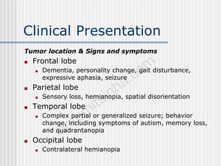
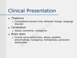
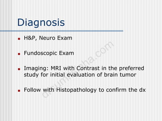
Primary CNS Tumors in
Adults
Statistics
■ Account fo 2% of all cancers in U.S. adults
■ 18,000 new diagnoses each year
■ Result in >12,000 deaths/year
■ Incidence higher in men than women
■ Lifetime risk: .65% in men and .5% in women
■ Incidence peaks at 65-79 y/o
■ 2 x incidence of GBM in Caucasions compared to
African-Americans
Risk Factors: Environmental
■ PROVEN:
■ High Dose Ionizing Radiation
■ UNPROVEN:
■ ETOH/Tobacco use
■ Cell Phones
■ Chemical Agents: Hair dye, pesticides
■ Head trauma
■ Infections: Toxoplasma gondii
■ Nitrosamine consumption
■ Occupational exposures: petroleum, vinyl chloride
Risk Factors: Genetic
■ LiFraumeni Syndrome (p53 mutation)
■ MEN Type 1
■ Neurofibromatosis 1 and 2
■ Neval Basal Cell CA Syndrome
■ Tuberous Sclerosis
■ Turcot’s Syndrome
■ Von Hippel-Lindau Syndrome
Types of Brain Neoplasms
■ Classified based on cellular origin and histologic
classification.
■ Neuroglial Tumors: 80% of all Brain Tumors
■ Divided into 4 Grades:
• Low Grade:I,II High Grade: III, IV
■ Astrocytes
■ Oligodendrocytes
■ Ependymal cells
■ GBM: Grade IV Astrocytoma
• Most common glioma and most common 1 brain tumor.
• Poor prognosis
• Found in Cerebral Hemispheres, can cross corpus callosum
Butterfly Glioma
Types
■ Oligodendroglioma
■ Rare, slow growing, Often found in Frontal
lobes.
■ Oligodendrocytes: often calcified in
oligodendroglioma, fried-egg appearance
Oligodendroglioma
Types
■ Schwannoma
■ 3rd most common Primary Brain Tumor
■ Often localized to 8th nerve: Acoustic
Schwannoma
■ Resectable
■ B/L Schwannoma associated with
Neurofibromatosis Type 2
Types
■ Meningiomas: 20% of all Primary Brain Tumors
■ Most often occurs in convexities of hemispheres and
parasagittal region
■ Arises from arachnoid cells external to brain
■ Resectable
■ Lymphomas
■ Incidence increasing in U.S.
■ Occurs in patients with Immunodeficiency Syndromes
Meningioma
Clinical Presentation
Sign or symptom/Percentage with the sign or sx
Headache 56
Memory loss 35
Cognitive changes 34
Motor deficit 33
Language deficit 32
Seizures 32
Clinical Presentation
Personality change 23
Visual problems 22
Changes in consciousness 16
Nausea or vomiting 13
Sensory deficit 13
Papilledema 5
Clinical Presentation
Tumor location & Signs and symptoms
■ Frontal lobe
■ Dementia, personality change, gait disturbance,
expressive aphasia, seizure
■ Parietal lobe
■ Sensory loss, hemianopia, spatial disorientation
■ Temporal lobe
■ Complex partial or generalized seizure; behavior
change, including symptoms of autism, memory loss,
and quadrantanopia
■ Occipital lobe
■ Contralateral hemianopia
Clinical Presentation
■ Thalamus
■ Contralateral sensory loss, behavior change, language
disorder
■ Cerebellum
■ Ataxia, dysmetria, nystagmus
■ Brain stem
■ Cranial nerve dysfunction, ataxia, pupillary
abnormalities, nystagmus, hemiparesis, autonomic
dysfunction
Diagnosis
■ H&P, Neuro Exam
■ Fundoscopic Exam
■ Imaging: MRI with Contrast in the preferred
study for initial evaluation of brain tumor
■ Follow with Histopathology to confirm the dx
Staging
■ No standard system of staging for
Primary Brain Tumors.
Treatment
■ Surgery
■ Radiation Therapy
■ Chemotherapy
Surgery
■ Obtain tissue for dx or Resection
■ Often curative for Pituitary Adenomas,
Meningioma, Schwannoma
■ The decision to perform a complete resection is
based on the location and extent of the tumor,
histopathology, and comorbid conditions
Radiation Tx
■ Postsurgical radiation is the standard treatment
for high-grade gliomas
■ Its role in the treatment of low-grade gliomas is
controversial
Chemotherapy
■ High Grade Gliomas:
■ Temozolomide and Radiation
■ GBM:
■ Silencing the MGMT gene: methyl guanine, methyl
transferase gene results in resistance to Alkylating and
Methylating agents.
■ Recurrent Malignant Gliomas:
■ Camptosar, Avastin, Gleevec, Tarceva

Cns tumors.ppt

  • 1.
  • 2.
    Statistics ■ Account fo2% of all cancers in U.S. adults ■ 18,000 new diagnoses each year ■ Result in >12,000 deaths/year ■ Incidence higher in men than women ■ Lifetime risk: .65% in men and .5% in women ■ Incidence peaks at 65-79 y/o ■ 2 x incidence of GBM in Caucasions compared to African-Americans
  • 3.
    Risk Factors: Environmental ■PROVEN: ■ High Dose Ionizing Radiation ■ UNPROVEN: ■ ETOH/Tobacco use ■ Cell Phones ■ Chemical Agents: Hair dye, pesticides ■ Head trauma ■ Infections: Toxoplasma gondii ■ Nitrosamine consumption ■ Occupational exposures: petroleum, vinyl chloride
  • 4.
    Risk Factors: Genetic ■LiFraumeni Syndrome (p53 mutation) ■ MEN Type 1 ■ Neurofibromatosis 1 and 2 ■ Neval Basal Cell CA Syndrome ■ Tuberous Sclerosis ■ Turcot’s Syndrome ■ Von Hippel-Lindau Syndrome
  • 5.
    Types of BrainNeoplasms ■ Classified based on cellular origin and histologic classification. ■ Neuroglial Tumors: 80% of all Brain Tumors ■ Divided into 4 Grades: • Low Grade:I,II High Grade: III, IV ■ Astrocytes ■ Oligodendrocytes ■ Ependymal cells ■ GBM: Grade IV Astrocytoma • Most common glioma and most common 1 brain tumor. • Poor prognosis • Found in Cerebral Hemispheres, can cross corpus callosum
  • 6.
  • 7.
    Types ■ Oligodendroglioma ■ Rare,slow growing, Often found in Frontal lobes. ■ Oligodendrocytes: often calcified in oligodendroglioma, fried-egg appearance
  • 8.
  • 9.
    Types ■ Schwannoma ■ 3rdmost common Primary Brain Tumor ■ Often localized to 8th nerve: Acoustic Schwannoma ■ Resectable ■ B/L Schwannoma associated with Neurofibromatosis Type 2
  • 10.
    Types ■ Meningiomas: 20%of all Primary Brain Tumors ■ Most often occurs in convexities of hemispheres and parasagittal region ■ Arises from arachnoid cells external to brain ■ Resectable ■ Lymphomas ■ Incidence increasing in U.S. ■ Occurs in patients with Immunodeficiency Syndromes
  • 11.
  • 12.
    Clinical Presentation Sign orsymptom/Percentage with the sign or sx Headache 56 Memory loss 35 Cognitive changes 34 Motor deficit 33 Language deficit 32 Seizures 32
  • 13.
    Clinical Presentation Personality change23 Visual problems 22 Changes in consciousness 16 Nausea or vomiting 13 Sensory deficit 13 Papilledema 5
  • 14.
    Clinical Presentation Tumor location& Signs and symptoms ■ Frontal lobe ■ Dementia, personality change, gait disturbance, expressive aphasia, seizure ■ Parietal lobe ■ Sensory loss, hemianopia, spatial disorientation ■ Temporal lobe ■ Complex partial or generalized seizure; behavior change, including symptoms of autism, memory loss, and quadrantanopia ■ Occipital lobe ■ Contralateral hemianopia
  • 15.
    Clinical Presentation ■ Thalamus ■Contralateral sensory loss, behavior change, language disorder ■ Cerebellum ■ Ataxia, dysmetria, nystagmus ■ Brain stem ■ Cranial nerve dysfunction, ataxia, pupillary abnormalities, nystagmus, hemiparesis, autonomic dysfunction
  • 16.
    Diagnosis ■ H&P, NeuroExam ■ Fundoscopic Exam ■ Imaging: MRI with Contrast in the preferred study for initial evaluation of brain tumor ■ Follow with Histopathology to confirm the dx
  • 17.
    Staging ■ No standardsystem of staging for Primary Brain Tumors.
  • 18.
    Treatment ■ Surgery ■ RadiationTherapy ■ Chemotherapy
  • 19.
    Surgery ■ Obtain tissuefor dx or Resection ■ Often curative for Pituitary Adenomas, Meningioma, Schwannoma ■ The decision to perform a complete resection is based on the location and extent of the tumor, histopathology, and comorbid conditions
  • 20.
    Radiation Tx ■ Postsurgicalradiation is the standard treatment for high-grade gliomas ■ Its role in the treatment of low-grade gliomas is controversial
  • 21.
    Chemotherapy ■ High GradeGliomas: ■ Temozolomide and Radiation ■ GBM: ■ Silencing the MGMT gene: methyl guanine, methyl transferase gene results in resistance to Alkylating and Methylating agents. ■ Recurrent Malignant Gliomas: ■ Camptosar, Avastin, Gleevec, Tarceva