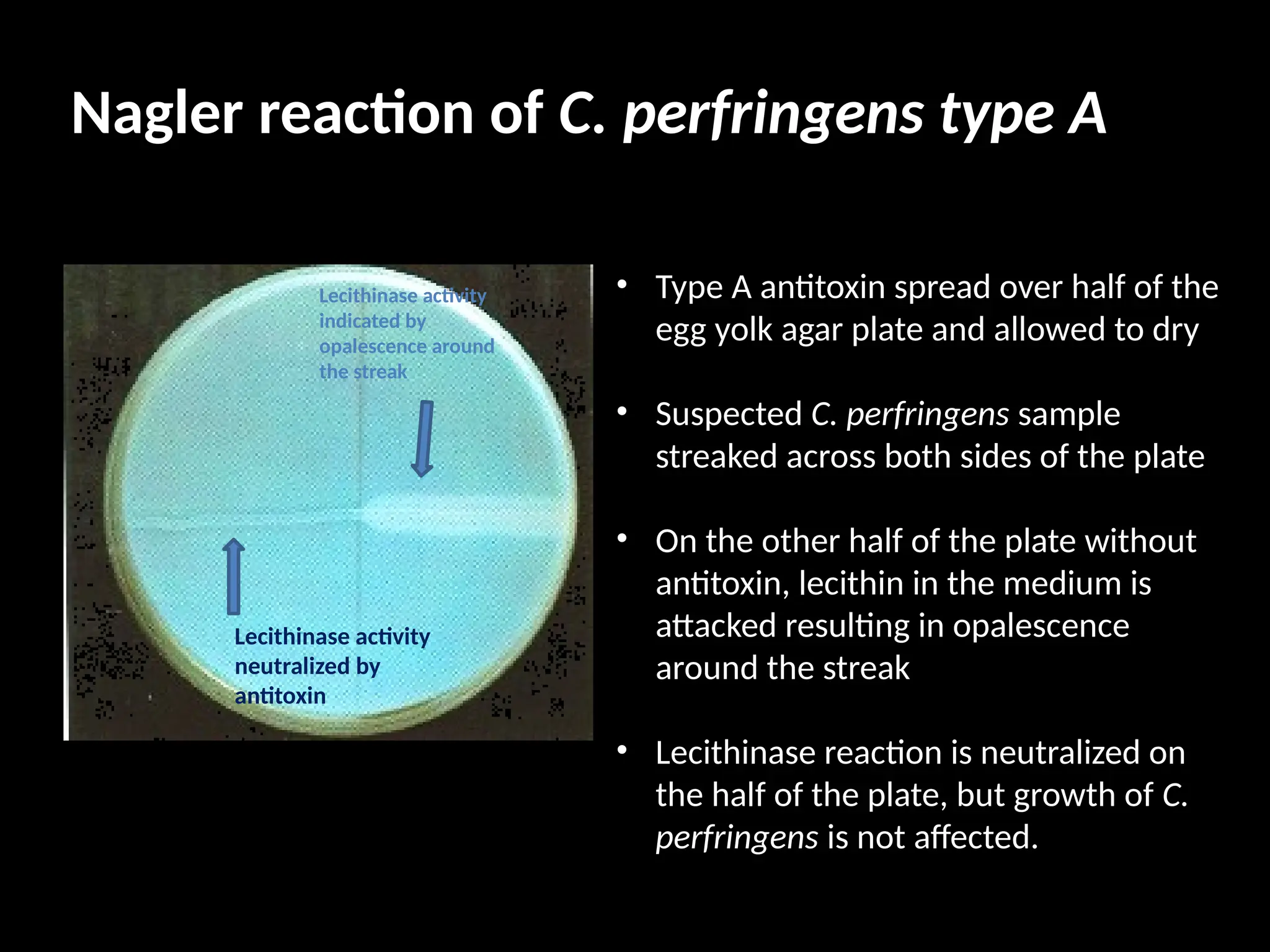
Clostridium spp
• Morphology
• Isolation, growth and colonial
characteristic
• Biochemical characteristic
• Antigenic characteristic
• Pathogenecity
• Diagnosis
Key points
• Gram positive
• Large anaerobic rods – 0.3-1.3 x 3-10 μm
• Endospore production
• Spores bulge the mother cell
• C. perfringens produces capsule in animal tissues
• Pathogenic species –straight rods
(C.spiroforme – curved/spiral)
• Motile by peritrichate flagella
(C. perfringens – non-motile)
Cont…
• Fermentative, oxidase-negative, catalase-negative
• Requires 2-10% CO2
• Enriched media – amino acids, carbohydrates,
vitamins and blood/serum.
• Optimum growth – 37°C
• Most pathogenic species produce one or more
exotoxins of varying potency.
• 80 Clostridium species – 11 species of veterinary
importance
Natural Habitat
• Soil, freshwater and marine sediments world over
• Pathogenic species - Intestinal tract of man and
animals- endogenous infection
• Other species – soil – exogenous infections –
contaminated wounds/ingestion.
Classification of Clostridium
• Neurotoxic group
• Histotoxic group
• Enteropathogenic and Enterotoxigenic group
• Clostridia associated with antibiotic induced
diseases.
A. Neurotoxic group
C. tetani – tennis racket/drumstick shaped spores due to terminal
endospores formation
C. botulinum – forms subterminal endospores
C. tetani
• Obligate anaerobe
• Stains Gram positive in fresh culture but may stain
gram negative in old cultures
• Endospores – terminal bulges “drumstick/tennis
racket” shaped
• Heat sensitive
– boiling for 15 mins – kills most strains
– autoclaving at 121° C for 15 mins – completely
sporicidal
• 11 serological type – flagellar antigen
C. Tetani - Exotoxins
• Tetanolysin (hemolysin)
• Tetanospasmin (neurotoxin)
• antigenically uniform
• plasmid-coded
• mainly responsible for clinical signs
• Primary symptom is rigidity and spastic paralysis
Pathogenecity
Endospores enters
Traumatised tissue/surgical wounds/umbilicus/uterus
Vegetative cells multiply at site of entry
Germination of spores
Ganglioside receptors of motor nerves terminals
Cells of ventral horn of spinal cord
produces tetanospasmin
Travels via peripheral nerves/bloodstream
Affects many groups of muscle at various level
Cont…
• Toxin acts presynaptically blocking synaptic inhibition
spastic paralysis
tetanic spasm
Ascending tetanus toxin travels up a regional motor nerve in a limb
tetanus develops first in the muscles of the limb
spreads to the opposite limb
moves upwards
Descending tetanus toxin circulating in bloodstream
affects susceptible motor nerve centres of
head
Neck
Limb
Eg. Dogs and cats
Eg. Humans and Horses
Laboratory Diagnosis
1. Direct Microscopy
2. Isolation
3. Identification
 Colonial morphology
 Biochemical
 Toxin identification
Direct Microscopy
Gram-staining – drumstick / tennis racket spores
Isolation
Sample – necrotic tissue/ exudates from wound
– heat to 80C for 20 mins
Blood agar (BA) plate /Stiff agar (BA with 3% agar)
or
Thioglycollate medium / cooked meat broth (CMB)
(subculture onto BA plates after 2-3 days of inoculation)
Plates incubated at 37°C for 3-4 days under H2 and
CO2 atmosphere
Colonial Morphology
Spreading growth and
narrow zone of beta -
hemolysis on sheep
blood agar
Stiff sheep blood agar
with 3 % agar to
prevent spreading
giving characteristic
rhizoid colonies
Spot inoculation
showing characteristic
spreading and
swarming growth on
normal blood agar with
1.5 % agar.
(Oblique illumination)
Biochemical identification
• Liquefies gelatin
• Does not ferment sugars
Toxin identification
• Toxin – animal serum/filtrate
from CMB or thioglycolate
medium
• Demonstration and
identification
• Neutralization test
• Protection test –
Animal showing no tetanospasmin activity
Inoculate antitoxin 2 hrs before
Inject toxin intramuscularly on the right hind leg
Animal showing tetanospasmin
activity
C. Botulinum
• Straight rods – 0.9-1.2 x 4-6 μm
• Obligate anaerobe – <2 % CO2 for growth
• Tolerates traces of oxygen due to Superoxide dismutase
• pH near or above neutral – produce oval subterminal spores
• Produces neurotoxins only during sporulation under anaerobic
environment
• Tryptose sulfite cyclosporine (TSC) growth media for isolation
• Lipase negative
• Lactose cannot be used as primary carbon source (characteristic
biochemical identification)
C. Botulinum - neurotoxins
• Eight toxigenic types of the organism exist, each producing an
immunologically distinct form of botulinum toxin.
• The toxins are designated A, B, C1, D, E, F, and G).
• Primary symptoms – weakness and flaccid paralysis
• Not all strains of C. botulinum produce the botulinum toxin.
Toxins are identical in action but differs in
potency, distribution and antigenecity
Pathogenecity
Intestinal tract bloodstream peripheral nerves
binds to susceptible cells
suppresses release of Ach at myoneural junction
Flaccid paralysis, death by circulatory failure and respiratory paralysis
Alternative mode of acquisition of toxin
1. Wound botulism/ toxicoinfection eg., shaker foal syndrome
2. Infant botulism – spores germinate in intestine eg. floppy baby
syndrome
3. Intraintestinal toxicoinfection
Toxin absorbed
Laboratory diagnosis
1. History and Clinical signs
2. Demonstration and identification of toxins
3. Direct Microscopy
4. Isolation
5. Identification
 Colonial morphology
 Biochemical
Direct Microscopy
Gram-staining – subterminal oval spore
Vegetative
rods
Subterminal
spore
Gram-positive C. botulinum
A. Demonstration of toxin from animals
Sample – Serum/centrifuged serous exudates
Inoculation into mice – i.v (0.3 ml) or i.p (0.5 ml)
“Wasp waist” appearance in mice
(abdominal breathing because of paralysis of respiratory muscles)
few hours – 5 days
PICTURE
A. Demonstration of toxin from foodstuffs
Food product – maceration overnight in saline
Centrifuge the suspension and filter through 0.45μm filter
Add 1% trypsin to filtrate (1:9)
(toxin can be in protoxin form hence treated)
Incubate the treated filtrate at 37C for 45 mins
Inoculation mice/guinea pigs – i.p (0.5 ml)
Isolation from foodstuffs
Food product – maceration in small amount of saline
Heat the suspension at 65-80°C for 30 mins
(this will kill most of contaminating organisms and induce spores to germinate)
Inoculate the suspension onto BA plates
Incubate under H2+ CO2 at 37°C for up to 5 days
Identification of toxin
• Mouse/guinea pigs neutralization tests
i. Polyvalent antitoxin – initially
ii. Monovalent antitoxin – specific neutralization of toxin
Determination of toxin-producing strain from isolates
suspected colonies inoculated into cooked meat broth
Incubate at 30°C for 5-10 days
Prepare filtrates from the suspension
Inoculate filtrates into laboratory animals to determine and identify
the toxin
Identification of toxin
Mouse/guinea pigs neutralization tests
i. Polyvalent antitoxin – initially
ii. Monovalent antitoxin – specific neutralization of toxin
C. botulinum type C on egg yolk medium
showing pearly layer around the colonies
due to lipase activity after 72 hrs
incubation
Colonial morphology
Optimum pH : 7.0 – 7.6
Optimum temperature : 30 – 37°C
C. botulinum type C on BA plate showing beta-
hemolysis and irregular heaped colony with
granular surface
Colonies on BA vary in appearance
from slightly domed with a ragged
edge to flat and rough or film-like
growth
Biochemical Tests
Suspect colonies are identified by biochemical tests
C. botulinum - 4 cultural types
Difference between tetanus toxin and botulinum toxin
Stiff legs, opisthotonus, raised tail
head, pyogenic infection of
umbilicus
Flaccid paralysis of wings and legs
HISTOTOXIC CLOSTRIDIUM GROUP
LECTURE - II
Pathogenesis
• Toxins produced not as potent as neurotoxic clostridium
• Gas gangrene bacteria are invasive
• Disease syndrome – simple wound infection , anaerobic
cellulitis to severe fatal gas-gangrene
• Infections – endogenous or exogenous
• Endogenous infection – C. chauvoei
• Exogenous infection – C. septicum
Endogenous infection
ingested endospores intestine lymphatics /bloodstream
muscle masses
(hindquarters/cardiac muscle)
tissue necrosis
germination of spore
Production of toxins
(localised damage)
Anaerobic condition, supply of amino
acids & other nutrients for vegetative cells
Exogenous infection
Endospores introduced into wounds
muscle masses
(hindquarters/cardiac muscle)
tissue necrosis
germination of spore
Production of toxins
(localised damage)
Terminal toxaemia and bacteraemia
Anaerobic condition, supply of amino
acids & other nutrients for vegetative
cells
Eg: Braxy – mucosa of
abomasum damaged
(Cold condn
from adjacent
rumen filled with frozen foods)
C. septicum spores germinate
Replication leads to toxin
production
Toxaemia & rapid death
Gas-Gangrene Group
Laboratory Diagnosis
1. Fluorescent antibody test
2. Gram-stained impression smears
3. Isolation and colonial appearance
4. Biochemical reactions
Fluorescent Antibody Test
smears of affected tissue or bone marrow from a rib
fixed in acetone
Stained with Fluorescein-labelled specific antisera
• C. chauvoei
• C. septicum
• C. novyi
• C. sordelli
Fluorescent antibody test (FAT) that detects C.
chauvoei in culture smears and tissue impressions
obtained directly during necropsy
Gram-stained impression smear
Isolation
• Sheep blood agar + liver extract – C. chauvoei
• Stiff blood agar/normal blood agar – C. septicum and
C. sordelli
• Normal blood agar – C. novyi type A and C.
perfringens
C. novyi – green halo on chocolate agar
– blackening on benzidine blood agar
Colonial appearance
C. perfringens with “target”
hemolysis
C. septicum with
swarming colonies
Nagler reaction of C. perfringens type A
• Type A antitoxin spread over half of the
egg yolk agar plate and allowed to dry
• Suspected C. perfringens sample
streaked across both sides of the plate
• On the other half of the plate without
antitoxin, lecithin in the medium is
attacked resulting in opalescence
around the streak
• Lecithinase reaction is neutralized on
the half of the plate, but growth of C.
perfringens is not affected.
Lecithinase activity
neutralized by
antitoxin
Lecithinase activity
indicated by
opalescence around
the streak
CAMP reaction of C. perfringens type A
S. agalactiae (vertical streak) enhances
partial hemolysis produced by alpha
toxin of C. perfringens on BA
A diffusible factor produced by S. agalactiae
(vertical streak) enhances partial hemolysis
produced by alpha toxin of C. perfringens on
BA
Complete zone of hemolysis seen
immediately around C. perfringens is caused
by theta toxin
Biochemical characteristics
Histotoxic clostridia affecting liver
Pathogenesis
Spores in intestine liver remain dormant in kupffer cells
trauma to liver
tissue damage to liver
Anaerobic condition
Spores germination
Toxin production
Migrating liver fluke
Replication of clostridia
Toxaemia/bacteraemia
Death
Pathogenesis
Toxins
Alpha Beta Gamma Delta Epsilon Zeta
C. novyi type A + - + + + -
C. novyi type B + + - - - +
C. novyi type C - - + - - -
C. Hemolyticum - +++
• Alpha toxin – lethal, necrotising, oedmatising
(threshold conc = 5ng/ml (5 parts per billion) with 50 % of cells rounded)
• Beta toxin – hemolytic, necrotising lecithinase (phospholipase)
• Gamma toxin – hemolytic, lecithinase
• Delta toxin – oxygen labile hemolysin
• Epsilon toxin – lecitho-vitelin and responsible for pearly layer
around cultures
• Zeta toxin – hemolysin
Toxins
Laboratory Diagnosis
1. Direct Gram Stained smear
2. Fluorescent antibody technique
3. Isolation
4. Biochemical reactions
5. Animal inoculation
Direct Gram-stained smear
C. novyi type B
• presence of characteristic liver lesion
• Large numbers of Gram-positive rods in liver
impression smears
C. hemolyticum
• Large gram-positive rods 0.8-1.0 x 3 – 10 μm
• Produce oval to cylindrical, subterminal spores
with bulging from mother cell.
C. colinum
• Gram-positive rods 1.0 μm diameter and 3 – 4 μm long
• Produce oval subterminal spores
• Sporulation infrequent
C. novyi type B and C. hemolyticum
• Acetone fixed liver impression smear
• Large numbers of Gram-positive rods
Fluorescent antibody technique
C. novyi type B and C. hemolyticum
• Very strict anaerobes – die
within 15 mins of exposure to oxygen
• Colonies are hemolytic,
small and usually rhizoid in nature
Isolation
Isolating and identifying C novyi is difficult due to its extreme
anaerobic nature. It is also fastidious and difficult to culture, requiring
the presence of thiols.
C. colinum
• Fastidious and difficult to isolate
• Polymixin B (25 μg/ml) to suppress contaminants
• Enriched media
• Tryptose-phosphate-glucose broth with 8% sterile citrated
horse plasma
• Thioglycollate broth with 3-10% horse serum
• 5-8 days fertile chicken eggs
• Several passage required
• Clostridium grown on Blood agar
Animal inoculation
Liver homogenate preparation
Homogenate + 5% calcium chloride
i.m injection of homogenates in guinea pigs
Death in 1-2 days
Enterotoxigenic/Enteropathogenic group
• Cause enterotoxaemias – acute, highly fatal intoxications
• Major exotoxins – C. perfringens type B, C and D
– occasionally type A and E
• Type A – intestinal tract of humans and animals, most soils.
• Type B and E – more adapted to survival in intestines
– survive in soil during outbreak to infect other
animals
Natural Habitat
C. perfringens
• Relatively aerotolerant
• Nonmotile
• Polysaccharide capsule in tissue
• Short fat Gram-positive rod (0.6 – 0.8 x 2 – 4 μm)
• Spores are oval, subterminal and bulge mother cell
• 5 types (A-E) based on different combination of
toxins
Characteristic reaction –
• Double zone hemolysis
• Stormy clot in litmus milk medium
• Nagler reaction
Double zone of
hemolysis
Stormy clot in litmus
milk medium
Nagler reaction
Growth on
Thioglycollate broth
Abrupt changes in feeding
(rich diet/overeating/ voracity on high protein and energy-rich foods)
1. slowing of peristalsis + retention of bacteria in intestines
absorption of toxins
2. Inadequately digested CHO
3. Provision of rich medium for proliferation of the bacteria
overgrowth of bacteria in large intestine
Spill over into the small intestine
Production of large amount of toxin and enterotoxaemia
Factors that precipitate enterotoxaemia
Pathogenesis:
based on the type of toxins produced
by the bacteria
1. Major toxins
1. alpha – Lecithinase (Phospholipase)
- zone of partial hemolysis on BA
- Nagler reaction based on
neutralization of lecithinase activity
2. beta – lethal and necrotizing
- labile toxin, sensitive to trypsin
(predilection of type B and C for neonates due to
colostral anti-trypsin activity)
- most important factor in
enterotoxaemia by type B
“epsilon” –
secreted as protoxin toxin
absorption of toxin gut permeability
Damages vascular endothelium loss of fluid, edema
trypsin
intestines
“iota” – secreted as protoxin toxin
also produced by C. spiroforme and C. difficile
trypsin
intestines
(including blood vessels in brain)
Enterotoxin + Neurotoxin
(Red water)
Minor toxin – contribute to tissue damage
• theta - hemolysin
• kappa - collagenase
• Mu - hyaluronidase
• Nu - DNase
Laboratory Diagnosis
Definitive test – demonstration and identification of toxin
Misc test – useful adjuncts for diagnosis
1. Gram-stained smear of S.I mucosa
2. Histopathology of brain section
3. Glycosuria
Miscellaneous Tests
• Gram-stained smear of S.I mucosa
• large no. of fat, Gram-positive rods
• Histopathology of brain section
• characteristic focal symmetrical encephalomalacia
• Glycosuria
• Presence of Glucose in urine
Demonstration of Toxins
20 – 30ml ileal content of recently dead animal
Centrifuge ileal contents
(if sample too mucoid, place cotton wool at the top of the centrifuge tube)
Collect clear supernatant
(epsilon and iota toxins are usually in active form in ileal sample)
Inoculate clarified ileal content into two mice each @ 0.4 ml, i.v
Death within 5 minutes : due to shock
Death within 10 hours : due to toxin
Identification of Toxins – neutralization test
Clarified ileal content of recently dead animal
Inoculate 0.5 ml clarified ileal content + 0.1 ml commercial antitoxins
i.v into mice @ 0.4ml
i.d into white, shaved, young guinea pigs @0.2 ml
Death: No neutralization
No death: toxin neutralization by antitoxin
Neutralization of toxins in ileal contents by antitoxins
Clostridia associated with antibiotic-induced diseases
• C. spiroforme
• C. difficile
Antibiotics
Disturbance to the normal intestinal microbiota
Depletion of the barrier effect
Colonization by pathogenic bacteria.
Full fledged infection
Clostridium spiroforme
• Blood agar – Loosely coiled, spiral Gram-positive form
• Fecal/caecal content – semicircular form
• Spores – terminal
• Cultural characteristic on BA
• Non-hemolytic
• Convex, circular, shiny, whitish to grey colonies
• Optimum temperature – 37°C
• Toxin –
• cytotoxin
• Exotoxin – identical to iota toxin of C. perfringens
• Disease – diarhoea in weaning rabbits
• Diagnosis – demonstration and identification of toxin in mice
Clostridium difficile
• Large Gram-positive rods (0.5 X 3-6μm)
• Spores – oval, subterminal
• Isolation – special BA enriched with
yeast extract, Haemin, Vitamin K, Cysteine, Antimicrobials
• Colony characteristic on BA
• Non-hemolytic
• Raised, rhizoid edged colony
• Toxin –
• Enterotoxin – designated as toxin A
• Cytotoxin – designated as toxin B
• Disease – enterocolitis in human, hamster, rabbit, guinea pig
– natural diarrhoea in dogs, foals and pigs
• Diagnosis –detection of toxin A and B in fecal samples by EIA
Biochemical reactions
BACK
Important features of spores Clinical implications
Spores are highly resistant to heating; spores
are not kiled by boiling (100C) but are killed
at 121C by autoclaving.
Medical supplies must be steam sterilized at
121C at least for 15 mins.
Spores are highly resistant to many
chemicals, inclulding most disinfectants.
Only solution designated as sporicidal will kill
spores
Spores can survive for many years in soil and
other inanimate objects.
Wound contamination with soils can be
infected with spores and cause diseases such
as tetanus and gas gangrene.
Spores do not exhibit measurable metabolic
activity.
Antibiotics are ineffective against spores.
Spores formed only when nutrients are
insufficient.
Spores are not often found at the site of
infection because nutrients are not limiting.

Clostridium ppt for undergraduate class

  • 1.
    Clostridium spp • Morphology •Isolation, growth and colonial characteristic • Biochemical characteristic • Antigenic characteristic • Pathogenecity • Diagnosis
  • 2.
    Key points • Grampositive • Large anaerobic rods – 0.3-1.3 x 3-10 μm • Endospore production • Spores bulge the mother cell • C. perfringens produces capsule in animal tissues • Pathogenic species –straight rods (C.spiroforme – curved/spiral) • Motile by peritrichate flagella (C. perfringens – non-motile)
  • 3.
    Cont… • Fermentative, oxidase-negative,catalase-negative • Requires 2-10% CO2 • Enriched media – amino acids, carbohydrates, vitamins and blood/serum. • Optimum growth – 37°C • Most pathogenic species produce one or more exotoxins of varying potency. • 80 Clostridium species – 11 species of veterinary importance
  • 4.
    Natural Habitat • Soil,freshwater and marine sediments world over • Pathogenic species - Intestinal tract of man and animals- endogenous infection • Other species – soil – exogenous infections – contaminated wounds/ingestion.
  • 5.
    Classification of Clostridium •Neurotoxic group • Histotoxic group • Enteropathogenic and Enterotoxigenic group • Clostridia associated with antibiotic induced diseases.
  • 6.
    A. Neurotoxic group C.tetani – tennis racket/drumstick shaped spores due to terminal endospores formation C. botulinum – forms subterminal endospores
  • 7.
    C. tetani • Obligateanaerobe • Stains Gram positive in fresh culture but may stain gram negative in old cultures • Endospores – terminal bulges “drumstick/tennis racket” shaped • Heat sensitive – boiling for 15 mins – kills most strains – autoclaving at 121° C for 15 mins – completely sporicidal • 11 serological type – flagellar antigen
  • 8.
    C. Tetani -Exotoxins • Tetanolysin (hemolysin) • Tetanospasmin (neurotoxin) • antigenically uniform • plasmid-coded • mainly responsible for clinical signs • Primary symptom is rigidity and spastic paralysis
  • 9.
    Pathogenecity Endospores enters Traumatised tissue/surgicalwounds/umbilicus/uterus Vegetative cells multiply at site of entry Germination of spores Ganglioside receptors of motor nerves terminals Cells of ventral horn of spinal cord produces tetanospasmin Travels via peripheral nerves/bloodstream Affects many groups of muscle at various level
  • 10.
    Cont… • Toxin actspresynaptically blocking synaptic inhibition spastic paralysis tetanic spasm Ascending tetanus toxin travels up a regional motor nerve in a limb tetanus develops first in the muscles of the limb spreads to the opposite limb moves upwards Descending tetanus toxin circulating in bloodstream affects susceptible motor nerve centres of head Neck Limb Eg. Dogs and cats Eg. Humans and Horses
  • 11.
    Laboratory Diagnosis 1. DirectMicroscopy 2. Isolation 3. Identification  Colonial morphology  Biochemical  Toxin identification
  • 12.
    Direct Microscopy Gram-staining –drumstick / tennis racket spores
  • 13.
    Isolation Sample – necrotictissue/ exudates from wound – heat to 80C for 20 mins Blood agar (BA) plate /Stiff agar (BA with 3% agar) or Thioglycollate medium / cooked meat broth (CMB) (subculture onto BA plates after 2-3 days of inoculation) Plates incubated at 37°C for 3-4 days under H2 and CO2 atmosphere
  • 14.
    Colonial Morphology Spreading growthand narrow zone of beta - hemolysis on sheep blood agar Stiff sheep blood agar with 3 % agar to prevent spreading giving characteristic rhizoid colonies Spot inoculation showing characteristic spreading and swarming growth on normal blood agar with 1.5 % agar. (Oblique illumination)
  • 15.
    Biochemical identification • Liquefiesgelatin • Does not ferment sugars
  • 16.
    Toxin identification • Toxin– animal serum/filtrate from CMB or thioglycolate medium • Demonstration and identification • Neutralization test • Protection test – Animal showing no tetanospasmin activity Inoculate antitoxin 2 hrs before Inject toxin intramuscularly on the right hind leg Animal showing tetanospasmin activity
  • 17.
    C. Botulinum • Straightrods – 0.9-1.2 x 4-6 μm • Obligate anaerobe – <2 % CO2 for growth • Tolerates traces of oxygen due to Superoxide dismutase • pH near or above neutral – produce oval subterminal spores • Produces neurotoxins only during sporulation under anaerobic environment • Tryptose sulfite cyclosporine (TSC) growth media for isolation • Lipase negative • Lactose cannot be used as primary carbon source (characteristic biochemical identification)
  • 18.
    C. Botulinum -neurotoxins • Eight toxigenic types of the organism exist, each producing an immunologically distinct form of botulinum toxin. • The toxins are designated A, B, C1, D, E, F, and G). • Primary symptoms – weakness and flaccid paralysis • Not all strains of C. botulinum produce the botulinum toxin. Toxins are identical in action but differs in potency, distribution and antigenecity
  • 19.
    Pathogenecity Intestinal tract bloodstreamperipheral nerves binds to susceptible cells suppresses release of Ach at myoneural junction Flaccid paralysis, death by circulatory failure and respiratory paralysis Alternative mode of acquisition of toxin 1. Wound botulism/ toxicoinfection eg., shaker foal syndrome 2. Infant botulism – spores germinate in intestine eg. floppy baby syndrome 3. Intraintestinal toxicoinfection Toxin absorbed
  • 20.
    Laboratory diagnosis 1. Historyand Clinical signs 2. Demonstration and identification of toxins 3. Direct Microscopy 4. Isolation 5. Identification  Colonial morphology  Biochemical
  • 21.
    Direct Microscopy Gram-staining –subterminal oval spore Vegetative rods Subterminal spore Gram-positive C. botulinum
  • 22.
    A. Demonstration oftoxin from animals Sample – Serum/centrifuged serous exudates Inoculation into mice – i.v (0.3 ml) or i.p (0.5 ml) “Wasp waist” appearance in mice (abdominal breathing because of paralysis of respiratory muscles) few hours – 5 days PICTURE
  • 23.
    A. Demonstration oftoxin from foodstuffs Food product – maceration overnight in saline Centrifuge the suspension and filter through 0.45μm filter Add 1% trypsin to filtrate (1:9) (toxin can be in protoxin form hence treated) Incubate the treated filtrate at 37C for 45 mins Inoculation mice/guinea pigs – i.p (0.5 ml)
  • 24.
    Isolation from foodstuffs Foodproduct – maceration in small amount of saline Heat the suspension at 65-80°C for 30 mins (this will kill most of contaminating organisms and induce spores to germinate) Inoculate the suspension onto BA plates Incubate under H2+ CO2 at 37°C for up to 5 days Identification of toxin • Mouse/guinea pigs neutralization tests i. Polyvalent antitoxin – initially ii. Monovalent antitoxin – specific neutralization of toxin
  • 25.
    Determination of toxin-producingstrain from isolates suspected colonies inoculated into cooked meat broth Incubate at 30°C for 5-10 days Prepare filtrates from the suspension Inoculate filtrates into laboratory animals to determine and identify the toxin Identification of toxin Mouse/guinea pigs neutralization tests i. Polyvalent antitoxin – initially ii. Monovalent antitoxin – specific neutralization of toxin
  • 26.
    C. botulinum typeC on egg yolk medium showing pearly layer around the colonies due to lipase activity after 72 hrs incubation Colonial morphology Optimum pH : 7.0 – 7.6 Optimum temperature : 30 – 37°C C. botulinum type C on BA plate showing beta- hemolysis and irregular heaped colony with granular surface Colonies on BA vary in appearance from slightly domed with a ragged edge to flat and rough or film-like growth
  • 27.
    Biochemical Tests Suspect coloniesare identified by biochemical tests C. botulinum - 4 cultural types
  • 28.
    Difference between tetanustoxin and botulinum toxin Stiff legs, opisthotonus, raised tail head, pyogenic infection of umbilicus Flaccid paralysis of wings and legs
  • 29.
  • 30.
    Pathogenesis • Toxins producednot as potent as neurotoxic clostridium • Gas gangrene bacteria are invasive • Disease syndrome – simple wound infection , anaerobic cellulitis to severe fatal gas-gangrene • Infections – endogenous or exogenous • Endogenous infection – C. chauvoei • Exogenous infection – C. septicum
  • 31.
    Endogenous infection ingested endosporesintestine lymphatics /bloodstream muscle masses (hindquarters/cardiac muscle) tissue necrosis germination of spore Production of toxins (localised damage) Anaerobic condition, supply of amino acids & other nutrients for vegetative cells
  • 32.
    Exogenous infection Endospores introducedinto wounds muscle masses (hindquarters/cardiac muscle) tissue necrosis germination of spore Production of toxins (localised damage) Terminal toxaemia and bacteraemia Anaerobic condition, supply of amino acids & other nutrients for vegetative cells Eg: Braxy – mucosa of abomasum damaged (Cold condn from adjacent rumen filled with frozen foods) C. septicum spores germinate Replication leads to toxin production Toxaemia & rapid death
  • 34.
  • 35.
    Laboratory Diagnosis 1. Fluorescentantibody test 2. Gram-stained impression smears 3. Isolation and colonial appearance 4. Biochemical reactions
  • 36.
    Fluorescent Antibody Test smearsof affected tissue or bone marrow from a rib fixed in acetone Stained with Fluorescein-labelled specific antisera • C. chauvoei • C. septicum • C. novyi • C. sordelli Fluorescent antibody test (FAT) that detects C. chauvoei in culture smears and tissue impressions obtained directly during necropsy
  • 37.
  • 38.
    Isolation • Sheep bloodagar + liver extract – C. chauvoei • Stiff blood agar/normal blood agar – C. septicum and C. sordelli • Normal blood agar – C. novyi type A and C. perfringens C. novyi – green halo on chocolate agar – blackening on benzidine blood agar
  • 39.
    Colonial appearance C. perfringenswith “target” hemolysis C. septicum with swarming colonies
  • 40.
    Nagler reaction ofC. perfringens type A • Type A antitoxin spread over half of the egg yolk agar plate and allowed to dry • Suspected C. perfringens sample streaked across both sides of the plate • On the other half of the plate without antitoxin, lecithin in the medium is attacked resulting in opalescence around the streak • Lecithinase reaction is neutralized on the half of the plate, but growth of C. perfringens is not affected. Lecithinase activity neutralized by antitoxin Lecithinase activity indicated by opalescence around the streak
  • 41.
    CAMP reaction ofC. perfringens type A S. agalactiae (vertical streak) enhances partial hemolysis produced by alpha toxin of C. perfringens on BA A diffusible factor produced by S. agalactiae (vertical streak) enhances partial hemolysis produced by alpha toxin of C. perfringens on BA Complete zone of hemolysis seen immediately around C. perfringens is caused by theta toxin
  • 42.
  • 43.
  • 44.
    Pathogenesis Spores in intestineliver remain dormant in kupffer cells trauma to liver tissue damage to liver Anaerobic condition Spores germination Toxin production Migrating liver fluke Replication of clostridia Toxaemia/bacteraemia Death
  • 45.
    Pathogenesis Toxins Alpha Beta GammaDelta Epsilon Zeta C. novyi type A + - + + + - C. novyi type B + + - - - + C. novyi type C - - + - - - C. Hemolyticum - +++
  • 46.
    • Alpha toxin– lethal, necrotising, oedmatising (threshold conc = 5ng/ml (5 parts per billion) with 50 % of cells rounded) • Beta toxin – hemolytic, necrotising lecithinase (phospholipase) • Gamma toxin – hemolytic, lecithinase • Delta toxin – oxygen labile hemolysin • Epsilon toxin – lecitho-vitelin and responsible for pearly layer around cultures • Zeta toxin – hemolysin Toxins
  • 47.
    Laboratory Diagnosis 1. DirectGram Stained smear 2. Fluorescent antibody technique 3. Isolation 4. Biochemical reactions 5. Animal inoculation
  • 48.
    Direct Gram-stained smear C.novyi type B • presence of characteristic liver lesion • Large numbers of Gram-positive rods in liver impression smears C. hemolyticum • Large gram-positive rods 0.8-1.0 x 3 – 10 μm • Produce oval to cylindrical, subterminal spores with bulging from mother cell. C. colinum • Gram-positive rods 1.0 μm diameter and 3 – 4 μm long • Produce oval subterminal spores • Sporulation infrequent
  • 49.
    C. novyi typeB and C. hemolyticum • Acetone fixed liver impression smear • Large numbers of Gram-positive rods Fluorescent antibody technique C. novyi type B and C. hemolyticum • Very strict anaerobes – die within 15 mins of exposure to oxygen • Colonies are hemolytic, small and usually rhizoid in nature Isolation Isolating and identifying C novyi is difficult due to its extreme anaerobic nature. It is also fastidious and difficult to culture, requiring the presence of thiols.
  • 50.
    C. colinum • Fastidiousand difficult to isolate • Polymixin B (25 μg/ml) to suppress contaminants • Enriched media • Tryptose-phosphate-glucose broth with 8% sterile citrated horse plasma • Thioglycollate broth with 3-10% horse serum • 5-8 days fertile chicken eggs • Several passage required • Clostridium grown on Blood agar
  • 51.
    Animal inoculation Liver homogenatepreparation Homogenate + 5% calcium chloride i.m injection of homogenates in guinea pigs Death in 1-2 days
  • 52.
    Enterotoxigenic/Enteropathogenic group • Causeenterotoxaemias – acute, highly fatal intoxications • Major exotoxins – C. perfringens type B, C and D – occasionally type A and E • Type A – intestinal tract of humans and animals, most soils. • Type B and E – more adapted to survival in intestines – survive in soil during outbreak to infect other animals Natural Habitat
  • 53.
    C. perfringens • Relativelyaerotolerant • Nonmotile • Polysaccharide capsule in tissue • Short fat Gram-positive rod (0.6 – 0.8 x 2 – 4 μm) • Spores are oval, subterminal and bulge mother cell • 5 types (A-E) based on different combination of toxins
  • 54.
    Characteristic reaction – •Double zone hemolysis • Stormy clot in litmus milk medium • Nagler reaction
  • 55.
  • 56.
    Stormy clot inlitmus milk medium
  • 57.
  • 58.
  • 59.
    Abrupt changes infeeding (rich diet/overeating/ voracity on high protein and energy-rich foods) 1. slowing of peristalsis + retention of bacteria in intestines absorption of toxins 2. Inadequately digested CHO 3. Provision of rich medium for proliferation of the bacteria overgrowth of bacteria in large intestine Spill over into the small intestine Production of large amount of toxin and enterotoxaemia Factors that precipitate enterotoxaemia
  • 60.
    Pathogenesis: based on thetype of toxins produced by the bacteria 1. Major toxins
  • 61.
    1. alpha –Lecithinase (Phospholipase) - zone of partial hemolysis on BA - Nagler reaction based on neutralization of lecithinase activity 2. beta – lethal and necrotizing - labile toxin, sensitive to trypsin (predilection of type B and C for neonates due to colostral anti-trypsin activity) - most important factor in enterotoxaemia by type B
  • 62.
    “epsilon” – secreted asprotoxin toxin absorption of toxin gut permeability Damages vascular endothelium loss of fluid, edema trypsin intestines “iota” – secreted as protoxin toxin also produced by C. spiroforme and C. difficile trypsin intestines (including blood vessels in brain) Enterotoxin + Neurotoxin
  • 63.
  • 64.
    Minor toxin –contribute to tissue damage • theta - hemolysin • kappa - collagenase • Mu - hyaluronidase • Nu - DNase
  • 65.
    Laboratory Diagnosis Definitive test– demonstration and identification of toxin Misc test – useful adjuncts for diagnosis 1. Gram-stained smear of S.I mucosa 2. Histopathology of brain section 3. Glycosuria
  • 66.
    Miscellaneous Tests • Gram-stainedsmear of S.I mucosa • large no. of fat, Gram-positive rods
  • 67.
    • Histopathology ofbrain section • characteristic focal symmetrical encephalomalacia • Glycosuria • Presence of Glucose in urine
  • 68.
    Demonstration of Toxins 20– 30ml ileal content of recently dead animal Centrifuge ileal contents (if sample too mucoid, place cotton wool at the top of the centrifuge tube) Collect clear supernatant (epsilon and iota toxins are usually in active form in ileal sample) Inoculate clarified ileal content into two mice each @ 0.4 ml, i.v Death within 5 minutes : due to shock Death within 10 hours : due to toxin
  • 69.
    Identification of Toxins– neutralization test Clarified ileal content of recently dead animal Inoculate 0.5 ml clarified ileal content + 0.1 ml commercial antitoxins i.v into mice @ 0.4ml i.d into white, shaved, young guinea pigs @0.2 ml Death: No neutralization No death: toxin neutralization by antitoxin
  • 70.
    Neutralization of toxinsin ileal contents by antitoxins
  • 71.
    Clostridia associated withantibiotic-induced diseases • C. spiroforme • C. difficile Antibiotics Disturbance to the normal intestinal microbiota Depletion of the barrier effect Colonization by pathogenic bacteria. Full fledged infection
  • 72.
    Clostridium spiroforme • Bloodagar – Loosely coiled, spiral Gram-positive form • Fecal/caecal content – semicircular form • Spores – terminal • Cultural characteristic on BA • Non-hemolytic • Convex, circular, shiny, whitish to grey colonies • Optimum temperature – 37°C • Toxin – • cytotoxin • Exotoxin – identical to iota toxin of C. perfringens • Disease – diarhoea in weaning rabbits • Diagnosis – demonstration and identification of toxin in mice
  • 73.
    Clostridium difficile • LargeGram-positive rods (0.5 X 3-6μm) • Spores – oval, subterminal • Isolation – special BA enriched with yeast extract, Haemin, Vitamin K, Cysteine, Antimicrobials • Colony characteristic on BA • Non-hemolytic • Raised, rhizoid edged colony • Toxin – • Enterotoxin – designated as toxin A • Cytotoxin – designated as toxin B • Disease – enterocolitis in human, hamster, rabbit, guinea pig – natural diarrhoea in dogs, foals and pigs • Diagnosis –detection of toxin A and B in fecal samples by EIA
  • 74.
  • 75.
  • 79.
    Important features ofspores Clinical implications Spores are highly resistant to heating; spores are not kiled by boiling (100C) but are killed at 121C by autoclaving. Medical supplies must be steam sterilized at 121C at least for 15 mins. Spores are highly resistant to many chemicals, inclulding most disinfectants. Only solution designated as sporicidal will kill spores Spores can survive for many years in soil and other inanimate objects. Wound contamination with soils can be infected with spores and cause diseases such as tetanus and gas gangrene. Spores do not exhibit measurable metabolic activity. Antibiotics are ineffective against spores. Spores formed only when nutrients are insufficient. Spores are not often found at the site of infection because nutrients are not limiting.

Editor's Notes

  • #76 Spore formation (sporulation) occurs when nutrients, such as sources of carbon and nitrogen are depleted.  Bacterial spores are highly resistant to Heat Dehydration Radiation and Chemicals. An endospore is structurally and chemically more complex than the vegetative cell.  It contains more layers than vegetative cells. Resistance of Bacterial spore may be mediated by dipicolinic acid, a calcium ion chelator found only in spores.  When the favorable condition prevail, (i.e. availability of water, appropriate nutrients)  spores germination occurs which forms vegetative cells of pathogenic bacteria. Following factors/constituents plays major role for the resistance of Bacterial Spore:  Calcium dipicolinate in core Keratin spore coat New enzymes (i.e., dipicolinic acid synthetase, heat-resistant catalase) Increases or decreases in other enzymes
  • #77 The shape and the position of spores vary in different species and can be useful for classification and identification purposes. Endospores may be located in the middle of the bacterium (central), at the end of the bacterium (terminal) and near the end of the bacteria (subterminal) and may be spherical or elliptical. Spores may be: Central or equatorial, giving the bacillus aspindle shape (eg. Clostridium bifermentans) Sub-terminal, the bacillus appearing Club shaped (eg. Clostridium perfringens) Oval and terminal, resembling a tennis racket (eg. Clostridium tertium) Spherical and terminal, giving a drumstick appearance (Clostridium tetani)
  • #78 Mature endospores are released from the vegetative cell to become free endospores.  When the free endospores are placed in an environment that supports growth, the endospores will revert back to a vegetative cell in a process called germination.  It should be noted that unlike the process of binary fission observed with vegetative cells, endospore formation is not a reproductive process but a process of differentiation that provides the bacteria with a mechanism for survival. Constituents of Bacterial Spores: Thick keratinlike coat Peptidoglycan Cell membrane A small amount of cytoplasm Very little water Bacterial DNA
  • #79 Spores are formed by only two genera of Gram positive rods of medical importance: Bacillus spp Clostridium spp. A variety of different microorganisms form "spores" or "cysts", but the endospores of low G+C Gram-positive bacteria are by far the most resistant to harsh conditions. .