Achieving High Yields in Scalable Xeno Free Culture Formats with Mesenchymal ...Merck Life Sciences
Watch the presentation of this webinar here: https://bit.ly/3ryE5ST
Optimize your mesenchymal stem cell growth. Join our webinar to learn more about our GMP-compliant xeno free media formulation that supports high performance expansions and compatibility with scalable xeno free manufacturing conditions.
Optimizing ex vivo cell expansion processes in preparation for clinical use is a critical step in cell therapy manufacturing. Given the curative and lifesaving impacts these therapies can have on patients, overcoming roadblocks with scalability and supply chain, using high quality raw materials are essential for therapeutic access.
The GMP-compliant Stemline® XF MSC Medium and cocktail promotes expansion of human mesenchymal stromal/stem cells (hMSCs) to high densities while maintaining cell identity and quality. This product was designed for derivation and expansion of MSCs using xeno free conditions in planar and microcarrier-based culture platforms, easing the transfer between research, clinical, and manufacturing scale cultures.
In this webinar, you will:
• Explore the current landscape and future trends of cell culture media for adult mesenchymal stem cells
• Discover ways to derive MSC's from Bone Marrow in Xeno Free conditions from static to microcarrier-based suspension culture platforms.
• Learn how Stemline® XF MSC Media provides robust performance and reduces scalability roadblocks
Presented by: Kathleen Ongena, Ph.D., Head of Customer Applications and Mark Ventresco, Cell Therapy Product Manager
Historically, genetic toxicology has been comprised of bacterial and cell based in vitro assays such as the Ames assay (a bacterial mutagenicity assay), Micronucleus and Chromosomal Aberration assays (mammalian cytogenetic assays), and Mouse Lymphoma Assay (in vitro mammalian cell gene mutation assay). These were routinely used for safety evaluation and are still part of the standard core battery. The emergence of new technologies has facilitated the development of in vitro methods for safe and effective drug and chemical testing.
This BioReliance® toxicology services webinar will explore alternative models, including 3D skin models that comply with the EC Scientific Committee on Consumer Safety (SCCS) recommendations. It will also discuss how the 3Rs (Replace, Reduce, Refine) Principle advocates the exploration of such alternative methods while achieving required goals.
In this webinar, you will learn:
• About in vitro alternatives to animal toxicity testing in pharma, chemical, tobacco, and personal care products.
• How the 3Rs (Replace, Reduce, Refine) Principle advocates exploring alternative methods without compromising the required goals.
• Alternatives to comply with the 7th Amendment to the EC Cosmetics Directive.
Risk Mitigation in Cell Line Development: Regulatory Considerations and Impac...Merck Life Sciences
In this webinar, you will learn about:
- Risk assessment approaches in upstream process development
- How early cell line development stage is linked to subsequent steps in the bioprocess to assure the quality of the final product
- Benefits of having a completely chemically defined cell line development process
Detailed description:
Chinese Hamster Ovary (CHO) cells are the preferred host for producing biotherapeutics where cell line development (CLD) is the foundation of the bioprocess. CLD processes are expected to be robust while meeting a myriad of regulatory requirements. The choice of production cell line, culture conditions, and having a chemically defined (CD) CLD process by using CD cloning media can impact the subsequent measures for the CMC (Chemistry, manufacturing, and controls).
In this presentation, we will discuss these choices and their impacts on subsequent bioprocess and CMC testing required by regulations and the benefits of incorporating CD cloning media into the CHOZN® expression platform.
Investing in Process Development for Increased MSC Production in Stirred Tank...MilliporeSigma
Interested in developing a robust cell therapy manufacturing platform? In this webinar we will share information in the form of case studies that highlight strategies to optimize your cell therapy production process.
Industry trends in regenerative medicine highlight a critical need for closed cell culture systems that support scalable manufacturing of adherent cell therapies. Typical static in vitro culture methods, however, are often too cumbersome and inefficient to support commercial scale production of mesenchymal stem/stromal cells (MSCs). Single-use stirred tank bioreactor systems are a platform that can address this limitation and have been proven effective for microcarrier-based production of adherent cell therapies. Implementation of optimized process control strategies for parameters such as dissolved oxygen (DO) and agitation rate are key to making an efficient transition from planar culture to stirred tank bioreactors. Herein, a stepwise approach to process development for MSC expansion in a small-scale single-use bioreactor is presented. Case studies focus on strategies to optimize DO control and agitation rates for bone marrow derived MSCs in microcarrier culture, highlighting improvements in process efficiency. In the first case study, the impact different gassing methods have on DO control and whether hypoxic growth conditions affect MSC function are examined. The second case study demonstrates the application of Zwietering’s equation for suspension of solids to overcome scaling challenges often associated with microcarrier culture in stirred tanks. Strategies to further improve the seeding process for bioreactor culture will also be reviewed. Identifying optimal seeding and process control strategies for microcarrier-based bioreactor expansion of adherent cells is paramount for the development of robust cell therapy manufacturing platforms.
In this webinar, you will learn about:
· Process development approaches for production scale-up of mesenchymal stem cells (MSCs)
· Implementing single-use, closed systems for manufacturing cell therapies
· Case studies focusing on strategies to optimize DO control and agitation rates for microcarrier-based cultures
Developing a single use adenovirus-vectored vaccine process through public-pr...Merck Life Sciences
This work highlights the importance of collaborations to accelerate vaccine process development and manufacturing under the constant pressure of emerging diseases and the growing need of global immunizations.
We are collaborating with the Jenner Institute of the University of Oxford to advance the development of a rapid, scalable and GMP compliant process for simian adenoviruses used as vector for vaccines such as Rabies and emerging threats like Zika and Ebola. This webinar will describe the transition from a labor and time intensive process development to one utilizing a maximum of disposable technologies such as single use bioreactors and filtration technologies, using the rabies vaccine as a first candidate. We will highlight the challenges and their corresponding solutions that in the end created a template that can be used for different types of adenoviral vectors-based vaccines manufacturing.
In this webinar, you will learn:
- The challenges of creating a rapid and scalable process for Adenovirus vector manufacturing.
- The solutions that overcame those challenges.
- How public-private collaborations can accelerate vaccine process development.
Setting up for successful lot release testing by Edmund AngMilliporeSigma
Is your lot release testing strategy ready for global commercialization?
In this webinar, you will learn:
• CMC testing requirements with CHO production platform for global commercialization
• Lot release testing of product intermediates and final product
• Product-specific qualification study
• Alternative rapid testing methods to advance lot release testing
CHO cells continue to serve as a key cell substrate for the manufacturing of recombinant proteins that span beyond therapeutic monoclonal antibodies and including subunit vaccines.
In this presentation, we will cover the CMC testing requirements with CHO production platform for global commercialization, Lot release testing of product intermediates and final product, product-specific qualification study and highlight the application of new testing methods and the benefits they bring to advance Lot Release Testing.
Does your cell line have a secret? Avoid surprises with characterizationMerck Life Sciences
Watch the recording of this webinar here: https://bit.ly/2Y05bV4
The first step to avoiding an unpleasant and costly contamination event is characterization of your cell banks.
Regardless of the biotech product, careful characterization of the cell banks used in its production is the first step in mitigating the risk of a contamination event. In fact, cell line characterization is an important component of the overall viral safety strategy for the product. We will describe the testing necessary to ensure cell banks are free from infectious and other adverse agents and that meets current regulatory expectations. Different levels of testing are performed for master, working, and end of production cell banks, and the differences in testing for each of these types of banks will be discussed.
In this webinar, you will learn:
• The types of tests that are needed to fully characterize your cell banks
• The best tests to use for your particular cell line
• Reasons why a viral contaminant may be missed
Stemline® XF MSC Medium has High Yield and Functionality in the 3 L Mobius® S...MilliporeSigma
Learn about process parameters and growth results of bone marrow-derived hMSCs cultured in Stemline® XF MSC Medium in a 3 L stirred tank bioreactor-microcarrier platform.
Releasing Your AAV Therapy with Confidence: Regulatory Considerations and Key...Merck Life Sciences
Watch the presentation of this webinar here: https://bit.ly/3icKkbZ
Ensuring the safety and quality of your AAV vector is of the utmost importance. Join this webinar for a high-level overview of the regulatory requirements for AAV testing throughout the manufacturing process, as well as a more detailed look at rcAAV and infectious titer assays.
Adeno-associated virus (AAV) vectors possess a number of advantages for use in human therapy including: high titer preparations, low immunogenicity, capacity to infect a wide range of cell types, and replication deficiency. Even with these advantages, there are biosafety concerns to consider when using AAV vectors.
This webinar will discuss key regulatory considerations across the manufacturing process, from the helper/packaging plasmids through to lot release testing. We will highlight critical assays that are required and delve into specifics on replication competent AAV testing and infectious titer determination by TCID50.
In this webinar, you will learn:
• Critical biosafety considerations for AAV vectors based on the latest regulatory guidance
• How replication competent AAV testing fits into your bulk and final release testing package
• The benefits of routine and platform assays over custom assay development
Presented by:
Steven McDade, Senior Technical Specialist, Field Technology Management
Alfonso Lavorgna, Ph.D., Operations Manager, Virology Services
Production and purification of Viral vectors for gene and cell therapy appli...Dr. Priyabrata Pattnaik
Presentation at "2016 Osong BioExcellence - Renaissance in Immunotherapy" at South Korea, an event jointly hosted by Kbio Health and Merck on 6th October 2016.
Emerging Viral Risks and Mitigation Strategies in Biologics ManufacturingMilliporeSigma
Emerging viruses represent a constant challenge to biopharmaceutical manufacturers, and therefore formal risk assessments and informed programs of safety testing are necessary to assure safety. Emerging viruses such as the Zika virus have the potential to contaminate raw materials of human origin, Schmallenberg virus is a contaminant of bovine serum, and the long-known, but often ignored, Hepatitis E virus represents further challenges to the safety of raw materials. Results of in vitro culture and molecular testing strategies of raw materials for viruses with diverse characteristics will be presented, and holistic approaches to mitigate the risk of novel viruses to the safety of raw materials will be outlined.
In this webinar, you will learn:
-The identity of emerging viruses and potential impact on the safety of raw materials and final products
-Testing strategies for specific viruses
-Holistic approaches to mitigate the risk of novel viruses in raw materials
Latest Updates in Biosafety Testing for Gene TherapyMilliporeSigma
The field of Gene Therapy is moving at a fast pace providing promise of lifesaving medicines to previously unmet clinical needs. Of significant importance in the development of these novel therapies is the ability to demonstrate their safety including freedom from adventitious agents originating from raw materials or introduced during the manufacturing process.
It can be challenging, in such a fast moving field, to identify and navigate the relevant regulatory requirements and expectations for biosafety testing of such therapies. So too it can be difficult to select the optimal test methods in light of limited product availability and shelf life. Encompassing current biosafety testing approaches for bacteria, fungi, mycoplasma and viruses on starting materials to drug product, this webinar will provide you with the fundamentals to design your own Gene Therapy testing strategy.
In this webinar, you will learn:
• The most up to date regulatory expectations for Gene Therapies
• How to design a testing strategy to meet US FDA and EMA requirements
• How selecting the right biosafety test can overcome some of the unique challenges with Gene Therapies
Learn about novel cell-based assays that enable improved immunotherapy drug development. See case studies utilizing checkpoint receptors such as PD-1, VISTA, and NIK.
Biosafety in Gene Therapy: Applying the latest regulatory guidance for RCL te...Merck Life Sciences
Watch the presentation of this webinar here: https://bit.ly/33WUiqE
Ensuring the safety and quality of your lentiviral vector is of the utmost importance. Attend this webinar to learn about testing strategies to monitor replication competent lentivirus. You will also hear about recent changes in regulatory guidance with regards to sample types and volumes tested.
The use of lentivirus vectors to produce groundbreaking gene therapies is on the rise. Ensuring the biosafety and quality of these vectors is achieved through a multi-tiered testing approach.
For lentivirus-based therapies, generation of replication competent particles is a potential risk. While improvements in design and manufacturing have decreased the probability of producing replication competent viruses, regulatory agencies provide guidelines to test for their presence at multiple stages in production. This webinar reviews the strategies for monitoring replication competent lentiviruses. We describe current methods and address: Sample types, testing volumes, and expected results.
In this webinar, you will learn about:
• The latest FDA regulatory guidelines on replication competent lentivirus (RCL) testing
• Methods used to monitor RCL
• Considerations on sample type and volume requirements
Platform Technologies to Accelerate Novel Vaccine Development and ManufacturingMerck Life Sciences
Watch the presentation of this webinar here: https://bit.ly/3jmLYHu
State-of-the-art vaccine technologies are transforming vaccine development, and solutions for fast and reliable production are needed.
The vaccine industry has undergone a revolution in technology resulting in a variety of novel therapeutic platforms that accelerate development and significantly reduce the duration for process optimization and scale-up. However, challenges in maintaining efficacy and improving process robustness remain. In this presentation, we present a comparison of these novel technologies, discuss key considerations for manufacturing and share selected case studies for platforms such as virus-like-particles, viral vectors, plasmid DNA, and mRNA platform.
In this webinar, you will learn:
• Benefits of platform technologies in vaccine development
• Key considerations when deciding between platforms
• Vaccine pipeline analysis and selected case studies
Presented by:
David Loong, Ph.D, Senior Consultant, Novel Modalities Asia Pacific, Bioprocessing Strategy
Josephine Cheng, Senior Consultant, Core Modalities Asia Pacific, Bioprocessing Strategy
Abstract:
Cell and gene therapies, well recognized as the drug revolution for this decade, are booming in Asian countries. Several cell and gene therapeutic products launched successfully in Europe and the US. The commercialization of these therapies is a hot topic, while ensuring product safety, especially quality for the new modalities, raises challenges within the industry. As a globally leading biosafety testing provider, Merck is committed to optimizing and advancing innovation and development of biosafety testing. As your reliable partner in CMC consideration, our comprehensive solutions for cell and gene therapy biosafety testing enable regulatory compliance. This presentation will cover rationale and methodologies for cell and gene therapy product testing from Merck’s BioReliance® testing portfolio, as well as provide an overview of our testing capabilities and services.
Developing a Scalable Upstream Bioreactor Process for Lentiviral Vector Produ...Merck Life Sciences
Watch the presentation of this webinar here: https://bit.ly/3b3Jc77
Gene therapies hold the promise to change lives. As your path to patients accelerates, how can you assure the robust process design, intensification and scalability that meets your evolving manufacturing needs? What benefits can a templated process bring to your commercial success?
As gene therapy progresses toward broader clinical and commercial success, the industry is shifting from treating rare conditions to those of larger populations. This requires scalable solutions for process intensification. In this webinar, we’ll discuss scale-up development for a common viral vector in gene therapy, lentivirus, using the VirusExpress™ Lentiviral Production Platform in Mobius® single-use bioreactors. We will highlight critical considerations when moving from bench-scale to clinical scale process design with manufacturability in mind to ensure commercial readiness. Finally, we’ll review the significant benefits of implementing a templated manufacturing process.
In this webinar you will learn:
• Scale-up development of a suspension-based lentivirus production process
• Designing a process that is manufacturing-friendly and supports commercialization
• The benefits of having a templated manufacturing process
Turning up the Compen-DIAL: Rapid Test Methods for Cell & Gene TherapiesMerck Life Sciences
Watch the presentation of this webinar here: https://bit.ly/3aeCPNB
Find out how we turn up the dial on quality control testing for cell and gene therapies through rapid methods for sterility, mycoplasma, and replication competent virus. We will review the current regulatory expectations as well as the benefits and limitations that come with each method.
Two of the biggest challenges with applying traditional quality control (QC) test methods to cell and gene therapies, is time to results, due to short shelf-life, and availability of sufficient sample, due to small production volumes.
So how can these challenges be overcome while still meeting regulatory expectations?
In this webinar we will discuss and review suitable methods for rapid testing of short-life cell and gene therapies that may also help conserve limited production material. We will look at benefits, limitations, and regulatory expectations for various QC needs including current and future rapid methods for sterility, mycoplasma and replication competent virus.
In this webinar, you will learn:
• Why the shelf life of a cell or gene therapy product may impact your QC testing strategy
• Current regulatory expectations surrounding rapid methods for sterility, mycoplasma and replication competent virus
• Potential impacts of pursuing a non-optimal QC testing strategy
Breaking the Status Quo: Using Mass Spectrometry to detect Host Cell ProteinsMerck Life Sciences
Watch the presentation of this webinar here: https://bit.ly/3b3Tbcd
Measurement of host cell proteins is vital to ensuring a biotherapy's purity and a patient's safety. Biotherapies treat diseases with products produced by living organisms, as a result, host cell components must be characterized and controlled. We'll review new methods within product characterization for detection.
Trace amounts of host cell proteins can be present after the production and purification of any biopharmaceutical. Detection of these species requires highly specific techniques to accurately quantify even low levels of contamination. Host cell protein impurities, present at PPM-levels in biotherapies, are a major immunogenicity risk because they can elicit an unpredictable immune response in patients. Their complex and diverse nature makes them challenging to detect or monitor. With acceptance criteria for host residual DNA usually set at a very low level (often =1.0 pg of DNA per mg of drug substance), effective removal techniques and sensitive methods of detection are critical.
Antibody-based techniques, like the enzyme-linked immunosorbent assay (ELISA), have been used to assess the HCP load of biotherapeutics before and after process changes. However, these techniques do not necessarily detect qualitative changes in the HCP population. In this webinar, we will discuss how mass spectrometry (MS)-based approaches coupled with ELISA methods help detect qualitative and quantitative differences in HCP populations.
In this webinar, you will learn:
• Comprehensive HCP ID and semi-quantitation
• HC agnostic process
• Creation of process specific database
• Differential clearance of specific HCPs throughout purification steps
• Monitoring of problematic species e.g. immunogenic (PLBL2), lipases and proteases
• Explanation about why 90% of BLAs filed included this HCP MS data
Large-scale Production of Stem Cells Utilizing MicrocarriersZohaib HUSSAIN
Large-scale Production of Stem Cells Utilizing MicrocarriersLarge-scale Production of Stem Cells Utilizing MicrocarriersLarge-scale Production of Stem Cells Utilizing MicrocarriersLarge-scale Production of Stem Cells Utilizing MicrocarriersLarge-scale Production of Stem Cells Utilizing MicrocarriersLarge-scale Production of Stem Cells Utilizing MicrocarriersLarge-scale Production of Stem Cells Utilizing MicrocarriersLarge-scale Production of Stem Cells Utilizing MicrocarriersLarge-scale Production of Stem Cells Utilizing MicrocarriersLarge-scale Production of Stem Cells Utilizing Microcarriers
An overview on docker and container technology behind it. Lastly, we discuss few tools that might come handy when dealing with large number of containers management.
Oil industry managed print sevices - (NIS case study) - Canon CEECanon Business CEE
The implemented Canon uniFLOW Enterprise print and scan management solution features one central server and 16 remote print servers. The solution also includes following modules: Secure Printing, Statistics, Desktop Accounting (for monitoring consumption and calculation of costs) and File Format Conversion (for converting various
office files into PDF format).
El presente documento busca Identificar las TICs para promover el trabajo colaborativo en el proceso de enseñanza y aprendizaje, diseñando artículos en Weblogs para el desarrollo de grupos de investigación. Incluye los siguientes contenidos: Marco general del sílabo,TIC para promover el y trabajo colaborativo.
• Weblogs para el desarrollo de grupos de investigación.
VMware remains the server virtualization leader, but competitors have emerged with attractive alternatives, especially for server consolidation and virtual infrastructure management. Focus first on business requirements so that current and future virtualization requirements can be met now and for years to come by the vendor that you choose. However, don’t automatically go with the vendor with the most market share, as many competitors have compelling offerings that may be good enough, especially if the internal cloud is not in your immediate plans.
Use this research to:
•Understand current capabilities of server virtualization vendors.
•Evaluate offerings of VMware, Microsoft, Citrix, Red Hat & Oracle for best fit.
•Use scenario analysis and case studies to shortlist vendors.
•Assess implementation recommendations and pitfalls.
Make best-fit server virtualization decisions to balance the best server virtualization capabilities with lowest TCO.
Chemotherapy Friends or Foe to Cancer Immunotherapy by Prof. Mohamed L. SalemProf. Mohamed Labib Salem
This talk is presented by Mohamed Labib Salem, Ph.D.; Prof. of Immunology; Director, Center of Excellence in Cancer Research, Tanta University, Egypt
at the 15TH INTERNATIONAL CONFERENCE ON CHEMISTRY AND ITS ROLE IN DEVELOPMENT (15TH ICCRD), August 9, 2021
Faculty of Science, Mansoura University, Egypt
For the development of cellular therapies it is essential to gain a profound insight into the specific function of individual cell types.
To this end, it is often crucial to separate the target cells from heterogeneous cell mixtures. Cell isolation and further cell processing for
clinical research has not always been as easy and effortless as it is now. Over the course of 25 years, Miltenyi Biotec revolutionized cell
processing for both basic research and clinical application.
We have 13 research and development projects within:
• Research
• Oncology
• Respiratory, Inflammation and Autoimmunity
• Cardiovascular and Metabolic Disease
• Antibody Discovery and Protein Engineering
• Pathology
• Biopharmaceutical Development
• Cell Culture and Fermentation Sciences
• Formulation Sciences
• Analytical Biotechnology Science
Neuromics base presentation 2020 with Virus Transport MediaPete Shuster
Neuromics' is a leader in providing Biopharmas, Academic and Government with CFR compliant 2 and 3-D human primary cell assays, media and supplements for discovery. We also provide antibodies, proteins/growth factors, apoptosis kits and genetic engineering/manipulation tools. We now have FDA registered Virus Transport Media (VTM).
Download Global cancer immunotherapy market outlook 2020KuicK Research
\"Global Cancer Immunotherapy Market Outlook 2020\" Report Highlight:
Introduction & Classification of Cancer Immunotherapy
Global Cancer Immunotherapy Pipeline by Company, Indication & Phase
Marketed Cancer Immunotherapies Clinical Insight & Patent Analysis by Company & Indication
Global Cancer Immunotherapy Pipeline: 1834 Drugs
Marketed Cancer Immunotherapies: 113 Drugs
Cancer Monoclonal Antibodies Pipeline: 622 Cancer mAb
Cancer Vaccines Pipeline: 312 Vaccines
Marketed Cancer mAb: 36 mAb
Marketed Cancer Vaccines: 12 Vaccines
Global cancer immunotherapy market outlook 2020KuicK Research
"Global Cancer Immunotherapy Market Outlook 2020" Report Highlight:
Introduction & Classification of Cancer Immunotherapy
Global Cancer Immunotherapy Pipeline by Company, Indication & Phase
Marketed Cancer Immunotherapies Clinical Insight & Patent Analysis by Company & Indication
Global Cancer Immunotherapy Pipeline: 1834 Drugs
Marketed Cancer Immunotherapies: 113 Drugs
Cancer Monoclonal Antibodies Pipeline: 622 Cancer mAb
Cancer Vaccines Pipeline: 312 Vaccines
Marketed Cancer mAb: 36 mAb
Marketed Cancer Vaccines: 12 Vaccines
Accelerate innovation and manufacturing in cell and gene therapy.pptxGenScript ProBio
The rapid expansion of the gene and cell therapy pipeline created constraints to accessing contract capacities around the globe. Innovation in gene and cell therapy expanded many drug development pipelines, and startups that are lacking internal production capacities heavily rely on contract manufacturing organizations (CDMO).
Hydrogels Deliver CAR-T cells For Solid Tumor Treatment.pdfDoriaFang
In the treatment of solid tumors, CAR-T has not yet achieved substantial breakthroughs. Could hydrogel-loaded CAT-T cells bring CAR-T therapy one step closer to solid tumors?
Cell Engineering and Molecular Pharming in Biopharmaceuticals.pptxAngela Abraham
Biopharmaceuticals are often produced by recombinant E. coli or mammalian cell lines. This is usually
achieved by the introduction of a gene or cDNA coding for the protein of interest into a well-characterized strain of producer
cells. Naturally, each recombinant production system has its own unique advantages and disadvantages. This paper
examines the current practices, developments, and future trends in the production of biopharmaceuticals. Platform technologies
for rapid screening and analyses of biosystems are reviewed. Strategies to improve productivity via metabolic
and integrated engineering are also highlighted.
Bioprocessing of Advanced Cellular Therapies CongressTony Couch
Last opportunity to get onto the programme.
Key sessions:
• Upstream process development for gene therapies
• Upstream process development for cell based therapies
• Downstream processing considerations
• Analytics - tackling measurement assurance for advanced cellular therapeutics
• Insights into Business Development and Reimbursement
• Regulatory perspectives
Recomendações da OMS sobre cuidados maternos e neonatais para uma experiência pós-natal positiva.
Em consonância com os ODS – Objetivos do Desenvolvimento Sustentável e a Estratégia Global para a Saúde das Mulheres, Crianças e Adolescentes, e aplicando uma abordagem baseada nos direitos humanos, os esforços de cuidados pós-natais devem expandir-se para além da cobertura e da simples sobrevivência, de modo a incluir cuidados de qualidade.
Estas diretrizes visam melhorar a qualidade dos cuidados pós-natais essenciais e de rotina prestados às mulheres e aos recém-nascidos, com o objetivo final de melhorar a saúde e o bem-estar materno e neonatal.
Uma “experiência pós-natal positiva” é um resultado importante para todas as mulheres que dão à luz e para os seus recém-nascidos, estabelecendo as bases para a melhoria da saúde e do bem-estar a curto e longo prazo. Uma experiência pós-natal positiva é definida como aquela em que as mulheres, pessoas que gestam, os recém-nascidos, os casais, os pais, os cuidadores e as famílias recebem informação consistente, garantia e apoio de profissionais de saúde motivados; e onde um sistema de saúde flexível e com recursos reconheça as necessidades das mulheres e dos bebês e respeite o seu contexto cultural.
Estas diretrizes consolidadas apresentam algumas recomendações novas e já bem fundamentadas sobre cuidados pós-natais de rotina para mulheres e neonatos que recebem cuidados no pós-parto em unidades de saúde ou na comunidade, independentemente dos recursos disponíveis.
É fornecido um conjunto abrangente de recomendações para cuidados durante o período puerperal, com ênfase nos cuidados essenciais que todas as mulheres e recém-nascidos devem receber, e com a devida atenção à qualidade dos cuidados; isto é, a entrega e a experiência do cuidado recebido. Estas diretrizes atualizam e ampliam as recomendações da OMS de 2014 sobre cuidados pós-natais da mãe e do recém-nascido e complementam as atuais diretrizes da OMS sobre a gestão de complicações pós-natais.
O estabelecimento da amamentação e o manejo das principais intercorrências é contemplada.
Recomendamos muito.
Vamos discutir essas recomendações no nosso curso de pós-graduação em Aleitamento no Instituto Ciclos.
Esta publicação só está disponível em inglês até o momento.
Prof. Marcus Renato de Carvalho
www.agostodourado.com
Lung Cancer: Artificial Intelligence, Synergetics, Complex System Analysis, S...Oleg Kshivets
RESULTS: Overall life span (LS) was 2252.1±1742.5 days and cumulative 5-year survival (5YS) reached 73.2%, 10 years – 64.8%, 20 years – 42.5%. 513 LCP lived more than 5 years (LS=3124.6±1525.6 days), 148 LCP – more than 10 years (LS=5054.4±1504.1 days).199 LCP died because of LC (LS=562.7±374.5 days). 5YS of LCP after bi/lobectomies was significantly superior in comparison with LCP after pneumonectomies (78.1% vs.63.7%, P=0.00001 by log-rank test). AT significantly improved 5YS (66.3% vs. 34.8%) (P=0.00000 by log-rank test) only for LCP with N1-2. Cox modeling displayed that 5YS of LCP significantly depended on: phase transition (PT) early-invasive LC in terms of synergetics, PT N0—N12, cell ratio factors (ratio between cancer cells- CC and blood cells subpopulations), G1-3, histology, glucose, AT, blood cell circuit, prothrombin index, heparin tolerance, recalcification time (P=0.000-0.038). Neural networks, genetic algorithm selection and bootstrap simulation revealed relationships between 5YS and PT early-invasive LC (rank=1), PT N0—N12 (rank=2), thrombocytes/CC (3), erythrocytes/CC (4), eosinophils/CC (5), healthy cells/CC (6), lymphocytes/CC (7), segmented neutrophils/CC (8), stick neutrophils/CC (9), monocytes/CC (10); leucocytes/CC (11). Correct prediction of 5YS was 100% by neural networks computing (area under ROC curve=1.0; error=0.0).
CONCLUSIONS: 5YS of LCP after radical procedures significantly depended on: 1) PT early-invasive cancer; 2) PT N0--N12; 3) cell ratio factors; 4) blood cell circuit; 5) biochemical factors; 6) hemostasis system; 7) AT; 8) LC characteristics; 9) LC cell dynamics; 10) surgery type: lobectomy/pneumonectomy; 11) anthropometric data. Optimal diagnosis and treatment strategies for LC are: 1) screening and early detection of LC; 2) availability of experienced thoracic surgeons because of complexity of radical procedures; 3) aggressive en block surgery and adequate lymph node dissection for completeness; 4) precise prediction; 5) adjuvant chemoimmunoradiotherapy for LCP with unfavorable prognosis.
The prostate is an exocrine gland of the male mammalian reproductive system
It is a walnut-sized gland that forms part of the male reproductive system and is located in front of the rectum and just below the urinary bladder
Function is to store and secrete a clear, slightly alkaline fluid that constitutes 10-30% of the volume of the seminal fluid that along with the spermatozoa, constitutes semen
A healthy human prostate measures (4cm-vertical, by 3cm-horizontal, 2cm ant-post ).
It surrounds the urethra just below the urinary bladder. It has anterior, median, posterior and two lateral lobes
It’s work is regulated by androgens which are responsible for male sex characteristics
Generalised disease of the prostate due to hormonal derangement which leads to non malignant enlargement of the gland (increase in the number of epithelial cells and stromal tissue)to cause compression of the urethra leading to symptoms (LUTS
Title: Sense of Taste
Presenter: Dr. Faiza, Assistant Professor of Physiology
Qualifications:
MBBS (Best Graduate, AIMC Lahore)
FCPS Physiology
ICMT, CHPE, DHPE (STMU)
MPH (GC University, Faisalabad)
MBA (Virtual University of Pakistan)
Learning Objectives:
Describe the structure and function of taste buds.
Describe the relationship between the taste threshold and taste index of common substances.
Explain the chemical basis and signal transduction of taste perception for each type of primary taste sensation.
Recognize different abnormalities of taste perception and their causes.
Key Topics:
Significance of Taste Sensation:
Differentiation between pleasant and harmful food
Influence on behavior
Selection of food based on metabolic needs
Receptors of Taste:
Taste buds on the tongue
Influence of sense of smell, texture of food, and pain stimulation (e.g., by pepper)
Primary and Secondary Taste Sensations:
Primary taste sensations: Sweet, Sour, Salty, Bitter, Umami
Chemical basis and signal transduction mechanisms for each taste
Taste Threshold and Index:
Taste threshold values for Sweet (sucrose), Salty (NaCl), Sour (HCl), and Bitter (Quinine)
Taste index relationship: Inversely proportional to taste threshold
Taste Blindness:
Inability to taste certain substances, particularly thiourea compounds
Example: Phenylthiocarbamide
Structure and Function of Taste Buds:
Composition: Epithelial cells, Sustentacular/Supporting cells, Taste cells, Basal cells
Features: Taste pores, Taste hairs/microvilli, and Taste nerve fibers
Location of Taste Buds:
Found in papillae of the tongue (Fungiform, Circumvallate, Foliate)
Also present on the palate, tonsillar pillars, epiglottis, and proximal esophagus
Mechanism of Taste Stimulation:
Interaction of taste substances with receptors on microvilli
Signal transduction pathways for Umami, Sweet, Bitter, Sour, and Salty tastes
Taste Sensitivity and Adaptation:
Decrease in sensitivity with age
Rapid adaptation of taste sensation
Role of Saliva in Taste:
Dissolution of tastants to reach receptors
Washing away the stimulus
Taste Preferences and Aversions:
Mechanisms behind taste preference and aversion
Influence of receptors and neural pathways
Impact of Sensory Nerve Damage:
Degeneration of taste buds if the sensory nerve fiber is cut
Abnormalities of Taste Detection:
Conditions: Ageusia, Hypogeusia, Dysgeusia (parageusia)
Causes: Nerve damage, neurological disorders, infections, poor oral hygiene, adverse drug effects, deficiencies, aging, tobacco use, altered neurotransmitter levels
Neurotransmitters and Taste Threshold:
Effects of serotonin (5-HT) and norepinephrine (NE) on taste sensitivity
Supertasters:
25% of the population with heightened sensitivity to taste, especially bitterness
Increased number of fungiform papillae
Tom Selleck Health: A Comprehensive Look at the Iconic Actor’s Wellness Journeygreendigital
Tom Selleck, an enduring figure in Hollywood. has captivated audiences for decades with his rugged charm, iconic moustache. and memorable roles in television and film. From his breakout role as Thomas Magnum in Magnum P.I. to his current portrayal of Frank Reagan in Blue Bloods. Selleck's career has spanned over 50 years. But beyond his professional achievements. fans have often been curious about Tom Selleck Health. especially as he has aged in the public eye.
Follow us on: Pinterest
Introduction
Many have been interested in Tom Selleck health. not only because of his enduring presence on screen but also because of the challenges. and lifestyle choices he has faced and made over the years. This article delves into the various aspects of Tom Selleck health. exploring his fitness regimen, diet, mental health. and the challenges he has encountered as he ages. We'll look at how he maintains his well-being. the health issues he has faced, and his approach to ageing .
Early Life and Career
Childhood and Athletic Beginnings
Tom Selleck was born on January 29, 1945, in Detroit, Michigan, and grew up in Sherman Oaks, California. From an early age, he was involved in sports, particularly basketball. which played a significant role in his physical development. His athletic pursuits continued into college. where he attended the University of Southern California (USC) on a basketball scholarship. This early involvement in sports laid a strong foundation for his physical health and disciplined lifestyle.
Transition to Acting
Selleck's transition from an athlete to an actor came with its physical demands. His first significant role in "Magnum P.I." required him to perform various stunts and maintain a fit appearance. This role, which he played from 1980 to 1988. necessitated a rigorous fitness routine to meet the show's demands. setting the stage for his long-term commitment to health and wellness.
Fitness Regimen
Workout Routine
Tom Selleck health and fitness regimen has evolved. adapting to his changing roles and age. During his "Magnum, P.I." days. Selleck's workouts were intense and focused on building and maintaining muscle mass. His routine included weightlifting, cardiovascular exercises. and specific training for the stunts he performed on the show.
Selleck adjusted his fitness routine as he aged to suit his body's needs. Today, his workouts focus on maintaining flexibility, strength, and cardiovascular health. He incorporates low-impact exercises such as swimming, walking, and light weightlifting. This balanced approach helps him stay fit without putting undue strain on his joints and muscles.
Importance of Flexibility and Mobility
In recent years, Selleck has emphasized the importance of flexibility and mobility in his fitness regimen. Understanding the natural decline in muscle mass and joint flexibility with age. he includes stretching and yoga in his routine. These practices help prevent injuries, improve posture, and maintain mobilit
These simplified slides by Dr. Sidra Arshad present an overview of the non-respiratory functions of the respiratory tract.
Learning objectives:
1. Enlist the non-respiratory functions of the respiratory tract
2. Briefly explain how these functions are carried out
3. Discuss the significance of dead space
4. Differentiate between minute ventilation and alveolar ventilation
5. Describe the cough and sneeze reflexes
Study Resources:
1. Chapter 39, Guyton and Hall Textbook of Medical Physiology, 14th edition
2. Chapter 34, Ganong’s Review of Medical Physiology, 26th edition
3. Chapter 17, Human Physiology by Lauralee Sherwood, 9th edition
4. Non-respiratory functions of the lungs https://academic.oup.com/bjaed/article/13/3/98/278874
- Video recording of this lecture in English language: https://youtu.be/lK81BzxMqdo
- Video recording of this lecture in Arabic language: https://youtu.be/Ve4P0COk9OI
- Link to download the book free: https://nephrotube.blogspot.com/p/nephrotube-nephrology-books.html
- Link to NephroTube website: www.NephroTube.com
- Link to NephroTube social media accounts: https://nephrotube.blogspot.com/p/join-nephrotube-on-social-media.html
Couples presenting to the infertility clinic- Do they really have infertility...Sujoy Dasgupta
Dr Sujoy Dasgupta presented the study on "Couples presenting to the infertility clinic- Do they really have infertility? – The unexplored stories of non-consummation" in the 13th Congress of the Asia Pacific Initiative on Reproduction (ASPIRE 2024) at Manila on 24 May, 2024.
3. 3
Editorial
Editorial
Dear Friends and Colleagues,
One of our core competencies as an organization
is to provide tools for your use in order to trans
late your research into clinical practice. Academic-
and investigator-driven research is a key innovator
when it comes to trying and establishing new
therapiestohelppatients.Oftentimessuchprogress
is hampered by the inability to transform research
ideas into clinical treatment regimens because of
the lack of adequate methods or components that
fulfill necessary regulatory requirements.
The reports in this issue highlight some of our
collaborative efforts to refine techniques and
develop products to enable translational research
andtoautomateandstandardizethemanufactureof
cellular therapies for clinical use. New and existing
regulations set the bar higher and higher. Standards
for the quality of materials used in the manufacture
of cell therapies are rising. A big focus is set on the
ability to generate reproducible cellular products,
which makes the translation of personalized autolo
gous or allogeneic therapies even more challenging.
The first report by Parker et al. highlights the
strength of automation to standardize and speed
up production of a tumor-infiltrating lymphocyte
therapyinpatientswithmetastaticmelanoma.The
use of the gentleMACS™ Dissociator in this case
can support the preparation of tumor material
to generate viable lymphocytes for reinfusion
after expansion to attack the metastasized
tumor. Incorporating an automated dissociator
helps reduce variability due to slight differences
in manual methods of dissection or handling
and therefore reduces potential differences and
variability in the potency of cellular therapies.
Koehl et al. utilized the CliniMACS® Cell Separa
tion System for the generation of a highly pure
NK cell product through depletion of CD3+
cells
and the further enrichment of CD56+
NK cells.
NK cells are coming more and more into focus
due to their ability to kill tumor cells and fight
infections in patients after hematopoetic stem cell
transplantation. Since several cell populations
can be found within a leukapheresis, with some
that might not be beneficial for a recipient,
cell separation provides the ability to remove
such unwanted cells. The report by Koehl et al.
highlights the ability of the CliniMACS System
to highly purify NK cell populations in order to
enable the best starting material for expansion
purposes and later treatment.
In the following report by Nabil Ahmed, the
added value of improved cell culture expansion
methods is shown through the use of GMP-grade
CD3 and CD28 antibodies. Faster expansion rates
were observed that do lead to a shorter production
time of the cellular product. This could speed up
the time to treatment for the patient and reduce
GMP-related costs.
All the reports in this issue, as well as the product
information on some of our new developments in
our GMP antibody and cell culture bag port
folio, show the advances made in the field of
cellular therapies to translate these promising
approaches into the clinic and make them widely
available through step-by-step improvement and
simplification.
You will find summary notes on the Stem Cell
Meeting Cologne 2010 and the DC 2010: Forum
on Vaccine Science with some of the highlights
of those expert meetings. We hope you enjoy
reading this newsletter and find inspiration
for your research, tackling some of the most
devastating diseases today.
With best regards,
Kai Pinkernell
Miltenyi Biotec GmbH
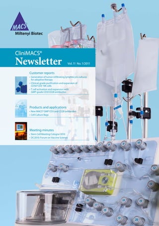
4. 4 CliniMACS Newsletter 1/2011
Customer report
The incidence of metastatic melanoma has
increased over the past three decades and
currently there are only two treatment options
that have received approval by the US Food
and Drug Administration (FDA): application of
interleukin-2 or dacarbazine result in response
rates of 13–16 % and 15 %, respectively¹, ².
Adoptive cell therapy (ACT) combining
lymphodepletionwiththeapplicationofexpanded
autologous tumor-infiltrating lymphocytes (TIL)
hasdemonstratedpromisingresultsinseveralnon-
randomized trials³, ⁴. Current efforts are mainly
directed towards simplification of the procedures
for individualized patient-specific TIL infusions,
and ultimately effective ACT of advanced
melanoma that could be applied by laboratories
in multiple centers. In a first clinical trial CD8+
young TIL, enriched with the CliniMACS®System,
wereadministeredtomelanomapatientsfollowing
lymphodepletion⁵.
Key steps during the preparation of young TIL
are the dissociation of fresh tumor tissue, TIL
enrichment and rapid expansion⁶. In this report
we present a new tissue dissociation procedure.
The primary method for initiation of TIL has
traditionally been the overnight digestion of fresh
tumor tissues in a triple enzyme medium. This
methodisopen,usesnon–FDA-approvedenzymes
(hyaluronidase) and is operator-dependent. The
gentleMACS™ Dissociator from Miltenyi Biotec
is a mechanical tissue dissociation device, which
potentially simplifies and standardizes tissue
dissociation for the generation of TIL. The
gentleMACS Dissociator works by disrupting
the extracellular matrix and cell adhesion
components without harming the integrity of the
gentleMACS™ Dissociation of melanoma
tumors for the generation of tumor-infiltrating
lymphocyte cultures for adoptive cell therapy
Linda L. Parker1, Olaf Hardt2, Alexander Scheffold2, Andreas Bosio2, and Mark E. Dudley1
1 Tumor Immunology Section, Surgery Branch, National Cancer Institute, National Institutes of Health, Bethesda, MD 20892, USA.
2 Miltenyi Biotec GmbH, 51429 Bergisch Gladbach, Germany
cell membrane. This is achieved by a combination
of varying enzyme mixes, mechanical forces,
incubation periods, and temperatures. The
automation of all mechanical steps, using the
gentleMACS Dissociator, has led to reproducible
results with reduced overall processing times. It
is a closed system, utilizes single-use, disposable
supplies, has rapid processing times, and can be
standardized.
We examined the gentleMACS Dissociator for
generationofTILculturesforresearchandclinical
use. Our early experiments using the instrument
explored multiple variables. Qualitative and
quantitative aspects of tissue processing were
optimized, including cell yield and viability,
flexibility for diverse clinical samples, technical
simplicity, and regulatory compliance. Some
variables considered included gentleMACS
Program settings, inclusion of enzymes, the
composition of enzymes, incubation times, and
sample size.
This analysis resulted in a standard operating
procedure (SOP) and the identification of a
minimum number of variables to be optimized.
We next focused on optimizing an SOP for the
gentleMACS Dissociator and comparing it with
the current clinical SOP to process fresh tumor
for the generation of TIL. Multiple head-to-head
comparisons were performed to evaluate:
i) the initial dissociation product for the total
cell yield, lymphocyte/tumor cell ratio, and
percentage of viable cells;
ii) the resulting TIL cultures for initial expansion,
phenotype, and cell function;
iii) clinical-scale expansion and clinical efficacy.
Introduction
5. 5
Customer report
Dissociation of fresh tumor tissues
All samples collected and used were derived from
patientswhosignedaninformedconsentapproved
by the institutional review board of the NIH. All
patients receiving treatment on this study were
treated as part of a clinical protocol.⁶
On the day of tumor resection, the specimen was
received in the laboratory immediately following
surgery. The specimen was bathed in sterile
saline in a sterile container. Viable tumor tissue
was dissected away from the majority of non-
viable tissue and healthy (non-tumor) tissue in
a laminar flow hood using a scalpel and sterile
forceps. Research samples were collected during
this dissection, including procurement of tissue
for confirmation of diagnosis by pathology.
Other research specimens included flash frozen
tissue for RNA isolation, tissue cryopreserved
in OCT for histopathology, an FNA sample for
cytopathology, and separate samples for tumor
tissueculturelines.Thetumorsamplewasweighed
and kept in a 50-mL centrifuge tube containing a
smallamountofsterileHBSSifprocessedthesame
day, or in 10-mL sterile Complete Medium (CM)
at 4 °C if it was to be processed after overnight
storage. The complete medium for culturing TIL
was prepared by supplementing RPMI 1640 with
10 % human serum (heat-inactivated at 56 °C for
30 minutes), and with final concentrations of
penicillin G (100 units/mL), streptomycin (100 μg/
mL), gentamicin (50 μg/mL), Hepes (25 mM),
and 2-mercaptoethanol (5.5 × 10⁻⁵ M). Selected
antibiotics for relevant allergies were omitted.
The tissue specimen was placed on a sterile cutting
surface (cutting board or an open Petri dish).
Using sterile scalpel and forceps the specimen
was cut into small (3–5 mm) fragments. Cut
Materials and methods
Figure 1: Comparison of the gentleMACS Procedure with the overnight digestion procedure with regard to cell numbers, viability,
and cell yield. For comparison, samples from 18 patients were processed using the gentleMACS Dissociator or overnight digestion.
Relative cell numbers of TIL (A) and tumor cells (B) as well as viability (C) and cell yield (D) were determined as described in Materials
and methods.
120
100
80
60
40
20
0
Cellsinprocessedtumor(%)
3217
3311-2
3244-2
3294
3281
3304
3269
3314-1
3271
3287
3291
3289
3297
3223
3210-2
3270
3238-1
3207
Lymphocytes
Digest gentleMACS
Dissociator
A
3217
3311-2
3244-2
3294
3281
3304
3269
3314-1
3271
3287
3291
3289
3297
3223
3210-2
3270
3238-1
3207
120
100
80
60
40
20
0
Cellsinprocessedtumor(%)
Tumor cells
Digest gentleMACS
Dissociator
B
3217
3311-2
3244-2
3294
3281
3304
3269
3314-1
3271
3287
3291
3289
3297
3223
3210-2
3270
3238-1
3207
120
100
80
60
40
20
0
Cellsinprocessedtumor(%)
Viability
Digest gentleMACS
Dissociator
C
3217
3311-2
3244-2
3294
3281
3304
3269
3314-1
3271
3287
3291
3289
3297
3223
3210-2
3270
3238-1
3207
200
180
160
140
120
100
80
40
60
20
0
Cellsinprocessedtumor(×10⁶)
Cell yield/g
Digest gentleMACS
Dissociator
D
Cellsinprocessedtumor(%)
Cellsinprocessedtumor(%)
Cellsinprocessedtumor(%)
Cellsinprocessedtumor(× 106)
Digest gentleMACS
Dissociator
Digest gentleMACS
Dissociator
Digest gentleMACS
Dissociator
Digest gentleMACS
Dissociator
3217
3311-2
3244-2
3294
3281
3304
3269
3314-1
3271
3287
3291
3289
3297
3223
3210-2
3270
3238-1
3207
120
100
80
60
40
20
0
Cellsinprocessedtumor(%)
6. 6 CliniMACS Newsletter 1/2011
Customer report
fragments were transferred into a gentleMACS
C Tube. Enzyme medium (EM) was added to the
tube (5 mL for 0.2–2 g of tissue and 10 mL for
2–5 g) and the tube was securely closed by turn
ing the cap until it clicks. EM was used to help
disperse tumor cells from the surgical specimen
during the short incubation periods between
the gentleMACS Program runs. The enzyme-
containing medium, RPMI 1640, did not contain
serum. It had the following added components
(final concentrations): penicillin G (100 units/mL),
streptomycin (100 μg/mL), gentamicin (50 μg/mL),
Fungizone® (1.25 μg/mL), Collagenase (1 mg/mL)
and Pulmozyme® (~30 units/mL). The enzyme
stocks were dry powders stored at –20 °C. The
C Tubes were installed vertically and cap-side
down into the gentleMACS Sleeve. Proper
installation ensured that the C Tubes were held in
position against rotational and axial forces. Pre-
defined programs are provided by the internal
gentleMACS Dissociator memory. The programs
vary in intensity, so an appropriate program was
selected depending on the texture of the tissue to
be processed. Softer tissues required a more gentle
rotation (lower speed) and harder tissues required
a more vigorous, longer rotation. For the human
tumor tissues received by this lab, we have found
the following series of programs and incubations
to be optimal:
· h_tumor_01.01 program
· 30 minutes incubation at 37 °C
· h_tumor_02.01 program
· 30 minutes incubation at 37 °C
· h_tumor_03.01 program
After the first program had run (h_tumor_01.01),
the C Tube was removed from the gentleMACS
Dissociator and placed in a 37 °C incubator for
30 minutes.Subsequently,theCTubewasinstalled
into the gentleMACS Dissociator for its second
program run (h_tumor_02.01). After another
30 minutes incubation step the third program run
(h_tumor_03.01) was performed. After the
final program run, the tissue appeared mostly
dissociated. If significant chunks of tissue were
macroscopically visible, one or two additional
Melanoma cell line
A2- A2+
888 938 526 624
None A1, 24 A1, 24 A2, 3 A2, 3
TIL 3207 (A1, A30) Std. Digest d22 322 247 390 45
TIL 3207 (A1, A30) gentleMACS d23 78 443 861 116
TIL 3211 (A1, A3) Std. Digest d24 29 23 18 20 33
TIL 3211 (A1, A3) gentleMACS d25 40 253 119 69 150
TIL 3212 (A1, A2) Std. Digest d19 46 82 658 488 322
TIL 3212 (A1, A2) gentleMACS d20 23 191 482 328 284
TIL 3210-2 (A1, A30) Std. Digest d28 45 34 33 20 33
TIL 3207 (A1, A30) gentleMACS d28 6 28 23 11 21
TIL 3223 (A2) Std. Digest d15 19 93 154 32890 17980
TIL 3223 (A2) gentleMACS d16 14 71 112 38210 27440
TIL 3217 (A24, A68) Std. Digest d21 15 332 180 87 289
TIL 3217 (A24, A68) gentleMACS d22 11 206 134 37 131
Table 1: Function of TIL generated by using the gentleMACS Dissociator or overnight digestion. Function of TIL was evaluated by
coculturing TIL with melanoma cell lines or primary melanoma cells (FrTu) and analysis of IFN-γ release by TIL.
7. 7
Customer report
gentleMACS vs. digest treatments
(responders and non-responders)
Foldexpansion
0
500
1000
1500
2000
2500
3000
3500
gentleMACS
digest
average
Figure 3: Clinical-scale expansion of TIL generated by using the
gentleMACS Dissociator or overnight digestion. TIL generated
by the gentleMACS Procedure or the standard digest were
expanded to clinically relevant cell numbers. The analysis
included both TIL that resulted in an objective clinical response
and TIL that did not induce a response.
FrTu
3104 3210 3003 3210-2 3217 3089 3010-2 3257-1
A11, 26 A2, 3 A1,2 A2, 30 A24, 68 A1, 0201 A 30, 33 A 24, 32
46 61
59 67
88 353
97 322
44 141
97 315
424 151 93
298 69 92
17
133
Fisher’s exact test: OR NR
gentleMACS
Dissociator
10 (38 %) 16
digest 7 (41 %) 10
Figure 4: Efficacy of TIL generated by using the gentleMACS
Dissociator or overnight digestion. Fisher’s exact test was
performed on the percentage of objective responses (OR)
to compare efficacies of TIL generated by the gentleMACS
Procedure or overnight digestion (p= 0.69).
gentleMACSDissociationrunswereappliedtothe
tissue, with or without one additional 30-minute
incubation step at 37 °C.
The dissociated tissue from the C Tube was
filtered through an autoclaved wire mesh placed in
an autoclaved funnel on top of a 250 mL centrifuge
tube. The wire mesh was rinsed with sterile HBSS
and the tube was filled with additional sterile
HBSS. The dissociated tissue was then washed
once by centrifuging 10 minutes at 1,500 rpm.
The supernatant was aspirated and the pellet
resuspended in a known volume of HBSS.
Processing dissociated single-cell tissue
samples and generation of TIL cultures
The resulting single-cell suspension was counted
using a hemocytometer with trypan blue exclusion
to analyze cell yield. Total numbers of lymphocytes,
tumor cells, and red blood cells (RBC), as well
as overall cell viability were calculated. If the cell
suspension had extensive cellular debris, or the
RBC : nucleated cell ratio exceeded 6 : 1, or the
overall viability was lower than 50 %, the sample
Numbers indicate IFN-γ concentrations in pg/mL. Bold numbers indicate results that were significantly different from the controls
that included no tumor cells.
Foldexpansion
8. 8 CliniMACS Newsletter 1/2011
Customer report
was further processed over a Ficoll® gradient. If
the cell suspension did not need to be cleaned up
with a Ficoll gradient, then the final wash was
completed as follows: the cells were centrifuged
(10 minutes, 1,500 rpm), and resuspended in CM.
Cultures were set up in 24-well tissue culture
plates at 0.5 × 10⁶ total live cells/mL (2 mL/well)
in CM containing 10% human AB serum and
IL-2 (6000 IU/mL). Remaining cells were frozen at
107 live cells/vial. TIL were cultured as previously
described.7
TIL cultures were monitored daily for expansion,
which was determined by microscopically
counting viable cells using trypan blue exclusion
and a hemocytometer.
Functional analysis of TIL
Selected TIL were evaluated for tumor recognition
by interferon-γ (IFN-γ) release assay as previously
described.⁷
Clinical-scale expansion
and therapeutic administration
Successfully generated TIL were used to treat
patients in ongoing surgery branch clinical
protocols. These TIL were rapidly expanded and
administered to metastatic melanoma patients.⁶
Statistical comparisons
Head-to-head comparisons were tested with a
paired t-test. Populations were compared using a
student’s2-sidedt-testassumingunequalvariance.
Fisher’s exact test was used to evaluate responses
vs. source of TIL. All responses were considered
significant for P 0.05. Patients treated between
May 2009 and May 2010 (inclusive) under Surgery
Branch, NCI Protocols #07-C-0176, were included
in this analysis.
Figure 2: Expansion potential of TIL generated by using the
gentleMACS Dissociator or overnight digestion. For comparison,
samples from 15 patients were processed using the gentleMACS
Dissociator or overnight digestion. Expansion of TIL was
measured as described in Materials and methods.
3207
3210-2
3211
3212
3215
3217
3223
3224
3225
3225
3230
3238-1
3244-2
3257-2
3269
40
30
20
10
0
Timetofirstdoubling(days)
Expansion potential (p=0.001)
Timetofirstdoubling(days)
3207
3210-2
3211
3212
3215
3217
3223
3224
3225
3225
3230
3238-1
3244-2
3257-2
3269
40
30
20
10
0
Timetofirstdoubling(days)
gentleMACS Digest
Dissociator
9. 9
Customer report
Results
The initial gentleMACS Dissociation product,
when compared to that of the current clinical
method of overnight enzymatic digestion,
showed no significant difference in the cellular
composition, the yield of viable cells, or the overall
viability (fig. 1).
For both methods, the initial dissociation
products could be efficiently expanded in culture
and were functional in vitro and in vivo. There
was similar TIL generation and TIL attributes in
most respects. Results do show, however, that
the resulting TIL cultures from the standardized,
semi-automated protocol using the gentleMACS
Dissociator expanded significantly faster than
those from manually dissociated tumors (fig. 2).
Further analysis of these expanded TIL showed
full functionality of the cells in vitro. There were
few evaluable samples, but there was no obvious
difference in function between cells obtained
through the gentleMACS Procedure or standard
digest (table 1).
There was no significant difference in lymphocyte
subsets of the resulting TIL. 12 of 16 TIL cultures
derived from the gentleMACS Protocol had more
CD8+
cells than those of a standard digest, but this
trend was not statistically significant. There was
also a slight trend towards more CD4+
cells in the
TIL of a standard digest (data not shown).
The optimized gentleMACS Dissociation
programs were compared with the standard
digest method for clinical expansion and efficacy.
The cultures derived from the gentleMACS
Protocol expanded well (fig. 3) and caused tumor
regression in patients when used in melanoma
immunotherapy protocols (fig. 4).
Conclusion
An optimal SOP for the preparation of TIL using
the gentleMACS Dissociator was established.
The gentleMACS Dissociator from Miltenyi
Biotec provides a semi-automated protocol for
the dissociation of melanoma tissue leading to
single-cell suspensions with yields and viabilities
comparable to that of overnight enzymatic
digestion. Both methods provide similar initial
TIL/tumor cell ratios, viability, and total cell yield
formosttumortissuesamples.However,TILfrom
gentleMACS Dissociations showed significantly
enhanced initial expansion in culture.
The gentleMACS Procedure is faster, more
standardized, and more efficient for tumor
samples up to 5 grams, whereas we have found
that the standard digest is more convenient for
processing larger tumors. Samples prepared using
the gentleMACS Dissociator were efficiently
expanded to clinically relevant cell numbers
(~5.0×10¹⁰) and were able to mediate tumor
regression in advanced melanoma patients.
Based on our results, we have incorporated the
gentleMACS Dissociator into our clinical SOPs
and use it interchangeably with the current
overnight enzymatic digestion for the generation
of TIL for adoptive cell therapy trials.
The authors would like to acknowledge Michelle
Langhan, Kate Hogan, Tom Shelton, and John
Wunderlich of the Surgery Branch, NCI for their
expert technical help with all aspects of this project.
References
1. Smith, F.O. et al. (2008) Clin. Cancer Res. 14: 5610–5618.
2. Tawbi, H.A. et al. (2007) Sem. Oncol. 34: 532–545.
3. Dudley, M.E. et al. (2008) J. Clin. Oncol. 26: 5233–5239.
4. Besser, M.J. et al. (2009) J Immunother. 32: 415–423.
5. Dudley, M.E. et al. (2010) Clin. Cancer Res. 16: 6122–6131.
6. Prieto, P.A. et al. (2010) J. Immunother. 33: 547–556.
7. Tran, K.Q. et al. (2008) J. Immunother. 31: 742–751.
10. 10 CliniMACS Newsletter 1/2011
Customer report
Cell therapy may represent a promising treatment
option for patients suffering from leukemia and
tumors who have a high risk for relapse after
allogeneic, especially haploidentical, stem cell
transplantation (SCT). While established T cell
therapies, such as donor lymphocyte infusions,
are associated with the risk of graft-versus-
host disease (GvHD), natural killer (NK) cells
may mediate graft-versus-leukemia/tumor
effects without induction of GvHD. Therefore,
immunotherapy with highly purified NK cells in
recipients of haploidentical SCT could serve as an
attractive alternative cell therapy 1, 2.
Human NK cells are lymphocytes of the innate
immune system involved in the early defense
against infectious pathogens and against MHC
class-I negative or low-expressing malignant
targets without the requirement for prior immune
sensitization of the host 3, 4. They reside mainly in
the marrow, spleen, and peripheral blood, where
they account for approximately 2–18 % of the
peripheralbloodlymphocytes.NKcellsareusually
the first lymphoid subpopulation to recover after
SCT 5. Phenotypically they can be defined by the
expression of CD56, an isoform of the neural cell
adhesionmoleculeandthelackoftheCD3antigen
on the surface. Further characterization allows
the major CD56dim
CD16+
(around 90%) to be
distinguishedfromtheminorCD56bright
CD16–
NK
subpopulation. The immunoregulatory CD56bright
NK cell subsets express the high-affinity
interleukin-2 (IL-2) receptor, which enables
them to proliferate in response to IL-2 and to
produce high amounts of cytokines, such as IFN-γ,
Clinical-grade purification and expansion of
CD56+
CD3–
NK cells for adoptive immuno
therapy of solid tumors and leukemia
Ulrike Koehl1, Ruth Esser1, Lutz Uharek2, and Jakob Passweg3
1 Pediatric Hematology and Oncology, University Hospital of Frankfurt, Germany.
2 Internal Medicine, Hematology and Oncology, Charite Berlin, Germany
3 Division of Hematology, University Hospital Geneva, Switzerland
TNF-α, TNF-β, GM-CSF, IL-10, and IL-13. The
CD56dim
NK cells are essentially cytotoxic cells
that express low levels of the IL-2 receptor. NK
cells are able to lyse targets by releasing cytotoxic
granules containing perforin and granzymes and
using antibody-dependent cellular cytotoxicity
pathways via membrane receptor binding to the
Fc portion of IgG antibody, and by the induction
of apoptosis through molecules of the TNF super
family (Fas/CD95, TRAIL).
Killing activity of NK cells is regulated by a set of
surface receptors that either induce or inhibit the
cytotoxic response 3, 6, 7. Activation of NK cells is
facilitated by the engagement of activating surface
receptors through interaction with stimulatory
ligands expressed by malignant cells. These
immune recognition receptors include NKG2D,
the natural cytotoxicity receptors (NCR) NKp30,
NKp44, and NKp46, CD16, NKp80, DNAM-1,
and 2B4 (CD244) 6, 8. Activation with cytokines,
such as IL-2, leads to a strong up-regulation
of the NCRs and NKG2D and this correlates
with increased NK cell cytotoxicity against
malignant cells 9. Inhibitory receptors comprise
both killer cell immunoglobulin-like receptors
(KIRs) and the heterodimeric C-type lectine
receptor CD94-NKG2A/B 10. In addition, several
activating KIR and the C-type lectin receptor
CD94-NKG2C/E/F are known 3. A number of
studies have demonstrated NK cell–based killing
of many different mouse and human tumors and
leukemias and have led to the initiation of the first
clinical phase I/II trials using allogeneic NK cells
for treatment of cancer.
Introduction
11. 11
Customer report
Figure 1: Purification, ex vivo expansion, and infusion of donor NK cells. In an ongoing clinical phase I/II trial patients receive freshly
isolated NK cells on day (+3), +40, +100 or IL-2–activated NK cells on day +40 and +100 post haploidentical SCT.
Clinical phase I/II study with allogeneic NK cells post haploidentical SCT:
Patients with high-risk leukemia and malignant tumors
Donor / Parents Recipient
NK cell purification
CD3+
depletion followed
by CD56+
cell enrichment,
according to GMP
Leukapheresis NK cell expansion
IL-2–dependent,
according to GMP
NK cell infusion
haploidentical
SCT
0 +3 +40 +100 (days post SCT)
NK cell
infusion
NK cell
infusion
NK cell
infusion
Materials and methods
Clinical-scale NK cell enrichment
Protocols for the enrichment of NK cells from non-
stimulated leukapheresis products using good
manufacturing practice (GMP) procedures have
already been established (table1;fig.1).The aimof
these procedures is to obtain a highly purified NK
cell product with minimal T cell contamination
and conserved NK cell cytotoxicity. NK cell
enrichment usually consists of one or two rounds
of CD3+
cell depletion with subsequent CD56+
cell
enrichment11, 12.
In our procedure, after steady-state leukapheresis
of unstimulated donors, the cells were washed
twice for platelet reduction with CliniMACS® PBS/
EDTA Buffer (Miltenyi Biotec, Bergisch Gladbach,
Germany) supplemented with 0.4 % human serum
albumin (Red Cross Blood Donor Service, Baden-
Württemberg-Hessen, Germany). Thereafter, 5 mL
of Intraglobin (Biotest, Dreieich, Germany) were
added and incubated for five minutes to reduce
non-specific antibody binding. Cells were labeled
for 30 minutes with CliniMACS CD3 Reagent
(Miltenyi Biotec), using one vial of reagent in case
oftotalnucleatedcell(TNC)numbersupto40×109or
CD3+
cellnumbersupto15×109andtwovialsincase
of TNC numbers up to 80×109 or CD3+
cell num
bers up to 30×109. After washing twice, CD3+
cells
were depleted with the CliniMACS Plus Instrument
using the separation program “DEPLETION 2.1”.
If necessary, the T cell depletion step was repeated
to further remove residual T cells. Thereafter, the
T cell–depleted harvests were concentrated and
labeled with CliniMACS CD56 Reagent (Miltenyi
Biotec) for 30 minutes (one vial CD56 Reagent
for TNC numbers up to 40×109 and CD56+
cell
numbersupto10×109).Afterwashing,CD56+
CD3–
NK cells were enriched using the separation
program “ENRICHMENT 1.1”. All steps were
performed in a closed system observing GMP. The
study protocol was approved by the local ethics
committee in Frankfurt and Basel and informed
consent of the donors has been obtained.
Expansion and activation of NK cells
ThepurifiedCD56+
CD3–
NKcellsweresuspended
and seeded at a concentration of 1–2×10⁶ cells/mL
in X-vivo™ 10 media (BioWhittaker, Verviers,
Belgium) supplemented with 5 % heat-inactivated
human fresh frozen plasma and 1,000 U/mL
rhIL-2 (Proleukin®, Novartis, Germany) under
12. 12 CliniMACS Newsletter 1/2011
Customer report
GMP-compliant conditions (fig.1). In an early
phase of the study, cells had been expanded and
activated using both 175 cm2 culture flasks (Nunc,
Wiesbaden, Germany) and VueLife™ cell culture
bags (CellGenix, Freiburg, Germany); for the
ongoing phase I/II trial only VueLife cell culture
bags were used 11. Fresh medium was added every
threedays andsamplesformonitoringcellcontent
andviabilityweretakendirectlyafterleukapheresis,
after each depletion/enrichment step, and every
second day during stimulation. Phenotyping
and evaluation for cytotoxicity was performed
by flow cytometry. After 10 days, stimulated
NK cells were administered to the patients or
cryopreserved in X-vivo™ 10 medium supple
mented with 10 % DMSO.
Phenotyping and functional characterization
of the product for quality control
The absolute number of CD56+
CD3–
NK cells and
the number of residual T cells were determined by
flowcytometryperformedonafour-orafive-color
flow cytometer (Epics XL or FC 500, Beckman
Coulter, Krefeld, Germany) in a single-platform
technique. The gating strategy was based on the
ISHAGE single-platform stem cell enumeration
method using low scatter, high expression
of CD3 and CD45 antigens, CD16 and CD56
expression and 7-AAD staining, in a no-wash
preparation with counting beads. Our previously
described four-color panels13 were extended
to the following five-color panels: CD45-FITC/
CD56-PE/CD3-ECD/7-AAD/CD16-PC7 and
45-FITC/CD3-PE/CD14-ECD/7AAD/CD56-PC7.
Samples were prepared in triplicate and 45-FITC/
IgG1-PE/CD14-ECD/7AAD/CD56-PC7 served as
a control. In addition, cells were labeled with
appropriate combinations of fluorochrome-
conjugated antibodies (MAb) to monitor NK cell
subsets, activating and inhibitory NK cell receptors,
and activation status. MAbs used were CD16
(clone 3G8), CD45 (clone J.33), CD56 (clone N901),
HLA-DR(cloneImmu-357),CD69(cloneTP1.55.3),
CD158a,h(KIR2DL1/S1,cloneEB6B),CD158b1/b2,j
(KIR2DL2/3/S2, clone GL183), CD158e1/e2 (KIRp70,
KIR3DL1/S1, clone Z27.3.7), CD158i (KARp50.3,
KIR2DS4, clone FES172), CD337 (NKp30, clone
Z25), CD336 (NKp44, clone Z231), CD335 (NKp46,
clone BAB281) and CD314 (NKG2D, clone ON72),
all supplied by Beckman Coulter (Marseille, France),
and CD3 (clone SK7) supplied by BD Bioscience.
The cytotoxicity of the highly enriched NK cells
before and after IL-2 stimulation was tested
against the MHC class I–negative cell line K562
or against the patient’s individual leukemic cells
using an antibody-based flow cytometric assay as
described previously 14, 15. NK cells and leukemic
cells were co-cultured for four hours at effector-
to-target ratios between 0.5 : 1 and 10 : 1. Absolute
cell counts were determined using Flow-Count®
beads. Cytotoxicity was defined as the loss of
viable target cells relative to the control. In other
studies NK cell functionality was measured by a
CD107a degranulation assay 16, 17.
Author Method Donors
(n)
NK cell purity
(%)
Recovery
(%)
Log T cell
depletion
18
Lang et al. (2002) CD56 enrichment, followed by CD3 depletion 4 98.6 (m) 42.0 (m) 3.6×10⁴-fold
19
McKenna et al. (2007) CD3 depletion 36 37.7 (x) 78.8 2.7
19
McKenna et al. (2007) CD3 depletion, followed by CD56 enrichment 13 89.7 (x) 19.4 4.3
20
Koehl et al. (2004) CD3 depletion, followed by CD56 enrichment 6 95.0 (m) 37.0 (m) 4.5 (m)
11
Koehl et al. (2005) CD3 depletion twice, followed by CD56 enrichment 15 94.9 (m) 33.0 (m) 5.0 (m)
21
Iyengar et al. (2003) CD3 depletion, followed by CD56 enrichment 12 91.0 (m) 48.7 (m) 5.3 (m)
22
Uharek et al. (2003) CD34 neg fraction: CD3 depletion, CD56 enrichment 7 75.0 (m) 42.0 (m) 4.0
23
Passweg et al. (2004) CD3 depletion, followed by CD56 enrichment 6 97.3 (m) 35.5 (m) 3.6
12
Meyer-Monard et al. (2009) CD3 depletion, followed by CD56 enrichment 24 94.5 (m) 58.0 (m) 4.2 (m)
24
Rizzieri et al. (2010) CD56 enrichment 51 96.5 80
m: median; x: mean
Table 1: Clinical-grade NK cell enrichment using the CliniMACS® System.
13. 13
Customer report
Results and discussion
Technical aspects—clinical-scale NK cell
product manufacture and NK cell activation
A number of studies have shown that clinical-
scale NK cell product manufacture from non-
stimulated leukapheresis products, using a
CD3+
cell depletion step followed by CD56+
cell
enrichment, leads to highly purified CD56+
CD3–
NK cell products with a purity ranging from
89.7–98.6 % (table 1) 11, 12, 18-24. Purity of NK cell
products was lower if the starting product was the
negative fraction after CD34+
cell selection 22. The
high NK cell purity and extensive T cell depletion
is at the expense of a considerable loss of NK cells
duringisolation.ThefinalrecoveryofCD3–
CD56+
NKcellsrangedbetween19.4 %and58 %.Overnight
storage of the leukapheresis product led to a greater
loss of NK cells during the NK cell enrichment
process compared to processing of fresh harvests 12.
A much higher NK cell recovery was obtained
by using only a CD3+
cell depletion step, without
further CD56+
cell enrichment. However, such
a product was associated with low purity and
less T cell depletion 19. Similarly, the final T cell
number was much higher if a CD56+
cell selection
was used alone, but this kind of purification was
associated with an increased NK cell recovery and
purity 24. In contrast, the two-step NK cell product
manufacture described here led to efficient T cell
depletion of 3.6 to 5.3 orders of magnitude (table 1),
and this could be further increased by performing
the CD3+
cell depletion step twice 11. A residual T cell
contamination between 0.01 and 0.09% in the final
product allows the infusion of NK cell products
of more than 1.0×10⁷ CD56+
CD3–
NK cells/kg BW
with less than 5.0×10⁴ CD3+
cells/kg BW and often
less than 2.5×10⁴CD3+
T cells/kg BW 1.
The objective of NK cell purification is not only
to remove potentially unwanted T cells but also to
enable activation and expansion of the NK cells.
Indeed, enriched NK cells can be infused without
any additional manipulation, or after overnight
culture in high-dose IL-2. They can also be
expanded in IL-2 or other cytokines, such as IL-15,
alone or in combination for two to several weeks
in cell culture bags or in a bioreactor 11, 25. Similarly,
it is possible to expand single KIR+
NK cells 26.
In vitro expansion has two aims, to activate the
selected CD56+
CD3–
cells, and to increase the total
number of NK cells. Using CD69 as an activation
marker, activation of NK cells was found to occur
within 1–3 days of incubation with IL-2 9, 20. When
enriched CD56+
CD3–
NK cells were cultured
with IL-2, a significant expansion was observed
although there was a lag of 3–5 days before the
NK cells started to proliferate 11. On day five,
expansion occurred and led to a two- to tenfold
increase of CD56+
CD3–
NK cells after 10–14
days. Although NK cells were viable immediately
after purification ( 90 %), the vital NK cell count
decreased by 30–50 % during the first three to five
days following IL-2 stimulation. Afterwards, cell
viability recovered to 98 %, and by day 10–14, a
maximal NK cell expansion was obtained. No
overgrowth of the remaining T cells was observed
during expansion and activation with IL-2. With
a protocol that enables the generation of NK cells
on a clinical scale, using a closed system that
conforms to GMP guidelines, the expanded
NK cells were highly cytotoxic against different
leukemic and tumor target cells 9, 20. Importantly, no
non-specific activation against normal allogeneic
lymphocytes occurred 18. In addition we were
able to demonstrate that IL-2 stimulation led to
up-regulation of all natural cytotoxicity receptors
(NCRs) and the activating receptor NKG2D,
which might explain the observed increased
cytotoxicity against MHC-Inegative
targets 9. There is
evidence that a combination of cytokines, such as
IL-2, IL-12, IL-15, and IL-21, may further increase
cytotoxic activity of NK cells. In addition to
NK cell enrichment from leukapheresis products
as summarized in table 1, NK cells can also be
generated from cord blood 27.
Clinical-scale collection, enrichment, activation,
and expansion of purified NK cells are feasible.
Most of the technical aspects for adoptive
NK cell therapy have been developed for
clinical applications. However these laboratory
procedures are time-consuming and expensive,
need particular skills, and must be performed
according to a GMP-compliant protocol.
Clinical studies using freshly purified
or IL-2–activated NK cells
Previous trials and ongoing clinical phase I/II
studies have shown the feasibility of using freshly
purified or IL-2–activated donor NK cells for the
treatment of high-risk patients suffering from
14. 14 CliniMACS Newsletter 1/2011
Customer report
schedule of the NK administration based on
results of ongoing phase I/II studies. Given the
plausible benefit of IL-2–stimulated NK cells
compared to freshly isolated, resting NK cells
with regard to cytotoxicity, it may be possible to
increase cytotoxicity by activation with cytokine
combinations like IL-2/IL-15 or by cross-talk with
dendritic cells (DCs). Additional investigation
is necessary to develop strategies to overcome
tumor immune escape mechanisms. Options may
encompass development of MAb against sMICA,
genetic engineering of NK cells by introduction
of chimeric receptors for tumor retargeting, or
enhancing tumor cell recognition by using small
interfering RNA to silence inhibitory receptors.
Open issues in clinical studies also include NK cell
dose rate, time schedule, appropriate selection of
donor/recipients, and also the types of tumors to
be considered for treatment, because it is already
known that certain types of malignant cells may
be more responsive to NK cell therapy than others.
Ultimately expansion of tumor-reactive NK cells
within the patient might prove to be feasible. It
is possible to transfuse NK cells simultaneously
with the transplants, and the first clinical trials
indicate that an early NK application post-SCT
may be most effective in attacking minimal
residual disease.
References
1. Passweg, J. R. et al. (2006) Best Pract. Res. Clin. Haematol. 19: 811–824.
2. Passweg, J. R. et al. (2005) Bone Marrow Transplant. 35: 637–643.
3. Farag, S. S. and Caligiuri, M.A. (2006) Blood Rev. 20: 123–137.
4. Ljunggren, H. G. and Malmberg, K. J. (2007) Nat. Rev. Immunol. 7:
329–339.
5. Koehl, U. et al. (2007) Bone Marrow Transplant. 39: 269–278.
6. Moretta,A. et al. (2001) Annu. Rev. Immunol. 19: 197–223.
7. Passweg, J. R. et al. (2007) Trends Immunol. 28: 437–441.
8. Lanier, L. L. (2005) Annu. Rev. Immunol. 23: 225–274.
9. Huenecke, S. et al. (2010) J. Immunother. 33: 200–210.
10. Karre, K. (2008) Nat. Immunol. 9: 477–480.
11. Koehl, U. et al. (2005) Klin. Padiatr. 217: 345–350.
12. Meyer-Monard, S. et al. (2009) Transfusion 49: 362–371.
13. Koehl, U. et al. (2008) Int. J. Hematol. 87: 98–105.
14. Zimmermann, S. Y. et al. (2005) J. Immunol. Methods 296: 63–76.
15. Kloess, S. et al. (2007) J. Immunol. Methods, 325: 140–147.
16. Penack, O. et al. (2005) Leukemia 19: 835–840.
17. Fischer, L. et al. (2006) Exp. Hematol. 34: 753–759.
18. Lang, P. et al. (2002) Bone Marrow Transplant. 29: 497–502.
19. McKenna, D. H. et al. (2007) Transfusion 47: 520–528.
20. Koehl, U. et al. (2004) Blood Cells Mol. Dis. 33: 261–266.
21. Iyengar, R. et al. (2003) Cytotherapy 5: 479–484.
22. Uharek, L. et al. (2003) Onkologie 26 (suppl. 5): 38.
23. Passweg, J. R. et al. (2004) Leukemia 18: 1835–1838.
24. Rizzieri, D.A. et al. (2010) Biol. Blood Marrow Transplant. 16:
1107–1114.
25. Sutlu, T. et al. (2010) Cytotherapy 12: 1044–1055.
26. Siegler, U. et al. (2010) Cytotherapy 12: 750–763.
27. Spanholtz, J. et al. (2010) PLoS One 5: e9221.
28. Miller, J. S. et al. (2005) Blood 105: 3051–3057.
29. Rubnitz, J. E., et al. (2010) J. Clin. Oncol. 28: 955–959.
30. Ruggeri, L. et al. (2002) Science 295: 2097–2100.
31. Kloess, S. et al. (2010) Eur. J. Immunol. 40: 3255–3267.
leukemia or tumors in both non-transplant settings
and after haploidentical SCT as an additional
immunotherapy 20, 23, 24, 28, 29. NK cell products
were infused as a single dose rate or as multiple
applications with doses between 0.2×10⁷/kg
and 8.1×10⁷ CD56+
CD3–
NK cells/kg BW, mostly
with less than 2.5×10⁴ CD3+
T cells/kg BW 1, 9, 24, 29.
These first immunotherapy trials show that NK
cells can be administered without immediate
adverse events, that they are well-tolerated by
the patients and do not induce GvHD grade II.
However, some cases of GvHD have been observed
after NK cell infusion. In some instances this
has been associated with a less efficient T cell
depletion. Whether GvHD is attributable to
contamination by T cells or is due to the effects of
NK cells cannot be determined on the basis of this
clinical data. The fact that (at least in some cases
of GvHD) the T cell content was higher than in
cases without GvHD, seems to favor a T cell effect.
With regard to NK cell efficiency, Rubnitz et al.29
recently reported that NK cell administration to
10 pediatric patients with AML in first complete
remission led to a two-year event-free survival of
100 %, with all patients still in complete remission.
An earlier study demonstrated that patients with
AMLhadalowerrateofleukemiarelapsecompared
to the expected rate, a lower rate of graft rejection,
and a paradoxical reduction in GvHD post-
haploidentical SCT, when the NK cells possessed
inhibitory KIRs for which the recipient had no
ligand 30. We could show an increased cytotoxic
activity of stimulated NK cells against high-
risk neuroblastoma (NB) due to IL-2–mediated
up-regulation of the activating receptors NKp30,
NKp44, NKp46, and NKG2D 9. However, we
have also been able to demonstrate tumor escape
from immune surveillance by release of soluble
MICA (ligand MHC class I–chain-related gene A)
compromising NKG2D-dependent NK cell
cytotoxicity in patients with NB. Elevated sMICA
levels in patients’ plasma correlated significantly
with impaired NK-cell–mediated cytotoxicity of
the infused donor NK cells 31.
Future perspectives
Future studies should improve NK cell immuno
therapy by increasing the understanding of the
conditions leading to tumor cell kill by NK cells,
by increasing the cytotoxicity of NK cells against
various malignancies, and by optimizing the
15. 15
Background
The presence of a CD28 costimulatory signal in
combination with CD3 activation represents a
more physiological way in which T lymphocytes
can be stimulated to become T lymphoblasts.
This could result in more robust T cell
activation, proliferation, and consequently more
efficient expression of a retroviral transgene,
while simultaneously preserving a favorable
differentiation phenotype.
Methodology
Peripheral blood mononuclear cells (PBMC) were
isolated from 3 donors by Lymphoprep gradient
centrifugation.
5×10⁵ PBMC per well of a 24-well plate were
activated with CD3 (Ortho Biotech, referred
to as “OB”) or CD3 +/– CD28 (Miltenyi Biotec,
Bergisch Gladbach, Germany, referred to as
“MB”) at a final concentration of 1 μg/mL
each. On day two, recombinant human IL-2
(Chiron, Emeryville, CA) was added at a final
concentration of 50 units/mL, and on day three
cells were harvested for retroviral transduction.
For transduction, we pre-coated a non-tissue,
culture-treated, 24-well plate with a recombinant
fibronectin fragment (FN CH-296; Retronectin;
Takara Shuzo, Otsu, Japan). Wells were washed
with phosphate-buffered saline (PBS; Sigma,
St. Louis, MO) and incubated twice for 30 minutes
with a high-titer retrovirus supernatant, encoding
a CD19-specific Chimeric Antigen Receptor (CAR).
Subsequently, 3×10⁵ T cells per well were trans
duced with retrovirus in the presence of 50 units
of IL-2 per milliliter. After 48–72 hours, cells were
removed and expanded in the presence of 50–100
units of IL-2 per milliliter for 10–15 days prior to
use. Conditions were compared for proliferation,
GMP-grade CD28 monoclonal antibody
induces more robust expression of
retroviral transgene while preserving a
favorable T cell phenotype
Nabil M. Ahmed, MD, MPH
Assistant Professor, Department of Pediatrics,
Center for Cell and Gene Therapy (CAGT),
Baylor College of Medicine, 1102 Bates Street, Suite 1770, MC3-3320, Houston, TX 77030
percentage and density of T cell transduction with
CAR as well as effector memory T cell markers.
The above outlined methodology is the current
approach to generate CAR-transduced activated
T lymphocytes at the CAGT for clinical use.¹
Results
In all three donors, there was an advantage to
using the combination of CD3 and CD28 Ab
in T cell proliferation (fig. 1). Consequently, a
significantly higher degree of transduction of
T cells (as evidenced by a higher percentage of
T cells transduced, fig. 2) and density of T cell
transduction (as evidenced by consistently higher
mean fluorescence intensity [MFI] values, fig. 3)
were observed under these conditions. The use of
CD3 and CD28 Ab together was associated with
an effector memory phenotype pattern that was
similar to that of CD3 alone (fig. 4).
Figure 1: Mean expansion rate of CD19-CAR transduced T cells
from three different donors after activation with the three
different antibodies.
Lymphoblast expansion
0
2
4
6
8
10
12
14
Day 2 Day 4 Day 7
Time
Fold Increase
CD3 (OB)
CD3 (MB)
CD3 + CD28 (MB)
FoldIncrease
Lymphoblast expansion
0
2
4
6
8
10
12
14
Day 2 Day 4 Day 7
Time
Fold Increase
CD3 (OB)
CD3 (MB)
CD3 + CD28 (MB)
FoldIncrease
Lymphoblast expansion
0
2
4
6
8
10
12
14
Day 2 Day 4 Day 7
Time
Fold Increase
CD3 (OB)
CD3 (MB)
CD3 + CD28 (MB)
FoldIncrease
Lymphoblast expansion
0
2
4
6
8
10
12
14
Day 2 Day 4 Day 7
Time
Fold Increase
CD3 (OB)
CD3 (MB)
CD3 + CD28 (MB)
FoldIncreaseFoldincrease
Time
Customer report
Lymphoblast expansion
16. 16 CliniMACS Newsletter 1/2011
Conclusion
The noted improvement in expansion could
reduce production time by approximately 5 days,
saving precious time for patients with progressive
tumors as well as presumably saving reagents and
technician time by achieving clinically relevant
cell numbers early. The improved transduction
rate and density could theoretically predict
better T cell activation upon encountering target
molecules, a fact that is particularly important in
the context where target molecules are expressed
only modestly on the cell surface. Lastly, the
presence of a similar effector memory phenotype
indicates that the improvement in activation and
expansion is not associated with a more exhausted
T cell phenotype.
Researchers at the CAGT have adapted the
protocol to generate CAR-transduced activated
T lympocytes for clinical use to include this
GMP‑grade CD28 monoclonal antibody.
Customer report
Figure 2: Individual transduction rate for each donor
%ofgated
Donor 1 Donor 2 Donor 3
Figure 3: Density of T cell transduction for each donor
Meanfluorescenceintensity
Figure 4: Expression of effector memory T cell markers (mean
percentage of CD3-gated cells from three different donors)
Reference
1. Pule, M. A. et al. (2008) Nat. Med. 14: 1264–1270.
0
10
20
30
40
50
60
70
80
90
100
CD3 (OB) CD3 (MB) CD3 + CD28 (MB)
Transduction rate CD 62L
CCR7
CD 27
CD 28
CD 45RA
CD 45RO
Transduction density
Donor 1 Donor 2 Donor 3
0
100
200
300
400
500
600
700
CD3 (OB) CD3 (MB) CD3 + CD28 (MB)
0
20
40
60
80
100
0
20
40
60
80
100
0
20
40
60
80
100
0
20
40
60
80
100
0
20
40
60
80
100
0
20
40
60
80
100
CD3 (OB) CD3 (MB) CD3 + CD28 (MB)
CD3 (OB) CD3 (MB) CD3 + CD28 (MB)
CD3 (OB) CD3 (MB) CD3 + CD28 (MB)
CD3 (OB) CD3 (MB) CD3 + CD28 (MB)
CD3 (OB) CD3 (MB) CD3 + CD28 (MB)
CD3 (OB) CD3 (MB) CD3 + CD28 (MB)
Mean%ofgatedMean%ofgatedMean%ofgatedMean%ofgatedMean%ofgatedMean%ofgated
17. 17
MACS® GMP Antibodies
GMP-grade soluble antibodies are essential for
powerful in vitro T cell stimulation protocols.
Miltenyi Biotec introduces two antibodies
optimized for high quality T cell cultures: MACS
GMP CD3 pure and MACS GMP CD28 pure.*
Optimized T cell expansion
for clinical-grade cell cultures
Stimulation of the CD3 part of the T cell receptor-
CD3 complex with agonistic antibodies induces
T cell activation and expansion. To amplify the
response, activation via CD3 is often combined
with additional stimulation of CD28 . Combined
stimulation with MACS GMP CD3 pure and
MACS GMP CD28 pure markedly increases
T cell expansion as compared to stimulation
with CD3 alone (fig. 1). The two signals provide
a physiological stimulus and optimize T cell
expansion for clinical-grade cell cultures.
Consistent, high quality
All MACS GMP Products, including our GMP-
grade antibodies, are manufactured in our state-
of-the-art GMP facility in Teterow, Germany, in
standardized and strictly controlled industrial
processing steps. They are designed following
the recommendations of the United States
Pharmacopeia, chapter 1043 on ancillary
materials for cell-, gene-, and tissue-engineered
products. Consequently, they are free of animal-
and human-derived contaminants, and product
specifications are confirmed by batch-specific
certificates of analysis to ensure consistency of
New tools for ex vivo T cell stimulation:
Miltenyi Biotec is now offering GMP-grade
soluble antibodies
CD3 (ng/mL)
Expansionrate(x-fold)
5
10
w/o CD28
1 μg CD28
15
20
25
30
35
40
45
50
0
0 0.1 1 10 100 1000
Figure 1: Optimized T cell expansion by a combination of
MACS GMP CD3 pure and MACS GMP CD28 pure. T cells were
co-cultured with APC (T cell–depleted PBMC) at a ratio of 1:10
for 7 days. MACS GMP CD3 pure and MACS GMP CD28 pure
antibodies were added at the indicated concentrations.
Figure 2: The Miltenyi Biotec GMP facility allows standardized
and stricly controlled industrial processing.
Products and applications
quality. By using GMP-compliant manufacturing
processes, the products are then ideally suited
for research use and in vitro cell processing in a
clinical research setting.
*) MACS GMP Products are for research use and ex vivo cell culture processing only, and are not intended for human in vivo applications.
For regulatory status in the USA, please contact your local representative.
18. 18 CliniMACS Newsletter 1/2011
Products and applications
The complete portfolio of Miltenyi Biotec Cell
Culture Bags will be simplified and harmonized.
With the release of new production lots in May/
June 2011, the Cell Differentiation and Expansion
Bags for research use and the CE-marked Cell
Differentiation and Expansion Bags will become
one product group: MACS®GMP Cell Culture Bags.
The features of the bags, including volumes,
connectors, and bag material, will remain
unchanged. We guarantee the high product
quality that you can expect from Miltenyi Biotec.
MACS GMP Cell Culture Bags will be available
worldwide. Please note that new order numbers
will be effective as of August 2011.
The gas-permeable and transparent bags are
intendedforthein vitrocultivation,differentiation,
or expansion of human cells from heterogenous
hematologic cell populations, e.g., expansion of
T cells or generation of dendritic cells.
Harmonization and expansion
of the Cell Culture Bag portfolio
The Cell Culture Bag portfolio will soon be
expanded by the addition of Cell Differentiation
Bags – 1000 and – 3000, with maximum culture
volumes of 1000 mL and 3000 mL, respectively.
Product Content Capacity Regulatory
status
Availability Order no.
MACS GMP Cell Differentiation Bag – 100 5 bags 100 mL *) Worldwide 1) 170-076-400
MACS GMP Cell Differentiation Bag – 250 5 bags 250 mL *) Worldwide 1) 170-076-401
MACS GMP Cell Differentiation Bag – 500 5 bags 500 mL *) Worldwide 1) 170-076-402
MACS GMP Cell Expansion Bag 5 bags up to 100 mL *) Worldwide 1) 170-076-403
MACS GMP Cell Differentiation Bag – 1000 2 bags 1000 mL *) Worldwide 1) Coming soon 170-076-404
MACS GMP Cell Differentiation Bag – 3000 2 bags 3000 mL *) Worldwide 1) Coming soon 170-076-405
*) MACS GMP Products are for research use and ex vivo cell culture processing only, and are not intended for human in vivo applications.
For regulatory status in the USA, please contact your local representative.
1) For availability in your country please contact your local representative.
21. 21
Meeting minutes
remodeling, reperfusion of the infarcted tissue,
accompanied by infusion of enriched stem cells
into the border zone, might support the recovery
of cardiac function.
Alexander Kaminski reported on the first
phase III trial (PERFECT) on intramyocardial
transplantation of autologous stem cells in
addition to bypass surgery. The trial postulates
that the use of bone marrow–derived CD133+
cells as an adjunct to coronary artery bypass
grafting will improve recovery of the myocardium
following infarction.
Eric Duckers outlined the double-blind APOLLO
study where post-infarction patients were treated
with adipose tissue–derived mesenchymal
stromal cells with the premise that these cells will
improve restoration of cardiac function.
Hans-Michael Klein showed first results of
the INSTEM trial aiming at evaluating safety
and feasibility of the perioperative isolation
and subsequent myocardial transplantation
of CD133+
cells in combination with coronary
bypass surgery and transmyocardial laser
revascularization.
Pilar Jimenéz-Quevedo elaborated on the
PROGENITOR trial, a randomized, blinded,
multicenter, controlled study to assess safety and
feasibility of the transendocardial injection of
peripheral blood–derived CD133+
cells in patients
with refractory angina.
Rachel Ruckdeschel Smith explicated the
CADUCEUS trial that examines the safety and
preliminary efficacy of autologous cardiosphere-
derived stem cells to reverse ventricular
dysfunction in patients with recent myocardial
infarction.
Klaus Wagner reported on the development of a
current therapy program employing CD133+
stem
cells for the treatment of end-stage peripheral
vascular disease. A trial is currently in the
preparation stage aimed at showing the safety
and feasibility of treatment with isolated bone
marrow–derived CD133+
endothelial progenitor
cells.
Consuelo del Cañizo showed first results of a
non-blinded, randomized study on the safety and
efficacy of autologous peripheral blood–derived
CD133+
cells in the treatment of patients with
critical limb ischemia.
Jan Schulte am Esch presented data from a study
on the use of CD133+
bone marrow–derived cells
for liver regeneration upon liver resection. The
treatment is based on the evidence that hepatic
stem cells provide a stimulus for liver regenera
tion. A controlled multicenter trial is currently
being designed to test this novel form of treatment
more rigorously.
In the second session of the “Clinical study
concepts and patient cohorts” symposium
various underlying mechanisms in stem cell
therapy were presented.
Johannes Waltenberger explicated that diabetes
mellitus impairs CD133+
progenitor function after
myocardial infarction, which potentially affects
responses to progenitor cell transplantation.
Wolfgang-MichaelFranzpresentedfirstresultsof
the multicenter, randomized, placebo-controlled
SITAGRAMI trial in acute myocardial patients.
The trial examines the efficacy and safety of a
combined administration of G-CSF (to mobilize
stem cells) and sitagliptin (to stimulate homing
by inhibiting the cleavage of SDF-1α) after acute
myocardial infarction.
Stefanie Dimmeler elaborated on a potential
cell therapeutic approach to cardiac disease by
augmenting functionality of autologous bone
marrow cells. Cardiac repair by bone marrow–
derived cells is currently limited due to impaired
homing and survival of progenitor cells. However,
modulation of microRNA expression in bone
marrow–derived cells holds potential to enhance
cell function and homing.
22. 22 CliniMACS Newsletter 1/2011
Meeting minutes
DC2010: Forum on Vaccine Science
September 26–30, Lugano, Switzerland
The 11th International Symposium on Dendritic Cells in Fundamental and Clinical Immunology
has seen numerous highlights. Here are a few of them.
Tumor immunology and vaccines
Ira Mellman from Genentech Inc. provided
an overview of some important developments
in cancer therapy that were achieved in 2010.
Positive results in a large prostate cancer trial
using the presumptive dendritic cell–based
vaccine Provenge (sipuleucel-T) led to the first
US-FDA approval for an active immunotherapy.
Ipilimumab (an anti-CTLA4 antibody) treatment
showed promising results in advanced, metastatic
melanoma patients. Both therapies showed
increases in overall survival.
While these developments help to establish proof
of principle in humans, a realistic view reveals
that the benefits are modest: The ipilimumab trial
showed a twofold increase in overall survival and
some complete remission. However, there are also
serious on-target toxicities leading to grade 3–4
adverse events, enterocolitis or hypophysitis, in
23% of the patients.
The mechanisms of action are not resolved; in
the case of Provenge it is not even clear whether
it functions as a vaccine. Effective cancer
immunotherapy seems like a reachable goal,
although a lot more investigation will be required.
Based on common experience with other targeted
therapies, immunotherapy will most likely
have to be developed in combination with non-
immunological agents.
23. 23
Meeting minutes
Plasmacytoid dendritic cells for immunotherapy in melanoma patients
DNGR-1+
BDCA3+
leukocytes
as putative equivalents of mouse CD8α+
dendritic cells
Fifteen stage IV melanoma patients received three
intranodal injections of 3×10⁶ PDCs each at
two‑week intervals.
The results indicate that vaccination with
peptide-loaded, TBE-activated PDCs is feasible
and that even small numbers of PDCs resulted
in TBE-specific T cell proliferation and antibody
responses. In two out of 15 patients tumor
antigen–specific T cells were detected in delayed-
type hypersensitivity reactions after only one
round of vaccination, without vaccine-related
toxicity. These results break new ground for
trials, in which DC subsets are used to optimize
DC vaccination.
itive, TLR7-negative, and (unlike CD8α+
DCs)
TLR9-negative. Upon stimulation with poly(I:C)
andTLR8agoniststhecellsproduceIL-12.ThisDC
subset efficiently takes up material derived from
dead cells and cross-presents exogenous antigens
to CD8+
T cells. The characterization of human
DNGR-1+
CD141 (BDCA-3)high
DCs paves the way
for utilizing this subset in immunotherapy.
Wilms’ tumor 1 antigen–targeted dendritic cell
Active immunization by tumor antigen-loaded
dendritic cells (DCs) represents a promising
approach towards adjuvant cancer treatment to
eliminate or control residual disease. In most DC
trials, however, end-stage cancer patients with
high tumor loads were treated.
ViggovanTendeloofromtheUniversityofAntwerp,
Belgium, and collaborators performed a phase I/II
trial in ten acute myeloid leukemia (AML)
patients investigating the effect of autologous DC
vaccination. As an immunotherapeutic target,
theyselectedWilms’tumor1protein(WT1),which
is a virtually universal tumor antigen. Its role in
leukemogenesis is established and it possesses
highly immunogenic characteristics.
DCs were matured with TNF-α/PGE2 and mature
DCs (mDCs) were electroporated in the presence
of WT1 mRNA. The DC vaccine was injected
intradermally biweekly four times. Until now,
17 patients in remission with a high risk for relapse
were treated. In five patients the AML-associated
tumormarkerreturnedtonormaluponvaccination.
Moreover, two patients in partial remission after
chemotherapy went into complete remission. Van
Tendeloo et al. monitored various immunological
parameters upon vaccination, which correlated
with the clinical responses: WT-1–specific
IFN-γ–producing CD8+
T cells increased, IL-2
was up-regulated, and NK cells were elevated.
There was no correlation between Treg cells or
IFN-α+
T cells and the clinical response.
Future approaches will include tagging mRNA
with LAMP sequences in order to target tumor
antigen to MHC II. This will allow the induction
of a CD4+
long-term memory response.
Jolanda de Vries from Radboud University
Nijmegen, The Netherlands, reported the
results of the first phase I clinical trial using
plasmacytoid dendritic cells (PDCs) for DC-based
immunotherapy of melanoma patients. PDCs
represent a highly specialized naturally occurring
DC subset and are main producers of type I
interferons (IFN) in response to viral infections.
For vaccination, PDCs were isolated directly from
blood in a closed isolation system (CliniMACS®
System). After overnight culture, cells were
activated by the commonly used TBE (tick-borne
encephalitis)–preventative vaccine and loaded
with tumor peptides. After six more hours of
culture, cells were used for vaccination.
Lionel Poulin from C. Reis e Sousa’s laboratory
at the London Research Institute, UK, presented
data on the characterization of a human dendritic
cell (DC) population, which expresses DNGR-1
(CLEC9A) and high levels of CD141 (BDCA-3).
These DCs resemble mouse CD8α+
DCs in pheno
typeandfunction.LikemouseCD8α+
DCs,human
DNGR-1+
CD141 (BDCA-3)high
DCs are TLR3-pos
24. 24 CliniMACS Newsletter 1/2011
FAQs
Why does Miltenyi Biotec offer MACS® Cytokines
at different quality levels ? What are the
differences between the different grades ?
MACS® Cytokines are available at four different
quality levels in order to suit the specific
requirements of different types of research.
MACS Cytokine grades are suitable for the
following applications:
Research grade is the standard quality for all our
products. Research-grade cytokines are sterile-
filtered and pass strict quality control measures.
Generally, endotoxin levels are below 0.1 ng / μg
( 1 EU/μg) and purities are higher than 95 %.
Their biological activity has usually been tested
employing appropriate bioassays, and minimum
activity levels are assured. The ED 50 value is
also provided.
Cell culture grade cytokines and growth factors
are lyophilized with DMEM/F12 and fetal bovine
serum or human serum albumin. These cytokines
FAQs
are best-suited for applications where direct
reconstitution in water is preferable, e.g., where
small volumes and/or tests in 96-well plates are
involved. Selected MACS Cytokines can also
be produced at premium-to-GMP grades, with
premium grade offering the convenience of
determined lot-specific biological activities to
eliminate laborious pre-testing.
Premium-grade cytokines share major charac
teristics with MACS GMP Cytokines for relevant
pre-clinical studies.
MACS GMP Cytokines boast both superior
quality and GMP compliance attributes: They
are manufactured and tested under a certified
ISO 9001 quality system and compliant with
relevant GMP guidelines.
MACS GMP Cytokines are designed in accordance
with the recommendations of USP 1043 on
ancillary materials; They are free of all human-
and animal-derived components.
25. 25
FAQs
Where can I find more information on the bio
logical activities of MACS GMP-grade Cytokine ?
The activity of GMP-grade cytokines is deter
mined for each lot, and can be found in the
lot‑specific Certificate of Analysis with which
they are delivered.
Which GMP-grade antigens are available
for use with the Cytokine Capture System ?
The following GMP-grade antigens may be used
with the Cytokine Capture System:
· MACS GMP HCMV pp65 – Recombinant
protein
· MACS GMP PepTivator® HCMV pp65
· MACS GMP PepTivator AdV5 Hexon
· MACS GMP PepTivator EBV LMP2a
· MACS GMP PepTivator WT1
What are the main characteristics of
Miltenyi Biotec Cell Culture Bags ?
Miltenyi Biotec Cell Culture Bags are individually
packed, sterile, and make use of non-pyrogenic
fluid paths. They are transparent for easy moni
toring of cell culturing processes by microscopy,
and they are gas-permeable.
I have completed a CliniMACS® Separation and
switched off the machine without copying down
the process code. Is it still possible to retrieve it?
Yes, the CliniMACS® Plus Instrument auto
matically stores the process codes for the last
15 separations performed on it. To recall them,
simply switch on the device and press “5”, and
then “2”, instead of “Enter” when the starting
screen is displayed. The most recent process code
will be at the very top of the resulting list.
We would greatly
appreciate your feedback.
Please e-mail us at
macs@miltenyibiotec.de
26. 26 CliniMACS Newsletter 1/2011
Date Congress Webpage
September 4–7, 2011 ESOT – European Society for Organ Transplantation Congress,
Glasgow, UK
www.esot.org/
Congresses/Glasgow/
September 8–11, 2011 COSTEM – International Congress on Controversies in
Stem Cell Transplantation and Cellular Therapies,
Berlin, Germany
www.comtecmed.com/costem
September 12–15, 2011 Stem Cells USA Regenerative Medicine Congress 2011,
Boston, MA, USA
www.terrapinn.com/
2011/stem-cells-usa-and-
regenerative-medicine-congress/
September 30 –
October 4, 2011
DGHO, ÖGHO, SGH und SGMO – Gemeinsame Jahrestagung
der Deutschen, Österreichischen und Schweizerischen
Gesellschaften für Hämatologie und Onkologie,
Basel, Switzerland
www.haematologie-
onkologie-2011.ch/
October 22–25, 2011 AABB – American Association of Blood Banks Annual Meeting,
San Diego, CA, USA
www.aabb.org/
events/annualmeeting/
October 29–30, 2011 Advanced EFIS-EJI Course on:
Innovative strategies to prevent transplant rejection,
Sorrento, Italy
www.ceppellini.it/
November 3–6, 2011 LC 2011 – 12th International Workshop on Langerhans Cells,
Innsbruck, Austria
www.lc2011.org/
November 3–6, 2011 iSBTc/SITC – Society for Immunotherapy of Cancer,
26th Annual Meeting,
Bethesda, MD, USA
www.sitcancer.org/
meetings/am11/
November 10–12, 2011 WBMT – Scientific Symposium of the Worldwide Network for
Blood Marrow Transplantation,
Hanoi, Vietnam
www.wbmt.org/
December 10–13, 2011 ASH – American Society of Hematology Annual Meeting,
San Diego, CA, USA
www.hematology.org/
Meetings/Annual-Meeting/
Calendar
Conference calendar
Meet us at the booth!
27. 27
Fax reply form
Fax reply form
CliniMACS® Newsletter Vol. 11 No. 1/2011
Please mark below and fax to: Miltenyi Biotec, Marketing Department, Attn.: Brigitte Borchert
Fax no. +49 2204 85197
My research focus is
Surname, First name
Institute, Department
Street
City/Postal code/Country
Phone
Fax
E-mail
Stem Cell Meeting Cologne
130-096-441
Highlights booklet
Clinical Product Catalog 2011/2012
130-090-666.10
MACS GMP Products Product information
MACS Products for DC work Product information
MACS Products for NK cell work Product information
MACS Cell Culture Bags Product information
Please check the box if you wish to receive further information.
Your local Miltenyi Biotec representative is available to visit your site and discuss our products in more detail.