Clinical anatomy of the GIT.pptx
•Download as PPTX, PDF•
0 likes•76 views

This document provides an overview of gastrointestinal anatomy and related diseases. It begins with descriptions of the esophagus, stomach, and large and small intestines. Key points include the layers of muscle in the esophagus, applications of vagotomy and gastrectomy, and the functions of the stomach, pancreas, and large intestine. Common gastrointestinal issues are then summarized such as esophageal varices, achalasia, hiatal hernia, peptic ulcers, gastritis, rectal varices, hemorrhoids, anal fistulas, and fissures. The document concludes with a clinical case of a potential anal fissure.

Recommended
More Related Content
Similar to Clinical anatomy of the GIT.pptx
Similar to Clinical anatomy of the GIT.pptx (20)
More from Dr Ndayisaba Corneille
More from Dr Ndayisaba Corneille (20)
Recently uploaded
Recently uploaded (20)
Clinical anatomy of the GIT.pptx
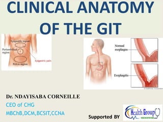
- 1. Dr. NDAYISABA CORNEILLE CEO of CHG MBChB,DCM,BCSIT,CCNA Supported BY CLINICAL ANATOMY OF THE GIT
- 2. ESOPHAGUS • The esophagus is a muscular tube that connects the pharynx to the stomach. • It begins in the neck where it is continuous with the laryngopharynx at the pharyngo-esophageal junction. • The esophagus consists of striated (voluntary) muscle in its upper third, smooth (involuntary) muscle in its lower third, and a mixture of striated and smooth muscle in between. Dr Ndayisaba Corneille
- 3. APPLIED ANATOMY: Esophageal Varices: • This results in cases of portal hypertension due to backward flow of blood into the systemic veins • This results in mark dilation of the lower esophageal veins. • In cases of increased pressure these varicosed veins will burst and this will lead to hematemesis and even fatal hemorrhage. Dr Ndayisaba Corneille
- 5. Achalasia Cardia: • A rare disorder making it difficult for food and liquid to pass into the stomach. • There is neuromuscular incoordination of the esophageal musculature as a result the difficulty in the passage of food through the esophagus this condition is known as dysphagia • It may be caused by an abnormal immune system response. • Symptoms include a backflow of food in the throat (regurgitation), chest pain and weight loss. • there would be accumulation of food especially at the lower part of the esophagus (abdominal part) and it will result in mark dilatation of the esophagus forming the achalasia cardia. Dr Ndayisaba Corneille
- 6. Esophagitis 1. The abdominal part of esophagus is most prone to esophageal carcinoma, peptic ulceration and inflammation this is due to regurgitation of the gastric acid. This is mostly found in patients regurgitative esophagitis. 2. Referred pain due to esophagitis is felt at the precordium and epigastric region. Dr Ndayisaba Corneille
- 7. Sliding hiatus hernia. • Sometimes the esophagus ends above the diaphragm as a result of this a hour glass stomach is formed which will lead to a sliding hiatus hernia is formed. • In most cases, a small hiatal hernia doesn't cause problems. • But a large hiatal hernia can allow food and acid to go up into the esophagus, leading to heartburn. • a very large hiatal hernia sometimes requires surgery. Dr Ndayisaba Corneille
- 8. The stomach • The stomach is a dilated part of the alimentary canal between the esophagus and the small intestine. • It is a muscular sac. • It is a J-shaped. Dr Ndayisaba Corneille
- 9. APPLIED ANATOMY of the Stomach • Vagotomy • This is the process of cutting up the vagus nerve so as to abolish the neurogenic gastric juice supply of the stomach. • The vagus nerves play a dominant role in the stimulation of gastric secretion. The basal or continuous secretion of gastric juice is almost entirely caused by tonic impulses in the vagus nerves. • Vagotomy also makes the stomach to becomes flaccid and this will result to difficulty in the passage of food into the duodenum since the action of the pyloric sphincter is compromised. • There are two types of vagotomy. Dr Ndayisaba Corneille
- 10. Total or complete vagotomy: • Where by the vagus nerve is totally cut off as a result of this the sphincteric action is compromised and passage of food is disrupted but to save the situation some procedures are carried out to help in the passage of food into the small intestine. • One of such procedures is the pyloroplasty where a tube is forced into the pyloric orifice thereby making the sphincteric action ineffective and through the tube food will gradually enter into the duodenum. Dr Ndayisaba Corneille
- 11. SELECTIVE VAGOTOMY: Dr Ndayisaba Corneille
- 13. Gastrectomy • Gastrectomy is surgery to remove part or all of the stomach. If only part of the stomach is removed, it is called partial gastrectomy. If the whole stomach is removed, it is called total gastrectomy. • About half of the patients subjected to total gastrectomy experience weight loss. • Malabsorption, particularly fat malabsorption, is a common feature after total gastrectomy. This may be due to shortened intestinal transit time, but is less often due to diarrhea or pancreatic exocrine insufficiency. Dr Ndayisaba Corneille
- 14. Gastritis • Gastritis: This is caused vagal stimulation which leads to high release of secretin which stimulates high acid secretion. Drugs like aspirin and others steroids can also lead to gastritis. • Gastritis pains are most times referred to the epigastric region (5th to 7th dermatome) Dr Ndayisaba Corneille
- 15. Peptic ulceration • Peptic ulcers occur when acid in the digestive tract eats away at the inner surface of the stomach or small intestine • Peptic ulceration usually occurs at the lesser curvature as a result of presence of the gastric canals. Dr Ndayisaba Corneille
- 16. Stomach Ulcer • Stomach ulcers, also known as gastric ulcers, are sores that develop on the lining of the stomach • Stomach ulcers are usually caused by Helicobacter pylori (H. pylori) bacteria or non- steroidal anti-inflammatory drugs (NSAIDs). • These can break down the stomach's defense against the acid it produces to digest food. The stomach lining then becomes damaged causing an ulcer to form Dr Ndayisaba Corneille
- 17. Stomach ulcer ……………… • when the ulceration becomes perforated it will result to peritonitis and the pain in the situation will be localized at the point of irritation. • Stomach ulcer pain usually begins in the upper middle part of the abdomen, above the umbilicus and below the sternum. • The pain may feel like burning sensation that may go through to the back. The onset of the pain may occur several hours after a meal when the stomach is empty. Dr Ndayisaba Corneille
- 18. PANCREAS • Soft, lobulated elongated gland with both exocrine and endocrine functions • Exocrine –pancreatic juice • Endocrine-insulin Dr Ndayisaba Corneille
- 20. Annular pancreas: • Developmental anomaly where ring of pancreatic tissue surrounds and obstruct duodenum Dr Ndayisaba Corneille
- 21. LARGE INTESTINE The large intestine constitutes the terminal part of the digestive system. It is divided into three main sections: cecum including the he primary function of the large intestine is the secretion is mucus, acts as a lubricant during the transport of the intestinal contents. Dr Ndayisaba Corneille
- 22. The vermiform Appendix/Abdominal Tonsil • Worm like narrow tubular diverticulum • Arises from posteromedial wall of the Caecum, 2 cm below the ileo-caecal junction • Suspended by a peritoneal fold – mesoappendix • It is a vestigial organ • Devoid of taenia coli, sacculations, appendices epiploicae • 2 -20 cm in length. ( average -9 cm) Dr Ndayisaba Corneille
- 24. Base of appendix Mc. Burney’s point maximum tenderness inflammation of appendix Spinoumbilical line Dr Ndayisaba Corneille
- 27. The rectum • The rectum is part of the GIT that lies between the sigmoid colon and anal canal. • it is about 12cm in length • It extends from the point where the sigmoid colon looses its mesentery which is anterior to the level to 3rd sacral vertebrae and terminates at the point where its muscular coat is replaced by internal anal sphincter at the anorectal junction, just as it passes through the pelvic floor behind the perineal body. Dr Ndayisaba Corneille
- 28. DISTINGUISHING FEATURES • Absence of plicae circularis. • There is absence of appendix epiplocae. • Absence of taenia coli, the taenia coli of the large intestine on getting to the rectum spreads out in a complete longitudinal muscular coat which is condensed anteriorly and posteriorly as the anterior and posterior band. In between these bands the longitudinal coat muscle is thin. • There is absence of mesenteric attachment as in other parts of the large intestine. • The rectum also presents sets of folds known as pilcae transversalis which also disappears as the rectum distends. • The rectum presents a dilated lower part known as the rectal ampulla which normally stores the resting feaces and flatus. Dr Ndayisaba Corneille
- 29. APPLIED ANATOMY: Digital examination of the Rectum • During digital examination or sigmoidoscopy, the patient is placed knee- chest position, lateral prone position, the modified lithotomy position, or the patient can bent over on a special table where the body is bent at an angle of 90O • These positions are mainly to draw the abdominal viscera upward into the upper abdominal cavity. PR - Per rectal examination Dr Ndayisaba Corneille
- 30. Rectal varies • Rectal varies occur because of portosystemic anastomosis which occurs between the superior rectal vein which is a tributary of the IMV (Portal) and middle rectal vein a systemic vein, • Portal obstruction will lead to pressure on the superior rectal vein as a result of this, there will be shunting of blood into middle rectal vein which will result to increased blood flow through the communicating venous channels which will become varicose giving rise to Rectal Hemorrhoids. Dr Ndayisaba Corneille
- 31. Cancer of the rectum: • The rectum is most prone to carcinoma and when it occurs it could spread extensive through lymphatic and venous channels. • It can affect the liver through the portal system, • it can affect the uterus and the ovary in females, it can also involve the prostate, the urinary bladder in males through lymphatic channels, • when the cancer occurs posteriorly it could involve the sacral plexus thereby causing pain across the lower limb and pain around the perineum. Dr Ndayisaba Corneille
- 32. Anal canal Terminal part of alimentary tract it begins at ano-rectal junction Rectal ampulla suddenly narrows at ano-rectal junction 2-3 cms infront and slightly below tip of coccyx From ano-rectal junction the canal passes Downwards & backwards through Pelvic diaphragm Dr Ndayisaba Corneille
- 33. INTERIOR OF ANAL CANAL Divided by pectineal line & Hilton’s line into 3 areas 1. Upper (15 mm) 2. Intermediate (15 mm) 3. Lower (8 mm) (Anal verge) Pectinate / dentate line Hilton’s line Dr Ndayisaba Corneille
- 34. ANORECTAL DISEASES • Hemorrhoids • Ischiorectal Abscess • Fistula in ano • Fissure in ano Dr Ndayisaba Corneille
- 36. in anal canal which may or may not bleed Piles pila (a ball) swelling Dr Ndayisaba Corneille
- 37. Pathogenesis • Various theories are : 1. Portal hypertension and varicose veins 2. Upright posture of human beings 3. Erosion and weakening of wall of veins due to infection secondary to trauma 5. Hard faecal matter obstructing venous return 6. Raised anal canal resting pressure Dr Ndayisaba Corneille
- 38. External hemorrhoid Internal hemorrhoid Below dentate line Above dentate line Varicosities of veins draining inferior rectal vein Varicosities of veins draining superior rectal vein Lined by Stratified squamous epithelium Lined by columnar epithelium Painful Pain insensitive Prone to thrombosis if vein ruptures (Thrombosed pile) May prolapse outside anal canal (prolapsed hemorrhoid) Dr Ndayisaba Corneille
- 40. HEMORRHOIDAL DISEASE A P L R Primary Locations 3-7-11 o’clock positons (Left Lateral, Right Anterior and Right Posterior) Right Posterior Right Anterior Left Lateral 3 MAJOR PILES Dr Ndayisaba Corneille
- 41. Anorectal Abscess and Fistula • Anal fistula? This is an abnormal communication between anal canal &skin Approx. 50% of anal abscess occur secondary to anal fistula. Abscess is the acute sign. • Anorectal abscess is presented as an Inflamed and tender perianal swelling Dr Ndayisaba Corneille
- 43. Anal Fissure • Anal fissure Linear ulcer (anal canal) dentate to anus • Symptoms of anal fissure Bleeding / anal pain • Physical findings Split in anal canal, posterior midline, sentinel pile, Digital Rectal Examination (DRE)is extremely painful Dr Ndayisaba Corneille
- 44. Sentinal pile is a tag formed by a ruptured anal valve Dr Ndayisaba Corneille
- 45. Sentinel skin tag Anal fissure () CLINICAL CASE: A 34 year old white female presents to your office experiencing painful "hemorrhoids" for 4 weeks. She states that the pain is associated with her bowel movements and is severe. She denies any blood per rectum. Anal fissure CASE STUDY Dr Ndayisaba Corneille
- 46. END Dr Ndayisaba Corneille THANKS FOR LISTENING By DR NDAYISABA CORNEILLE MBChB,DCM,BCSIT,CCNA Contact us: amentalhealths@gmail.com/ ndayicoll@gmail.com whatsaps :+256772497591 /+250788958241