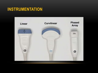
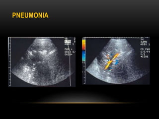
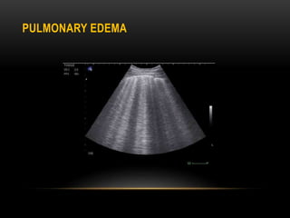
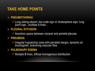
3. INTRODUCTION
The portable chest ultrasound is a routine diagnostic tool used in
the ICU setting to assess patient lung function
Chest ultrasound is useful only for assessing superficial pulmonary,
pleural-based and chest wall lesions
Chest ultrasound is increasingly used to guide interventional
procedures of the chest, such as biopsy and placement of
intercostal chest drain
4. Chest ultrasound necessitates the understanding of some basic
signs for interpreting normal and abnormal lung conditions.
SONOGRAPHIC ANATOMY
15. PLEURAL EFFUSION
The classic appearance of a pleural effusion is an echo free area
between the visceral and parietal portions of the pleura
Four different appearances are recognized at ultrasound
1. Anechoic 3. Complex and septated
2. Complex but not septated 4. Echogenic
17. PLEURAL THICKENING
Pleural thickening appears as hypoechoic broadening of the pleura
Pleural thickening most frequently related to scarring, fibrosis,
empyema and pleuritis
19. PLEURAL MASS
Malignant masses of the pleura include mesothelioma, lymphoma
and metastases
Mesothelioma is seen as irregular thickening of the pleura that may
appear nodular and is frequently associated with a large pleural
effusion
Color Doppler ultrasound of a malignant pleural mass may reveal
neovascularity with irregular tortuous vessels
21. PNEUMOTHORAX
Use of a combination of absent lung sliding and the loss of comet
tail artifacts has a sensitivity of 100% and specificity of 96.5%
The key sonographic signs used to diagnose pneumothorax
include
Absent lung sliding
Exaggerated horizontal artifacts/A lines
Loss of comet tail artifacts/B lines
Broadening of the pleural line to a band
23. LUNG POINT SIGN
The most specific finding of pneumothorax at ultrasound is the lung
point sign
The lung point sign represents the boundary between the aerated
lung and pneumothorax
26. BAR CODE SIGN
At M mode ultrasound , the area underneath soft tissue-
pneumothorax interface demonstrates multiple horizontal bands of
hyperechoic artifacts caused by the lack of visible lung motion
This M-mode appearance mimics bar code or stratosphere sign
29. DISEASES OF THE LUNG PARENCHYMA
In healthy individuals, visualization of the lung parenchyma is not
possible because the large difference in acoustic impedance
between the chest wall and the air with in the lung results in near
total reflection of the ultrasound waves
However, in parenchymal diseases that extend to the pleural
surface, replacement of air with in the lung creates an acoustic
window, allowing assessment of lung tissue
30. PNEUMONIA
Consolidation can be visualized on ultrasound
Appears as hypoechoic area with irregular margins
Branching echogenic structures are often seen within the
consolidation and represent air bronchograms
Air bronchogram may be seen as hyperechoic foci moving through
the bronchi(dynamic air bronchogram)within the consolidation
The branching pattern of vascular flow with in the consolidated
lung segment can be observed by using color Doppler
32. LUNG ABSCESS
A lung abscess can be identified as a hypoechoic lesion with a well
defined or irregular wall
The center of the abscess is usually anechoic but may contain
internal echoes and septations
34. ALVEOLAR-INTERSTITIAL SYNDROME
Abnormally increased lung water content and reduced air in the
alveoli result in fluid leakage into the pulmonary interstitium and
alveolar spaces
Patient with interstitial disease have an increased number of
vertical, comet tail artifacts called B lines
35. PULMONARY EDEMA
Pulmonary edema is the most common cause of acute alveolar
interstitial syndrome
In pulmonary edema, multiple B lines are seen having diffuse
distribution
At least 3 lines with curvilinear and 6 lines with linear transducer
are considered multiple and consistent with pathologic B lines
37. COPD AND ASTHMA
Multiple B lines at ultrasound are suggestive of alveolar interstitial
syndrome, which can be used to rule out acute COPD exacerbation
In general, the ultrasound appearance of the lungs and pleura in
asthmatic and COPD patients demonstrates multiple A lines with
normal lung sliding
38. PULMONARY MASS
PRIMARY LUNG NEOPLASMS
Appears as a homogenous, well defined mass that is usually
hypoechoic, but may be slightly echogenic
There is usually posterior acoustic enhancement
Color Doppler ultrasound is useful in distinguishing malignant
from benign pulmonary masses
Malignant masses are associated with neovascularity
40. METASTATIC
Peripheral pulmonary metastasis may be detected at sonography,
appearing as multiple sub pleural echogenic nodule measuring 1–2
cm in diameter
Color Doppler demonstrates the high vascularity of these lesions
42. DISEASES OF THE CHEST WALL
Soft tissue disease
Ultrasound is sensitive for the detection of soft tissue masses
arising within the chest wall. most of these are benign, such as
lipoma, sebaceous cyst, hematoma and abscess
Lymph nodes, particularly within the axilla and supraclavicular
fossa are easily examined with ultrasound
43. LIMITATIONS
Although the role of chest ultrasound has been well recognized in
evaluation of pleural effusion, pneumothorax, pulmonary edema,
and guidance for thoracentesis or biopsy, the value of ultrasound in
evaluation of the lung remains limited
Several lung sign at ultrasound are artifacts
Artifacts are affected by machine factors such as focal zone,
frequency and gain setting
45. TAKE HOME POINTS
PNEUMOTHORAX:
Lung sliding absent, bar code sign or stratosphere sign, lung
point sign , multiple A-lines
PLEURAL EFFUSION
Anechoic space between visceral and parietal pleurae
PNEUMOIA
Irregular hypoechoic area with serrated margin, dynamic air
brochogram, branching vascular flow
PULMONARY EDEMA
Multiple B lines, diffuse homogenous distribution
Xxxx
Ultrasound cannot penetrate into the substance of aerated lung, it is not a primary modality in evaluating the lung
However analysis of lung motion and the pattern of artifacts arising from the lung can help to investigate parenchymal lung disease
Normal chest US shows bat sign…when the probe is placed in the sagittal and longitudinal plane on the the chest in between the ribs…bat sign is produce
here the cortices and posterior acoustic shadowing of the ribs are the wings, the hyperechoic pleural line is the belly….showing bat sign
In the other image a. Subcutaneous fat , b. muscle, c.ribs , between the arrows is the pleural line
Normal chest usg shows multiple A lines
A lines are multiple horizontal, hyperechoic, reverbration artifacts originating from the pleural line repeating at regular interval indicating air
A lines are Seen in normal lung and pneumothorax
Formation : air is not a good transducer of sound waves,so sound scatter at the air/pleura interface….some waves reverberate in the interface then back to the probe creating equidistant,parallel bright white arcs called A lines
Lung Sliding/Gliding sign : In a normal lung pleural line will slide back and forth
the visceral pleura and underlying aerated lung can be seen sliding along the parietal pleura due to respiratory motion……….
Video…
A lines and posterior acoustic shadowing of ribs
If we are unable to see the lung sliding,machine can do this for us. M mode is the way the machine see things that are moving
Here are M mode usg images through normal lung giving the appearance of a sea above a sandy shore…sea shore sign
Horizontal means static, granny means motion
xxxx….Here are two different distinct pattern created by the motion of lung pleura against the relatively more static chest wall
In some normal lungs there are a limited no of comet tail artifacts arising from the lung surface known as B lines
Lung ultrasound images showing B lines(arrow heads) and pleural line
B lines are vertical, hyperechoic, comet tail artifacts that erase A lines
More than 3 on curvillinear and 6 on linear probe are multiple b lines
Multiple diffuse B lines = abnormal lung
Seen when fluid replaces air
Formation: as ultrasound waves alternate between air and fluid in the interstitial space ,b lines are formed
High frequency linear probe is ideal for imaging superficial structures like pleura or for identifying lung slinding
Low frequency curvillinear probe is useful in the assessment of effusion,consolidation and diaphragm
A phased array transducer probe is useful because the footprint can be placed easily in the intercostal space
The lung ultrasound examination consists of 12 imaging zones, 6 on each side of thorax.
The upper and basal part of each hemithorax is divided into anterior, lateral and posterior by the anterior and posterior axillary lines.
Ant and post lung zones are examined in supine and sitting positions respectively
Supine and sitting for 1 and 3/ant and post zones respectively
The lateral zone can be examined in Supine or lateral position
Transudates are almost invariably anechoic
Exudates may appear anechoic, complex or echogenic
we do usg in Coronal/side and longitudinal view above diaphragm
Usg images showing pleural effusion
Minimal right sided pleural effuion on right sided image
Large volume anechoic effusion visualized superior to diaphragm and spleen on left sided image
xxxxxx
Unlike pleural effusion the pleural thickening does not exhibit the fluid color sign on color doppler
Usg image showing pleural thickening as a hypo echoic band just superficial to the echogenic pleural lung interface
Usg image showing A hypo /anechoic area causes doubling of the pleural line on right side
Pleural masses are generally visualized sonographically… they May be benign or malignant
Benign pleural masses such as fibroma, lipoma and neuroma are uncommon
xxx….These appear as well defined rounded masses of variable echogenicity, depending on the fat content of the cells
A biopsy is usually required to reach definitive diagnosis
last….The most common pleural metastases are from primary adenocarcinoma
Ultrasound image showing an irregular pleural mass and pleural fluid ……..xxxxxx representing mesothelioma
Bedside sonography is useful for excluding pneumothorax
Lung point is the tranzition zone between where normal lung is sliding and where the pneumothorax is not sliding
M mode USG image showing lung point sign which is the transition zone between normal lung and pneumothorax
M mode ultrasound image showing bar code sign/air artifacts…. multiple horizontal bands of hyperechoic artifacts caused by the lack of visible lung motion
M mode USG images
A…seashore sign
B…Bar code sign
C… Lung point sign
Consolidation resembles the sonographic texture of the liver
in pneumonia we lose A lines
In ateletasis the air bronchogram is usually static
USG image showing an area of consolidation with in the right lower lobe .the texture of the consolidated lung appear isoechoic to the liver. multiple echogenic foci are seen within the consolidated lung and correspond to air filled airways
On the color doppler scan, a pulmonary artery branch supplying the segment is clearly seen
in pneumonia we lose a lines
Pneumonia resulting from pyogenic organisms can undergo necrosis leading to lung abscess
Ultrasound image showing a rounded hypoechoic lesion within a left basal consolidation
B lines are due to reverberation artifact secondary to reflection of sound waves at the interlobular septa
ARDS is another example in which multiple b lines are distributed inhomgeously
Chest ultrasound image showing multiple B lines arising from the pleural line and erasing the A lines representig pulmonary edema
The primary role of usg in pulmonary mass is to provide guidence for needle biopy
Pulmonary masses can be visualized when there is no aerated lung between the mass and transducer
xxx…Consolidation and fluid bronchograms may be seen adjacent to the mass
Last….Ultrasound is a valuable tool in the assessment of pancoast or superior sulcus tumors
Usg image showing hypoechoic mass,slight inhomogenous with irregular surface but sharp delimited from ventilated lung…lung cancer
Color doppler usg of lung cancer showing vascularity without normal radial distribution of vessels and bronchi(destruction of normal lung structure)
Peripherally located metastatic lung tumor
Ultrasound guided biopsy proved this to be a metastasis of a renal cell carcinoma
Right….multiple horizontal bands of hyperechoic artifacts caused by the lack of visible lung motion….pneumothorax
appearance of a sea above a sandy shore…horizontal…static…granny …motion….normal lung
ultrasound image showing multiple B lines arising from the pleural line and erasing the A lines representig pulmonary edema