SlideShare a Scribd company logo
1
Multistep nature of cancer development
• Phenotypic progression
– loss of control over cell growth/death (neoplasm)
– invasiveness (carcinoma)
– distal spread (metastatic tumor)
• Genetic progression
– multiple genetic lesions required for cancer
– normal cell malignant cell
– how many independent genetic lesions are necessary for
development of a clinically obvious cancer?
– mutation rates, rate-limiting steps, genomic instability
Cancer genes
• What genes, when altered, promote cancer?
– tumor suppressor genes and proto-oncogenes
• Some genes are altered in a restricted set of tumor types
– e.g., APC in colorectal carcinoma
• Others are altered in a broad spectrum of tumor types
– e.g., p53 and the Ras genes
• The importance of tumor gene “pathways”
– the p53 and Rb tumor suppressor pathways
Proto-oncogenes vs. tumor suppressor genes
Proto-oncogenes promote cancer when
malignantly activated
– An activated proto-oncogene contributes to
tumorigenesis by "gain-of-function"
– Thus, an activated proto-oncogene is genetically
dominant at the cellular level
• an activated oncogene can elicit a new phenotype
(tumorigenesis) even in the presence of the corresponding
wildtype allele
Proto-oncogenes vs. tumor suppressor genes
Tumor suppressor genes promote cancer when
malignantly inactivated
– A tumor suppressor contributes to tumorigenesis by
"loss-of-function"
– In most instances, an inactivated tumor suppressor
gene is genetically recessive at the cellular level.
• it will not promote tumorigenesis in diploid cells unless
the other (wildtype) allele is also lost or inactivated
• some exceptions (e.g., dominant-negative p53 mutations)
Tumor suppressor genes
• in this lecture we will focus on…
– the retinoblastoma susceptibility (Rb) gene
– the p53 tumor suppressor gene
• genetic properties
• biochemical functions of their protein products
• the p53 and Rb tumor suppressor “pathways”
Cancer susceptibility syndromes
• What proportion of human cancers are heritable?
• Hereditary syndromes of cancer susceptibility are
usually caused by germline mutations of tumor
suppressor genes.
– familial retinoblastoma: Rb
– Li-Fraumeni syndrome: p53
– familial adenomatous polyposis coli: APC
– hereditary non-adenomatous c.c.: MLH1, MSH2
• Penetrance: fully penetrant mutations segregate as
dominant traits in a Mendelian fashion
2
Sporadic and Heritable forms
of Retinoblastoma
• age of tumor onset
– sporadic (~60% of cases): ~ 6 years
– heritable (~40% “ “ ): ~ 2 years
• number of independent tumors
– sporadic: single tumor (only one eye is affected)
– heritable: multiple tumors (both eyes are affected)
• tumor frequencies in children of patients
– sporadic: 1 in 105
– heritable: 1 in 2
patients with heritable retinoblastoma transmit
a “Rb susceptibility gene” to their children in a
dominant Mendelian fashion
Knudson's hypothesis (1970s)
• Two (rate-limiting) genetic lesions required…
• Sporadic retinoblastoma:
– both alterations are acquired somatically
– incidence: (10-6)(10-6)(107 cells) = 10-5 tumors/person
– very rare; involves only one eye
• Heritable retinoblastoma:
– one alteration is inherited in the germline
(i.e., the “Rb susceptibility gene”)
– the second alteration is acquired somatically
– incidence: (1)(10-6)(107 cells) = 10 tumors/person
– all carriers affected; involves both eyes!!
– one mutation is inherited in the germline
– second mutation is acquired somatically
– incidence: (1)(10-6)(107 cells) = 10 tumors/person
• All mutation carriers are affected
– Rb susceptibility is a highly penetrant trait
– tumor susceptibility is transmitted as a dominant trait in family
pedigrees (despite the fact that tumor suppression genes
function recessively at the cellular level).
Hereditary Retinoblastoma
The Retinoblastoma (Rb) gene
• What are the two rate-limiting genetic alterations?
• Cytogenetic abnormalities of chromosome 13:
– interstitial deletions of variable length
– always involve material from chromosome band 13q14
– sporadic patients: deletions in tumor cells only
– heritable patients: deletions in both normal & tumor cells
• Is Rb susceptibility due to genetic loss at 13q14?
If so, then the two mutations required for retinoblastoma might
represent inactivation of both alleles of a single gene at 13q14
Sporadic
Retinoblastoma
Familial
Retinoblastoma
tumor cells:
Rb, Rb
(normal)
Rbm1, Rb
(normal)
Rbm1, Rbm2
(neoplastic)
Rbm1, Rbm2
(neoplastic)
m1
m2
m2
malignant mutations of the Rb gene act recessively at the
cellular level, contributing to neoplasia by loss-of-function
retinal cells:
The Retinoblastoma gene (Rb)
• 1988: isolation of the Rb gene on 13q14
Familial retinoblastoma
• one Rb gene lesion in germline of familial patients
• other (normal) Rb allele lost or inactivated in tumors
Sporadic retinoblastoma
• both alleles of Rb are normal in germline
• both Rb alleles lost or inactivated in tumors
Important: genetic lesions in the same gene are
responsible for both the familial and sporadic forms
of retinoblastoma!!
3
chromosome 13
maternal
&
paternal
homologues
Inactivation of the second Rb allele (in somatic cells)
Rbm1
Rbm1 Rbm2
de novo
mutation
Rbm1
chromosome
loss
Rbm1 Rbm1
chromosome
loss
&
reduplication
gene
conversionRbm1 Rbm1
The penetrance of
germline Rb mutations
• Almost all carriers will develop retinoblastoma
– high penetrance
• (mutation rate)(target cells) = (10-6)(107 cells) = 10
• retinoblastoma susceptibility is transmitted as a
dominant Mendelian trait
• Some carriers will also develop osteosarcoma
– low penetrance
• (mutation rate)(target cells) < 1
• osteosarcoma susceptibility is not transmitted as a
dominant Mendelian trait
Tumor Suppressors: the p53 gene
• p53 encodes a transcription factor
• the p53 gene is altered in many human tumors (usually by
missense mutation).
• in vitro cell transformation by mutant p53 genes.
Is p53 a proto-oncogene?
• murine erythroleukemias induced by Friend Leukemia Virus:
a natural knockout of the p53 gene by proviral insertion!
• suppression of cell transformation by the wild-type p53 gene.
Is p53 a tumor suppressor gene?
Dominant-negative mutations
of a tumor suppressor gene
• dominant-positive mutation (e.g., missense
mutations in the Ras proto-oncogenes).
• recessive-negative mutation (e.g., Rb loss).
• dominant-negative mutation (e.g., many p53
missense mutations).
• note: dominant-negative mutations result in
functional inactivation of the protein products of
both alleles (including the normal allele).
Dominant-negative mutations of p53
• how do dominant-negative mutations work?
• p53 normally functions as a homo-tetramer
• consider p53 function in a cell with one wildtype
and one mutant p53 allele:
functional
p53 tetramer?
yes
yes
no
no
wildtype p53
mutant p53
Dominant-negative mutations of p53
• how do dominant-negative mutations work?
• p53 normally functions as a homo-tetramer
• consider p53 function in a cell with one wildtype
and one mutant p53 allele:
functional
p53 tetramer?
yes
yes
no
no
wildtype p53
mutant p53
• mutant p53 is more
stable than wildtype p53
4
p53 mutations in
hereditary and sporadic cancer
• Li-Fraumeni Syndrome (LFS)
– caused by germline mutations of p53
– LFS carriers develop many different forms
of cancer
• sporadic cancer
– often caused by somatic mutations of p53
– very common in human cancer
– found in many different forms of cancer
Tumor suppressor proteins
• proteins encoded by Rb and p53
• the normal functions of these proteins
• mechanisms of tumor suppression
• the Rb and p53 tumor suppressor pathways
Phosphorylation of the Rb protein
• The phosphorylation state of Rb changes during
normal cell cycle progression.
– Rb is hypophosphorylated in:
• G0 (resting cells)
• early G1 (cycling cells)
– Rb is hyperphosphorylated in:
• S phase
• G2 phase
– Rb is phosphorylated before the G1/S transition…
• by an enzymatic complex: CDK4 / cyclin D
M
G1 S
G2
restriction
point
G0
(resting cell)
cell cycle progression
Rb Rb~P
Cdk4 / cyclin D
The restriction point (in late G1)
• the major control point of cell cycle progression
• G1/S transition is mediated by the E2F family of
transcription factors
• E2F binds the promoters of genes required for cell
cycle progression (G1/S transition and S phase).
E2F
promoter of
S-phase genes
transcription
Some S phase genes regulated by E2F:
S phase gene Function
• thymidine kinase nucleotide synthesis
• DHFR (dihydrofolate reductase) “ “
• DNA polymerase α DNA synthesis
• ORC1 “ “
• histone H2A chromosome assembly
• cyclin E cell cycle progression
• cyclin A “ “ “
5
Resting cells and early G1 phase cells
• hypophosphorylated Rb binds promoter-bound E2F
• Rb inactivates transcription by E2F
• S phase genes are repressed
• G1/S transition is blocked
Rb
E2F
S-phase genes
repressed
restriction point
• CDK4/cyclin D phosphorylates Rb in the “pocket”
• hyperphosphorylated Rb dissociates from E2F
• E2F activates transcription of S phase genes
• cells enter S phase
Rb
P P P
E2F
Rb
S-phase genes
activated
E2F
S-phase genes
repressed
Cdk4/cyclin D
• In normal cells, phosphorylation of Rb by the
CDK4/cyclin D kinase is a highly regulated
• Focal point for the major signal transduction
pathways that control normal cell growth
Rb
P P P
E2F
Rb
S-phase genes
activated
E2F
S-phase genes
repressed
Cdk4/cyclin D
Rb
P P P
E2F
Rb
S-phase genes
activated
E2F
S-phase genes
repressed
Cdk4/cyclin D
diverse signaling pathways
The function of Rb
• hypophosphorylated Rb serves to restrain the
proliferation of normal cells.
• regulated phosphorylation of Rb allows normal
cells to proliferate at the correct time and place.
• therefore, imagine the consequences of losing
normal Rb function…
– deregulation of E2F (and the G1/S transition)!
• how might Rb become inactivated in cancer?
Inactivation of Rb function in tumors
(leaving E2F unregulated)
• Direct inactivation:
– Rb gene deletion (occurs in retinoblastoma)
– point mutations in the Rb pocket (in retinoblastoma)
– occupancy of the Rb pocket by early proteins of
DNA tumor viruses
• human papilloma virus (HPV), an etiological agent in
human cervical carcinomas
• HPV encodes two proteins required for tumorigenesis
• E7 binds the pocket of hypophosphorylated Rb
• Deregulation of E2F (and the G1/S transition)
6
The Rb tumor suppressor pathway
p16
Rb
P P P
E2F
Rb
S-phase genes
activated
E2F
S-phase genes
repressed
Cdk4 / cyclin D
Indirect inactivation of Rb function in tumors
• overexpression of cyclin D1
– breast cancer, B cell lymphoma
• loss of p16, an inhibitor of Cdk4
– many human cancers
• inherited point mutation in Cdk4 that renders it
insensitive to inhibition by p16
– familial melanoma
»Inactivation of the Rb pathway occurs in most, if not
all, human tumors!
Normal functions of the p53 protein
• p53 polypeptides are very unstable in normal cells
(1/2 life of ~30 minutes)
• the cellular response to genotoxic stress
– DNA damage by UV light, ionizing radiation, chemical
carcinogens, errors in replication, etc.)
– induction of certain signal transduction pathways
• post-translational modifications of p53 polypeptides:
– especially, phosphorylation and acetylation
– stabilize p53 (1/2 life of ~150 min.), leading to higher
steady-state levels
– increase the transcriptional activity of p53
consequences of p53 activation
• The transcriptional activity of p53 induces a
cellular response, the nature of which is dependent
on various factors, including the cell type.
– p53 induces G1 arrest (and DNA repair) in:
• normal fibroblasts
• certain epithelial cells
– p53 induces apoptosis in:
• thymocytes
p53 induction of
cell cycle arrest or apoptosis
• in either case, replication of damaged DNA ceases
• prevents accumulation of oncogenic mutations
• In essence, p53 suppresses tumor formation by
maintaining the integrity of the genetic material in
cells subjected to genotoxic stress.
Transcriptional targets of p53
• p21 CDK inhibitor
G1 and G2 arrest in fibroblasts
• 14-3-3σ
G2 arrest in epithelial cells
• PUMA
promotes apoptosis in thymocytes, fibroblasts, neurons
• p53R2 nuclear ribonucleotide reductase
required for DNA repair
• p48 subunit of the XPA complex
required for nucleotide excision repair
• etc…
7
Normal cell p53 mutant cell
p53 protein is activated
p53 induces target genes
DNA repair
growth arrest
Apoptosis
No response
DNA damage
persists
- proto-oncogenes activated
- tumor suppressors inactivated
- cancer -
Normal cell
genotoxic stress
The p53 tumor suppressor pathway
p53
mdm2
p21
ATM
»Inactivation of the p53 pathway occurs in most,
if not all, human tumors!
Sporadic colorectal carcinoma
• Accounts for >80% of colorectal carcinomas.
• Natural history of the sporadic disease
(viewed by colonoscopy):
– normal epithelium
– hyperproliferative epithelium (APC*)
– early adenoma (APC*)
– intermediate adenoma (APC*, Ras*)
– late adenoma (APC*, Ras*, SMAD2/4*)
– carcinoma (APC*, Ras*, SMAD2/4*, p53*)
– metastasis
Hereditary colorectal carcinoma
• A predisposition to colorectal carcinoma is
associated with two distinct syndromes.
– Familial adenomatous polyposis coli
(FAP)
– Heritable non-polyposis colorectal carcinoma
(HNPCC)
Familial adenomatous polyposis coli (FAP)
• FAP: rare, autosomal, dominantly-inherited
• Natural history of FAP:
– multiple polyps (hundreds or thousands) develop
throughout the colon by early adulthood.
– inevitably, one or more of these polyps will progress into
an invasive carcinoma.
• FAP results from inherited lesions in the APC gene
(which behaves as conventional tumor suppressor)
• FAP accounts for less than 1% of all colorectal
carcinomas. However, most sporadic cases of
colorectal carcinoma have somatic mutations of APC.
8
Hereditary non-polyposis colorectal carcinoma
(HNPCC).
• HNPCC: autosomal, dominantly-inherited
• Natural history of HNPCC:
– Carriers do not have an obvious pre-malignant condition
(e.g., the histology of their colon is normal).
– Carriers are predisposed to develop colorectal carcinoma.
• HNPCC results from inherited lesions in genes
encoding components of the DNA mismatch repair
system:
– hMSH2, hMLH1, hPMS1, or hPMS2
Mismatch repair defects in HNPCC
• mismatch repair incorrectly paired nucleotides
• malignant cells of HNPCC tumors exhibit genetic
instability at the nucleotide level
• this underlying genetic instability accelerates
malignant progression.
• HNPCC accounts for 2-4% of colorectal carcinomas
• In addition, ~13% of sporadic colorectal carcinomas
display mismatch repair deficiency; these all harbor
somatic mutations in one of the mismatch repair genes
implicated in HNPCC
Tumor Suppression
• tumor suppressors inhibit tumor formation by a
variety of different mechanisms:
• negative control of cell growth...
– by regulating cell cycle progression (e.g., Rb, p16)
– by regulating signal transduction pathways (APC)
• maintenance of genomic integrity...
– by regulating the cellular response to DNA damage
(p53, ATM, Chk2)
– by functioning as effectors of DNA repair
(hMSH2, hMLH1)
.
• Linkage analysis of family pedigrees: identify
polymorphic markers that co-segregate with tumor
susceptibility
• Loss of heterozygosity (LOH) in tumor cells:
identify polymorphic markers that exhibit tumor-
specific LOH
Positional cloning: search for transcription units
(candidate genes) and analyze each for:
– germline mutations that co-segregate with tumor
susceptibility (in familial tumors)
– somatic mutations that arise uniquely in the malignant
cells (in sporadic tumors).
Methods to isolate tumor suppressor genes
9
Chromosome 13
maternal
&
paternal
homologues
the second Rb mutation (in somatic cells)
Rbm1
Rbm1 Rbm2
de novo
mutation
Rbm1
chromosome
loss
Rbm1 Rbm1
chromosome
loss
&
reduplication
gene
conversionRbm1 Rbm1
LOH
LOH
LOH
 World Cancer Day – 4th February 2014
 By 2025, there will be more than 20 million
new cancer cases per year, compared with
14.1 million in 2012,
IARCWorld Cancer Report 2014
 Greek word : Karkinos (crab)
 Hippocrates : First coined the term
 Cancer is an overgrowth of cells bearing
cumulative genetic injuries that confer
growth advantage over the normal cells
 Accumulation of mutations and epigenomic
changes in somatic cells
Sporadic
Familial
Hereditary
77-80%
10-15 %
5-10%
Nagy R, Sweet K, EngC. Highly penetrant hereditary cancer syndromes. Oncogene. 2004 Aug 23;23(38):6445-70
• Early onset
• Bilateral/Multifocal
Disease
• Multiple Primary
cancer in single
individual
• Clusters of Cancer in
family
• Rare cancers
• Precursor lesions
Deregulating Cellular
Energetic
Avoiding immune
Destruction
Hanahan D, Weinberg RA. The hallmarks of cancer. Cell. 2000
 Gene : A hereditary unit consisting of a sequence of
DNA that occupies a specific location on a
chromosome and determines a particular
characteristic in an organism
 Trait : A distinguishing feature, a genetically
determined characteristic or condition.
 Locus : specific area on chromosome where the gene
is found
 Allele :Versions of a gene
 Genotype : the genetic makeup of an organism
 Phenotype : the physical appearance of an organism
 Pleiotropy : the ability of a gene to affect an
organism in many ways
 Penetrance
 The probability that a gene will have any
phenotypic expression
 Its an all or none concept
 Expressivity
 Severity of the manifestations of the phenotype
Driver Mutation :
• Contribute to
Oncogenesis
• Provide growth
advantages
• Occurs in
• Oncogenes
• Tumor SuppressorGenes
Passenger
Mutation :
• Neutral Mutation
• Carried along the
ride
• Does not cause or
propel cancer’s
growth or spread
 Proto-oncogenes – normally promote
normal cell growth; mutations convert them
to oncogenes.
 Tumor suppressor genes – normally restrain
cell growth; loss of function results in
unregulated growth.
 Oncogenes
 promote cell
proliferation
 dominant & highly
conserved
 types :
▪ viral oncogenes [ v-
oncs ]
▪ cellular oncogenes [ c-
oncs ]
Secreted Growth Factors
• c-sis, hst
Cell Surface Receptors
• c-sis, hst erb B, ret, trk, fes, fms
IntracellularTransducers
• c-src, c-abl, mst, ras
DNA-binding Nuclear Proteins
• myc, jun, fos
Regulators of the Cell Cycle
• bcl, bax, bad
 Tumor Suppressor Gene
 Inhibits growth and multiplication of mutated
cells
 Prevent neoplastic transformation
 Recessive and highly conserved
 Categories :
 Caretaker Gene
 Gatekeeper Gene
Category Gene Function Tumors
Gatekeeper p53 Transcription factor Li-Fraumeni
syndrome
Rb1 Transcriptional
regulator
Familial
retinoblastoma
APC Regulates β-
catenin function
Familial
Adenomatus
polyposis
Caretaker BRCA1 DNA repair Breast and ovarian
cancer
BRCA2 DNA repair Breast Cancer
MSH2 DNA mismatch
repair
HNPCC
Syndrome Incidence Genes
Involve
d
Molecular Pathway
affected
RenalType Others
characteristics
VHL 1:36,000 VHL Hypoxic
pathway(through
HIF)
Clear cell
RCC
pancreatic cysts
and
neuroendocrine
tumors,
pheochromocyto
ma,
retina I angiomas,
hemangioblastom
as
Birt-
Hogg-
Dube
rare FLCN m-TOR Variable
subtypes
cutaneous lesions,
pulmonary cvsts
and
spontaneous
pneumothorax
Syndrome Incidence Genes
Involved
Molecular
Pathway
affected
RenalType Others
characteristics
HPRC rare (Iess
than
1:1,500.00
MET C-MET Type 1 papillary
RCC
not specific
HLRCC Rare
(unknown)
FH Krebs cycle HLRC related
RCC
Multiples
cutaneous and
uterine
leyomiomas
SDH-RCC rare(unknow
n)
SDHB/SD
HC/
SDHD
Krebs cycle SDH related
RCC
Paraganglioma
s/
Pheocromocyt
oma GIST
 Hereditary kidney cancer accounts for 3 to 5%
of all kidney cancer
 Ten inherited cancer susceptibility syndromes
are associated with inherited risk of kidney
cancer and 12 genes have been identified
Genetics
• Autosomal Dominant
• Gene Locus on Chromosome 3p25
Incidence
• 1 in 35000 live births
Genetics
• Tumor Suppressor gene
• VHL gene product pVHL regulates function of HIF
• Reduced pVHL– no HIF degradation—increasedVEGF
 Sites involved
Organ Tumor Non-tumor lesions
Brain Hemangioblastoma
Eye Hemangioblastoma
Kidney CRCC Cyst
Adrenal Phaemochromocytoma
Pancreas NET Cyst
Inner Ear Endolymphatic Sac tumor
Epididymis PapillaryCystadenoma
Type 1 VHL loss or mutation that
affects the protein
folding
Haemangioblastoma
Renal Cell Carcinoma
Low risk of
phaeocromocytoma
Type 2A VHL missence mutation Haemangioblastoma
phaeocromocytoma
Low risk of Renal Cell
Carcinoma
Type 2B VHL missence mutation Haemangioblastoma
Renal Cell Carcinoma
Phaeocromocytoma
Type 2C VHL missence mutation Phaeocromocytoma only
Haemangioblastoma Clear Cell RCC Phaeochromocytoma
Papillary Cystadenoma
Diagnostic Criteria
Known Positive Family Hx – presence of
one:
Single retinal or cerebellar
hemangioblastoma
RCC
Pheochromocytoma
Negative Family Hx –
2 retinal or cerebellar
hemangioblastomas
Single hemangioblastoma and
additional characteristic lesion.
Screening and Follow-up
• Annual ophthalmologic evaluation and
visual field testing starting around age 2
• MRI of the brain and spine every 2 years
starting in early adolescence
• Annual abdominal US starting at age 5
• Abdominal CT or MRI starting at age 20
• Frequent blood pressure monitoring and
measurement of urinary catecholamine
levels or plasma metanephrine levels every
1–2 years starting at age 2
Treatment
• Hemangioblastomas
• Complete surgical resection
• Incomplete resection + external beam RT
•VEGF and PDGF pathway inhibitors (sunitinib and sorafinib)
Genetics :
• Autosomal dominant
• Gene : FLCN, Chromosome 17p, codes for protein “folliculin”
Clinical manifestations :
• Fibrofolliculomas (dysplastic hair follicules)
• Lung cysts
• Spontaneous pneumothorax
• Renal cancer
Incidence
• 1 of 200,000 people
• Hybrid oncocytic-
chromophobe type
• Chromophobe tumors
• Clear cell
• Oncocytomas
• Papillary renal cancers
Fibofolliculoma
Renal
Oncocytoma
Chromophobe
RCC
Major criteria
• At least five fibrofolliculomas, at least one
histologically confirmed, of adult onset or
• Pathogenic FLCN mutation
Minor criteria
• multiple lung cysts: bilateral basally located
lung cysts with no other apparent cause,
with or without spontaneous
pneumothorax
• renal cancer: early onset
Genetic Screening:
Germline FLCN testing is
recommended beginning
at age 21
Imaging Surveillance:
• Annual MRI or low dose
CT is recommended
beginning at age 20
• In patients without a
renal lesion MRI every 3
years is recommended
Hereditary Papillary
Renal Cancer
• Genetics
• Autosomal
Dominant
• Gene : MET
(activating mutation)
Chromosome 7q31.1
• Manifestations
• Papillary RCC type 1
Hereditary Leiomyoma
Renal Cell carcinoma
• Genetics
• Gene : FH,
chromosome 1q42
• Manifestations
• Papillary RCC type II
• Uterine Leiomyoma
Genetic Mechanism :
• SDH- kreb’s cycle enzyme
• Germline mutation in SDH B,C,D gene (SDHB has strongest association)
• SDHB gene locus- chromosome 1p36
• Succinate stabilises HIF
Incidence
• 0.1-2% of all renal cancers
Manifestation
• Paraganglioma
• Phaeochromocytoma
• Renal cell Carcinoma (Clear cell or chromophobe)
 BAP1 mutations and familial renal cancer
 Predispose to
▪ Familial clear cell renal cancer
▪ Uveal and cutaneous melanoma
▪ Mesothelioma
 Chromosome 3 translocations
 PTEN hamartoma tumor syndrome (Cowden
disease)
 Urothelial carcinoma :
 Associated with Lynch Syndrome
 Prostate Carcinoma :
 Polygenic Disorder
 BRCA1 and BRCA2 mutation :
▪ Increased risk of Prostate Cancer
▪ High grade and Advanced Stage
Syndrome Inheritance Gene Locus Gene Incidence Histology
FAP AD 5q21 APC 2-12% PTC_CMV
Cowden AD 10q23.3 PTEN,SDH,PIK3CA,
AKT1
35% PTC (follicular
variant), FTC,
Adenomatous
Nodule, C cell
hyperplasia
Carney AD 2p16,17q24 PPKAR1α 15% PTC,FTC,
adenomatous
nodule,Follicular
Adenoma
Wermer AR 8p11-p12 WRN 18% PTC,FTC,ATC
Mc-cune
Albright
20q13.2-13.3 GNAS PTC,FTC, Follicular
adenoma
DICER-1
syndrome
AD 14q32.13 DICER1 Rare Nodular Hyperplasia
 Lifetime risk for epithelial thyroid cancer is
approximately 10%
 Median age of onset was 37 years
 Youngest age at diagnosis was 7 years
 Thyroid lesions in CS
 Adenomatous Nodule
▪ Multiple, unencapsulated, homogenous, firm
 C cell hyperplasia
 Follicular Carcinoma
 PTC (Rare)
Adenomatous nodules in Cowden syndrome, routine stain.
(A) Solid adenomatous nodules surrounded by a thin rim of fibrous capsule, closely
dispersed in the thyroid gland.
(B) Each adenomatous nodule is composed of small follicles lacking abundant colloid
(C) Immunohistochemistry for PTEN shows loss of PTEN expression in an
adenomatous nodule in Cowden’s disease.
 160 fold increased risk of PTC
 Incidence : ~ 0.4% to 6.1% (Steinhagen E, et
al. 2012)
 Most thyroid cancer in individuals with FAP is
Cribriform-morular variant of PTC
Cribriform–morular variant of papillary thyroid carcinoma in familial adenomatous
polyposis, routine stain. (A, B)Typical cribriform arrangement composed of fused
follicles lined by tall cells and lumina lacking colloid. (C) Morular formation.
 Rare condition characterized
by
 Skin pigmentary abnormalities
 Myxomas
 Endocrine tumors or
overactivity
 Genetics
 Autosomal dominant
 Gene – PPKAR1A gene on
chromosome 17q
 Clinical Features/Major Diagnostic Criteria
 Spotty skin pigmentation
 Myxoma (cutaneous and mucosal)
 Cardiac myxoma
 Breast myxomatosis
 Primary pigmented nodular adrenocortical disease (PPNAD)
 Acromegaly as a result of growth hormone (GH)-producing
adenoma
 Large-cell calcifying Sertoli cell tumor (LCCSCT)
 Thyroid carcinoma or multiple, hypoechoic nodules on thyroid
ultrasound in a child younger than age 18 years
 Psammomatous melanotic schwannomas (PMS)
 Blue nevus, epithelioid blue nevus
 Breast ductal adenoma
 Osteochondromyxoma
 Cell cycle checkpoint kinase 2 (CHEK2)
 Tumor suppressor gene
 CHEK2 mutations associated with a
moderate increase in the risk for various
types of cancer
 Breast cancer - 2-4x increased risk
 Colorectal
 Prostate
 Siolek et al 2015 studied CHEK2 gene in
thyroid cancer patients
 468 cases and controls (Polish population)
 CHEK2 mutations seen in 15% of unselected
PTC patients and 6% of controls (p=0.006)
 7/11 women with breast and thyroid cancers
had CHEK2 mutations
 Tumor susceptibility
syndrome
 Confers increased risk for-
 Pleuropulmonary blastoma
(PPB)
 Ovarian sex cord-stromal
tumors (such as Sertoli- Leydig
cell tumor or juvenile granulosa
cell tumor)
 Cystic nephroma
 Thyroid gland neoplasia
 Rhabdomyosarcoma
 Pituitary blastoma
 Wilm’sTumor
 Increased risk of developing thyroid cysts
and/or multinodular goiter (MNG) in families
with a germline DICER1 mutation
 Risk of developing syndrome-associated
thyroid cancer (papillary or follicular)
 Birt-Hogg-Dube syndrome – cutaneous
manifestations, pulmonary cysts/history of
pneumothorax, and various types of renal
tumors
 Hyperparathyroidism-jaw tumor syndrome
(HPT-JT)
 Hereditary
Paraganglioma/Pheochromocytoma (SDHB
and SDHD)
 Multiple Endocrine NeoplasiaType 1
MEN 1 MEN 2
•Parathyroid hyperplasia or adenoma
•Islet cell hyperplasia, adenoma, or
carcinoma
•Pituitary hyperplasia or adenoma
•Other less common manifestations:
foregut carcinoid, pheochromocytoma,
subcutaneous or visceral lipomas
MEN2A
• MTC
•Pheochromocytoma
•Parathyroid adenoma
•MEN2A with cutaneous lichen amyloidosis
• MEN2A with Hirschsprung disease
MEN2B
• MTC
• Pheochromocytoma
• Mucosal and gastrointestinal neuromas
• Marfanoid features
Chr. 10q11.2
Medullary thyroid carcinoma in multiple endocrine neoplasia syndrome, routine stain. (A) Medullary thyroid
carcinoma featuring lobular and solid architectures with stromal calcitonin amyloid deposits.Tumour cells are
variable in size and shape, with slightly granular cytoplasm and punctate coarse chromatin. (B) Medullary thyroid
microcarcinoma in a young patient subjected to prophylactic thyroidectomy.
Syndrome Chromosome/Gene CNS tumors
Ataxia telangiectasia (AR) 11q22/ATM Meningioma, glioblastoma,
pilocytic astrocytoma,
medulloblastoma
Cowden (AR) 10q23/PTEN Dysplastic cerebellar
gangliocytoma, glial tumors
FAP (AD) 5q21/APC Medulloblastoma, astrocytoma,
ependymoma
Hereditary non– polyposis-
related colorectal cancer
(AD)
3/MLH1
7/PMS2
2/MSH2
2/MSH6
Glioblastoma, other glial tumors
Gorlin (AD) 9q/PTCH
10q24/SUFU
Desmoplastic medulloblastoma,
meningioma, oligodendroglioma,
craniopharyngioma
Syndrome Chromosome/Gene CNS tumors
Multiple endocrine
neoplasia 1 (AD)
11q13/MEN1 Parathyroid adenoma, pancreatic
and duodenal neuroendocrine
tumors
Neurofibromatosis type 1 17q11/NF1 Optic/chiasmatic gliomas,
plexiform neurofibromas, pilocytic
astrocytoma, glioblastoma
Neurofibromatosis type 2 22q12/NF2 Vestibular schwannoma,
meningioma, ependymoma
Tuberous sclerosis
complex
9q34/TSC1
16q13/TSC2
Subependymal giant cell
astrocytoma
von Hippel–Lindau 3p25/VHL Hemangioblastoma.
endolymphatic sac tumor
Genetics
• Autosomal dominant
• Gene locus on Chromosomes 9q31 and 16q13
• 70% cases sporadic
• Wide variety of germline mutations
MOLECULAR
• TSC1 -> Hamartin
• TSC2 ->Tubulin
• Tumor suppressor genes
• Heterodimerize to inhibit mTOR pathway (mammalian target of rapamyacin)
• Cell proliferation and growth through mRNA translation and ribosome synthesis
INCIDENCE1
• in 6,000 to 10,000 live births
Manifestations
Giant Cell Astrocytoma
Ependymoma
Bilateral Renal Angiomyolipomas
Renal Cell Carcinoma (Childhood)
Cardiac Rhabdomyoma
Other CNS Abnormalities:Tubers,
Psychomotor delay, Seizures
Definite diagnosis: 2 major or 1 major and 2 minor criteria
Probable diagnosis: 1 major and 1 minor criteria
Possible diagnosis: 1 major or 2 minor criteria
Major Criteria:
• Skin manifestation (facial angiofibroma, ungual fibroma, > hypomelanotic macules, shagreen
patch)
• Brain and eye lesions (cortical tuber, subependymal nodules, subependymal giant cell
astrocytoma, multiple retinal nodular hamartomas)
• Tumors in other organs (cardiac, rhabdomyoma, lymphangioleiomyomatosis, renal
angiomyolipoma)
Minor Criteria:
• Pits in dental enamel
• Rectal polyps
• Bone cysts
• Cerebral white matter migration abnormalities
• Gingival fibromas
• Nonrenal hamartomas
• Retinal achromic patches
• Confetti skin lesions
• Multiple renal cysts
Screening and Follow-up :
• Funduscopic examination
• Renal US or CT
• Brain MR imaging
• Echocardiography/Electrocardiography
Treatment
• SEGA
• Surgical resection
• Rapamycin can cause regression and
prevention of epilepsy
Angiofibroma Angiomyolipoma SEGA
GENETICS
• Autosomal dominant
• Gene locus on Chromosome 17q11.2
• Penetrance = 100%
• ~50% new mutations
MOLECULAR
• NF gene -> Neurofibromin
• Negative regulator of RAS oncogene
• Tumor suppressor gene
• Inactivation causes cell growth and tumor
development
• Regulates neuroglial progenitor function
INCIDENCE
• 1 in 2,500 to 3,000 live births
Manifestations
OpticGlioma (Nerve and
Chiasmatic)
Neurofibrosarcoma
GBM
PilocyticAstrocytoma
Plexiform Neurofibroma
Pheochromocytoma
Embryonal RMS
Leukemia (chronic
myelogenous)
MPNST
Neuroblastoma
GIST
 Diagnostic Criteria
 Requires the presence of at least 2 of the following criteria
 > 6 Café-au- lait macules (>5 mm in children and >15 mm
in adults)
 > 2 Cutaneous or subcutaneous neurofibromas or one
plexiform neurofibroma
 Axillary or inguinal freckling
 Optic pathway glioma
 > 2 Lisch nodules (small elevated hamartomas of the iris)
 > 1 Distinctive osseous lesion (sphenoid wing dysplasia or
thinning of long bone cortex)
 First-degree relative with NF1
Screening and Follow-up
• Yearly physical exam
• Yearly ophthalmologic exam in early childhood ( up to age 5)
• Regular developmental assessment
• Routine blood pressure monitoring
• Appropriate monitoring by a specialist according to CNS, skeletal, or
cardiovascular abnormalities
Treatment
Optic glioma
• Surgery
• RT (after age 5)
• Chemotherapy (carboplatin + vincristine)
Plexiform Neurofibromas
• Tipifarnib (RAS inhibitor)
GENETICS
• Autosomal dominant
• Gene locus on Chromosome 22q11
• 100% penetrance
MOLECULAR
• Gene -> Merlin
• Cytoskeleton organizing protein
• Tumor suppressor protein
• Involved in cell proliferation via the PAK/Rac signaling system
• Cell membrane stability, motility, and intracellular adhesion
INCIDENCE
• 1 in 25,000 to 40,000 live births
Manifestations
• Vestibular Schwannoma (Bilateral)
• Meningioma
• Ependymoma
• Pilocytic Astrocytoma
Diagnostic Criteria
 Definite diagnosis if either condition is fulfilled
 Bilateral vestibular schwannomas
 1st-degree relative with NF2 and either
▪ Unilateral vestibular schwannoma <30 years
▪ Any 2 of the following: meningioma, schwannoma, glioma,
posterior subcapsular lens opacity or cerebral calcifications
 Probable diagnosis if either condition is fulfilled
 1) Unilateral vestibular schwannoma at <30 years and at
least one of the following: meningioma, schwannoma,
glioma, posterior subcapsular lens opacity
 2) Multiple meningiomas and either of the following:
schwannoma, glioma, posterior subcapsular lens
opacity
Screening and Follow-up
• Annual MRI starting at age 10 to at least age 40
• Frequent hearing and vision evaluation
Treatment
Vestibular schwannoma
• Surgical resection (translabyrinthine or suboccipital
retrosigmoid)
• Radiation therapy
• Stereotactic radiosurgery vs.External beam
GENETICS
• Germline mutation in SMARCB1 (aka INI-I)
• Seen in 40-50% of familial cases
• Tumor suppressor gene
MOLECULAR DIAGNOSIS
• SMARCB1-associated schwannomatosis
• Two or more pathologically proved schwannomas or meningiomasAND
genetic studies of at least two tumors with loss of heterozygosity (LOH)
for chromosome 22 and two different NF2 mutations; if there is a
common SMARCB1 mutation
• Pathologically proved schwannoma or meningiomaAND germline
SMARCB1 pathogenic mutation
• Diagnostic Criteria
Two or more non-intradermal schwannomas, one with pathological
confirmation, including no bilateral vestibular schwannoma by MRI
• One pathologically confirmed schwannoma or intracranial meningioma
AND affected first-degree relative
• Exclusion criteria:
•Germline pathogenic NF2 mutation
•Fulfill diagnostic criteria for NF2
•First-degree relative with NF2
•Schwannomas in previous field of radiation therapy only
Turcot Syndrome
• GENETICS
• Autosomal dominant
• Gene locus on Chromosome 5
• MOLECULAR
• PMS2 -> Postmeiotic segragation
increased
• MLH1 -> MutL homolog 1
• MSH2 -> MutS homolog 2
• DNA mismatch repair (MMR) genes
• Disruption of the WNT signalling pathway
• Manifestations
• Medulloblastoma
• Glioblastoma Multiforme
• Anaplastic Astrocytoma
• Ependymoma
• Colon Cancer/Multiple Colonic Polyps
Gorlin Syndrome
• GENETICS
• Autosomal dominant
• Gene locus on Chromosome 9q
• MOLECULAR
• PTCH -> Drosophila Melanogaster Patched
GeneEncodes a transmembrane receptor
for secreted ligand sonic hedgehog
(SHH)Essential for cerebellar development
• Manifestations
• Desmoplastic Medulloblastoma
• Multiple Basal Cell Carcinomas
GENETICS
• Autosomal recessive
• Gene locus on Chromosome 11q22-23
MOLECULAR
• ATM –
• Ataxia telangiectasia mutated
• Protein kinase
• DNA damage response and associated cell-cycle checkpoint regulation
INCIDENCE
• 1 in 40,000 to 100,000 live births
Manifestations
PilocyticAstrocytoma
Medulloblastoma
Glioblastoma Multiforme
Lymphoma/Leukemia
Lung Cancer
Breast Cancer
Ovarian Cancer
Stomach Cancer
Other CNS
Abnormalities:Vascular
telangiectasiasCerebellar
atrophy
Diagnostic Criteria
Progressive cerebellar dysfunction
between ages one and four years.
Presenting as:
Gait and truncal ataxia
Head tilting
Slurred speech
Oculomotor apraxia and uneven
(interrupted or “bumpy”)
tracking across a visual field
Genetics
• Germline mutation in SMARCB1 in
chromosome 22q11.23 (RTPS 1)
• Mutation of in SMARCA4 in chromosome
19p13.2
Sites of involvement
• CNS
• AT/RT
• Medulloblastoma, Choroid Plexus Ca,
Supratentorial PNET
• Extraneural
• Bilateral malignant rhabdoid tumors of
Kidney
Syndrome Mode of
Inheritance
Genes Component
Malignancies
Wiskott-Aldrich
syndrome
X-linked recessive WAS NHL
Severe combined
immune deficiency
X-linked recessive,
recessive
IL2RG,ADA, JAK3,
RAG1, RAG2, IL7R,
PTPRC, DCLRE1C
B-cell lymphoma
X-linked
lymphoproliferativ
e syndrome
X-linked recessive SH2D1A Lymphoma
Lynch Syndrome and
Colorectal Cancer
1
Lynch Syndrome (LS)
• Familial Predisposition to Colorectal and other cancers
• Autosomal Dominant
• Henry Lynch, MD 1966
• Hereditary Nonpolyposis Colorectal Cancer (HNPCC)
2
Colorectal Cancer in LS
• Cancers more frequently
• Cancers at younger age (40’s)
• Right sided cancers
• Adenoma to carcinoma sequence more rapid
• Synchronous and Metachronous Cancers
• Lifetime Risk 10-74% (vs. 5.5% without)
• Better Prognosis
3
Genetics of CRC
Sporadic
(65%–85%)
Familial
(10%–30%)
Lynch syndrome
(3%)
Familial adenomatous
polyposis (FAP) (1%)
Rare CRC
syndromes
(<0.1%)
MYH associated
polyposis (MAP)
(1%)
Other LS Cancers
Cancer Incidence (%) Incidence LS (%)
Endometrium 2.7 14-71
Stomach <1 0.2-13
Ovary 1.6 4-20
Hepatobiliary Tract <1 0.02-4
Urinary Tract <1 0.2-25
Small Bowel <1 0.4-12
Brain/CNS <1 1-4
Sebaceous Neoplasm <1 1-9
Pancreas 1.5 0.4-4
Prostate 16.2 9-30
Breast 12.4 5-18
A Classic HNPCC/Lynch Family
CRC
dx 50s
CRC
dx 45
CRC
dx 61
CRC
dx 75
Ovarian
Ca, dx 64
CRC
dx 48
CRC
dx 52
Endometrial
Ca, dx 59
CRC
dx 42
45
Clinical Diagnosis of LS
Amsterdam I Criteria (1991)
1. Three or more relatives with histologically verified colorectal cancer,
1 of which is a first-degree relative of the other two. Familial
adenomatous polyposis should be excluded.
2. Two or more generations with colorectal cancer.
3. One or more colorectal cancer cases diagnosed before the age of 50
years.
Clinical Diagnosis of LS
Amsterdam II Criteria (1999)
1. Three or more relatives with histologically verified HNPCC-
associated cancer (colorectal cancer, cancer of the endometrium, small
bowel, ureter, or renal pelvis), 1 of which is a first-degree relative of
the other 2. Familial adenomatous polyposis should be excluded.
2. Cancer involving at least 2 generations.
3. One or more cancer cases diagnosed before the age of 50 years.
Clinical Diagnosis of LS
Revised Bathesda Guidelines (2004)
1. CRC diagnosed at younger than 50 years.
2. Presence of synchronous or metachronous CRC or other LS-associated
tumors.*
3. CRC with MSI-high pathologic-associated features (Crohn-like lymphocytic
reaction, mucinous/signet cell differentiation, or medullary growth pattern)
diagnosed in an individual younger than 60 years old.
4. Patient with CRC and CRC or LS-associated tumor* diagnosed in at least 1
first-degree relative younger than 50 years old.
5. Patient with CRC and CRC or LS-associated tumor* at any age in 2 first-
degree or second-degree relatives.
* LS-associated tumors include tumor of the colorectum, endometrium,
stomach, ovary, pancreas, ureter, renal pelvis, biliary tract, brain, small bowel,
sebaceous glands, and kerotoacanthomas.
LS Genetic Alterations
• Microsatellite Instability (MSI)
• MSI - High
• MSI - Low
• MS - Stable
• MSI – High Better Prognosis
• Most MSI Colorectal Cancers are not LS (12% of sporadic CRC)
10
Microsatellite Instability
-CG-
-CGCGCGCG
-CG-
-CGCGCGCG
-CG-
-CGCGCGCG
-CG
-CGCGCGCG-
-CG-
-CGCGCGCGCG-
-CG-
-CGCGCG-
-CG-
-CGCG-
-CG-
-CGCG-
-CGCGCG-
-CGCGCGCGCG-
Normal Cells
Tumor Cells
Microsatellite Instability
Normal Microsatellites
Mismatch Repair (MMR) Genes and Proteins
• Proofread Replicated DNA
• Problem will be most obvious in repetitive sequences
• Defect in both copies leads to cancer
• If already carries one defect, at high risk to develop a second defect –
Lynch Syndrome
• LS is Autosomal Dominant
Carrier Parent
Non-carrier Parent
Autosomal Dominant Inheritance
Aa aa
Aa Aa aa aa
Carrier Carrier Non-carrier Non-carrier
1/2 1/2
Mismatch Repair (MMR) Genes
• MLH1
• MSH2
• MSH6
• PMS2
• EPCAM (Promotor for MSH2)
14
MLH1
MSH2
MSH6
PMS2
Universal Tumor Testing for LS
• MSI Testing
• Inexpensive
• Prognostic and Treatment Information
• MMR Protein Testing
• Inexpensive
• Directs which gene to look at
• Confirm Positive Results with Gene Analysis
16
Followup
• Genetic Counseling
• Gene Testing Appropriate Family Members
• Familial Colorectal Cancer Type X (FCRCTX)
17
Treatment of Colon Cancer in LS Patients
• Partial Colectomy
• Risk of Metachronous Cancer 16-19% at 10 years
• Total or Subtotal Colectomy with Ileorectal/Ileosigmoid Anastomosis
• Risk of Metachronous Cancer 0-3.4% at 10 years
• Diarrhea
• Need to consider age and sphincter function
18
Treatment of Rectal Cancer in LS Patients
• Resection of Rectum with Anastomosis
• Risk of Metachronous Cancer 69% at 30 years with colonoscopy every 1.6
years
• Total Proctocolectomy with Ileal Pouch-Anal Anastomosis (IPAA)
• Standard of Care for cancer with UC or FAP
• LS patients older
• Total Proctocolectomy with End Ileostomy
19
CRC Screening in LS Patients
• Colonoscopy every 1-3 years leads to fewer CRC and at a later age
than unscreened
• Colonoscopy every 1-3 years leads to similar CRC mortality compared
to those without LS, although more CRC diagnosed
• More frequent colonoscopy (≤ 2 years) better
20
CRC Screening in LS Patients
Guideline: Screening for CRC by colonoscopy is recommended in
persons at risk (first-degree relatives of those affected) or affected with
LS every 1 to 2 years, beginning between ages 20−25 years or 2−5
years before the youngest age of diagnosis of CRC in the family if
diagnosed before age 25 years.
May need to adjust based on exact family history and which gene is
mutated.
21
Endometrial Cancer in LS
• Second most common cancer
• 75% Stage I and 88% 5 year survival
• Hard to prove screening helps
• Annual Pelvic Exam and Endometrial Sampling Offered Starting at
Age 30-35
22
Ovarian Cancer in LS
• No data as to screening
• Transvaginal Ultrasound and CA-125 Screening does not seem to help
with BRCA1 and BRCA2 patients
• Annual Transvaginal Ultrasound Offered Starting at Age 30-35
23
Prophylactic Hysterectomy and
Oophorectomy in LS
• Retrospective analysis of 315 women with MMR mutations
• 33% Uterine cancer without surgery
• No uterine cancer with surgery
• 5.5% Ovarian cancer without surgery
• No ovarian cancer with surgery
• Guideline: Hysterectomy and Oophorectomy after childbearing or at
age 40
24
Gastric Cancer in LS
• Lifetime Risk 0.2-13%
• EGD every 2-3 years beginning age 30-35
• Treat H.pylori if found
• Modify based on family history and gene mutation
25
Urinary Cancers in LS
• Not much data screening (urinalysis, urine cytology) helps
• Inexpensive
• Noninvasive
• Easy
• Consider annually starting age 30-35
26
Other Cancers in LS
• No screening or no increased screening beyond that for usual
population
• Pancreatic
• Small Intestine
• Prostate
• Breast
• Either no clear increased risk or no good screening test
27
Questions?
28

More Related Content

What's hot

Gene expression profiling in breast carcinoma
Gene expression profiling in breast carcinomaGene expression profiling in breast carcinoma
Gene expression profiling in breast carcinomaghoshparthanrs
 
Oncotype dx presentation
Oncotype dx presentationOncotype dx presentation
Oncotype dx presentation
ahmed mjali
 
Clinical response of skin and mucosa to radiation
Clinical response of skin and mucosa to radiationClinical response of skin and mucosa to radiation
Clinical response of skin and mucosa to radiation
Anil Gupta
 
PENTEC GUIDELINES FOR PAEDIATRIC RADIOTHERAPY
PENTEC GUIDELINES FOR PAEDIATRIC RADIOTHERAPYPENTEC GUIDELINES FOR PAEDIATRIC RADIOTHERAPY
PENTEC GUIDELINES FOR PAEDIATRIC RADIOTHERAPY
Kanhu Charan
 
Prophylactic cranial irradiation
Prophylactic cranial irradiationProphylactic cranial irradiation
Prophylactic cranial irradiation
spa718
 
Molecular biology of colo rectal cancers
Molecular biology of colo rectal cancersMolecular biology of colo rectal cancers
Molecular biology of colo rectal cancers
Neha Seth
 
Radiotherapy in leukemias kiran
Radiotherapy  in leukemias kiranRadiotherapy  in leukemias kiran
Radiotherapy in leukemias kiran
Kiran Ramakrishna
 
Cancer genome (2)
Cancer genome (2)Cancer genome (2)
Cancer genome (2)
Nilesh Kucha
 
Hormone therapy in prostate cancer 1
Hormone therapy in prostate cancer 1Hormone therapy in prostate cancer 1
Hormone therapy in prostate cancer 1
Deepika Malik
 
Prophylactic cranial irradiation
Prophylactic cranial irradiationProphylactic cranial irradiation
Prophylactic cranial irradiation
Shreya Singh
 
Radiation carcinogenesis
Radiation carcinogenesis Radiation carcinogenesis
Radiation carcinogenesis
PRAGATHEESWARI
 
EWINGS SARCOMA & RADIOTHERAPY
EWINGS SARCOMA & RADIOTHERAPYEWINGS SARCOMA & RADIOTHERAPY
EWINGS SARCOMA & RADIOTHERAPY
Paul George
 
Carcinoma prostate stampede trial
Carcinoma  prostate stampede trialCarcinoma  prostate stampede trial
Carcinoma prostate stampede trial
Rohit Kabre
 
Chapter 38 role of surgery in cancer prevention
Chapter 38 role of surgery in cancer preventionChapter 38 role of surgery in cancer prevention
Chapter 38 role of surgery in cancer prevention
Nilesh Kucha
 
Radiotherapy in benign disease.
Radiotherapy in benign disease.Radiotherapy in benign disease.
Radiotherapy in benign disease.
Parag Roy
 
Hormonal therapy in breast cancer
Hormonal therapy in breast cancerHormonal therapy in breast cancer
Hormonal therapy in breast cancer
DrAyush Garg
 
RADIATION CARCINOGENESIS
RADIATION CARCINOGENESISRADIATION CARCINOGENESIS
RADIATION CARCINOGENESIS
Isha Jaiswal
 
Management of Low Grade Glioma
Management of Low Grade GliomaManagement of Low Grade Glioma
Management of Low Grade Glioma
Shreya Singh
 
HEREDITARY BREAST and OVARY CANCER [HBOC] SYNDROME, Dr BUI DAC CHI.
HEREDITARY BREAST and OVARY CANCER [HBOC] SYNDROME, Dr BUI DAC CHI.HEREDITARY BREAST and OVARY CANCER [HBOC] SYNDROME, Dr BUI DAC CHI.
HEREDITARY BREAST and OVARY CANCER [HBOC] SYNDROME, Dr BUI DAC CHI.
hungnguyenthien
 
Molecular diagnosis in breast cancer
Molecular diagnosis in breast cancerMolecular diagnosis in breast cancer
Molecular diagnosis in breast cancer
Ankit Lalchandani
 

What's hot (20)

Gene expression profiling in breast carcinoma
Gene expression profiling in breast carcinomaGene expression profiling in breast carcinoma
Gene expression profiling in breast carcinoma
 
Oncotype dx presentation
Oncotype dx presentationOncotype dx presentation
Oncotype dx presentation
 
Clinical response of skin and mucosa to radiation
Clinical response of skin and mucosa to radiationClinical response of skin and mucosa to radiation
Clinical response of skin and mucosa to radiation
 
PENTEC GUIDELINES FOR PAEDIATRIC RADIOTHERAPY
PENTEC GUIDELINES FOR PAEDIATRIC RADIOTHERAPYPENTEC GUIDELINES FOR PAEDIATRIC RADIOTHERAPY
PENTEC GUIDELINES FOR PAEDIATRIC RADIOTHERAPY
 
Prophylactic cranial irradiation
Prophylactic cranial irradiationProphylactic cranial irradiation
Prophylactic cranial irradiation
 
Molecular biology of colo rectal cancers
Molecular biology of colo rectal cancersMolecular biology of colo rectal cancers
Molecular biology of colo rectal cancers
 
Radiotherapy in leukemias kiran
Radiotherapy  in leukemias kiranRadiotherapy  in leukemias kiran
Radiotherapy in leukemias kiran
 
Cancer genome (2)
Cancer genome (2)Cancer genome (2)
Cancer genome (2)
 
Hormone therapy in prostate cancer 1
Hormone therapy in prostate cancer 1Hormone therapy in prostate cancer 1
Hormone therapy in prostate cancer 1
 
Prophylactic cranial irradiation
Prophylactic cranial irradiationProphylactic cranial irradiation
Prophylactic cranial irradiation
 
Radiation carcinogenesis
Radiation carcinogenesis Radiation carcinogenesis
Radiation carcinogenesis
 
EWINGS SARCOMA & RADIOTHERAPY
EWINGS SARCOMA & RADIOTHERAPYEWINGS SARCOMA & RADIOTHERAPY
EWINGS SARCOMA & RADIOTHERAPY
 
Carcinoma prostate stampede trial
Carcinoma  prostate stampede trialCarcinoma  prostate stampede trial
Carcinoma prostate stampede trial
 
Chapter 38 role of surgery in cancer prevention
Chapter 38 role of surgery in cancer preventionChapter 38 role of surgery in cancer prevention
Chapter 38 role of surgery in cancer prevention
 
Radiotherapy in benign disease.
Radiotherapy in benign disease.Radiotherapy in benign disease.
Radiotherapy in benign disease.
 
Hormonal therapy in breast cancer
Hormonal therapy in breast cancerHormonal therapy in breast cancer
Hormonal therapy in breast cancer
 
RADIATION CARCINOGENESIS
RADIATION CARCINOGENESISRADIATION CARCINOGENESIS
RADIATION CARCINOGENESIS
 
Management of Low Grade Glioma
Management of Low Grade GliomaManagement of Low Grade Glioma
Management of Low Grade Glioma
 
HEREDITARY BREAST and OVARY CANCER [HBOC] SYNDROME, Dr BUI DAC CHI.
HEREDITARY BREAST and OVARY CANCER [HBOC] SYNDROME, Dr BUI DAC CHI.HEREDITARY BREAST and OVARY CANCER [HBOC] SYNDROME, Dr BUI DAC CHI.
HEREDITARY BREAST and OVARY CANCER [HBOC] SYNDROME, Dr BUI DAC CHI.
 
Molecular diagnosis in breast cancer
Molecular diagnosis in breast cancerMolecular diagnosis in breast cancer
Molecular diagnosis in breast cancer
 

Similar to Chapter 5 cancer susceptibility syndromes

Genetics of cancer
Genetics of cancerGenetics of cancer
Genetics of cancer
Dr. Ishan Y. Pandya
 
tumor suppressor gene, prb, p53 gene
tumor suppressor gene, prb, p53 genetumor suppressor gene, prb, p53 gene
tumor suppressor gene, prb, p53 gene
KAUSHAL SAHU
 
Tumor suppressorgenes
Tumor suppressorgenesTumor suppressorgenes
Tumor suppressorgenes
Prasad CSBR
 
Molecular basis of Cancer
Molecular basis of CancerMolecular basis of Cancer
Molecular basis of Cancer
Nethravathi Siri
 
15_tumorvir.ppt
15_tumorvir.ppt15_tumorvir.ppt
15_tumorvir.ppt
KeerthanaBoopathi3
 
Tumor supressor genes
Tumor supressor genesTumor supressor genes
Tumor supressor genes
Vijay Shankar
 
ONCOGENES
ONCOGENESONCOGENES
ONCOGENES
Amany Elsayed
 
Cancer susceptibility syndromes.pptx
Cancer susceptibility syndromes.pptxCancer susceptibility syndromes.pptx
Cancer susceptibility syndromes.pptx
Kiran Ramakrishna
 
14 march seminar
14 march seminar14 march seminar
14 march seminar
Pooja Goswami
 
Molecular mechanism of neoplasia
Molecular mechanism of neoplasiaMolecular mechanism of neoplasia
Molecular mechanism of neoplasia
Utkarsh Sharma
 
ELGP 21_Molecular basis of cancer.pdf
ELGP 21_Molecular basis of cancer.pdfELGP 21_Molecular basis of cancer.pdf
ELGP 21_Molecular basis of cancer.pdf
DanySamuel4
 
Epigenetics: Introduction and Definition and the mechanism
Epigenetics: Introduction and Definition and the mechanismEpigenetics: Introduction and Definition and the mechanism
Epigenetics: Introduction and Definition and the mechanism
Mohit Adhikary
 
Cancer and genetic influences
Cancer and genetic influences Cancer and genetic influences
Cancer and genetic influences
Shalimar Shadeed
 
Signal transduction proteins and pathways in oncogenesis
Signal transduction proteins and pathways in oncogenesisSignal transduction proteins and pathways in oncogenesis
Signal transduction proteins and pathways in oncogenesis
Shashidhara TS
 
Cancer genes
Cancer genesCancer genes
Cancer genes
Mohammed Fathy
 
16. Biology of Cancer
16. Biology of Cancer16. Biology of Cancer
16. Biology of Cancer
Shailendra shera
 
Chapter 20
Chapter 20Chapter 20
Chapter 20
Aftab Badshah
 
Retinoblastoma
RetinoblastomaRetinoblastoma
Retinoblastoma
Mohammed Fathy
 
Cytogenetic and molecular pathology of soft tissue tumours
Cytogenetic and molecular pathology of soft tissue tumoursCytogenetic and molecular pathology of soft tissue tumours
Cytogenetic and molecular pathology of soft tissue tumours
kavvya Ragu
 
cytogeneticandmolecularpathologyofsofttissuetumours-210412064652.pptx
cytogeneticandmolecularpathologyofsofttissuetumours-210412064652.pptxcytogeneticandmolecularpathologyofsofttissuetumours-210412064652.pptx
cytogeneticandmolecularpathologyofsofttissuetumours-210412064652.pptx
drnoorngs2021
 

Similar to Chapter 5 cancer susceptibility syndromes (20)

Genetics of cancer
Genetics of cancerGenetics of cancer
Genetics of cancer
 
tumor suppressor gene, prb, p53 gene
tumor suppressor gene, prb, p53 genetumor suppressor gene, prb, p53 gene
tumor suppressor gene, prb, p53 gene
 
Tumor suppressorgenes
Tumor suppressorgenesTumor suppressorgenes
Tumor suppressorgenes
 
Molecular basis of Cancer
Molecular basis of CancerMolecular basis of Cancer
Molecular basis of Cancer
 
15_tumorvir.ppt
15_tumorvir.ppt15_tumorvir.ppt
15_tumorvir.ppt
 
Tumor supressor genes
Tumor supressor genesTumor supressor genes
Tumor supressor genes
 
ONCOGENES
ONCOGENESONCOGENES
ONCOGENES
 
Cancer susceptibility syndromes.pptx
Cancer susceptibility syndromes.pptxCancer susceptibility syndromes.pptx
Cancer susceptibility syndromes.pptx
 
14 march seminar
14 march seminar14 march seminar
14 march seminar
 
Molecular mechanism of neoplasia
Molecular mechanism of neoplasiaMolecular mechanism of neoplasia
Molecular mechanism of neoplasia
 
ELGP 21_Molecular basis of cancer.pdf
ELGP 21_Molecular basis of cancer.pdfELGP 21_Molecular basis of cancer.pdf
ELGP 21_Molecular basis of cancer.pdf
 
Epigenetics: Introduction and Definition and the mechanism
Epigenetics: Introduction and Definition and the mechanismEpigenetics: Introduction and Definition and the mechanism
Epigenetics: Introduction and Definition and the mechanism
 
Cancer and genetic influences
Cancer and genetic influences Cancer and genetic influences
Cancer and genetic influences
 
Signal transduction proteins and pathways in oncogenesis
Signal transduction proteins and pathways in oncogenesisSignal transduction proteins and pathways in oncogenesis
Signal transduction proteins and pathways in oncogenesis
 
Cancer genes
Cancer genesCancer genes
Cancer genes
 
16. Biology of Cancer
16. Biology of Cancer16. Biology of Cancer
16. Biology of Cancer
 
Chapter 20
Chapter 20Chapter 20
Chapter 20
 
Retinoblastoma
RetinoblastomaRetinoblastoma
Retinoblastoma
 
Cytogenetic and molecular pathology of soft tissue tumours
Cytogenetic and molecular pathology of soft tissue tumoursCytogenetic and molecular pathology of soft tissue tumours
Cytogenetic and molecular pathology of soft tissue tumours
 
cytogeneticandmolecularpathologyofsofttissuetumours-210412064652.pptx
cytogeneticandmolecularpathologyofsofttissuetumours-210412064652.pptxcytogeneticandmolecularpathologyofsofttissuetumours-210412064652.pptx
cytogeneticandmolecularpathologyofsofttissuetumours-210412064652.pptx
 

More from Nilesh Kucha

Chapter 39 role of radiotherapy in benign diseases.pptx [read only]
Chapter 39 role of radiotherapy in benign diseases.pptx [read only]Chapter 39 role of radiotherapy in benign diseases.pptx [read only]
Chapter 39 role of radiotherapy in benign diseases.pptx [read only]
Nilesh Kucha
 
Chapter 39 role of radiotherapy in benign diseases
Chapter 39 role of radiotherapy in benign diseasesChapter 39 role of radiotherapy in benign diseases
Chapter 39 role of radiotherapy in benign diseases
Nilesh Kucha
 
Chapter 39 role of radiotherapy in benign diseases
Chapter 39 role of radiotherapy in benign diseasesChapter 39 role of radiotherapy in benign diseases
Chapter 39 role of radiotherapy in benign diseases
Nilesh Kucha
 
Chapter 37 svco
Chapter 37 svcoChapter 37 svco
Chapter 37 svco
Nilesh Kucha
 
Chapter 36 t reg cells
Chapter 36 t reg cellsChapter 36 t reg cells
Chapter 36 t reg cells
Nilesh Kucha
 
Chapter 35 tumor lysis syndrome
Chapter 35 tumor lysis syndromeChapter 35 tumor lysis syndrome
Chapter 35 tumor lysis syndrome
Nilesh Kucha
 
Chapter 34 medical stat
Chapter 34 medical statChapter 34 medical stat
Chapter 34 medical stat
Nilesh Kucha
 
Chapter 33 isolated tumor cells
Chapter 33 isolated tumor cellsChapter 33 isolated tumor cells
Chapter 33 isolated tumor cells
Nilesh Kucha
 
Chapter 32 invasion and metastasis
Chapter 32 invasion and metastasisChapter 32 invasion and metastasis
Chapter 32 invasion and metastasis
Nilesh Kucha
 
Chapter 31 genetic counselling
Chapter 31 genetic counsellingChapter 31 genetic counselling
Chapter 31 genetic counselling
Nilesh Kucha
 
Chapter 30 febrile neutropenia
Chapter 30 febrile neutropeniaChapter 30 febrile neutropenia
Chapter 30 febrile neutropenia
Nilesh Kucha
 
Chapter 29 dendritic cells
Chapter 29 dendritic cellsChapter 29 dendritic cells
Chapter 29 dendritic cells
Nilesh Kucha
 
Chapter 28 clincal trials
Chapter 28 clincal trials Chapter 28 clincal trials
Chapter 28 clincal trials
Nilesh Kucha
 
Chapter 27 chemotherapy side effects dr lms
Chapter 27 chemotherapy side effects  dr lmsChapter 27 chemotherapy side effects  dr lms
Chapter 27 chemotherapy side effects dr lms
Nilesh Kucha
 
Chapter 26 chemoprevention of cancer
Chapter 26 chemoprevention of cancerChapter 26 chemoprevention of cancer
Chapter 26 chemoprevention of cancer
Nilesh Kucha
 
Chapter 25 assessment of clincal responses
Chapter 25 assessment of clincal responsesChapter 25 assessment of clincal responses
Chapter 25 assessment of clincal responses
Nilesh Kucha
 
Chapter 24.3 metronomic chemotherapy
Chapter 24.3 metronomic chemotherapyChapter 24.3 metronomic chemotherapy
Chapter 24.3 metronomic chemotherapy
Nilesh Kucha
 
Chapter 24.2 lmwh in cancer asso thrombosis
Chapter 24.2 lmwh in cancer asso thrombosisChapter 24.2 lmwh in cancer asso thrombosis
Chapter 24.2 lmwh in cancer asso thrombosis
Nilesh Kucha
 
Chapter 24.1 kinase inhibitors and monoclonal antibodies
Chapter 24.1 kinase inhibitors and monoclonal antibodiesChapter 24.1 kinase inhibitors and monoclonal antibodies
Chapter 24.1 kinase inhibitors and monoclonal antibodies
Nilesh Kucha
 
Chapter 24 tyrosine kinase inhibitors
Chapter 24 tyrosine kinase inhibitorsChapter 24 tyrosine kinase inhibitors
Chapter 24 tyrosine kinase inhibitors
Nilesh Kucha
 

More from Nilesh Kucha (20)

Chapter 39 role of radiotherapy in benign diseases.pptx [read only]
Chapter 39 role of radiotherapy in benign diseases.pptx [read only]Chapter 39 role of radiotherapy in benign diseases.pptx [read only]
Chapter 39 role of radiotherapy in benign diseases.pptx [read only]
 
Chapter 39 role of radiotherapy in benign diseases
Chapter 39 role of radiotherapy in benign diseasesChapter 39 role of radiotherapy in benign diseases
Chapter 39 role of radiotherapy in benign diseases
 
Chapter 39 role of radiotherapy in benign diseases
Chapter 39 role of radiotherapy in benign diseasesChapter 39 role of radiotherapy in benign diseases
Chapter 39 role of radiotherapy in benign diseases
 
Chapter 37 svco
Chapter 37 svcoChapter 37 svco
Chapter 37 svco
 
Chapter 36 t reg cells
Chapter 36 t reg cellsChapter 36 t reg cells
Chapter 36 t reg cells
 
Chapter 35 tumor lysis syndrome
Chapter 35 tumor lysis syndromeChapter 35 tumor lysis syndrome
Chapter 35 tumor lysis syndrome
 
Chapter 34 medical stat
Chapter 34 medical statChapter 34 medical stat
Chapter 34 medical stat
 
Chapter 33 isolated tumor cells
Chapter 33 isolated tumor cellsChapter 33 isolated tumor cells
Chapter 33 isolated tumor cells
 
Chapter 32 invasion and metastasis
Chapter 32 invasion and metastasisChapter 32 invasion and metastasis
Chapter 32 invasion and metastasis
 
Chapter 31 genetic counselling
Chapter 31 genetic counsellingChapter 31 genetic counselling
Chapter 31 genetic counselling
 
Chapter 30 febrile neutropenia
Chapter 30 febrile neutropeniaChapter 30 febrile neutropenia
Chapter 30 febrile neutropenia
 
Chapter 29 dendritic cells
Chapter 29 dendritic cellsChapter 29 dendritic cells
Chapter 29 dendritic cells
 
Chapter 28 clincal trials
Chapter 28 clincal trials Chapter 28 clincal trials
Chapter 28 clincal trials
 
Chapter 27 chemotherapy side effects dr lms
Chapter 27 chemotherapy side effects  dr lmsChapter 27 chemotherapy side effects  dr lms
Chapter 27 chemotherapy side effects dr lms
 
Chapter 26 chemoprevention of cancer
Chapter 26 chemoprevention of cancerChapter 26 chemoprevention of cancer
Chapter 26 chemoprevention of cancer
 
Chapter 25 assessment of clincal responses
Chapter 25 assessment of clincal responsesChapter 25 assessment of clincal responses
Chapter 25 assessment of clincal responses
 
Chapter 24.3 metronomic chemotherapy
Chapter 24.3 metronomic chemotherapyChapter 24.3 metronomic chemotherapy
Chapter 24.3 metronomic chemotherapy
 
Chapter 24.2 lmwh in cancer asso thrombosis
Chapter 24.2 lmwh in cancer asso thrombosisChapter 24.2 lmwh in cancer asso thrombosis
Chapter 24.2 lmwh in cancer asso thrombosis
 
Chapter 24.1 kinase inhibitors and monoclonal antibodies
Chapter 24.1 kinase inhibitors and monoclonal antibodiesChapter 24.1 kinase inhibitors and monoclonal antibodies
Chapter 24.1 kinase inhibitors and monoclonal antibodies
 
Chapter 24 tyrosine kinase inhibitors
Chapter 24 tyrosine kinase inhibitorsChapter 24 tyrosine kinase inhibitors
Chapter 24 tyrosine kinase inhibitors
 

Recently uploaded

Preventing Pickleball Injuries & Treatment
Preventing Pickleball Injuries & TreatmentPreventing Pickleball Injuries & Treatment
Preventing Pickleball Injuries & Treatment
LAB Sports Therapy
 
Haridwar ❤CALL Girls 🔝 89011★83002 🔝 ❤ℂall Girls IN Haridwar ESCORT SERVICE❤
Haridwar ❤CALL Girls 🔝 89011★83002 🔝 ❤ℂall Girls IN Haridwar ESCORT SERVICE❤Haridwar ❤CALL Girls 🔝 89011★83002 🔝 ❤ℂall Girls IN Haridwar ESCORT SERVICE❤
Haridwar ❤CALL Girls 🔝 89011★83002 🔝 ❤ℂall Girls IN Haridwar ESCORT SERVICE❤
ranishasharma67
 
The Docs PPG - 30.05.2024.pptx..........
The Docs PPG - 30.05.2024.pptx..........The Docs PPG - 30.05.2024.pptx..........
The Docs PPG - 30.05.2024.pptx..........
TheDocs
 
Navigating Women's Health: Understanding Prenatal Care and Beyond
Navigating Women's Health: Understanding Prenatal Care and BeyondNavigating Women's Health: Understanding Prenatal Care and Beyond
Navigating Women's Health: Understanding Prenatal Care and Beyond
Aboud Health Group
 
Global launch of the Healthy Ageing and Prevention Index 2nd wave – alongside...
Global launch of the Healthy Ageing and Prevention Index 2nd wave – alongside...Global launch of the Healthy Ageing and Prevention Index 2nd wave – alongside...
Global launch of the Healthy Ageing and Prevention Index 2nd wave – alongside...
ILC- UK
 
GLOBAL WARMING BY PRIYA BHOJWANI @..pptx
GLOBAL WARMING BY PRIYA BHOJWANI @..pptxGLOBAL WARMING BY PRIYA BHOJWANI @..pptx
GLOBAL WARMING BY PRIYA BHOJWANI @..pptx
priyabhojwani1200
 
Introduction to Forensic Pathology course
Introduction to Forensic Pathology courseIntroduction to Forensic Pathology course
Introduction to Forensic Pathology course
fprxsqvnz5
 
A Community health , health for prisoners
A Community health  , health for prisonersA Community health  , health for prisoners
A Community health , health for prisoners
Ahmed Elmi
 
Navigating Healthcare with Telemedicine
Navigating Healthcare with  TelemedicineNavigating Healthcare with  Telemedicine
Navigating Healthcare with Telemedicine
Iris Thiele Isip-Tan
 
India Clinical Trials Market: Industry Size and Growth Trends [2030] Analyzed...
India Clinical Trials Market: Industry Size and Growth Trends [2030] Analyzed...India Clinical Trials Market: Industry Size and Growth Trends [2030] Analyzed...
India Clinical Trials Market: Industry Size and Growth Trends [2030] Analyzed...
Kumar Satyam
 
Navigating Challenges: Mental Health, Legislation, and the Prison System in B...
Navigating Challenges: Mental Health, Legislation, and the Prison System in B...Navigating Challenges: Mental Health, Legislation, and the Prison System in B...
Navigating Challenges: Mental Health, Legislation, and the Prison System in B...
Guillermo Rivera
 
GURGAON Call Girls ❤8901183002❤ #ℂALL# #gIRLS# In GURGAON ₹,2500 Cash Payment...
GURGAON Call Girls ❤8901183002❤ #ℂALL# #gIRLS# In GURGAON ₹,2500 Cash Payment...GURGAON Call Girls ❤8901183002❤ #ℂALL# #gIRLS# In GURGAON ₹,2500 Cash Payment...
GURGAON Call Girls ❤8901183002❤ #ℂALL# #gIRLS# In GURGAON ₹,2500 Cash Payment...
ranishasharma67
 
Roti bank chennai PPT [Autosaved].pptx1
Roti bank  chennai PPT [Autosaved].pptx1Roti bank  chennai PPT [Autosaved].pptx1
Roti bank chennai PPT [Autosaved].pptx1
roti bank
 
HEAT WAVE presented by priya bhojwani..pptx
HEAT WAVE presented by priya bhojwani..pptxHEAT WAVE presented by priya bhojwani..pptx
HEAT WAVE presented by priya bhojwani..pptx
priyabhojwani1200
 
Performance Standards for Antimicrobial Susceptibility Testing
Performance Standards for Antimicrobial Susceptibility TestingPerformance Standards for Antimicrobial Susceptibility Testing
Performance Standards for Antimicrobial Susceptibility Testing
Nguyễn Thị Vân Anh
 
ABDOMINAL COMPARTMENT SYSNDROME
ABDOMINAL COMPARTMENT SYSNDROMEABDOMINAL COMPARTMENT SYSNDROME
ABDOMINAL COMPARTMENT SYSNDROME
Rommel Luis III Israel
 
一比一原版纽约大学毕业证(NYU毕业证)成绩单留信认证
一比一原版纽约大学毕业证(NYU毕业证)成绩单留信认证一比一原版纽约大学毕业证(NYU毕业证)成绩单留信认证
一比一原版纽约大学毕业证(NYU毕业证)成绩单留信认证
o6ov5dqmf
 
Contact Now 89011**83002 Dehradun ℂall Girls By Full Service ℂall Girl In De...
Contact Now  89011**83002 Dehradun ℂall Girls By Full Service ℂall Girl In De...Contact Now  89011**83002 Dehradun ℂall Girls By Full Service ℂall Girl In De...
Contact Now 89011**83002 Dehradun ℂall Girls By Full Service ℂall Girl In De...
aunty1x2
 
Myopia Management & Control Strategies.pptx
Myopia Management & Control Strategies.pptxMyopia Management & Control Strategies.pptx
Myopia Management & Control Strategies.pptx
RitonDeb1
 
Nursing Care of Client With Acute And Chronic Renal Failure.ppt
Nursing Care of Client With Acute And Chronic Renal Failure.pptNursing Care of Client With Acute And Chronic Renal Failure.ppt
Nursing Care of Client With Acute And Chronic Renal Failure.ppt
Rommel Luis III Israel
 

Recently uploaded (20)

Preventing Pickleball Injuries & Treatment
Preventing Pickleball Injuries & TreatmentPreventing Pickleball Injuries & Treatment
Preventing Pickleball Injuries & Treatment
 
Haridwar ❤CALL Girls 🔝 89011★83002 🔝 ❤ℂall Girls IN Haridwar ESCORT SERVICE❤
Haridwar ❤CALL Girls 🔝 89011★83002 🔝 ❤ℂall Girls IN Haridwar ESCORT SERVICE❤Haridwar ❤CALL Girls 🔝 89011★83002 🔝 ❤ℂall Girls IN Haridwar ESCORT SERVICE❤
Haridwar ❤CALL Girls 🔝 89011★83002 🔝 ❤ℂall Girls IN Haridwar ESCORT SERVICE❤
 
The Docs PPG - 30.05.2024.pptx..........
The Docs PPG - 30.05.2024.pptx..........The Docs PPG - 30.05.2024.pptx..........
The Docs PPG - 30.05.2024.pptx..........
 
Navigating Women's Health: Understanding Prenatal Care and Beyond
Navigating Women's Health: Understanding Prenatal Care and BeyondNavigating Women's Health: Understanding Prenatal Care and Beyond
Navigating Women's Health: Understanding Prenatal Care and Beyond
 
Global launch of the Healthy Ageing and Prevention Index 2nd wave – alongside...
Global launch of the Healthy Ageing and Prevention Index 2nd wave – alongside...Global launch of the Healthy Ageing and Prevention Index 2nd wave – alongside...
Global launch of the Healthy Ageing and Prevention Index 2nd wave – alongside...
 
GLOBAL WARMING BY PRIYA BHOJWANI @..pptx
GLOBAL WARMING BY PRIYA BHOJWANI @..pptxGLOBAL WARMING BY PRIYA BHOJWANI @..pptx
GLOBAL WARMING BY PRIYA BHOJWANI @..pptx
 
Introduction to Forensic Pathology course
Introduction to Forensic Pathology courseIntroduction to Forensic Pathology course
Introduction to Forensic Pathology course
 
A Community health , health for prisoners
A Community health  , health for prisonersA Community health  , health for prisoners
A Community health , health for prisoners
 
Navigating Healthcare with Telemedicine
Navigating Healthcare with  TelemedicineNavigating Healthcare with  Telemedicine
Navigating Healthcare with Telemedicine
 
India Clinical Trials Market: Industry Size and Growth Trends [2030] Analyzed...
India Clinical Trials Market: Industry Size and Growth Trends [2030] Analyzed...India Clinical Trials Market: Industry Size and Growth Trends [2030] Analyzed...
India Clinical Trials Market: Industry Size and Growth Trends [2030] Analyzed...
 
Navigating Challenges: Mental Health, Legislation, and the Prison System in B...
Navigating Challenges: Mental Health, Legislation, and the Prison System in B...Navigating Challenges: Mental Health, Legislation, and the Prison System in B...
Navigating Challenges: Mental Health, Legislation, and the Prison System in B...
 
GURGAON Call Girls ❤8901183002❤ #ℂALL# #gIRLS# In GURGAON ₹,2500 Cash Payment...
GURGAON Call Girls ❤8901183002❤ #ℂALL# #gIRLS# In GURGAON ₹,2500 Cash Payment...GURGAON Call Girls ❤8901183002❤ #ℂALL# #gIRLS# In GURGAON ₹,2500 Cash Payment...
GURGAON Call Girls ❤8901183002❤ #ℂALL# #gIRLS# In GURGAON ₹,2500 Cash Payment...
 
Roti bank chennai PPT [Autosaved].pptx1
Roti bank  chennai PPT [Autosaved].pptx1Roti bank  chennai PPT [Autosaved].pptx1
Roti bank chennai PPT [Autosaved].pptx1
 
HEAT WAVE presented by priya bhojwani..pptx
HEAT WAVE presented by priya bhojwani..pptxHEAT WAVE presented by priya bhojwani..pptx
HEAT WAVE presented by priya bhojwani..pptx
 
Performance Standards for Antimicrobial Susceptibility Testing
Performance Standards for Antimicrobial Susceptibility TestingPerformance Standards for Antimicrobial Susceptibility Testing
Performance Standards for Antimicrobial Susceptibility Testing
 
ABDOMINAL COMPARTMENT SYSNDROME
ABDOMINAL COMPARTMENT SYSNDROMEABDOMINAL COMPARTMENT SYSNDROME
ABDOMINAL COMPARTMENT SYSNDROME
 
一比一原版纽约大学毕业证(NYU毕业证)成绩单留信认证
一比一原版纽约大学毕业证(NYU毕业证)成绩单留信认证一比一原版纽约大学毕业证(NYU毕业证)成绩单留信认证
一比一原版纽约大学毕业证(NYU毕业证)成绩单留信认证
 
Contact Now 89011**83002 Dehradun ℂall Girls By Full Service ℂall Girl In De...
Contact Now  89011**83002 Dehradun ℂall Girls By Full Service ℂall Girl In De...Contact Now  89011**83002 Dehradun ℂall Girls By Full Service ℂall Girl In De...
Contact Now 89011**83002 Dehradun ℂall Girls By Full Service ℂall Girl In De...
 
Myopia Management & Control Strategies.pptx
Myopia Management & Control Strategies.pptxMyopia Management & Control Strategies.pptx
Myopia Management & Control Strategies.pptx
 
Nursing Care of Client With Acute And Chronic Renal Failure.ppt
Nursing Care of Client With Acute And Chronic Renal Failure.pptNursing Care of Client With Acute And Chronic Renal Failure.ppt
Nursing Care of Client With Acute And Chronic Renal Failure.ppt
 

Chapter 5 cancer susceptibility syndromes

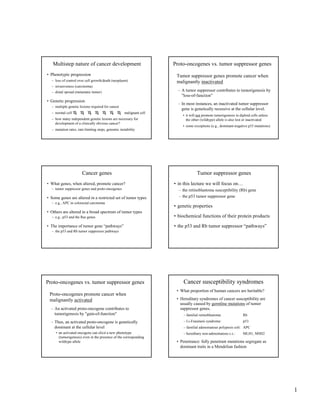
  • 1. 1 Multistep nature of cancer development • Phenotypic progression – loss of control over cell growth/death (neoplasm) – invasiveness (carcinoma) – distal spread (metastatic tumor) • Genetic progression – multiple genetic lesions required for cancer – normal cell malignant cell – how many independent genetic lesions are necessary for development of a clinically obvious cancer? – mutation rates, rate-limiting steps, genomic instability Cancer genes • What genes, when altered, promote cancer? – tumor suppressor genes and proto-oncogenes • Some genes are altered in a restricted set of tumor types – e.g., APC in colorectal carcinoma • Others are altered in a broad spectrum of tumor types – e.g., p53 and the Ras genes • The importance of tumor gene “pathways” – the p53 and Rb tumor suppressor pathways Proto-oncogenes vs. tumor suppressor genes Proto-oncogenes promote cancer when malignantly activated – An activated proto-oncogene contributes to tumorigenesis by "gain-of-function" – Thus, an activated proto-oncogene is genetically dominant at the cellular level • an activated oncogene can elicit a new phenotype (tumorigenesis) even in the presence of the corresponding wildtype allele Proto-oncogenes vs. tumor suppressor genes Tumor suppressor genes promote cancer when malignantly inactivated – A tumor suppressor contributes to tumorigenesis by "loss-of-function" – In most instances, an inactivated tumor suppressor gene is genetically recessive at the cellular level. • it will not promote tumorigenesis in diploid cells unless the other (wildtype) allele is also lost or inactivated • some exceptions (e.g., dominant-negative p53 mutations) Tumor suppressor genes • in this lecture we will focus on… – the retinoblastoma susceptibility (Rb) gene – the p53 tumor suppressor gene • genetic properties • biochemical functions of their protein products • the p53 and Rb tumor suppressor “pathways” Cancer susceptibility syndromes • What proportion of human cancers are heritable? • Hereditary syndromes of cancer susceptibility are usually caused by germline mutations of tumor suppressor genes. – familial retinoblastoma: Rb – Li-Fraumeni syndrome: p53 – familial adenomatous polyposis coli: APC – hereditary non-adenomatous c.c.: MLH1, MSH2 • Penetrance: fully penetrant mutations segregate as dominant traits in a Mendelian fashion
  • 2. 2 Sporadic and Heritable forms of Retinoblastoma • age of tumor onset – sporadic (~60% of cases): ~ 6 years – heritable (~40% “ “ ): ~ 2 years • number of independent tumors – sporadic: single tumor (only one eye is affected) – heritable: multiple tumors (both eyes are affected) • tumor frequencies in children of patients – sporadic: 1 in 105 – heritable: 1 in 2 patients with heritable retinoblastoma transmit a “Rb susceptibility gene” to their children in a dominant Mendelian fashion Knudson's hypothesis (1970s) • Two (rate-limiting) genetic lesions required… • Sporadic retinoblastoma: – both alterations are acquired somatically – incidence: (10-6)(10-6)(107 cells) = 10-5 tumors/person – very rare; involves only one eye • Heritable retinoblastoma: – one alteration is inherited in the germline (i.e., the “Rb susceptibility gene”) – the second alteration is acquired somatically – incidence: (1)(10-6)(107 cells) = 10 tumors/person – all carriers affected; involves both eyes!! – one mutation is inherited in the germline – second mutation is acquired somatically – incidence: (1)(10-6)(107 cells) = 10 tumors/person • All mutation carriers are affected – Rb susceptibility is a highly penetrant trait – tumor susceptibility is transmitted as a dominant trait in family pedigrees (despite the fact that tumor suppression genes function recessively at the cellular level). Hereditary Retinoblastoma The Retinoblastoma (Rb) gene • What are the two rate-limiting genetic alterations? • Cytogenetic abnormalities of chromosome 13: – interstitial deletions of variable length – always involve material from chromosome band 13q14 – sporadic patients: deletions in tumor cells only – heritable patients: deletions in both normal & tumor cells • Is Rb susceptibility due to genetic loss at 13q14? If so, then the two mutations required for retinoblastoma might represent inactivation of both alleles of a single gene at 13q14 Sporadic Retinoblastoma Familial Retinoblastoma tumor cells: Rb, Rb (normal) Rbm1, Rb (normal) Rbm1, Rbm2 (neoplastic) Rbm1, Rbm2 (neoplastic) m1 m2 m2 malignant mutations of the Rb gene act recessively at the cellular level, contributing to neoplasia by loss-of-function retinal cells: The Retinoblastoma gene (Rb) • 1988: isolation of the Rb gene on 13q14 Familial retinoblastoma • one Rb gene lesion in germline of familial patients • other (normal) Rb allele lost or inactivated in tumors Sporadic retinoblastoma • both alleles of Rb are normal in germline • both Rb alleles lost or inactivated in tumors Important: genetic lesions in the same gene are responsible for both the familial and sporadic forms of retinoblastoma!!
  • 3. 3 chromosome 13 maternal & paternal homologues Inactivation of the second Rb allele (in somatic cells) Rbm1 Rbm1 Rbm2 de novo mutation Rbm1 chromosome loss Rbm1 Rbm1 chromosome loss & reduplication gene conversionRbm1 Rbm1 The penetrance of germline Rb mutations • Almost all carriers will develop retinoblastoma – high penetrance • (mutation rate)(target cells) = (10-6)(107 cells) = 10 • retinoblastoma susceptibility is transmitted as a dominant Mendelian trait • Some carriers will also develop osteosarcoma – low penetrance • (mutation rate)(target cells) < 1 • osteosarcoma susceptibility is not transmitted as a dominant Mendelian trait Tumor Suppressors: the p53 gene • p53 encodes a transcription factor • the p53 gene is altered in many human tumors (usually by missense mutation). • in vitro cell transformation by mutant p53 genes. Is p53 a proto-oncogene? • murine erythroleukemias induced by Friend Leukemia Virus: a natural knockout of the p53 gene by proviral insertion! • suppression of cell transformation by the wild-type p53 gene. Is p53 a tumor suppressor gene? Dominant-negative mutations of a tumor suppressor gene • dominant-positive mutation (e.g., missense mutations in the Ras proto-oncogenes). • recessive-negative mutation (e.g., Rb loss). • dominant-negative mutation (e.g., many p53 missense mutations). • note: dominant-negative mutations result in functional inactivation of the protein products of both alleles (including the normal allele). Dominant-negative mutations of p53 • how do dominant-negative mutations work? • p53 normally functions as a homo-tetramer • consider p53 function in a cell with one wildtype and one mutant p53 allele: functional p53 tetramer? yes yes no no wildtype p53 mutant p53 Dominant-negative mutations of p53 • how do dominant-negative mutations work? • p53 normally functions as a homo-tetramer • consider p53 function in a cell with one wildtype and one mutant p53 allele: functional p53 tetramer? yes yes no no wildtype p53 mutant p53 • mutant p53 is more stable than wildtype p53
  • 4. 4 p53 mutations in hereditary and sporadic cancer • Li-Fraumeni Syndrome (LFS) – caused by germline mutations of p53 – LFS carriers develop many different forms of cancer • sporadic cancer – often caused by somatic mutations of p53 – very common in human cancer – found in many different forms of cancer Tumor suppressor proteins • proteins encoded by Rb and p53 • the normal functions of these proteins • mechanisms of tumor suppression • the Rb and p53 tumor suppressor pathways Phosphorylation of the Rb protein • The phosphorylation state of Rb changes during normal cell cycle progression. – Rb is hypophosphorylated in: • G0 (resting cells) • early G1 (cycling cells) – Rb is hyperphosphorylated in: • S phase • G2 phase – Rb is phosphorylated before the G1/S transition… • by an enzymatic complex: CDK4 / cyclin D M G1 S G2 restriction point G0 (resting cell) cell cycle progression Rb Rb~P Cdk4 / cyclin D The restriction point (in late G1) • the major control point of cell cycle progression • G1/S transition is mediated by the E2F family of transcription factors • E2F binds the promoters of genes required for cell cycle progression (G1/S transition and S phase). E2F promoter of S-phase genes transcription Some S phase genes regulated by E2F: S phase gene Function • thymidine kinase nucleotide synthesis • DHFR (dihydrofolate reductase) “ “ • DNA polymerase α DNA synthesis • ORC1 “ “ • histone H2A chromosome assembly • cyclin E cell cycle progression • cyclin A “ “ “
  • 5. 5 Resting cells and early G1 phase cells • hypophosphorylated Rb binds promoter-bound E2F • Rb inactivates transcription by E2F • S phase genes are repressed • G1/S transition is blocked Rb E2F S-phase genes repressed restriction point • CDK4/cyclin D phosphorylates Rb in the “pocket” • hyperphosphorylated Rb dissociates from E2F • E2F activates transcription of S phase genes • cells enter S phase Rb P P P E2F Rb S-phase genes activated E2F S-phase genes repressed Cdk4/cyclin D • In normal cells, phosphorylation of Rb by the CDK4/cyclin D kinase is a highly regulated • Focal point for the major signal transduction pathways that control normal cell growth Rb P P P E2F Rb S-phase genes activated E2F S-phase genes repressed Cdk4/cyclin D Rb P P P E2F Rb S-phase genes activated E2F S-phase genes repressed Cdk4/cyclin D diverse signaling pathways The function of Rb • hypophosphorylated Rb serves to restrain the proliferation of normal cells. • regulated phosphorylation of Rb allows normal cells to proliferate at the correct time and place. • therefore, imagine the consequences of losing normal Rb function… – deregulation of E2F (and the G1/S transition)! • how might Rb become inactivated in cancer? Inactivation of Rb function in tumors (leaving E2F unregulated) • Direct inactivation: – Rb gene deletion (occurs in retinoblastoma) – point mutations in the Rb pocket (in retinoblastoma) – occupancy of the Rb pocket by early proteins of DNA tumor viruses • human papilloma virus (HPV), an etiological agent in human cervical carcinomas • HPV encodes two proteins required for tumorigenesis • E7 binds the pocket of hypophosphorylated Rb • Deregulation of E2F (and the G1/S transition)
  • 6. 6 The Rb tumor suppressor pathway p16 Rb P P P E2F Rb S-phase genes activated E2F S-phase genes repressed Cdk4 / cyclin D Indirect inactivation of Rb function in tumors • overexpression of cyclin D1 – breast cancer, B cell lymphoma • loss of p16, an inhibitor of Cdk4 – many human cancers • inherited point mutation in Cdk4 that renders it insensitive to inhibition by p16 – familial melanoma »Inactivation of the Rb pathway occurs in most, if not all, human tumors! Normal functions of the p53 protein • p53 polypeptides are very unstable in normal cells (1/2 life of ~30 minutes) • the cellular response to genotoxic stress – DNA damage by UV light, ionizing radiation, chemical carcinogens, errors in replication, etc.) – induction of certain signal transduction pathways • post-translational modifications of p53 polypeptides: – especially, phosphorylation and acetylation – stabilize p53 (1/2 life of ~150 min.), leading to higher steady-state levels – increase the transcriptional activity of p53 consequences of p53 activation • The transcriptional activity of p53 induces a cellular response, the nature of which is dependent on various factors, including the cell type. – p53 induces G1 arrest (and DNA repair) in: • normal fibroblasts • certain epithelial cells – p53 induces apoptosis in: • thymocytes p53 induction of cell cycle arrest or apoptosis • in either case, replication of damaged DNA ceases • prevents accumulation of oncogenic mutations • In essence, p53 suppresses tumor formation by maintaining the integrity of the genetic material in cells subjected to genotoxic stress. Transcriptional targets of p53 • p21 CDK inhibitor G1 and G2 arrest in fibroblasts • 14-3-3σ G2 arrest in epithelial cells • PUMA promotes apoptosis in thymocytes, fibroblasts, neurons • p53R2 nuclear ribonucleotide reductase required for DNA repair • p48 subunit of the XPA complex required for nucleotide excision repair • etc…
  • 7. 7 Normal cell p53 mutant cell p53 protein is activated p53 induces target genes DNA repair growth arrest Apoptosis No response DNA damage persists - proto-oncogenes activated - tumor suppressors inactivated - cancer - Normal cell genotoxic stress The p53 tumor suppressor pathway p53 mdm2 p21 ATM »Inactivation of the p53 pathway occurs in most, if not all, human tumors! Sporadic colorectal carcinoma • Accounts for >80% of colorectal carcinomas. • Natural history of the sporadic disease (viewed by colonoscopy): – normal epithelium – hyperproliferative epithelium (APC*) – early adenoma (APC*) – intermediate adenoma (APC*, Ras*) – late adenoma (APC*, Ras*, SMAD2/4*) – carcinoma (APC*, Ras*, SMAD2/4*, p53*) – metastasis Hereditary colorectal carcinoma • A predisposition to colorectal carcinoma is associated with two distinct syndromes. – Familial adenomatous polyposis coli (FAP) – Heritable non-polyposis colorectal carcinoma (HNPCC) Familial adenomatous polyposis coli (FAP) • FAP: rare, autosomal, dominantly-inherited • Natural history of FAP: – multiple polyps (hundreds or thousands) develop throughout the colon by early adulthood. – inevitably, one or more of these polyps will progress into an invasive carcinoma. • FAP results from inherited lesions in the APC gene (which behaves as conventional tumor suppressor) • FAP accounts for less than 1% of all colorectal carcinomas. However, most sporadic cases of colorectal carcinoma have somatic mutations of APC.
  • 8. 8 Hereditary non-polyposis colorectal carcinoma (HNPCC). • HNPCC: autosomal, dominantly-inherited • Natural history of HNPCC: – Carriers do not have an obvious pre-malignant condition (e.g., the histology of their colon is normal). – Carriers are predisposed to develop colorectal carcinoma. • HNPCC results from inherited lesions in genes encoding components of the DNA mismatch repair system: – hMSH2, hMLH1, hPMS1, or hPMS2 Mismatch repair defects in HNPCC • mismatch repair incorrectly paired nucleotides • malignant cells of HNPCC tumors exhibit genetic instability at the nucleotide level • this underlying genetic instability accelerates malignant progression. • HNPCC accounts for 2-4% of colorectal carcinomas • In addition, ~13% of sporadic colorectal carcinomas display mismatch repair deficiency; these all harbor somatic mutations in one of the mismatch repair genes implicated in HNPCC Tumor Suppression • tumor suppressors inhibit tumor formation by a variety of different mechanisms: • negative control of cell growth... – by regulating cell cycle progression (e.g., Rb, p16) – by regulating signal transduction pathways (APC) • maintenance of genomic integrity... – by regulating the cellular response to DNA damage (p53, ATM, Chk2) – by functioning as effectors of DNA repair (hMSH2, hMLH1) . • Linkage analysis of family pedigrees: identify polymorphic markers that co-segregate with tumor susceptibility • Loss of heterozygosity (LOH) in tumor cells: identify polymorphic markers that exhibit tumor- specific LOH Positional cloning: search for transcription units (candidate genes) and analyze each for: – germline mutations that co-segregate with tumor susceptibility (in familial tumors) – somatic mutations that arise uniquely in the malignant cells (in sporadic tumors). Methods to isolate tumor suppressor genes
  • 9. 9 Chromosome 13 maternal & paternal homologues the second Rb mutation (in somatic cells) Rbm1 Rbm1 Rbm2 de novo mutation Rbm1 chromosome loss Rbm1 Rbm1 chromosome loss & reduplication gene conversionRbm1 Rbm1 LOH LOH LOH
  • 10.
  • 11.  World Cancer Day – 4th February 2014  By 2025, there will be more than 20 million new cancer cases per year, compared with 14.1 million in 2012, IARCWorld Cancer Report 2014
  • 12.  Greek word : Karkinos (crab)  Hippocrates : First coined the term  Cancer is an overgrowth of cells bearing cumulative genetic injuries that confer growth advantage over the normal cells
  • 13.
  • 14.  Accumulation of mutations and epigenomic changes in somatic cells
  • 15. Sporadic Familial Hereditary 77-80% 10-15 % 5-10% Nagy R, Sweet K, EngC. Highly penetrant hereditary cancer syndromes. Oncogene. 2004 Aug 23;23(38):6445-70
  • 16. • Early onset • Bilateral/Multifocal Disease • Multiple Primary cancer in single individual • Clusters of Cancer in family • Rare cancers • Precursor lesions
  • 17.
  • 18. Deregulating Cellular Energetic Avoiding immune Destruction Hanahan D, Weinberg RA. The hallmarks of cancer. Cell. 2000
  • 19.  Gene : A hereditary unit consisting of a sequence of DNA that occupies a specific location on a chromosome and determines a particular characteristic in an organism  Trait : A distinguishing feature, a genetically determined characteristic or condition.  Locus : specific area on chromosome where the gene is found  Allele :Versions of a gene  Genotype : the genetic makeup of an organism  Phenotype : the physical appearance of an organism  Pleiotropy : the ability of a gene to affect an organism in many ways
  • 20.  Penetrance  The probability that a gene will have any phenotypic expression  Its an all or none concept  Expressivity  Severity of the manifestations of the phenotype
  • 21. Driver Mutation : • Contribute to Oncogenesis • Provide growth advantages • Occurs in • Oncogenes • Tumor SuppressorGenes Passenger Mutation : • Neutral Mutation • Carried along the ride • Does not cause or propel cancer’s growth or spread
  • 22.  Proto-oncogenes – normally promote normal cell growth; mutations convert them to oncogenes.  Tumor suppressor genes – normally restrain cell growth; loss of function results in unregulated growth.
  • 23.  Oncogenes  promote cell proliferation  dominant & highly conserved  types : ▪ viral oncogenes [ v- oncs ] ▪ cellular oncogenes [ c- oncs ]
  • 24. Secreted Growth Factors • c-sis, hst Cell Surface Receptors • c-sis, hst erb B, ret, trk, fes, fms IntracellularTransducers • c-src, c-abl, mst, ras DNA-binding Nuclear Proteins • myc, jun, fos Regulators of the Cell Cycle • bcl, bax, bad
  • 25.
  • 26.  Tumor Suppressor Gene  Inhibits growth and multiplication of mutated cells  Prevent neoplastic transformation  Recessive and highly conserved  Categories :  Caretaker Gene  Gatekeeper Gene
  • 27. Category Gene Function Tumors Gatekeeper p53 Transcription factor Li-Fraumeni syndrome Rb1 Transcriptional regulator Familial retinoblastoma APC Regulates β- catenin function Familial Adenomatus polyposis Caretaker BRCA1 DNA repair Breast and ovarian cancer BRCA2 DNA repair Breast Cancer MSH2 DNA mismatch repair HNPCC
  • 28.
  • 29.
  • 30.
  • 31.
  • 32.
  • 33.
  • 34.
  • 35.
  • 36.
  • 37. Syndrome Incidence Genes Involve d Molecular Pathway affected RenalType Others characteristics VHL 1:36,000 VHL Hypoxic pathway(through HIF) Clear cell RCC pancreatic cysts and neuroendocrine tumors, pheochromocyto ma, retina I angiomas, hemangioblastom as Birt- Hogg- Dube rare FLCN m-TOR Variable subtypes cutaneous lesions, pulmonary cvsts and spontaneous pneumothorax
  • 38. Syndrome Incidence Genes Involved Molecular Pathway affected RenalType Others characteristics HPRC rare (Iess than 1:1,500.00 MET C-MET Type 1 papillary RCC not specific HLRCC Rare (unknown) FH Krebs cycle HLRC related RCC Multiples cutaneous and uterine leyomiomas SDH-RCC rare(unknow n) SDHB/SD HC/ SDHD Krebs cycle SDH related RCC Paraganglioma s/ Pheocromocyt oma GIST
  • 39.  Hereditary kidney cancer accounts for 3 to 5% of all kidney cancer  Ten inherited cancer susceptibility syndromes are associated with inherited risk of kidney cancer and 12 genes have been identified
  • 40. Genetics • Autosomal Dominant • Gene Locus on Chromosome 3p25 Incidence • 1 in 35000 live births Genetics • Tumor Suppressor gene • VHL gene product pVHL regulates function of HIF • Reduced pVHL– no HIF degradation—increasedVEGF
  • 41.
  • 42.  Sites involved Organ Tumor Non-tumor lesions Brain Hemangioblastoma Eye Hemangioblastoma Kidney CRCC Cyst Adrenal Phaemochromocytoma Pancreas NET Cyst Inner Ear Endolymphatic Sac tumor Epididymis PapillaryCystadenoma
  • 43. Type 1 VHL loss or mutation that affects the protein folding Haemangioblastoma Renal Cell Carcinoma Low risk of phaeocromocytoma Type 2A VHL missence mutation Haemangioblastoma phaeocromocytoma Low risk of Renal Cell Carcinoma Type 2B VHL missence mutation Haemangioblastoma Renal Cell Carcinoma Phaeocromocytoma Type 2C VHL missence mutation Phaeocromocytoma only
  • 44. Haemangioblastoma Clear Cell RCC Phaeochromocytoma Papillary Cystadenoma
  • 45. Diagnostic Criteria Known Positive Family Hx – presence of one: Single retinal or cerebellar hemangioblastoma RCC Pheochromocytoma Negative Family Hx – 2 retinal or cerebellar hemangioblastomas Single hemangioblastoma and additional characteristic lesion. Screening and Follow-up • Annual ophthalmologic evaluation and visual field testing starting around age 2 • MRI of the brain and spine every 2 years starting in early adolescence • Annual abdominal US starting at age 5 • Abdominal CT or MRI starting at age 20 • Frequent blood pressure monitoring and measurement of urinary catecholamine levels or plasma metanephrine levels every 1–2 years starting at age 2 Treatment • Hemangioblastomas • Complete surgical resection • Incomplete resection + external beam RT •VEGF and PDGF pathway inhibitors (sunitinib and sorafinib)
  • 46. Genetics : • Autosomal dominant • Gene : FLCN, Chromosome 17p, codes for protein “folliculin” Clinical manifestations : • Fibrofolliculomas (dysplastic hair follicules) • Lung cysts • Spontaneous pneumothorax • Renal cancer Incidence • 1 of 200,000 people • Hybrid oncocytic- chromophobe type • Chromophobe tumors • Clear cell • Oncocytomas • Papillary renal cancers
  • 48. Major criteria • At least five fibrofolliculomas, at least one histologically confirmed, of adult onset or • Pathogenic FLCN mutation Minor criteria • multiple lung cysts: bilateral basally located lung cysts with no other apparent cause, with or without spontaneous pneumothorax • renal cancer: early onset Genetic Screening: Germline FLCN testing is recommended beginning at age 21 Imaging Surveillance: • Annual MRI or low dose CT is recommended beginning at age 20 • In patients without a renal lesion MRI every 3 years is recommended
  • 49. Hereditary Papillary Renal Cancer • Genetics • Autosomal Dominant • Gene : MET (activating mutation) Chromosome 7q31.1 • Manifestations • Papillary RCC type 1 Hereditary Leiomyoma Renal Cell carcinoma • Genetics • Gene : FH, chromosome 1q42 • Manifestations • Papillary RCC type II • Uterine Leiomyoma
  • 50. Genetic Mechanism : • SDH- kreb’s cycle enzyme • Germline mutation in SDH B,C,D gene (SDHB has strongest association) • SDHB gene locus- chromosome 1p36 • Succinate stabilises HIF Incidence • 0.1-2% of all renal cancers Manifestation • Paraganglioma • Phaeochromocytoma • Renal cell Carcinoma (Clear cell or chromophobe)
  • 51.  BAP1 mutations and familial renal cancer  Predispose to ▪ Familial clear cell renal cancer ▪ Uveal and cutaneous melanoma ▪ Mesothelioma  Chromosome 3 translocations  PTEN hamartoma tumor syndrome (Cowden disease)
  • 52.  Urothelial carcinoma :  Associated with Lynch Syndrome  Prostate Carcinoma :  Polygenic Disorder  BRCA1 and BRCA2 mutation : ▪ Increased risk of Prostate Cancer ▪ High grade and Advanced Stage
  • 53.
  • 54. Syndrome Inheritance Gene Locus Gene Incidence Histology FAP AD 5q21 APC 2-12% PTC_CMV Cowden AD 10q23.3 PTEN,SDH,PIK3CA, AKT1 35% PTC (follicular variant), FTC, Adenomatous Nodule, C cell hyperplasia Carney AD 2p16,17q24 PPKAR1α 15% PTC,FTC, adenomatous nodule,Follicular Adenoma Wermer AR 8p11-p12 WRN 18% PTC,FTC,ATC Mc-cune Albright 20q13.2-13.3 GNAS PTC,FTC, Follicular adenoma DICER-1 syndrome AD 14q32.13 DICER1 Rare Nodular Hyperplasia
  • 55.  Lifetime risk for epithelial thyroid cancer is approximately 10%  Median age of onset was 37 years  Youngest age at diagnosis was 7 years  Thyroid lesions in CS  Adenomatous Nodule ▪ Multiple, unencapsulated, homogenous, firm  C cell hyperplasia  Follicular Carcinoma  PTC (Rare)
  • 56. Adenomatous nodules in Cowden syndrome, routine stain. (A) Solid adenomatous nodules surrounded by a thin rim of fibrous capsule, closely dispersed in the thyroid gland. (B) Each adenomatous nodule is composed of small follicles lacking abundant colloid (C) Immunohistochemistry for PTEN shows loss of PTEN expression in an adenomatous nodule in Cowden’s disease.
  • 57.  160 fold increased risk of PTC  Incidence : ~ 0.4% to 6.1% (Steinhagen E, et al. 2012)  Most thyroid cancer in individuals with FAP is Cribriform-morular variant of PTC
  • 58. Cribriform–morular variant of papillary thyroid carcinoma in familial adenomatous polyposis, routine stain. (A, B)Typical cribriform arrangement composed of fused follicles lined by tall cells and lumina lacking colloid. (C) Morular formation.
  • 59.  Rare condition characterized by  Skin pigmentary abnormalities  Myxomas  Endocrine tumors or overactivity  Genetics  Autosomal dominant  Gene – PPKAR1A gene on chromosome 17q
  • 60.  Clinical Features/Major Diagnostic Criteria  Spotty skin pigmentation  Myxoma (cutaneous and mucosal)  Cardiac myxoma  Breast myxomatosis  Primary pigmented nodular adrenocortical disease (PPNAD)  Acromegaly as a result of growth hormone (GH)-producing adenoma  Large-cell calcifying Sertoli cell tumor (LCCSCT)  Thyroid carcinoma or multiple, hypoechoic nodules on thyroid ultrasound in a child younger than age 18 years  Psammomatous melanotic schwannomas (PMS)  Blue nevus, epithelioid blue nevus  Breast ductal adenoma  Osteochondromyxoma
  • 61.  Cell cycle checkpoint kinase 2 (CHEK2)  Tumor suppressor gene  CHEK2 mutations associated with a moderate increase in the risk for various types of cancer  Breast cancer - 2-4x increased risk  Colorectal  Prostate
  • 62.  Siolek et al 2015 studied CHEK2 gene in thyroid cancer patients  468 cases and controls (Polish population)  CHEK2 mutations seen in 15% of unselected PTC patients and 6% of controls (p=0.006)  7/11 women with breast and thyroid cancers had CHEK2 mutations
  • 63.  Tumor susceptibility syndrome  Confers increased risk for-  Pleuropulmonary blastoma (PPB)  Ovarian sex cord-stromal tumors (such as Sertoli- Leydig cell tumor or juvenile granulosa cell tumor)  Cystic nephroma  Thyroid gland neoplasia  Rhabdomyosarcoma  Pituitary blastoma  Wilm’sTumor
  • 64.  Increased risk of developing thyroid cysts and/or multinodular goiter (MNG) in families with a germline DICER1 mutation  Risk of developing syndrome-associated thyroid cancer (papillary or follicular)
  • 65.  Birt-Hogg-Dube syndrome – cutaneous manifestations, pulmonary cysts/history of pneumothorax, and various types of renal tumors  Hyperparathyroidism-jaw tumor syndrome (HPT-JT)  Hereditary Paraganglioma/Pheochromocytoma (SDHB and SDHD)  Multiple Endocrine NeoplasiaType 1
  • 66.
  • 67. MEN 1 MEN 2 •Parathyroid hyperplasia or adenoma •Islet cell hyperplasia, adenoma, or carcinoma •Pituitary hyperplasia or adenoma •Other less common manifestations: foregut carcinoid, pheochromocytoma, subcutaneous or visceral lipomas MEN2A • MTC •Pheochromocytoma •Parathyroid adenoma •MEN2A with cutaneous lichen amyloidosis • MEN2A with Hirschsprung disease MEN2B • MTC • Pheochromocytoma • Mucosal and gastrointestinal neuromas • Marfanoid features
  • 69.
  • 70. Medullary thyroid carcinoma in multiple endocrine neoplasia syndrome, routine stain. (A) Medullary thyroid carcinoma featuring lobular and solid architectures with stromal calcitonin amyloid deposits.Tumour cells are variable in size and shape, with slightly granular cytoplasm and punctate coarse chromatin. (B) Medullary thyroid microcarcinoma in a young patient subjected to prophylactic thyroidectomy.
  • 71.
  • 72.
  • 73. Syndrome Chromosome/Gene CNS tumors Ataxia telangiectasia (AR) 11q22/ATM Meningioma, glioblastoma, pilocytic astrocytoma, medulloblastoma Cowden (AR) 10q23/PTEN Dysplastic cerebellar gangliocytoma, glial tumors FAP (AD) 5q21/APC Medulloblastoma, astrocytoma, ependymoma Hereditary non– polyposis- related colorectal cancer (AD) 3/MLH1 7/PMS2 2/MSH2 2/MSH6 Glioblastoma, other glial tumors Gorlin (AD) 9q/PTCH 10q24/SUFU Desmoplastic medulloblastoma, meningioma, oligodendroglioma, craniopharyngioma
  • 74. Syndrome Chromosome/Gene CNS tumors Multiple endocrine neoplasia 1 (AD) 11q13/MEN1 Parathyroid adenoma, pancreatic and duodenal neuroendocrine tumors Neurofibromatosis type 1 17q11/NF1 Optic/chiasmatic gliomas, plexiform neurofibromas, pilocytic astrocytoma, glioblastoma Neurofibromatosis type 2 22q12/NF2 Vestibular schwannoma, meningioma, ependymoma Tuberous sclerosis complex 9q34/TSC1 16q13/TSC2 Subependymal giant cell astrocytoma von Hippel–Lindau 3p25/VHL Hemangioblastoma. endolymphatic sac tumor
  • 75. Genetics • Autosomal dominant • Gene locus on Chromosomes 9q31 and 16q13 • 70% cases sporadic • Wide variety of germline mutations MOLECULAR • TSC1 -> Hamartin • TSC2 ->Tubulin • Tumor suppressor genes • Heterodimerize to inhibit mTOR pathway (mammalian target of rapamyacin) • Cell proliferation and growth through mRNA translation and ribosome synthesis INCIDENCE1 • in 6,000 to 10,000 live births Manifestations Giant Cell Astrocytoma Ependymoma Bilateral Renal Angiomyolipomas Renal Cell Carcinoma (Childhood) Cardiac Rhabdomyoma Other CNS Abnormalities:Tubers, Psychomotor delay, Seizures
  • 76. Definite diagnosis: 2 major or 1 major and 2 minor criteria Probable diagnosis: 1 major and 1 minor criteria Possible diagnosis: 1 major or 2 minor criteria Major Criteria: • Skin manifestation (facial angiofibroma, ungual fibroma, > hypomelanotic macules, shagreen patch) • Brain and eye lesions (cortical tuber, subependymal nodules, subependymal giant cell astrocytoma, multiple retinal nodular hamartomas) • Tumors in other organs (cardiac, rhabdomyoma, lymphangioleiomyomatosis, renal angiomyolipoma) Minor Criteria: • Pits in dental enamel • Rectal polyps • Bone cysts • Cerebral white matter migration abnormalities • Gingival fibromas • Nonrenal hamartomas • Retinal achromic patches • Confetti skin lesions • Multiple renal cysts Screening and Follow-up : • Funduscopic examination • Renal US or CT • Brain MR imaging • Echocardiography/Electrocardiography Treatment • SEGA • Surgical resection • Rapamycin can cause regression and prevention of epilepsy Angiofibroma Angiomyolipoma SEGA
  • 77. GENETICS • Autosomal dominant • Gene locus on Chromosome 17q11.2 • Penetrance = 100% • ~50% new mutations MOLECULAR • NF gene -> Neurofibromin • Negative regulator of RAS oncogene • Tumor suppressor gene • Inactivation causes cell growth and tumor development • Regulates neuroglial progenitor function INCIDENCE • 1 in 2,500 to 3,000 live births Manifestations OpticGlioma (Nerve and Chiasmatic) Neurofibrosarcoma GBM PilocyticAstrocytoma Plexiform Neurofibroma Pheochromocytoma Embryonal RMS Leukemia (chronic myelogenous) MPNST Neuroblastoma GIST  Diagnostic Criteria  Requires the presence of at least 2 of the following criteria  > 6 Café-au- lait macules (>5 mm in children and >15 mm in adults)  > 2 Cutaneous or subcutaneous neurofibromas or one plexiform neurofibroma  Axillary or inguinal freckling  Optic pathway glioma  > 2 Lisch nodules (small elevated hamartomas of the iris)  > 1 Distinctive osseous lesion (sphenoid wing dysplasia or thinning of long bone cortex)  First-degree relative with NF1 Screening and Follow-up • Yearly physical exam • Yearly ophthalmologic exam in early childhood ( up to age 5) • Regular developmental assessment • Routine blood pressure monitoring • Appropriate monitoring by a specialist according to CNS, skeletal, or cardiovascular abnormalities Treatment Optic glioma • Surgery • RT (after age 5) • Chemotherapy (carboplatin + vincristine) Plexiform Neurofibromas • Tipifarnib (RAS inhibitor)
  • 78. GENETICS • Autosomal dominant • Gene locus on Chromosome 22q11 • 100% penetrance MOLECULAR • Gene -> Merlin • Cytoskeleton organizing protein • Tumor suppressor protein • Involved in cell proliferation via the PAK/Rac signaling system • Cell membrane stability, motility, and intracellular adhesion INCIDENCE • 1 in 25,000 to 40,000 live births Manifestations • Vestibular Schwannoma (Bilateral) • Meningioma • Ependymoma • Pilocytic Astrocytoma Diagnostic Criteria  Definite diagnosis if either condition is fulfilled  Bilateral vestibular schwannomas  1st-degree relative with NF2 and either ▪ Unilateral vestibular schwannoma <30 years ▪ Any 2 of the following: meningioma, schwannoma, glioma, posterior subcapsular lens opacity or cerebral calcifications  Probable diagnosis if either condition is fulfilled  1) Unilateral vestibular schwannoma at <30 years and at least one of the following: meningioma, schwannoma, glioma, posterior subcapsular lens opacity  2) Multiple meningiomas and either of the following: schwannoma, glioma, posterior subcapsular lens opacity Screening and Follow-up • Annual MRI starting at age 10 to at least age 40 • Frequent hearing and vision evaluation Treatment Vestibular schwannoma • Surgical resection (translabyrinthine or suboccipital retrosigmoid) • Radiation therapy • Stereotactic radiosurgery vs.External beam
  • 79. GENETICS • Germline mutation in SMARCB1 (aka INI-I) • Seen in 40-50% of familial cases • Tumor suppressor gene MOLECULAR DIAGNOSIS • SMARCB1-associated schwannomatosis • Two or more pathologically proved schwannomas or meningiomasAND genetic studies of at least two tumors with loss of heterozygosity (LOH) for chromosome 22 and two different NF2 mutations; if there is a common SMARCB1 mutation • Pathologically proved schwannoma or meningiomaAND germline SMARCB1 pathogenic mutation • Diagnostic Criteria Two or more non-intradermal schwannomas, one with pathological confirmation, including no bilateral vestibular schwannoma by MRI • One pathologically confirmed schwannoma or intracranial meningioma AND affected first-degree relative • Exclusion criteria: •Germline pathogenic NF2 mutation •Fulfill diagnostic criteria for NF2 •First-degree relative with NF2 •Schwannomas in previous field of radiation therapy only
  • 80. Turcot Syndrome • GENETICS • Autosomal dominant • Gene locus on Chromosome 5 • MOLECULAR • PMS2 -> Postmeiotic segragation increased • MLH1 -> MutL homolog 1 • MSH2 -> MutS homolog 2 • DNA mismatch repair (MMR) genes • Disruption of the WNT signalling pathway • Manifestations • Medulloblastoma • Glioblastoma Multiforme • Anaplastic Astrocytoma • Ependymoma • Colon Cancer/Multiple Colonic Polyps Gorlin Syndrome • GENETICS • Autosomal dominant • Gene locus on Chromosome 9q • MOLECULAR • PTCH -> Drosophila Melanogaster Patched GeneEncodes a transmembrane receptor for secreted ligand sonic hedgehog (SHH)Essential for cerebellar development • Manifestations • Desmoplastic Medulloblastoma • Multiple Basal Cell Carcinomas
  • 81. GENETICS • Autosomal recessive • Gene locus on Chromosome 11q22-23 MOLECULAR • ATM – • Ataxia telangiectasia mutated • Protein kinase • DNA damage response and associated cell-cycle checkpoint regulation INCIDENCE • 1 in 40,000 to 100,000 live births Manifestations PilocyticAstrocytoma Medulloblastoma Glioblastoma Multiforme Lymphoma/Leukemia Lung Cancer Breast Cancer Ovarian Cancer Stomach Cancer Other CNS Abnormalities:Vascular telangiectasiasCerebellar atrophy Diagnostic Criteria Progressive cerebellar dysfunction between ages one and four years. Presenting as: Gait and truncal ataxia Head tilting Slurred speech Oculomotor apraxia and uneven (interrupted or “bumpy”) tracking across a visual field
  • 82. Genetics • Germline mutation in SMARCB1 in chromosome 22q11.23 (RTPS 1) • Mutation of in SMARCA4 in chromosome 19p13.2 Sites of involvement • CNS • AT/RT • Medulloblastoma, Choroid Plexus Ca, Supratentorial PNET • Extraneural • Bilateral malignant rhabdoid tumors of Kidney
  • 83. Syndrome Mode of Inheritance Genes Component Malignancies Wiskott-Aldrich syndrome X-linked recessive WAS NHL Severe combined immune deficiency X-linked recessive, recessive IL2RG,ADA, JAK3, RAG1, RAG2, IL7R, PTPRC, DCLRE1C B-cell lymphoma X-linked lymphoproliferativ e syndrome X-linked recessive SH2D1A Lymphoma
  • 85. Lynch Syndrome (LS) • Familial Predisposition to Colorectal and other cancers • Autosomal Dominant • Henry Lynch, MD 1966 • Hereditary Nonpolyposis Colorectal Cancer (HNPCC) 2
  • 86. Colorectal Cancer in LS • Cancers more frequently • Cancers at younger age (40’s) • Right sided cancers • Adenoma to carcinoma sequence more rapid • Synchronous and Metachronous Cancers • Lifetime Risk 10-74% (vs. 5.5% without) • Better Prognosis 3
  • 87. Genetics of CRC Sporadic (65%–85%) Familial (10%–30%) Lynch syndrome (3%) Familial adenomatous polyposis (FAP) (1%) Rare CRC syndromes (<0.1%) MYH associated polyposis (MAP) (1%)
  • 88. Other LS Cancers Cancer Incidence (%) Incidence LS (%) Endometrium 2.7 14-71 Stomach <1 0.2-13 Ovary 1.6 4-20 Hepatobiliary Tract <1 0.02-4 Urinary Tract <1 0.2-25 Small Bowel <1 0.4-12 Brain/CNS <1 1-4 Sebaceous Neoplasm <1 1-9 Pancreas 1.5 0.4-4 Prostate 16.2 9-30 Breast 12.4 5-18
  • 89. A Classic HNPCC/Lynch Family CRC dx 50s CRC dx 45 CRC dx 61 CRC dx 75 Ovarian Ca, dx 64 CRC dx 48 CRC dx 52 Endometrial Ca, dx 59 CRC dx 42 45
  • 90. Clinical Diagnosis of LS Amsterdam I Criteria (1991) 1. Three or more relatives with histologically verified colorectal cancer, 1 of which is a first-degree relative of the other two. Familial adenomatous polyposis should be excluded. 2. Two or more generations with colorectal cancer. 3. One or more colorectal cancer cases diagnosed before the age of 50 years.
  • 91. Clinical Diagnosis of LS Amsterdam II Criteria (1999) 1. Three or more relatives with histologically verified HNPCC- associated cancer (colorectal cancer, cancer of the endometrium, small bowel, ureter, or renal pelvis), 1 of which is a first-degree relative of the other 2. Familial adenomatous polyposis should be excluded. 2. Cancer involving at least 2 generations. 3. One or more cancer cases diagnosed before the age of 50 years.
  • 92. Clinical Diagnosis of LS Revised Bathesda Guidelines (2004) 1. CRC diagnosed at younger than 50 years. 2. Presence of synchronous or metachronous CRC or other LS-associated tumors.* 3. CRC with MSI-high pathologic-associated features (Crohn-like lymphocytic reaction, mucinous/signet cell differentiation, or medullary growth pattern) diagnosed in an individual younger than 60 years old. 4. Patient with CRC and CRC or LS-associated tumor* diagnosed in at least 1 first-degree relative younger than 50 years old. 5. Patient with CRC and CRC or LS-associated tumor* at any age in 2 first- degree or second-degree relatives. * LS-associated tumors include tumor of the colorectum, endometrium, stomach, ovary, pancreas, ureter, renal pelvis, biliary tract, brain, small bowel, sebaceous glands, and kerotoacanthomas.
  • 93. LS Genetic Alterations • Microsatellite Instability (MSI) • MSI - High • MSI - Low • MS - Stable • MSI – High Better Prognosis • Most MSI Colorectal Cancers are not LS (12% of sporadic CRC) 10
  • 95. Mismatch Repair (MMR) Genes and Proteins • Proofread Replicated DNA • Problem will be most obvious in repetitive sequences • Defect in both copies leads to cancer • If already carries one defect, at high risk to develop a second defect – Lynch Syndrome • LS is Autosomal Dominant
  • 96. Carrier Parent Non-carrier Parent Autosomal Dominant Inheritance Aa aa Aa Aa aa aa Carrier Carrier Non-carrier Non-carrier 1/2 1/2
  • 97. Mismatch Repair (MMR) Genes • MLH1 • MSH2 • MSH6 • PMS2 • EPCAM (Promotor for MSH2) 14
  • 99. Universal Tumor Testing for LS • MSI Testing • Inexpensive • Prognostic and Treatment Information • MMR Protein Testing • Inexpensive • Directs which gene to look at • Confirm Positive Results with Gene Analysis 16
  • 100. Followup • Genetic Counseling • Gene Testing Appropriate Family Members • Familial Colorectal Cancer Type X (FCRCTX) 17
  • 101. Treatment of Colon Cancer in LS Patients • Partial Colectomy • Risk of Metachronous Cancer 16-19% at 10 years • Total or Subtotal Colectomy with Ileorectal/Ileosigmoid Anastomosis • Risk of Metachronous Cancer 0-3.4% at 10 years • Diarrhea • Need to consider age and sphincter function 18
  • 102. Treatment of Rectal Cancer in LS Patients • Resection of Rectum with Anastomosis • Risk of Metachronous Cancer 69% at 30 years with colonoscopy every 1.6 years • Total Proctocolectomy with Ileal Pouch-Anal Anastomosis (IPAA) • Standard of Care for cancer with UC or FAP • LS patients older • Total Proctocolectomy with End Ileostomy 19
  • 103. CRC Screening in LS Patients • Colonoscopy every 1-3 years leads to fewer CRC and at a later age than unscreened • Colonoscopy every 1-3 years leads to similar CRC mortality compared to those without LS, although more CRC diagnosed • More frequent colonoscopy (≤ 2 years) better 20
  • 104. CRC Screening in LS Patients Guideline: Screening for CRC by colonoscopy is recommended in persons at risk (first-degree relatives of those affected) or affected with LS every 1 to 2 years, beginning between ages 20−25 years or 2−5 years before the youngest age of diagnosis of CRC in the family if diagnosed before age 25 years. May need to adjust based on exact family history and which gene is mutated. 21
  • 105. Endometrial Cancer in LS • Second most common cancer • 75% Stage I and 88% 5 year survival • Hard to prove screening helps • Annual Pelvic Exam and Endometrial Sampling Offered Starting at Age 30-35 22
  • 106. Ovarian Cancer in LS • No data as to screening • Transvaginal Ultrasound and CA-125 Screening does not seem to help with BRCA1 and BRCA2 patients • Annual Transvaginal Ultrasound Offered Starting at Age 30-35 23
  • 107. Prophylactic Hysterectomy and Oophorectomy in LS • Retrospective analysis of 315 women with MMR mutations • 33% Uterine cancer without surgery • No uterine cancer with surgery • 5.5% Ovarian cancer without surgery • No ovarian cancer with surgery • Guideline: Hysterectomy and Oophorectomy after childbearing or at age 40 24
  • 108. Gastric Cancer in LS • Lifetime Risk 0.2-13% • EGD every 2-3 years beginning age 30-35 • Treat H.pylori if found • Modify based on family history and gene mutation 25
  • 109. Urinary Cancers in LS • Not much data screening (urinalysis, urine cytology) helps • Inexpensive • Noninvasive • Easy • Consider annually starting age 30-35 26
  • 110. Other Cancers in LS • No screening or no increased screening beyond that for usual population • Pancreatic • Small Intestine • Prostate • Breast • Either no clear increased risk or no good screening test 27