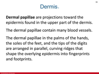
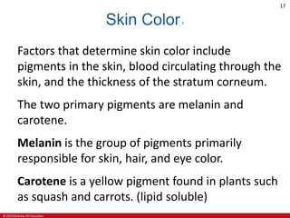
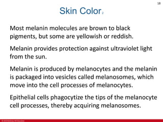
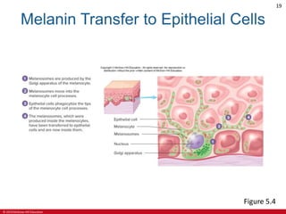
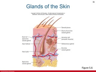
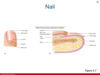
SlideShare a Scribd company logo
1 of 60
Download to read offline
Seeley’s
ESSENTIALS OF
Anatomy &
Physiology
Tenth Edition
Cinnamon Vanputte
Jennifer Regan
Andrew Russo
See separate PowerPoint slides for all figures and tables
pre-inserted into PowerPoint without notes.
© 2019 McGraw-Hill Education. All rights reserved. Authorized only for instructor use in the classroom. No reproduction or further distribution permitted without the prior written consent of McGraw-Hill Education.
© 2019 McGraw-Hill Education
2
Chapter 5
Integumentary System
Lecture Outline
© 2019 McGraw-Hill Education
3
Integumentary System1
The integumentary system consists of the skin
and accessory structures, such as hair, glands,
and nails.
Integument means covering.
The appearance of the integumentary system
can indicate physiological imbalances in the
body.
© 2019 McGraw-Hill Education
4
Integumentary System2
Figure 5.1
© 2019 McGraw-Hill Education
5
Integumentary System Functions1
1. Protection. The skin provides protection
against abrasion and ultraviolet light.
2. Sensation. The integumentary system has
sensory receptors that can detect heat, cold,
touch, pressure, and pain.
3. Vitamin D production. When exposed to
ultraviolet light, the skin produces a
molecule that can be transformed into
vitamin D.
© 2019 McGraw-Hill Education
6
Integumentary System Functions2
4. Temperature regulation. The amount of
blood flow beneath the skin’s surface and the
activity of sweat glands in the skin both help
regulate body temperature.
5. Excretion. Small amounts of waste products
are lost through the skin and in gland
secretions.
© 2019 McGraw-Hill Education
7
Skin
The skin is made up of two major tissue layers: the
epidermis and the dermis.
The epidermis is the most superficial layer of skin.
It is a layer of epithelial tissue that rests on the
dermis.
The dermis is a layer of dense connective tissue.
The skin rests on the subcutaneous tissue, which is
a layer of connective tissue.
The subcutaneous tissue is not part of the skin.
© 2019 McGraw-Hill Education
8
Epidermis and Dermis
Figure 5.2
(b) ©Victor Eroschenko
© 2019 McGraw-Hill Education
9
Epidermis1
The epidermis prevents water loss and resists
abrasion.
The epidermis, known as the cutaneous
membrane, is a keratinized stratified squamous
epithelium.
The epidermis is composed of distinct layers
called strata.
© 2019 McGraw-Hill Education
10
The deepest stratum, stratum basale consists of
cuboidal or columnar cells that undergo mitotic
division about every 19 days.
One daughter cell becomes a new stratum
basale cell and can divide again.
The other daughter cell is pushed toward the
surface, a journey that takes about 40-56 days.
Epidermis2
© 2019 McGraw-Hill Education
11
Epidermis3
The stratum corneum, the most superficial
stratum of the epidermis, consists of dead
squamous cells filled with keratin.
Keratin gives the stratum corneum its structural
strength.
Composed of 25 or more layers of dead
squamous cells joined by desmosomes.
As new cells form, they push older cells to the
surface, where they slough, or flake off.
© 2019 McGraw-Hill Education
12
Epidermis4
Excessive sloughing of stratum corneum cells
from the surface of the scalp is called dandruff.
In skin subjected to friction, the number of
layers in the stratum corneum greatly increases,
producing a thickened area called a callus.
Over a bony prominence, the stratum corneum
can thicken to form a cone-shaped structure
called a corn.
© 2019 McGraw-Hill Education
13
Dermis1
The dermis is composed of dense collagenous
connective tissue containing fibroblasts,
adipocytes, and macrophages.
Nerves, hair follicles, smooth muscles, glands,
and lymphatic vessels extend into the dermis.
Collagen fibers, oriented in many directions, and
elastic fibers are responsible for the structural
strength of the dermis and resistance to stretch.
© 2019 McGraw-Hill Education
14
Dermis2
Some collagen fibers are oriented more
directions than others, forming cleavage lines.
Cleavage lines, or tension lines, in the skin, are
more resistant to stretch.
An incision made parallel with these lines tends
to gap less and produce less scar tissue.
If the skin is overstretched for any reason, the
dermis can be damaged, leaving stretch marks.
© 2019 McGraw-Hill Education
15
Cleavage Lines
Figure 5.3
© 2019 McGraw-Hill Education
16
Dermis3
Dermal papillae are projections toward the
epidermis found in the upper part of the dermis.
The dermal papillae contain many blood vessels.
The dermal papillae in the palms of the hands,
the soles of the feet, and the tips of the digits
are arranged in parallel, curving ridges that
shape the overlying epidermis into fingerprints
and footprints.
© 2019 McGraw-Hill Education
17
Skin Color1
Factors that determine skin color include
pigments in the skin, blood circulating through the
skin, and the thickness of the stratum corneum.
The two primary pigments are melanin and
carotene.
Melanin is the group of pigments primarily
responsible for skin, hair, and eye color.
Carotene is a yellow pigment found in plants such
as squash and carrots. (lipid soluble)
© 2019 McGraw-Hill Education
18
Skin Color2
Most melanin molecules are brown to black
pigments, but some are yellowish or reddish.
Melanin provides protection against ultraviolet light
from the sun.
Melanin is produced by melanocytes and the melanin
is packaged into vesicles called melanosomes, which
move into the cell processes of melanocytes.
Epithelial cells phagocytize the tips of the melanocyte
cell processes, thereby acquiring melanosomes.
© 2019 McGraw-Hill Education
19
Melanin Transfer to Epithelial Cells
Figure 5.4
© 2019 McGraw-Hill Education
20
Skin Color3
Large amounts of melanin form freckles or moles in
some regions of the skin.
Melanin production is determined by genetic factors,
exposure to light, and hormones.
Genetic factors are responsible for the amounts of
melanin produced in different races.
Since all races have about the same number of
melanocytes, racial variations in skin color are
determined by the amount, kind, and distribution of
melanin.
© 2019 McGraw-Hill Education
21
Skin Color4
Exposure to ultraviolet light—for example, in
sunlight— stimulates melanocytes to increase
melanin production. The result is a suntan.
Although many genes are responsible for skin
color, a single mutation can prevent the
production of melanin and cause albinism.
© 2019 McGraw-Hill Education
22
Skin Color5
Carotene is lipid-soluble; when consumed, it
accumulates in the lipids of the stratum corneum
and in the adipocytes of the dermis and
subcutaneous tissue.
If large amounts of carotene are consumed, the
skin can become quite yellowish.
© 2019 McGraw-Hill Education
23
Skin Color6
The color of blood in the dermis contributes to
skin color.
A decrease in blood flow, as occurs in shock, can
make the skin appear pale.
A decrease in the blood O2 content produces a
bluish color of the skin, called cyanosis.
© 2019 McGraw-Hill Education
24
Subcutaneous Tissue1
The skin (cutaneous membrane) rests on the
subcutaneous tissue.
The subcutaneous tissue, which is not part of the
skin, is sometimes called hypodermis.
The subcutaneous tissue attaches the skin to
underlying bone and muscle and supplies it with
blood vessels and nerves.
It is loose connective tissue, including adipose tissue
that contains about half the body’s stored lipids.
© 2019 McGraw-Hill Education
25
Subcutaneous Tissue2
The amount and location of adipose tissue vary
with age, sex, and diet.
Adipose tissue in the subcutaneous tissue
functions as padding and insulation.
The subcutaneous tissue can be used to estimate
total body fat.
The acceptable percentage of body fat varies
from 21% to 30% for females and from 13% to
25% for males.
© 2019 McGraw-Hill Education
26
Hair1
In humans, hair is found everywhere on the skin,
except on the palms, soles, lips, nipples, parts of
the genitalia, and the distal segments of the
fingers and toes.
Each hair arises from a hair follicle, an
invagination of the epidermis that extends deep
into the dermis.
A hair shaft protrudes above the surface of the
skin; the root is below the surface and the hair
bulb is the expanded base of the root.
© 2019 McGraw-Hill Education
27
Hair2
A hair has a hard cortex, which surrounds a
softer center, the medulla.
The cortex is covered by the cuticle, a single layer
of overlapping cells that holds the hair in the hair
follicle.
Hair is produced in the hair bulb, which rests on
the hair papilla.
The hair papilla is an extension of the dermis
that protrudes into the hair bulb and contains
blood vessels.
© 2019 McGraw-Hill Education
28
Hair3
Hair is produced in cycles, with a growth stage
and resting stage.
During the growth stage, hair is formed by
mitosis of epithelial cells within the hair bulb;
these cells divide and undergo keratinization.
During the resting stage, growth stops and the
hair is held in the hair follicle.
When the next growth stage begins, a new hair is
formed and the old hair falls out.
© 2019 McGraw-Hill Education
29
Hair4
The duration of each stage depends on the
individual hair.
Eyelashes grow for about 30 days and rest for
105 days, whereas scalp hairs grow for 3 years
and rest for 1 to 2 years.
The loss of hair normally means that the hair is
being replaced because the old hair falls out of
the hair follicle when the new hair begins to
grow.
© 2019 McGraw-Hill Education
30
Hair5
Hair color is determined by varying amounts and
types of melanin.
With age, the amount of melanin in hair can
decrease, causing the hair color to become
faded, or the hair can contain no melanin and be
white.
Each hair follicle is attached to smooth muscle
cells called the arrector pili muscle, which can
contract and cause the hair to become
perpendicular to the skin’s surface.
© 2019 McGraw-Hill Education
31
Hair Follicle
Figure 5.5
© 2019 McGraw-Hill Education
32
Glands1
The major glands of the skin are the sebaceous
glands and the sweat glands.
Sebaceous glands are simple, branched acinar
glands, with most being connected by a duct to the
superficial part of a hair follicle.
They produce sebum, an oily, white substance rich in
lipids.
The sebum is released by holocrine secretion and
lubricates the hair and the surface of the skin, which
prevents drying and protects against some bacteria.
© 2019 McGraw-Hill Education
33
Glands2
There are two kinds of sweat glands: eccrine and
apocrine.
Eccrine sweat glands are simple, coiled, tubular
glands and release sweat by merocrine secretion.
Eccrine glands are located in almost every part of
the skin but most numerous in the palms and
soles.
They produce a secretion that is mostly water
with a few salts.
© 2019 McGraw-Hill Education
34
Glands3
Eccrine sweat glands have ducts that open onto
the surface of the skin through sweat pores and
are for thermal regulation.
Sweat can also be released in the palms, soles,
armpits, and other places because of emotional
stress.
© 2019 McGraw-Hill Education
35
Glands4
Apocrine sweat glands are simple, coiled, tubular
glands that produce a thick secretion rich in organic
substances.
The glands open into hair follicles in the armpits and
genitalia.
Apocrine sweat glands become active at puberty
because of the influence of sex hormones.
The secretion generally is odorless, but when
released quickly breaks down by bacterial action
giving body odor.
© 2019 McGraw-Hill Education
36
Glands of the Skin
Figure 5.6
© 2019 McGraw-Hill Education
37
Nails1
The nail is a thin plate, consisting of layers of
dead stratum corneum cells that contain a very
hard type of keratin.
The visible part of the nail is the nail body, and
the part of the nail covered by skin is the nail
root.
The cuticle, or eponychium, is stratum corneum
that extends onto the nail body and the nail root
extends distally from the nail matrix.
© 2019 McGraw-Hill Education
38
Nails2
The nail also attaches to the underlying nail bed,
which is located distal to the nail matrix.
The nail matrix and bed are epithelial tissue with a
stratum basale that gives rise to the cells that form
the nail.
A small part of the nail matrix, the lunula, can be
seen through the nail body as a whitish, crescent-
shaped area at the base of the nail.
Cell production within the nail matrix causes the nail
to grow continuously.
© 2019 McGraw-Hill Education
39
Nail
Figure 5.7
© 2019 McGraw-Hill Education
40
Integumentary System Protection1
The integumentary system performs many
protective functions:
1. Reduction in body water loss
2. Acts as a barrier that prevents microorganisms
and other foreign substances from entering the
body
3. Protects underlying structures against abrasion
4. Melanin absorbs ultraviolet light and protects
underlying structures from its damaging effects
© 2019 McGraw-Hill Education
41
Integumentary System Protection2
5. Hair protection: The hair on the head acts as
a heat insulator, eyebrows keep sweat out of
the eyes, eyelashes protect the eyes from
foreign objects, and hair in the nose and ears
prevents the entry of dust and other
materials.
6. The nails protect the ends of the fingers and
toes from damage and can be used in
defense.
© 2019 McGraw-Hill Education
42
Sensory Receptor
Many sensory receptors are associated with the
skin.
Receptors in the epidermis and dermis can
detect pain, heat, cold, and pressure.
Although hair does not have a nerve supply,
sensory receptors around the hair follicle can
detect the movement of a hair.
© 2019 McGraw-Hill Education
43
Vitamin D Production
1. UV light causes the skin to produce a precursor
molecule of vitamin D.
2. The precursor molecule is carried by the blood
to the liver where it is enzymatically converted.
3. The enzymatically converted molecule is carried
by the blood to the kidneys where it is converted
again to the active form of vitamin D.
4. Vitamin D stimulates the small intestine to
absorb calcium and phosphate for many body
functions.
© 2019 McGraw-Hill Education
44
Temperature Regulation1
Regulation of body temperature is important
because the rate of chemical reactions within
the body can be increased or decreased by
changes in body temperature.
Even slight changes in temperature can make
enzymes operate less efficiently and disrupt the
normal rates of chemical changes in the body.
Exercise, fever, and an increase in environmental
temperature tend to raise body temperature.
© 2019 McGraw-Hill Education
45
Temperature Regulation2
In order to maintain homeostasis, the body
must rid itself of excess heat.
Blood vessels in the dermis dilate and enable
more blood to flow within the skin, thus causing
heat to dissipate from the body.
Sweat also assists in loss of heat through
evaporative cooling.
© 2019 McGraw-Hill Education
46
Temperature Regulation3
If body temperature begins to drop below
normal, heat can be conserved by the
constriction of dermal blood vessels, which
reduces blood flow to the skin.
Less heat is transferred from deeper structures
to the skin, and heat loss is reduced.
With smaller amounts of warm blood flowing
through the skin, the skin temperature
decreases.
© 2019 McGraw-Hill Education
47
Heat Exchange in the Skin
Figure 5.8
© 2019 McGraw-Hill Education
48
Excretion
The integumentary system plays a minor role in
excretion, the removal of waste products from
the body.
In addition to water and salts, sweat contains
small amounts of waste products, such as urea,
uric acid, and ammonia.
Even though the body can lose large amounts of
sweat, the sweat glands do not play a significant
role in the excretion of waste products.
© 2019 McGraw-Hill Education
49
Diagnostic Aid
The integumentary system is useful in diagnosis
because it is observed easily.
Cyanosis, a bluish color to the skin caused by
decreased blood O2 content, is an indication of
impaired circulatory or respiratory function.
A yellowish skin color, called jaundice, can occur
when the liver is damaged by a disease, such as
viral hepatitis.
Rashes and lesions in the skin can be symptoms of
problems elsewhere in the body.
© 2019 McGraw-Hill Education
50
Burns1
A burn is injury to a tissue caused by heat, cold,
friction, chemicals, electricity, or radiation.
Burns are classified according to their depth.
Partial-thickness burns are classified as first-
degree and second-degree.
A full-thickness burn is a third-degree burn.
© 2019 McGraw-Hill Education
51
First-Degree Burn
A first-degree (superficial) burn involves only
the epidermis and is red and painful.
Slight edema, or swelling, may be present.
They can be caused by sunburn or brief
exposure to very hot or very cold objects, and
they heal without scarring in about a week.
© 2019 McGraw-Hill Education
52
Second-Degree Burn
Second-degree (partial-thickness) burns damage
both the epidermis and the dermis.
If dermal damage is minimal, symptoms include
redness, pain, edema, and blisters.
Healing takes about 2 weeks, and no scarring
results.
If the burn goes deep into the dermis, the
wound appears red, tan, or white; can take
several months to heal and might scar.
© 2019 McGraw-Hill Education
53
Third-Degree Burn
Third-degree (full-thickness) burns damage the
complete epidermis and dermis.
The region of third-degree burn is usually
painless because sensory receptors in the
epidermis and dermis have been destroyed.
Third-degree burns appear white, tan, brown,
black, or deep cherry red.
© 2019 McGraw-Hill Education
54
Burn Healing
In all second-degree burns, the epidermis,
including the stratum basale where the stem
cells are found, is damaged.
The epidermis regenerates from epithelial tissue
in hair follicles and sweat glands, as well as from
the edges of the wound.
Deep partial-thickness and full-thickness burns
take a long time to heal, and they form scar
tissue with disfiguring and debilitating wound
contractures.
© 2019 McGraw-Hill Education
55
Treatment of Burns
To prevent complications of deep partial-thickness
and full-thickness burns and to speed healing, skin
grafts are often performed.
In a procedure called a split skin graft, the
epidermis and part of the dermis are removed from
another part of the body and placed over the burn
When it is not possible or practical to move skin
from one part of the body to a burn site, physicians
sometimes use artificial skin or grafts from human
cadavers.
© 2019 McGraw-Hill Education
56
Burns2
Figure 5.9
© 2019 McGraw-Hill Education
57
Skin Cancer
Most common cancer
Mainly caused by UV light exposure
Fair-skinned people more prone
Prevented by limiting sun exposure and using
sunscreens
UVA rays cause tan and is associated with malignant
melanomas
UVB rays cause sunburns
Sunscreens should block UVA and UVB rays
© 2019 McGraw-Hill Education
58
Types of Skin Cancer
Basal cell carcinoma:
• cells in stratum basale affected
• cancer removed by surgery
Squamous cell carcinoma:
• cells above stratum basale affected
• can cause death
Malignant melanoma:
• arises from melanocytes in a mole
• rare type
• can cause death
© 2019 McGraw-Hill Education
59
Cancer of the Skin
Figure 5.13
(a) ©Dr. P. Marazzi/Science Source RF; (b) ©Dr. P. Marazzi/Science Source; (c) Source: National Cancer Institute
© 2019 McGraw-Hill Education
60
Aging and the Integument
Blood flow decreases and skin becomes thinner
due to decreased amounts of collagen
Decreased activity of sebaceous and sweat
glands make temperature regulation more
difficult
Loss of elastic fibers cause skin to sag and
wrinkle

More Related Content

Similar to ch05lectureppta-210924134735.pdf

SKIN : Excretory system in Human
SKIN : Excretory system in HumanSKIN : Excretory system in Human
SKIN : Excretory system in Humannurul ikhsani
 
The Integumentary System Bethany, Susan
The Integumentary System Bethany, SusanThe Integumentary System Bethany, Susan
The Integumentary System Bethany, Susanwbuchberg
 
Integumentary system prep.pptx
Integumentary system prep.pptxIntegumentary system prep.pptx
Integumentary system prep.pptxSamanthaEllaine
 
Structure of skin, Dry Skin and Acne
Structure of skin, Dry Skin and AcneStructure of skin, Dry Skin and Acne
Structure of skin, Dry Skin and AcneJuhi Solanky
 
integmentary skin Dr Ahmed H Ahmed.pdf
integmentary skin Dr Ahmed H Ahmed.pdfintegmentary skin Dr Ahmed H Ahmed.pdf
integmentary skin Dr Ahmed H Ahmed.pdfSalahaddin University
 
INTEGUMENTARY SYSTEM BY EVA KIPTOO.pptx
INTEGUMENTARY SYSTEM BY EVA KIPTOO.pptxINTEGUMENTARY SYSTEM BY EVA KIPTOO.pptx
INTEGUMENTARY SYSTEM BY EVA KIPTOO.pptxNabiswaboazWangila
 
3. Integumentary system (Skin).pptx
3. Integumentary system (Skin).pptx3. Integumentary system (Skin).pptx
3. Integumentary system (Skin).pptxAbhiDabra
 
Phys Assess_Skin.pptx
Phys Assess_Skin.pptxPhys Assess_Skin.pptx
Phys Assess_Skin.pptxnonaaryan2
 
Ch6 Ppt Lect
Ch6 Ppt LectCh6 Ppt Lect
Ch6 Ppt Lectbholmes
 
Ch6 Ppt Lect
Ch6 Ppt LectCh6 Ppt Lect
Ch6 Ppt Lectbholmes
 
Skin structures and functions ppt Dr.Mumux
Skin structures and functions ppt Dr.MumuxSkin structures and functions ppt Dr.Mumux
Skin structures and functions ppt Dr.MumuxMumux Mirani
 
Skin Tissue - Histology and Embryrology.
Skin Tissue - Histology and Embryrology.Skin Tissue - Histology and Embryrology.
Skin Tissue - Histology and Embryrology.Denizasmaz
 
Skin and Appendages of skin
Skin and Appendages of skinSkin and Appendages of skin
Skin and Appendages of skinSajid Khan
 
ANATOMY AND PHYSIOLOGY OF SKIN
ANATOMY AND PHYSIOLOGY OF SKINANATOMY AND PHYSIOLOGY OF SKIN
ANATOMY AND PHYSIOLOGY OF SKINAlmasMajeeth
 
INTEGUMENTARY SYSTEM (1).pdf
INTEGUMENTARY SYSTEM (1).pdfINTEGUMENTARY SYSTEM (1).pdf
INTEGUMENTARY SYSTEM (1).pdfSaqibShaik2
 
Skin and its diseases. Dermatology description
Skin and its diseases. Dermatology descriptionSkin and its diseases. Dermatology description
Skin and its diseases. Dermatology descriptionKeshav177622
 

Similar to ch05lectureppta-210924134735.pdf (20)

SKIN : Excretory system in Human
SKIN : Excretory system in HumanSKIN : Excretory system in Human
SKIN : Excretory system in Human
 
The Integumentary System Bethany, Susan
The Integumentary System Bethany, SusanThe Integumentary System Bethany, Susan
The Integumentary System Bethany, Susan
 
Integumentary system prep.pptx
Integumentary system prep.pptxIntegumentary system prep.pptx
Integumentary system prep.pptx
 
Structure of skin, Dry Skin and Acne
Structure of skin, Dry Skin and AcneStructure of skin, Dry Skin and Acne
Structure of skin, Dry Skin and Acne
 
integmentary skin Dr Ahmed H Ahmed.pdf
integmentary skin Dr Ahmed H Ahmed.pdfintegmentary skin Dr Ahmed H Ahmed.pdf
integmentary skin Dr Ahmed H Ahmed.pdf
 
INTEGUMENTARY SYSTEM BY EVA KIPTOO.pptx
INTEGUMENTARY SYSTEM BY EVA KIPTOO.pptxINTEGUMENTARY SYSTEM BY EVA KIPTOO.pptx
INTEGUMENTARY SYSTEM BY EVA KIPTOO.pptx
 
3. Integumentary system (Skin).pptx
3. Integumentary system (Skin).pptx3. Integumentary system (Skin).pptx
3. Integumentary system (Skin).pptx
 
Phys Assess_Skin.pptx
Phys Assess_Skin.pptxPhys Assess_Skin.pptx
Phys Assess_Skin.pptx
 
Ch6 Ppt Lect
Ch6 Ppt LectCh6 Ppt Lect
Ch6 Ppt Lect
 
Ch6 Ppt Lect
Ch6 Ppt LectCh6 Ppt Lect
Ch6 Ppt Lect
 
Dermatology
DermatologyDermatology
Dermatology
 
Skin structures and functions ppt Dr.Mumux
Skin structures and functions ppt Dr.MumuxSkin structures and functions ppt Dr.Mumux
Skin structures and functions ppt Dr.Mumux
 
Skin
SkinSkin
Skin
 
Skin Tissue - Histology and Embryrology.
Skin Tissue - Histology and Embryrology.Skin Tissue - Histology and Embryrology.
Skin Tissue - Histology and Embryrology.
 
Skin and Appendages of skin
Skin and Appendages of skinSkin and Appendages of skin
Skin and Appendages of skin
 
Integumentary system.pptx
Integumentary system.pptxIntegumentary system.pptx
Integumentary system.pptx
 
ANATOMY AND PHYSIOLOGY OF SKIN
ANATOMY AND PHYSIOLOGY OF SKINANATOMY AND PHYSIOLOGY OF SKIN
ANATOMY AND PHYSIOLOGY OF SKIN
 
INTEGUMENTARY SYSTEM (1).pdf
INTEGUMENTARY SYSTEM (1).pdfINTEGUMENTARY SYSTEM (1).pdf
INTEGUMENTARY SYSTEM (1).pdf
 
Skin and its diseases. Dermatology description
Skin and its diseases. Dermatology descriptionSkin and its diseases. Dermatology description
Skin and its diseases. Dermatology description
 
TAM1
TAM1TAM1
TAM1
 

More from DiannaAsug

Aldehydes-and-Ketones-by-Group-6_20230920_072912_0000 (1).pdf
Aldehydes-and-Ketones-by-Group-6_20230920_072912_0000 (1).pdfAldehydes-and-Ketones-by-Group-6_20230920_072912_0000 (1).pdf
Aldehydes-and-Ketones-by-Group-6_20230920_072912_0000 (1).pdfDiannaAsug
 
CARBOXYLIC-ACID-DERIVATIVES_20230925_091857_0000.pdf
CARBOXYLIC-ACID-DERIVATIVES_20230925_091857_0000.pdfCARBOXYLIC-ACID-DERIVATIVES_20230925_091857_0000.pdf
CARBOXYLIC-ACID-DERIVATIVES_20230925_091857_0000.pdfDiannaAsug
 
ch09lectureppta1-210924134742.pdf
ch09lectureppta1-210924134742.pdfch09lectureppta1-210924134742.pdf
ch09lectureppta1-210924134742.pdfDiannaAsug
 
ch09lectureppta1-210924134742.pdf
ch09lectureppta1-210924134742.pdfch09lectureppta1-210924134742.pdf
ch09lectureppta1-210924134742.pdfDiannaAsug
 
ch08ppt-210924134741.pdf
ch08ppt-210924134741.pdfch08ppt-210924134741.pdf
ch08ppt-210924134741.pdfDiannaAsug
 
ch08ppt-210924134741.pdf
ch08ppt-210924134741.pdfch08ppt-210924134741.pdf
ch08ppt-210924134741.pdfDiannaAsug
 
ch08ppt-210924134741.pdf
ch08ppt-210924134741.pdfch08ppt-210924134741.pdf
ch08ppt-210924134741.pdfDiannaAsug
 
ch06lectureppta-210924134736.pdf
ch06lectureppta-210924134736.pdfch06lectureppta-210924134736.pdf
ch06lectureppta-210924134736.pdfDiannaAsug
 
ch07lectureppta-210924134739.pdf
ch07lectureppta-210924134739.pdfch07lectureppta-210924134739.pdf
ch07lectureppta-210924134739.pdfDiannaAsug
 
CHAPTER-4-TISSUES.pdf
CHAPTER-4-TISSUES.pdfCHAPTER-4-TISSUES.pdf
CHAPTER-4-TISSUES.pdfDiannaAsug
 

More from DiannaAsug (10)

Aldehydes-and-Ketones-by-Group-6_20230920_072912_0000 (1).pdf
Aldehydes-and-Ketones-by-Group-6_20230920_072912_0000 (1).pdfAldehydes-and-Ketones-by-Group-6_20230920_072912_0000 (1).pdf
Aldehydes-and-Ketones-by-Group-6_20230920_072912_0000 (1).pdf
 
CARBOXYLIC-ACID-DERIVATIVES_20230925_091857_0000.pdf
CARBOXYLIC-ACID-DERIVATIVES_20230925_091857_0000.pdfCARBOXYLIC-ACID-DERIVATIVES_20230925_091857_0000.pdf
CARBOXYLIC-ACID-DERIVATIVES_20230925_091857_0000.pdf
 
ch09lectureppta1-210924134742.pdf
ch09lectureppta1-210924134742.pdfch09lectureppta1-210924134742.pdf
ch09lectureppta1-210924134742.pdf
 
ch09lectureppta1-210924134742.pdf
ch09lectureppta1-210924134742.pdfch09lectureppta1-210924134742.pdf
ch09lectureppta1-210924134742.pdf
 
ch08ppt-210924134741.pdf
ch08ppt-210924134741.pdfch08ppt-210924134741.pdf
ch08ppt-210924134741.pdf
 
ch08ppt-210924134741.pdf
ch08ppt-210924134741.pdfch08ppt-210924134741.pdf
ch08ppt-210924134741.pdf
 
ch08ppt-210924134741.pdf
ch08ppt-210924134741.pdfch08ppt-210924134741.pdf
ch08ppt-210924134741.pdf
 
ch06lectureppta-210924134736.pdf
ch06lectureppta-210924134736.pdfch06lectureppta-210924134736.pdf
ch06lectureppta-210924134736.pdf
 
ch07lectureppta-210924134739.pdf
ch07lectureppta-210924134739.pdfch07lectureppta-210924134739.pdf
ch07lectureppta-210924134739.pdf
 
CHAPTER-4-TISSUES.pdf
CHAPTER-4-TISSUES.pdfCHAPTER-4-TISSUES.pdf
CHAPTER-4-TISSUES.pdf
 

Recently uploaded

Chandrapur Call girls 8617370543 Provides all area service COD available
Chandrapur Call girls 8617370543 Provides all area service COD availableChandrapur Call girls 8617370543 Provides all area service COD available
Chandrapur Call girls 8617370543 Provides all area service COD availableDipal Arora
 
VIP Call Girls Tirunelveli Aaradhya 8250192130 Independent Escort Service Tir...
VIP Call Girls Tirunelveli Aaradhya 8250192130 Independent Escort Service Tir...VIP Call Girls Tirunelveli Aaradhya 8250192130 Independent Escort Service Tir...
VIP Call Girls Tirunelveli Aaradhya 8250192130 Independent Escort Service Tir...narwatsonia7
 
Top Rated Bangalore Call Girls Richmond Circle ⟟ 8250192130 ⟟ Call Me For Gen...
Top Rated Bangalore Call Girls Richmond Circle ⟟ 8250192130 ⟟ Call Me For Gen...Top Rated Bangalore Call Girls Richmond Circle ⟟ 8250192130 ⟟ Call Me For Gen...
Top Rated Bangalore Call Girls Richmond Circle ⟟ 8250192130 ⟟ Call Me For Gen...narwatsonia7
 
Call Girls Service Surat Samaira ❤️🍑 8250192130 👄 Independent Escort Service ...
Call Girls Service Surat Samaira ❤️🍑 8250192130 👄 Independent Escort Service ...Call Girls Service Surat Samaira ❤️🍑 8250192130 👄 Independent Escort Service ...
Call Girls Service Surat Samaira ❤️🍑 8250192130 👄 Independent Escort Service ...CALL GIRLS
 
Night 7k to 12k Navi Mumbai Call Girl Photo 👉 BOOK NOW 9833363713 👈 ♀️ night ...
Night 7k to 12k Navi Mumbai Call Girl Photo 👉 BOOK NOW 9833363713 👈 ♀️ night ...Night 7k to 12k Navi Mumbai Call Girl Photo 👉 BOOK NOW 9833363713 👈 ♀️ night ...
Night 7k to 12k Navi Mumbai Call Girl Photo 👉 BOOK NOW 9833363713 👈 ♀️ night ...aartirawatdelhi
 
Call Girls Coimbatore Just Call 9907093804 Top Class Call Girl Service Available
Call Girls Coimbatore Just Call 9907093804 Top Class Call Girl Service AvailableCall Girls Coimbatore Just Call 9907093804 Top Class Call Girl Service Available
Call Girls Coimbatore Just Call 9907093804 Top Class Call Girl Service AvailableDipal Arora
 
Call Girls Faridabad Just Call 9907093804 Top Class Call Girl Service Available
Call Girls Faridabad Just Call 9907093804 Top Class Call Girl Service AvailableCall Girls Faridabad Just Call 9907093804 Top Class Call Girl Service Available
Call Girls Faridabad Just Call 9907093804 Top Class Call Girl Service AvailableDipal Arora
 
Call Girls Kochi Just Call 9907093804 Top Class Call Girl Service Available
Call Girls Kochi Just Call 9907093804 Top Class Call Girl Service AvailableCall Girls Kochi Just Call 9907093804 Top Class Call Girl Service Available
Call Girls Kochi Just Call 9907093804 Top Class Call Girl Service AvailableDipal Arora
 
VIP Russian Call Girls in Varanasi Samaira 8250192130 Independent Escort Serv...
VIP Russian Call Girls in Varanasi Samaira 8250192130 Independent Escort Serv...VIP Russian Call Girls in Varanasi Samaira 8250192130 Independent Escort Serv...
VIP Russian Call Girls in Varanasi Samaira 8250192130 Independent Escort Serv...Neha Kaur
 
Call Girls Service Jaipur Grishma WhatsApp ❤8445551418 VIP Call Girls Jaipur
Call Girls Service Jaipur Grishma WhatsApp ❤8445551418 VIP Call Girls JaipurCall Girls Service Jaipur Grishma WhatsApp ❤8445551418 VIP Call Girls Jaipur
Call Girls Service Jaipur Grishma WhatsApp ❤8445551418 VIP Call Girls Jaipurparulsinha
 
Call Girls Horamavu WhatsApp Number 7001035870 Meeting With Bangalore Escorts
Call Girls Horamavu WhatsApp Number 7001035870 Meeting With Bangalore EscortsCall Girls Horamavu WhatsApp Number 7001035870 Meeting With Bangalore Escorts
Call Girls Horamavu WhatsApp Number 7001035870 Meeting With Bangalore Escortsvidya singh
 
Russian Call Girls in Jaipur Riya WhatsApp ❤8445551418 VIP Call Girls Jaipur
Russian Call Girls in Jaipur Riya WhatsApp ❤8445551418 VIP Call Girls JaipurRussian Call Girls in Jaipur Riya WhatsApp ❤8445551418 VIP Call Girls Jaipur
Russian Call Girls in Jaipur Riya WhatsApp ❤8445551418 VIP Call Girls Jaipurparulsinha
 
Lucknow Call girls - 8800925952 - 24x7 service with hotel room
Lucknow Call girls - 8800925952 - 24x7 service with hotel roomLucknow Call girls - 8800925952 - 24x7 service with hotel room
Lucknow Call girls - 8800925952 - 24x7 service with hotel roomdiscovermytutordmt
 
💎VVIP Kolkata Call Girls Parganas🩱7001035870🩱Independent Girl ( Ac Rooms Avai...
💎VVIP Kolkata Call Girls Parganas🩱7001035870🩱Independent Girl ( Ac Rooms Avai...💎VVIP Kolkata Call Girls Parganas🩱7001035870🩱Independent Girl ( Ac Rooms Avai...
💎VVIP Kolkata Call Girls Parganas🩱7001035870🩱Independent Girl ( Ac Rooms Avai...Taniya Sharma
 
Bangalore Call Girls Nelamangala Number 7001035870 Meetin With Bangalore Esc...
Bangalore Call Girls Nelamangala Number 7001035870  Meetin With Bangalore Esc...Bangalore Call Girls Nelamangala Number 7001035870  Meetin With Bangalore Esc...
Bangalore Call Girls Nelamangala Number 7001035870 Meetin With Bangalore Esc...narwatsonia7
 
Top Rated Bangalore Call Girls Mg Road ⟟ 8250192130 ⟟ Call Me For Genuine Sex...
Top Rated Bangalore Call Girls Mg Road ⟟ 8250192130 ⟟ Call Me For Genuine Sex...Top Rated Bangalore Call Girls Mg Road ⟟ 8250192130 ⟟ Call Me For Genuine Sex...
Top Rated Bangalore Call Girls Mg Road ⟟ 8250192130 ⟟ Call Me For Genuine Sex...narwatsonia7
 
Russian Escorts Girls Nehru Place ZINATHI 🔝9711199012 ☪ 24/7 Call Girls Delhi
Russian Escorts Girls  Nehru Place ZINATHI 🔝9711199012 ☪ 24/7 Call Girls DelhiRussian Escorts Girls  Nehru Place ZINATHI 🔝9711199012 ☪ 24/7 Call Girls Delhi
Russian Escorts Girls Nehru Place ZINATHI 🔝9711199012 ☪ 24/7 Call Girls DelhiAlinaDevecerski
 
Top Rated Hyderabad Call Girls Erragadda ⟟ 6297143586 ⟟ Call Me For Genuine ...
Top Rated  Hyderabad Call Girls Erragadda ⟟ 6297143586 ⟟ Call Me For Genuine ...Top Rated  Hyderabad Call Girls Erragadda ⟟ 6297143586 ⟟ Call Me For Genuine ...
Top Rated Hyderabad Call Girls Erragadda ⟟ 6297143586 ⟟ Call Me For Genuine ...chandars293
 
Best Rate (Hyderabad) Call Girls Jahanuma ⟟ 8250192130 ⟟ High Class Call Girl...
Best Rate (Hyderabad) Call Girls Jahanuma ⟟ 8250192130 ⟟ High Class Call Girl...Best Rate (Hyderabad) Call Girls Jahanuma ⟟ 8250192130 ⟟ High Class Call Girl...
Best Rate (Hyderabad) Call Girls Jahanuma ⟟ 8250192130 ⟟ High Class Call Girl...astropune
 
Call Girls Ooty Just Call 9907093804 Top Class Call Girl Service Available
Call Girls Ooty Just Call 9907093804 Top Class Call Girl Service AvailableCall Girls Ooty Just Call 9907093804 Top Class Call Girl Service Available
Call Girls Ooty Just Call 9907093804 Top Class Call Girl Service AvailableDipal Arora
 

Recently uploaded (20)

Chandrapur Call girls 8617370543 Provides all area service COD available
Chandrapur Call girls 8617370543 Provides all area service COD availableChandrapur Call girls 8617370543 Provides all area service COD available
Chandrapur Call girls 8617370543 Provides all area service COD available
 
VIP Call Girls Tirunelveli Aaradhya 8250192130 Independent Escort Service Tir...
VIP Call Girls Tirunelveli Aaradhya 8250192130 Independent Escort Service Tir...VIP Call Girls Tirunelveli Aaradhya 8250192130 Independent Escort Service Tir...
VIP Call Girls Tirunelveli Aaradhya 8250192130 Independent Escort Service Tir...
 
Top Rated Bangalore Call Girls Richmond Circle ⟟ 8250192130 ⟟ Call Me For Gen...
Top Rated Bangalore Call Girls Richmond Circle ⟟ 8250192130 ⟟ Call Me For Gen...Top Rated Bangalore Call Girls Richmond Circle ⟟ 8250192130 ⟟ Call Me For Gen...
Top Rated Bangalore Call Girls Richmond Circle ⟟ 8250192130 ⟟ Call Me For Gen...
 
Call Girls Service Surat Samaira ❤️🍑 8250192130 👄 Independent Escort Service ...
Call Girls Service Surat Samaira ❤️🍑 8250192130 👄 Independent Escort Service ...Call Girls Service Surat Samaira ❤️🍑 8250192130 👄 Independent Escort Service ...
Call Girls Service Surat Samaira ❤️🍑 8250192130 👄 Independent Escort Service ...
 
Night 7k to 12k Navi Mumbai Call Girl Photo 👉 BOOK NOW 9833363713 👈 ♀️ night ...
Night 7k to 12k Navi Mumbai Call Girl Photo 👉 BOOK NOW 9833363713 👈 ♀️ night ...Night 7k to 12k Navi Mumbai Call Girl Photo 👉 BOOK NOW 9833363713 👈 ♀️ night ...
Night 7k to 12k Navi Mumbai Call Girl Photo 👉 BOOK NOW 9833363713 👈 ♀️ night ...
 
Call Girls Coimbatore Just Call 9907093804 Top Class Call Girl Service Available
Call Girls Coimbatore Just Call 9907093804 Top Class Call Girl Service AvailableCall Girls Coimbatore Just Call 9907093804 Top Class Call Girl Service Available
Call Girls Coimbatore Just Call 9907093804 Top Class Call Girl Service Available
 
Call Girls Faridabad Just Call 9907093804 Top Class Call Girl Service Available
Call Girls Faridabad Just Call 9907093804 Top Class Call Girl Service AvailableCall Girls Faridabad Just Call 9907093804 Top Class Call Girl Service Available
Call Girls Faridabad Just Call 9907093804 Top Class Call Girl Service Available
 
Call Girls Kochi Just Call 9907093804 Top Class Call Girl Service Available
Call Girls Kochi Just Call 9907093804 Top Class Call Girl Service AvailableCall Girls Kochi Just Call 9907093804 Top Class Call Girl Service Available
Call Girls Kochi Just Call 9907093804 Top Class Call Girl Service Available
 
VIP Russian Call Girls in Varanasi Samaira 8250192130 Independent Escort Serv...
VIP Russian Call Girls in Varanasi Samaira 8250192130 Independent Escort Serv...VIP Russian Call Girls in Varanasi Samaira 8250192130 Independent Escort Serv...
VIP Russian Call Girls in Varanasi Samaira 8250192130 Independent Escort Serv...
 
Call Girls Service Jaipur Grishma WhatsApp ❤8445551418 VIP Call Girls Jaipur
Call Girls Service Jaipur Grishma WhatsApp ❤8445551418 VIP Call Girls JaipurCall Girls Service Jaipur Grishma WhatsApp ❤8445551418 VIP Call Girls Jaipur
Call Girls Service Jaipur Grishma WhatsApp ❤8445551418 VIP Call Girls Jaipur
 
Call Girls Horamavu WhatsApp Number 7001035870 Meeting With Bangalore Escorts
Call Girls Horamavu WhatsApp Number 7001035870 Meeting With Bangalore EscortsCall Girls Horamavu WhatsApp Number 7001035870 Meeting With Bangalore Escorts
Call Girls Horamavu WhatsApp Number 7001035870 Meeting With Bangalore Escorts
 
Russian Call Girls in Jaipur Riya WhatsApp ❤8445551418 VIP Call Girls Jaipur
Russian Call Girls in Jaipur Riya WhatsApp ❤8445551418 VIP Call Girls JaipurRussian Call Girls in Jaipur Riya WhatsApp ❤8445551418 VIP Call Girls Jaipur
Russian Call Girls in Jaipur Riya WhatsApp ❤8445551418 VIP Call Girls Jaipur
 
Lucknow Call girls - 8800925952 - 24x7 service with hotel room
Lucknow Call girls - 8800925952 - 24x7 service with hotel roomLucknow Call girls - 8800925952 - 24x7 service with hotel room
Lucknow Call girls - 8800925952 - 24x7 service with hotel room
 
💎VVIP Kolkata Call Girls Parganas🩱7001035870🩱Independent Girl ( Ac Rooms Avai...
💎VVIP Kolkata Call Girls Parganas🩱7001035870🩱Independent Girl ( Ac Rooms Avai...💎VVIP Kolkata Call Girls Parganas🩱7001035870🩱Independent Girl ( Ac Rooms Avai...
💎VVIP Kolkata Call Girls Parganas🩱7001035870🩱Independent Girl ( Ac Rooms Avai...
 
Bangalore Call Girls Nelamangala Number 7001035870 Meetin With Bangalore Esc...
Bangalore Call Girls Nelamangala Number 7001035870  Meetin With Bangalore Esc...Bangalore Call Girls Nelamangala Number 7001035870  Meetin With Bangalore Esc...
Bangalore Call Girls Nelamangala Number 7001035870 Meetin With Bangalore Esc...
 
Top Rated Bangalore Call Girls Mg Road ⟟ 8250192130 ⟟ Call Me For Genuine Sex...
Top Rated Bangalore Call Girls Mg Road ⟟ 8250192130 ⟟ Call Me For Genuine Sex...Top Rated Bangalore Call Girls Mg Road ⟟ 8250192130 ⟟ Call Me For Genuine Sex...
Top Rated Bangalore Call Girls Mg Road ⟟ 8250192130 ⟟ Call Me For Genuine Sex...
 
Russian Escorts Girls Nehru Place ZINATHI 🔝9711199012 ☪ 24/7 Call Girls Delhi
Russian Escorts Girls  Nehru Place ZINATHI 🔝9711199012 ☪ 24/7 Call Girls DelhiRussian Escorts Girls  Nehru Place ZINATHI 🔝9711199012 ☪ 24/7 Call Girls Delhi
Russian Escorts Girls Nehru Place ZINATHI 🔝9711199012 ☪ 24/7 Call Girls Delhi
 
Top Rated Hyderabad Call Girls Erragadda ⟟ 6297143586 ⟟ Call Me For Genuine ...
Top Rated  Hyderabad Call Girls Erragadda ⟟ 6297143586 ⟟ Call Me For Genuine ...Top Rated  Hyderabad Call Girls Erragadda ⟟ 6297143586 ⟟ Call Me For Genuine ...
Top Rated Hyderabad Call Girls Erragadda ⟟ 6297143586 ⟟ Call Me For Genuine ...
 
Best Rate (Hyderabad) Call Girls Jahanuma ⟟ 8250192130 ⟟ High Class Call Girl...
Best Rate (Hyderabad) Call Girls Jahanuma ⟟ 8250192130 ⟟ High Class Call Girl...Best Rate (Hyderabad) Call Girls Jahanuma ⟟ 8250192130 ⟟ High Class Call Girl...
Best Rate (Hyderabad) Call Girls Jahanuma ⟟ 8250192130 ⟟ High Class Call Girl...
 
Call Girls Ooty Just Call 9907093804 Top Class Call Girl Service Available
Call Girls Ooty Just Call 9907093804 Top Class Call Girl Service AvailableCall Girls Ooty Just Call 9907093804 Top Class Call Girl Service Available
Call Girls Ooty Just Call 9907093804 Top Class Call Girl Service Available
 

ch05lectureppta-210924134735.pdf

  • 1. Seeley’s ESSENTIALS OF Anatomy & Physiology Tenth Edition Cinnamon Vanputte Jennifer Regan Andrew Russo See separate PowerPoint slides for all figures and tables pre-inserted into PowerPoint without notes. © 2019 McGraw-Hill Education. All rights reserved. Authorized only for instructor use in the classroom. No reproduction or further distribution permitted without the prior written consent of McGraw-Hill Education.
  • 2. © 2019 McGraw-Hill Education 2 Chapter 5 Integumentary System Lecture Outline
  • 3. © 2019 McGraw-Hill Education 3 Integumentary System1 The integumentary system consists of the skin and accessory structures, such as hair, glands, and nails. Integument means covering. The appearance of the integumentary system can indicate physiological imbalances in the body.
  • 4. © 2019 McGraw-Hill Education 4 Integumentary System2 Figure 5.1
  • 5. © 2019 McGraw-Hill Education 5 Integumentary System Functions1 1. Protection. The skin provides protection against abrasion and ultraviolet light. 2. Sensation. The integumentary system has sensory receptors that can detect heat, cold, touch, pressure, and pain. 3. Vitamin D production. When exposed to ultraviolet light, the skin produces a molecule that can be transformed into vitamin D.
  • 6. © 2019 McGraw-Hill Education 6 Integumentary System Functions2 4. Temperature regulation. The amount of blood flow beneath the skin’s surface and the activity of sweat glands in the skin both help regulate body temperature. 5. Excretion. Small amounts of waste products are lost through the skin and in gland secretions.
  • 7. © 2019 McGraw-Hill Education 7 Skin The skin is made up of two major tissue layers: the epidermis and the dermis. The epidermis is the most superficial layer of skin. It is a layer of epithelial tissue that rests on the dermis. The dermis is a layer of dense connective tissue. The skin rests on the subcutaneous tissue, which is a layer of connective tissue. The subcutaneous tissue is not part of the skin.
  • 8. © 2019 McGraw-Hill Education 8 Epidermis and Dermis Figure 5.2 (b) ©Victor Eroschenko
  • 9. © 2019 McGraw-Hill Education 9 Epidermis1 The epidermis prevents water loss and resists abrasion. The epidermis, known as the cutaneous membrane, is a keratinized stratified squamous epithelium. The epidermis is composed of distinct layers called strata.
  • 10. © 2019 McGraw-Hill Education 10 The deepest stratum, stratum basale consists of cuboidal or columnar cells that undergo mitotic division about every 19 days. One daughter cell becomes a new stratum basale cell and can divide again. The other daughter cell is pushed toward the surface, a journey that takes about 40-56 days. Epidermis2
  • 11. © 2019 McGraw-Hill Education 11 Epidermis3 The stratum corneum, the most superficial stratum of the epidermis, consists of dead squamous cells filled with keratin. Keratin gives the stratum corneum its structural strength. Composed of 25 or more layers of dead squamous cells joined by desmosomes. As new cells form, they push older cells to the surface, where they slough, or flake off.
  • 12. © 2019 McGraw-Hill Education 12 Epidermis4 Excessive sloughing of stratum corneum cells from the surface of the scalp is called dandruff. In skin subjected to friction, the number of layers in the stratum corneum greatly increases, producing a thickened area called a callus. Over a bony prominence, the stratum corneum can thicken to form a cone-shaped structure called a corn.
  • 13. © 2019 McGraw-Hill Education 13 Dermis1 The dermis is composed of dense collagenous connective tissue containing fibroblasts, adipocytes, and macrophages. Nerves, hair follicles, smooth muscles, glands, and lymphatic vessels extend into the dermis. Collagen fibers, oriented in many directions, and elastic fibers are responsible for the structural strength of the dermis and resistance to stretch.
  • 14. © 2019 McGraw-Hill Education 14 Dermis2 Some collagen fibers are oriented more directions than others, forming cleavage lines. Cleavage lines, or tension lines, in the skin, are more resistant to stretch. An incision made parallel with these lines tends to gap less and produce less scar tissue. If the skin is overstretched for any reason, the dermis can be damaged, leaving stretch marks.
  • 15. © 2019 McGraw-Hill Education 15 Cleavage Lines Figure 5.3
  • 16. © 2019 McGraw-Hill Education 16 Dermis3 Dermal papillae are projections toward the epidermis found in the upper part of the dermis. The dermal papillae contain many blood vessels. The dermal papillae in the palms of the hands, the soles of the feet, and the tips of the digits are arranged in parallel, curving ridges that shape the overlying epidermis into fingerprints and footprints.
  • 17. © 2019 McGraw-Hill Education 17 Skin Color1 Factors that determine skin color include pigments in the skin, blood circulating through the skin, and the thickness of the stratum corneum. The two primary pigments are melanin and carotene. Melanin is the group of pigments primarily responsible for skin, hair, and eye color. Carotene is a yellow pigment found in plants such as squash and carrots. (lipid soluble)
  • 18. © 2019 McGraw-Hill Education 18 Skin Color2 Most melanin molecules are brown to black pigments, but some are yellowish or reddish. Melanin provides protection against ultraviolet light from the sun. Melanin is produced by melanocytes and the melanin is packaged into vesicles called melanosomes, which move into the cell processes of melanocytes. Epithelial cells phagocytize the tips of the melanocyte cell processes, thereby acquiring melanosomes.
  • 19. © 2019 McGraw-Hill Education 19 Melanin Transfer to Epithelial Cells Figure 5.4
  • 20. © 2019 McGraw-Hill Education 20 Skin Color3 Large amounts of melanin form freckles or moles in some regions of the skin. Melanin production is determined by genetic factors, exposure to light, and hormones. Genetic factors are responsible for the amounts of melanin produced in different races. Since all races have about the same number of melanocytes, racial variations in skin color are determined by the amount, kind, and distribution of melanin.
  • 21. © 2019 McGraw-Hill Education 21 Skin Color4 Exposure to ultraviolet light—for example, in sunlight— stimulates melanocytes to increase melanin production. The result is a suntan. Although many genes are responsible for skin color, a single mutation can prevent the production of melanin and cause albinism.
  • 22. © 2019 McGraw-Hill Education 22 Skin Color5 Carotene is lipid-soluble; when consumed, it accumulates in the lipids of the stratum corneum and in the adipocytes of the dermis and subcutaneous tissue. If large amounts of carotene are consumed, the skin can become quite yellowish.
  • 23. © 2019 McGraw-Hill Education 23 Skin Color6 The color of blood in the dermis contributes to skin color. A decrease in blood flow, as occurs in shock, can make the skin appear pale. A decrease in the blood O2 content produces a bluish color of the skin, called cyanosis.
  • 24. © 2019 McGraw-Hill Education 24 Subcutaneous Tissue1 The skin (cutaneous membrane) rests on the subcutaneous tissue. The subcutaneous tissue, which is not part of the skin, is sometimes called hypodermis. The subcutaneous tissue attaches the skin to underlying bone and muscle and supplies it with blood vessels and nerves. It is loose connective tissue, including adipose tissue that contains about half the body’s stored lipids.
  • 25. © 2019 McGraw-Hill Education 25 Subcutaneous Tissue2 The amount and location of adipose tissue vary with age, sex, and diet. Adipose tissue in the subcutaneous tissue functions as padding and insulation. The subcutaneous tissue can be used to estimate total body fat. The acceptable percentage of body fat varies from 21% to 30% for females and from 13% to 25% for males.
  • 26. © 2019 McGraw-Hill Education 26 Hair1 In humans, hair is found everywhere on the skin, except on the palms, soles, lips, nipples, parts of the genitalia, and the distal segments of the fingers and toes. Each hair arises from a hair follicle, an invagination of the epidermis that extends deep into the dermis. A hair shaft protrudes above the surface of the skin; the root is below the surface and the hair bulb is the expanded base of the root.
  • 27. © 2019 McGraw-Hill Education 27 Hair2 A hair has a hard cortex, which surrounds a softer center, the medulla. The cortex is covered by the cuticle, a single layer of overlapping cells that holds the hair in the hair follicle. Hair is produced in the hair bulb, which rests on the hair papilla. The hair papilla is an extension of the dermis that protrudes into the hair bulb and contains blood vessels.
  • 28. © 2019 McGraw-Hill Education 28 Hair3 Hair is produced in cycles, with a growth stage and resting stage. During the growth stage, hair is formed by mitosis of epithelial cells within the hair bulb; these cells divide and undergo keratinization. During the resting stage, growth stops and the hair is held in the hair follicle. When the next growth stage begins, a new hair is formed and the old hair falls out.
  • 29. © 2019 McGraw-Hill Education 29 Hair4 The duration of each stage depends on the individual hair. Eyelashes grow for about 30 days and rest for 105 days, whereas scalp hairs grow for 3 years and rest for 1 to 2 years. The loss of hair normally means that the hair is being replaced because the old hair falls out of the hair follicle when the new hair begins to grow.
  • 30. © 2019 McGraw-Hill Education 30 Hair5 Hair color is determined by varying amounts and types of melanin. With age, the amount of melanin in hair can decrease, causing the hair color to become faded, or the hair can contain no melanin and be white. Each hair follicle is attached to smooth muscle cells called the arrector pili muscle, which can contract and cause the hair to become perpendicular to the skin’s surface.
  • 31. © 2019 McGraw-Hill Education 31 Hair Follicle Figure 5.5
  • 32. © 2019 McGraw-Hill Education 32 Glands1 The major glands of the skin are the sebaceous glands and the sweat glands. Sebaceous glands are simple, branched acinar glands, with most being connected by a duct to the superficial part of a hair follicle. They produce sebum, an oily, white substance rich in lipids. The sebum is released by holocrine secretion and lubricates the hair and the surface of the skin, which prevents drying and protects against some bacteria.
  • 33. © 2019 McGraw-Hill Education 33 Glands2 There are two kinds of sweat glands: eccrine and apocrine. Eccrine sweat glands are simple, coiled, tubular glands and release sweat by merocrine secretion. Eccrine glands are located in almost every part of the skin but most numerous in the palms and soles. They produce a secretion that is mostly water with a few salts.
  • 34. © 2019 McGraw-Hill Education 34 Glands3 Eccrine sweat glands have ducts that open onto the surface of the skin through sweat pores and are for thermal regulation. Sweat can also be released in the palms, soles, armpits, and other places because of emotional stress.
  • 35. © 2019 McGraw-Hill Education 35 Glands4 Apocrine sweat glands are simple, coiled, tubular glands that produce a thick secretion rich in organic substances. The glands open into hair follicles in the armpits and genitalia. Apocrine sweat glands become active at puberty because of the influence of sex hormones. The secretion generally is odorless, but when released quickly breaks down by bacterial action giving body odor.
  • 36. © 2019 McGraw-Hill Education 36 Glands of the Skin Figure 5.6
  • 37. © 2019 McGraw-Hill Education 37 Nails1 The nail is a thin plate, consisting of layers of dead stratum corneum cells that contain a very hard type of keratin. The visible part of the nail is the nail body, and the part of the nail covered by skin is the nail root. The cuticle, or eponychium, is stratum corneum that extends onto the nail body and the nail root extends distally from the nail matrix.
  • 38. © 2019 McGraw-Hill Education 38 Nails2 The nail also attaches to the underlying nail bed, which is located distal to the nail matrix. The nail matrix and bed are epithelial tissue with a stratum basale that gives rise to the cells that form the nail. A small part of the nail matrix, the lunula, can be seen through the nail body as a whitish, crescent- shaped area at the base of the nail. Cell production within the nail matrix causes the nail to grow continuously.
  • 39. © 2019 McGraw-Hill Education 39 Nail Figure 5.7
  • 40. © 2019 McGraw-Hill Education 40 Integumentary System Protection1 The integumentary system performs many protective functions: 1. Reduction in body water loss 2. Acts as a barrier that prevents microorganisms and other foreign substances from entering the body 3. Protects underlying structures against abrasion 4. Melanin absorbs ultraviolet light and protects underlying structures from its damaging effects
  • 41. © 2019 McGraw-Hill Education 41 Integumentary System Protection2 5. Hair protection: The hair on the head acts as a heat insulator, eyebrows keep sweat out of the eyes, eyelashes protect the eyes from foreign objects, and hair in the nose and ears prevents the entry of dust and other materials. 6. The nails protect the ends of the fingers and toes from damage and can be used in defense.
  • 42. © 2019 McGraw-Hill Education 42 Sensory Receptor Many sensory receptors are associated with the skin. Receptors in the epidermis and dermis can detect pain, heat, cold, and pressure. Although hair does not have a nerve supply, sensory receptors around the hair follicle can detect the movement of a hair.
  • 43. © 2019 McGraw-Hill Education 43 Vitamin D Production 1. UV light causes the skin to produce a precursor molecule of vitamin D. 2. The precursor molecule is carried by the blood to the liver where it is enzymatically converted. 3. The enzymatically converted molecule is carried by the blood to the kidneys where it is converted again to the active form of vitamin D. 4. Vitamin D stimulates the small intestine to absorb calcium and phosphate for many body functions.
  • 44. © 2019 McGraw-Hill Education 44 Temperature Regulation1 Regulation of body temperature is important because the rate of chemical reactions within the body can be increased or decreased by changes in body temperature. Even slight changes in temperature can make enzymes operate less efficiently and disrupt the normal rates of chemical changes in the body. Exercise, fever, and an increase in environmental temperature tend to raise body temperature.
  • 45. © 2019 McGraw-Hill Education 45 Temperature Regulation2 In order to maintain homeostasis, the body must rid itself of excess heat. Blood vessels in the dermis dilate and enable more blood to flow within the skin, thus causing heat to dissipate from the body. Sweat also assists in loss of heat through evaporative cooling.
  • 46. © 2019 McGraw-Hill Education 46 Temperature Regulation3 If body temperature begins to drop below normal, heat can be conserved by the constriction of dermal blood vessels, which reduces blood flow to the skin. Less heat is transferred from deeper structures to the skin, and heat loss is reduced. With smaller amounts of warm blood flowing through the skin, the skin temperature decreases.
  • 47. © 2019 McGraw-Hill Education 47 Heat Exchange in the Skin Figure 5.8
  • 48. © 2019 McGraw-Hill Education 48 Excretion The integumentary system plays a minor role in excretion, the removal of waste products from the body. In addition to water and salts, sweat contains small amounts of waste products, such as urea, uric acid, and ammonia. Even though the body can lose large amounts of sweat, the sweat glands do not play a significant role in the excretion of waste products.
  • 49. © 2019 McGraw-Hill Education 49 Diagnostic Aid The integumentary system is useful in diagnosis because it is observed easily. Cyanosis, a bluish color to the skin caused by decreased blood O2 content, is an indication of impaired circulatory or respiratory function. A yellowish skin color, called jaundice, can occur when the liver is damaged by a disease, such as viral hepatitis. Rashes and lesions in the skin can be symptoms of problems elsewhere in the body.
  • 50. © 2019 McGraw-Hill Education 50 Burns1 A burn is injury to a tissue caused by heat, cold, friction, chemicals, electricity, or radiation. Burns are classified according to their depth. Partial-thickness burns are classified as first- degree and second-degree. A full-thickness burn is a third-degree burn.
  • 51. © 2019 McGraw-Hill Education 51 First-Degree Burn A first-degree (superficial) burn involves only the epidermis and is red and painful. Slight edema, or swelling, may be present. They can be caused by sunburn or brief exposure to very hot or very cold objects, and they heal without scarring in about a week.
  • 52. © 2019 McGraw-Hill Education 52 Second-Degree Burn Second-degree (partial-thickness) burns damage both the epidermis and the dermis. If dermal damage is minimal, symptoms include redness, pain, edema, and blisters. Healing takes about 2 weeks, and no scarring results. If the burn goes deep into the dermis, the wound appears red, tan, or white; can take several months to heal and might scar.
  • 53. © 2019 McGraw-Hill Education 53 Third-Degree Burn Third-degree (full-thickness) burns damage the complete epidermis and dermis. The region of third-degree burn is usually painless because sensory receptors in the epidermis and dermis have been destroyed. Third-degree burns appear white, tan, brown, black, or deep cherry red.
  • 54. © 2019 McGraw-Hill Education 54 Burn Healing In all second-degree burns, the epidermis, including the stratum basale where the stem cells are found, is damaged. The epidermis regenerates from epithelial tissue in hair follicles and sweat glands, as well as from the edges of the wound. Deep partial-thickness and full-thickness burns take a long time to heal, and they form scar tissue with disfiguring and debilitating wound contractures.
  • 55. © 2019 McGraw-Hill Education 55 Treatment of Burns To prevent complications of deep partial-thickness and full-thickness burns and to speed healing, skin grafts are often performed. In a procedure called a split skin graft, the epidermis and part of the dermis are removed from another part of the body and placed over the burn When it is not possible or practical to move skin from one part of the body to a burn site, physicians sometimes use artificial skin or grafts from human cadavers.
  • 56. © 2019 McGraw-Hill Education 56 Burns2 Figure 5.9
  • 57. © 2019 McGraw-Hill Education 57 Skin Cancer Most common cancer Mainly caused by UV light exposure Fair-skinned people more prone Prevented by limiting sun exposure and using sunscreens UVA rays cause tan and is associated with malignant melanomas UVB rays cause sunburns Sunscreens should block UVA and UVB rays
  • 58. © 2019 McGraw-Hill Education 58 Types of Skin Cancer Basal cell carcinoma: • cells in stratum basale affected • cancer removed by surgery Squamous cell carcinoma: • cells above stratum basale affected • can cause death Malignant melanoma: • arises from melanocytes in a mole • rare type • can cause death
  • 59. © 2019 McGraw-Hill Education 59 Cancer of the Skin Figure 5.13 (a) ©Dr. P. Marazzi/Science Source RF; (b) ©Dr. P. Marazzi/Science Source; (c) Source: National Cancer Institute
  • 60. © 2019 McGraw-Hill Education 60 Aging and the Integument Blood flow decreases and skin becomes thinner due to decreased amounts of collagen Decreased activity of sebaceous and sweat glands make temperature regulation more difficult Loss of elastic fibers cause skin to sag and wrinkle