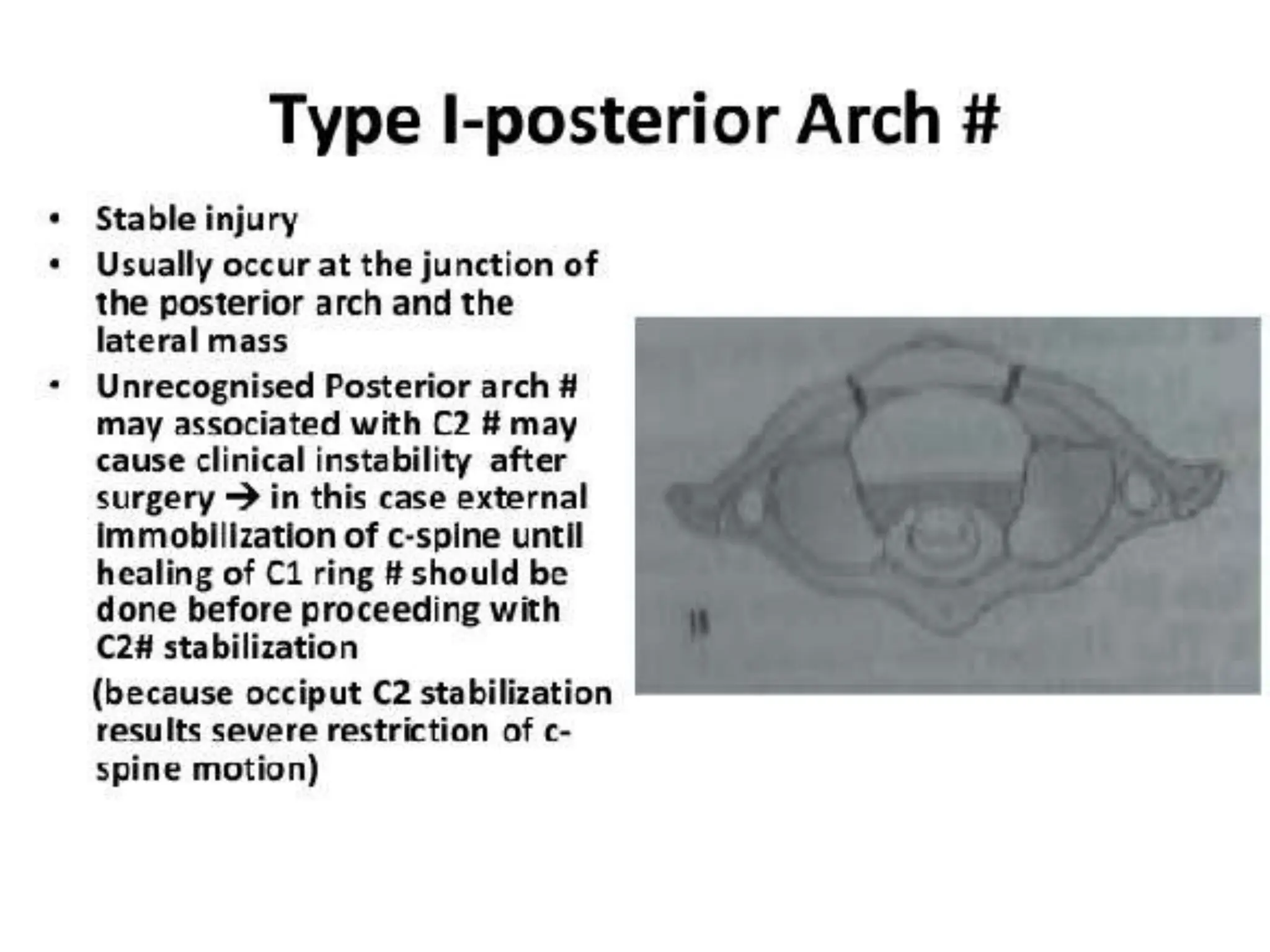
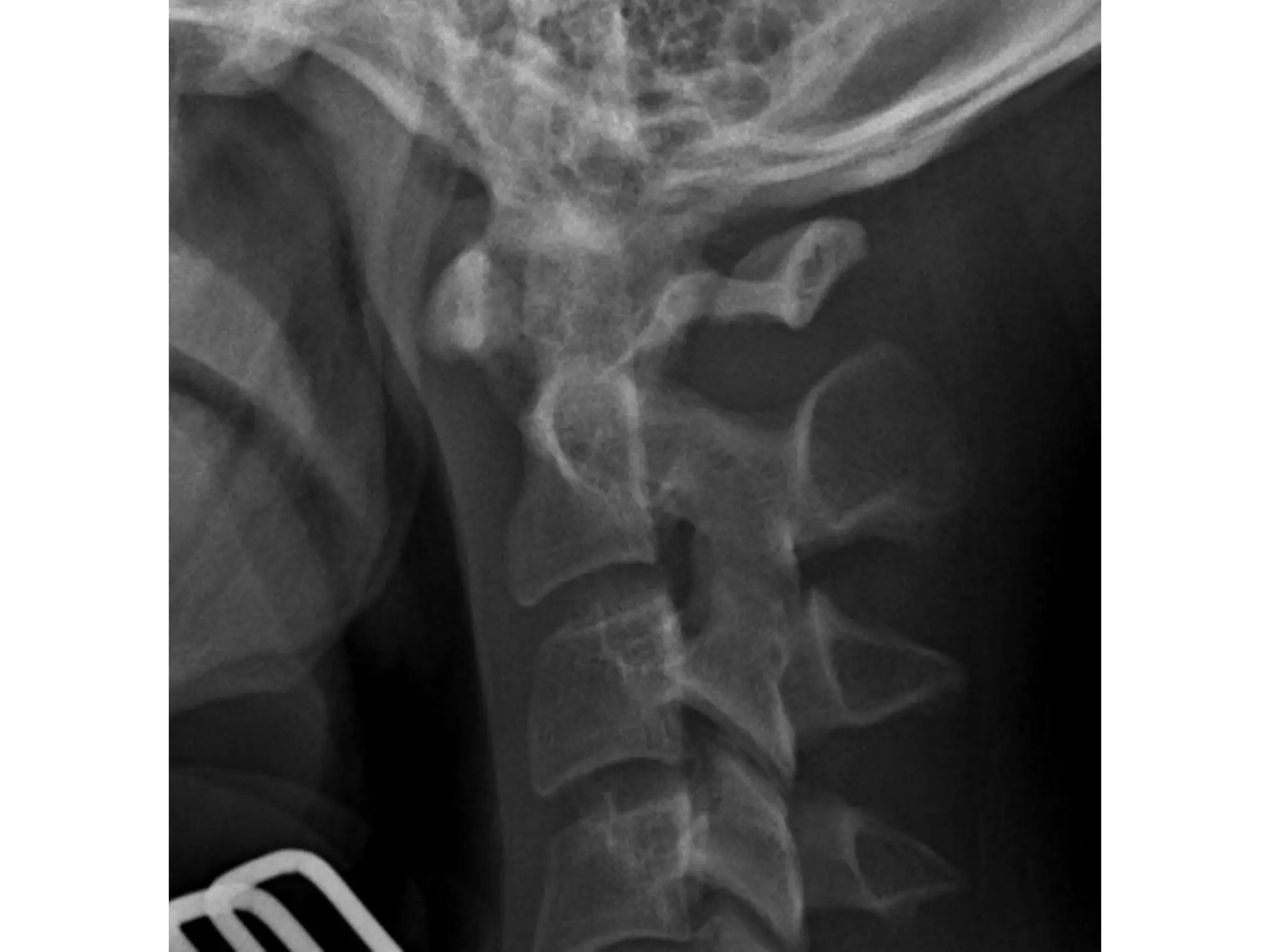
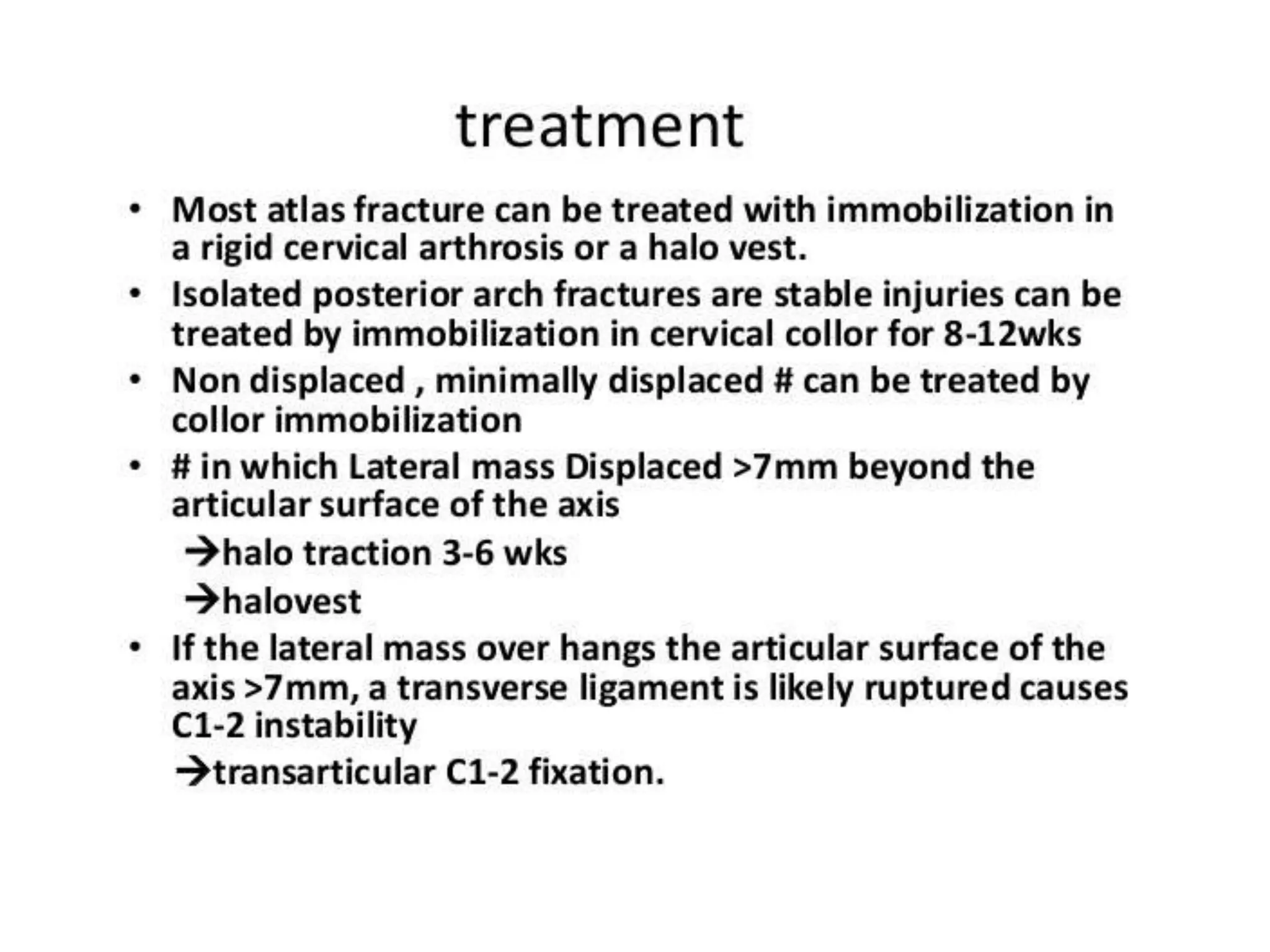
Atlas fracture
Nikita Sharma
BPT 7 SEM
Fracture of atlas
INTRODUCTION-
• Epidemiology
• make up ~7% of cervical spine fractures
• risk of neurologic injury is low
• commonly missed due to inadequate imaging of occipitocervical junction
• Pathophysiology
• mechanism
• includes hyperextension, lateral compression, and axial compression
• Associated conditions
• spine fracture
• 50% have an associated spine injury
• 40% associated with axis fx
• Prognosis
• stability dependent on degree of injury and healing potential of transverse ligament
Imaging
• Radiographs
• lateral radiographs
• CT
• study of choice to delineate fracture pattern and
identify associated injuries in the cervical spine
• MRI
• more sensitive at detecting injury to transverse
ligament
Conservative treatment
• hard collar vs. halo immobilization for 6-12 weeks
• indications
• stable Type I fx (intact transverse ligament)
• stable Jefferson fx (Type II) (intact transverse ligament)
• stable Type III (intact transverse ligament)
• technique
• controversy exists around optimal form of
immobilization
Surgical treatment
• posterior C1-C2 fusion vs. occipitocervical fusion
• indications
• unstable Type II (controversial)
• unstable Type III (controversial)
• technique
• may consider preoperative traction to reduce
displaced lateral masses

Cervical fracture and injuries Presentation

  • 1.
  • 2.
    Fracture of atlas INTRODUCTION- •Epidemiology • make up ~7% of cervical spine fractures • risk of neurologic injury is low • commonly missed due to inadequate imaging of occipitocervical junction • Pathophysiology • mechanism • includes hyperextension, lateral compression, and axial compression • Associated conditions • spine fracture • 50% have an associated spine injury • 40% associated with axis fx • Prognosis • stability dependent on degree of injury and healing potential of transverse ligament
  • 6.
    Imaging • Radiographs • lateralradiographs • CT • study of choice to delineate fracture pattern and identify associated injuries in the cervical spine • MRI • more sensitive at detecting injury to transverse ligament
  • 10.
    Conservative treatment • hardcollar vs. halo immobilization for 6-12 weeks • indications • stable Type I fx (intact transverse ligament) • stable Jefferson fx (Type II) (intact transverse ligament) • stable Type III (intact transverse ligament) • technique • controversy exists around optimal form of immobilization
  • 11.
    Surgical treatment • posteriorC1-C2 fusion vs. occipitocervical fusion • indications • unstable Type II (controversial) • unstable Type III (controversial) • technique • may consider preoperative traction to reduce displaced lateral masses