Medulla Oblongata
Lowest partof brain
stem
Connects to the spinal
cord
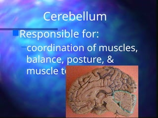
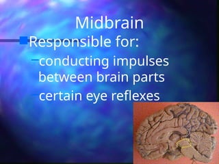
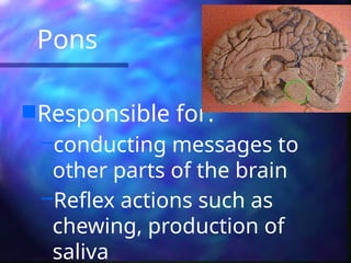
Responsible for:
–regulating heart beat,
respirations, swallowing,
14.
2. Spinal Cord
Goesdown back of body
from Medulla Oblongata
Surrounded and protected
by vertebrae
Responsible for reflex
actions
Carries sensory and motor
Three Membranes
C. Duramater
–thick, tough outer layer
D. Arachnoid membrane
–middle delicate weblike layer
E. Pia mater
–inner most layer with blood
vessels to nourish the nerves
17.
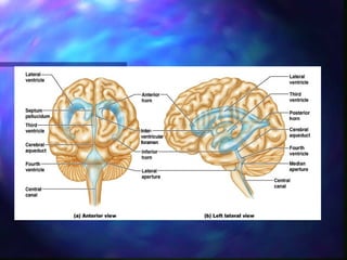
4. Ventricles
Four hallowspaces
located in the middle of
the brain.
Connected to each other
Filled with fluid called
cerebrospinal fluid
Carries nurients toparts
of brain and spinal cord
helps remove metabolic
products & wastes
after circulation,
absorbed into the blood
vessels of the dura mater.
21.
Simplified …
Backof brain: perception
Top of brain: movement
Front of brain: thinking
A. Cranial Nerves
12pairs & their branches
Some responsible for
special senses: sight,
hearing, taste, smell
Others receive sensations:
touch, pressure, pain,
temperature
24.
B. Spinal Nerves
31pairs & their branches
carries messages to &
from the spinal cord
Both sensory and motor
nerves
3. Autonomic Nervous
System
Helpsmaintain a balance
in involuntary functions of
the body. But allows the
body to react in times of
emergency.
27.
2 divisions ofANS
Sympathetic
nervous
system
–acts in
emergency
Parasympatheti
c
–counter acts
the
sympathetic ns
after the
emergency
28.
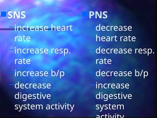
SNS
–increase heart
rate
–increaseresp.
rate
–increase b/p
–decrease
digestive
system activity
PNS
–decrease
heart rate
–decrease resp.
rate
–decrease b/p
–increase
digestive
system
29.
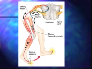
Reflex Arc
Areflex arc is a neural pathway that controls
an action reflex. In human, most sensory
neurons do not pass directly into the brain,
but synapse in the spinal cord. This
characteristic allows reflex actions to occur
relatively quickly by activating spinal motor
neurons without the delay of routing signals
through the brain, although the brain will
receive sensory input while the reflex action
occurs.
31.
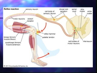
Knee Jerk
Whenthe patellar tendon is tapped just below the knee,
the patellar reflex is initiated and the lower leg kicks
forward (via contraction of the quadriceps). The tap
initiates an action potential in a specialized structure
known as a muscle spindle located within the quadriceps.
This action potential travels to the spinal cord, via a
sensory axon which chemically communicates by releasing
glutamate (see synapse) onto a motor nerve. The result of
this motor nerve activity is contraction of the quadriceps
muscle, leading to extension of the lower leg at the knee.
The sensory input from the quadriceps also activates local
interneurons that release the inhibitory neurotransmitter
glycine onto motor neurons, blocking the innervation of
the antagonistic (hamstring) muscle. The relaxation of the
opposing muscle facilitates extension of the lower leg.
33.
The Effects ofExercise on the
Nervous System
Although you are well aware that physical
exercise is essential to keep the body in
shape, what you may not know is that
physical exercise is also beneficial for the
brain and the nervous system.
34.
Walking Benefits theBrain
According to The Franklin Institute, walking is
good for the brain and the nervous system.
This is because as you walk, your blood
circulation is increased and more glucose and
oxygen reach your brain. As walking is not a
very strenuous activity, your leg muscles do
not use extra oxygen and glucose as in other
forms of more strenuous exercise. Also, as
more blood flows to the brain it helps remove
toxins and to improve concentration, learning
ability and memory.
35.
Sports Training
According toBritish sports coach Brian Mac, it
is possible to train the neurological system
by using repetitive exercises. This enables
athletes to develop quicker reactions,
balance and good coordination. The
repetitive exercises can gradually be built on
by adding new movements, allowing the
athlete to build up a system of learned
moves and skills.
Exercises for theNeuromuscular
System
Balance – prevention of falls
Coordination – smoothness of movement
Strength – moving body parts against
gravity or resistance
38.
Falls
More than one-thirdof adults
ages 65 years and older fall
each year
(Hornbrook 1994; Hausdorff 2001).
39.
Prevention of Falls
Exercise is one of the most important
things to do to reduce your chances of
falling.
Exercise increases:
Balance
Strength
Coordination
40.
Effects of Trainingon balance and sensory inputs
Practice of specific functional movements and complex tasks =
dynamic balance
static balance
righting reflexes
proprioception
vestibular function
simple and complex reaction and movement times
visual function
body awareness
posture and gait
41.
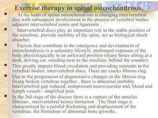
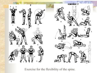
Exercise therapy inspinal osteochondrosis.
At the heart of spinal osteochondrosis is changing intervertebral
disc with subsequent involvement in the process of vertebral bodies
adjacent intervertebral joints and ligaments .
Intervertebral discs play an important role in the stable position of
the vertebrae, provide mobility of the spine, act as biological shock
absorber.
Factors that contribute to the emergence and development of
osteochondrosis is a sedentary lifestyle, prolonged exposure of the
body physiologically in an awkward position (many hours sitting at a
desk, driving car, standing next to the machine, behind the counter).
This greatly impairs blood circulation and providing nutrients to the
vertebral bodies, intervertebral discs. There are cracks fibrous ring.
Due to the progression of degenerative changes in the fibrous ring
fixing broken vertebrae together , there is abnormal mobility.
Intervertebral gap reduced, compressed neurovascular end, blood and
lymph vessels - amplified pain.
In the 3rd stage of the disease there is a rupture of the annulus
fibrosus , intervertebral hernia formation . The final stage is
characterized by a painful thickening and displacement of the
vertebrae, the formation of abnormal bone growths .
42.
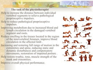
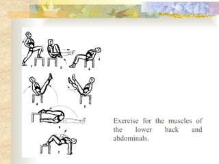
The task ofthe physiotherapist
Help to increase the distance between individual
vertebral segments to relieve pathological
proprioceptive impulses.
Help to reduce pathological proprioceptive
impulses.
Improving metabolism due to increased blood and
lymph circulation in the damaged vertebral
segment and roots.
Reduce swelling in the tissues located in the region
of the intervertebral foramen, improve blood
circulation in the affected limb .
Increasing and restoring full range of motion in the
extremities and spine, reducing static and
dynamic disturbances and compensatory
movements, restoration of impaired posture .
Help restore trophic, tone, muscle strength of the
trunk and extremities.
Improve overall physical performance.
43.
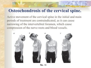
Osteochondrosis of thecervical spine.
Active movement of the cervical spine in the initial and main
periods of treatment are contraindicated, as it can cause
narrowing of the intervertebral foramen, which cause
compression of the nerve roots and blood vessels.