This document provides an overview of the nervous system, including its organization into the central and peripheral systems, which incorporate the somatic and autonomic divisions. It explains the roles of sympathetic and parasympathetic nervous systems and introduces the structure and function of neurons and glial cells. Additionally, it touches on conditions such as multiple sclerosis and details the effects of activation in both sympathetic and parasympathetic states.

![CHAPTER 2Section 2.2 The Structure of Neurons
information enters the dorsal areas. What can you predict about
damage to the spinal cord? Dam-
age to the ventral part of the spinal cord will produce motor
dysfunction, whereas damage to the
dorsal part will impair sensation. Obviously, damage to the
entire spinal cord, as when the spinal
cord is totally transected or torn apart, will affect both sensory
and motor functions.
Sensory neurons in the head and neck send their axons directly
to the brain, via cranial nerves,
which we will examine in Chapter 4. Therefore, damage to the
spinal cord does not affect sensory
or motor functions of the head. Damage to a cranial nerve will
impair the specific sense associ-
ated with that nerve. For example, damage to the olfactory
nerve, or cranial nerve I, will impair
the sense of smell, and injury to the optic nerve, also known as
cranial nerve II, will impair vision.
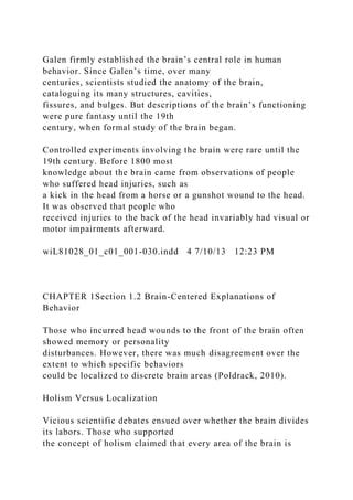
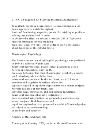
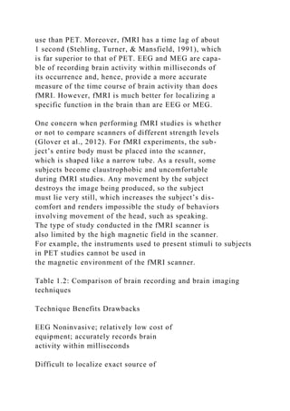
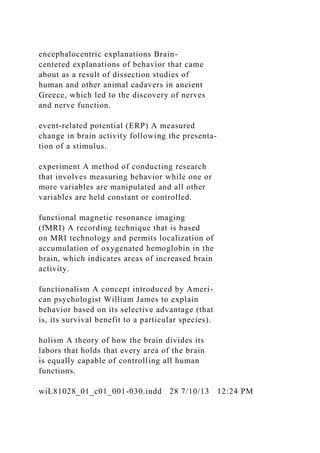
In the spinal cord an interneuron is a neuron that receives
information from one neuron and
passes it on to another neuron. That is, as its name implies
[inter means “between”], an inter-
neuron is situated between two other neurons. The axons and
dendrites of interneurons do not
extend beyond their cell clusters in the gray matter of the spinal
cord. See Figure 2.7 for their loca-
tion in the spinal cord.
Special Features of Neurons
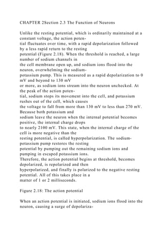
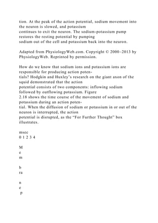
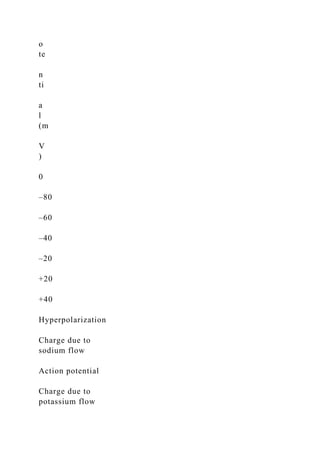
As you’ve just learned, neurons are a unique type of cell. They](https://image.slidesharecdn.com/images-221108013517-7df3a4d0/85/Images-comCorbisLearning-ObjectivesAfter-completing-t-docx-25-320.jpg)
![Baby Amanda was nearly 15 months old when she was admitted
to a metropolitan hospital in a coma.
Shortly before the ambulance brought her to the hospital,
Amanda had been toddling happily around
the living room of her home, where she lived with her mother
and father. Unfortunately, someone had
left a recliner in its laid-back position, with its back down and
its footstool up. Amanda threw herself
down on the footstool, giggling. The weight of her body pushed
the footstool down, causing the chair
to automatically assume its upright position.
Amanda’s neck was trapped in the space between the footstool
and the chair, and when the chair
folded upright, the footstool squeezed against her neck,
obstructing the flow of blood to her brain.
Amanda’s mother was in the living room at the time of this
incident, and she raced over to the chair as
soon as she realized what had happened. It took her nearly 20
seconds to get Amanda free from the
chair. However, by that time, Amanda was purple in the face
and unconscious. Unable to revive her
baby, Amanda’s mother dialed 911 and summoned an
ambulance.
When Amanda arrived at the hospital, she was breathing on her
own but did not respond to any
stimulation whatsoever. She was examined by a team of
neurologists who determined that she was in
a coma. The anoxia [an- means “without,” and -oxia means
“oxygen”] caused by the compression of
the chair on her neck, which stopped the flow of blood to her
brain, damaged Amanda’s brain to such
an extent that she was not expected to regain consciousness.
Amanda remained in a vegetative state](https://image.slidesharecdn.com/images-221108013517-7df3a4d0/85/Images-comCorbisLearning-ObjectivesAfter-completing-t-docx-28-320.jpg)