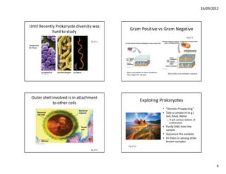
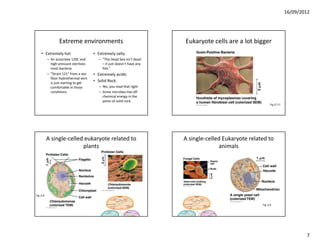
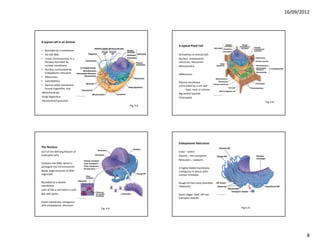
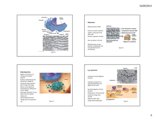
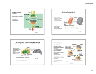
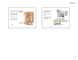
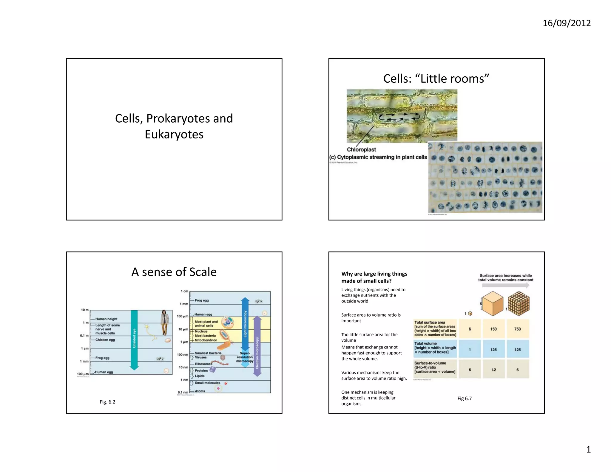
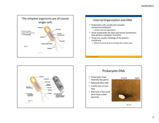
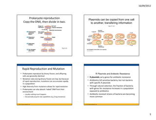
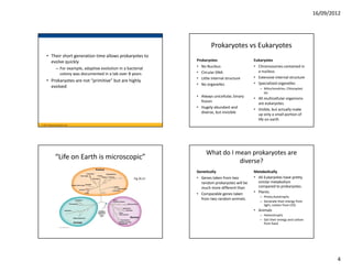
16/09/2012


                                                        Cells: “Little rooms”


       Cells, Prokaryotes and 
             Eukaryotes


           A sense of Scale      Why are large living things 
                                 made of small cells?
                                 Living things (organisms) need to 
                                 exchange nutrients with the 
                                 outside world

                                 Surface area to volume ratio is 
                                 important

                                 Too little surface area for the 
                                 volume
                                 Means that exchange cannot 
                                 happen fast enough to support 
                                 the whole volume.

                                 Various mechanisms keep the 
                                 surface area to volume ratio high.

                                 One mechanism is keeping 
                                 distinct cells in multicellular      Fig 6.7
Fig. 6.2                         organisms.


                                                                                        1
16/09/2012


The simplest organisms are of course                   Internal Organization and DNA
             single cells
                                             • Prokaryotic cells usually lack complex 
                                               compartmentalization
                                                    – Limited internal organization
And the simplest
Single celled organisms                      • Some prokaryotes do have specialized membranes 
Are Prokaryotes
                                               that perform metabolic functions
                                             • These are usually infoldings of the plasma 
                                               membrane
                                                    – Which of course serve to increase the surface area.
    Fig 6.5


                                        © 2011 Pearson Education, Inc.


                                                                         Prokaryote DNA
                                            • Prokaryotes have 
                                              relatively few genes
                                            • Relatively little DNA
                                            • Usually one circular 
                                              loop
                                            • May have a few small 
                                              extra loops called 
                                              plasmids.
              Fig. 6.5

                                                                                                 Fig 27.8


                                                                                                                    2
16/09/2012


                Prokaryote reproduction                                                  Plasmids can be copied from one cell
            Copy the DNA, then divide in two.                                              to another, transfering information
                                                                                                                                    Figure 27.13a-3


                                                                                                                  Bacterial
                                                                                                    F plasmid     chromosome
                                                                                                                                                      F cell

                                                                                          F cell
                                                                                        (donor)

                                                                                     Mating
                                                                                     bridge

                                                                                          F cell
                                                                                     (recipient)
                                                                                                                                                      F cell
                                                                                                                  Bacterial
                                                                                                                  chromosome
                                                                     Fig 12.12
                                                                                     (a) Conjugation and transfer of an F plasmid


          Rapid Reproduction and Mutation
     • Prokaryotes reproduce by binary fission, and offspring                                               R Plasmids and Antibiotic Resistance
       cells are generally identical
                                                                                      • R plasmids carry genes for antibiotic resistance
     • Mutation rates during binary fission are low, but because 
       of rapid reproduction, mutations can accumulate rapidly 
                                                                                      • Antibiotics kill sensitive bacteria, but not bacteria 
       in a population                                                                  with specific R plasmids
     • High diversity from mutations allows for rapid evolution                       • Through natural selection, the fraction of bacteria 
     • Prokaryotes can also absorb ‘naked’ DNA from their                               with genes for resistance increases in a population 
       environment                                                                      exposed to antibiotics
            – Usually nothing much happens                                            • Antibiotic‐resistant strains of bacteria are becoming 
            – Occasionally acquire new capabilities (e.g. Drug resistance)              more common


© 2011 Pearson Education, Inc.                                                   © 2011 Pearson Education, Inc.


                                                                                                                                                                        3
16/09/2012


                                                                               Prokaryotes vs Eukaryotes
     • Their short generation time allows prokaryotes to 
       evolve quickly                                                  Prokaryotes                     Eukaryotes
                – For example, adaptive evolution in a bacterial       • No Nucleus                    • Chromosomes contained in 
                                                                       • Circular DNA                    a nucleus.
                  colony was documented in a lab over 8 years
                                                                       • Little internal structure     • Extensive internal structure
     • Prokaryotes are not “primitive” but are highly 
                                                                       • No organelles                 • Specialized organelles
       evolved                                                                                            – Mitochondrion, Chloroplast 
                                                                                                            etc
                                                                       • Always unicellular, binary 
                                                                                                       • All multicellular organisms 
                                                                         fission
                                                                                                         are eukaryotes.
                                                                       • Hugely abundant and 
                                                                                                       • Visible, but actually make 
                                                                         diverse, but invisible
                                                                                                         up only a small portion of 
                                                                                                         life on earth
© 2011 Pearson Education, Inc.


                                                                            What do I mean prokaryotes are 
               “Life on Earth is microscopic”
                                                                                       diverse?
                                                                       Genetically                     Metabolically
                                                           Fig 26.21   • Genes taken from two          • All Eukaryotes have pretty 
                                                                         random prokaryotes will be      similar metabolism 
                                                                         much more different than        compared to prokaryotes.
                                                                       • Comparable genes taken        • Plants:
                                                                                                          – Photo,Autotrophs
                                                                         from two random animals.
                                                                                                          – Generate their energy from 
                                                                                                            light, carbon from CO2
                                                                                                       • Animals
                                                                                                          – Heterotrophs
                                                                                                          – Get their energy and carbon 
                                                                                                            from food.


                                                                                                                                                   4
16/09/2012


       There are prokaryotes that...
• Get their energy from                   • Aerobic (use Oxygen)
  inorganic chemicals                     • Anaerobic (cannot use 
• “Breath” iron                             oxygen)
    – Use iron as a final electron        • E.g., Methanogens
      acceptor in respiration               “exhale” methane
    – What we do with oxygen                  – What we do with CO2
• “Burn” iron
    – Use iron as an original 
      electron donor in 
      respiration
    – What we do with food.


     Prokaryotes in the living world                                              Exploring diversity of prokaryotes
• “The major biogeochemical               • “Our view of the natural world [is 
  cycles on which we depend were            changing] as radically as did our 
  in place three billion years ago,         view of the cosmos when we 
  long before the appearance of             began looking at it with 
  visible life, and are today               technologies that allowed us to 
  maintained by the ‘invisibles’ and        see more than can be seen with 
  their vast range of metabolisms.”         the naked eye.”
                                          • “Neglect of the invisible world is 
• “The contribution of visible life to      no longer any more acceptable 
  biodiversity is very small indeed.”       than, say, teaching astronomy but 
                                            ignoring the existence of galaxies 
                                            beyond the Milky Way, or 
                                            teaching physics while refusing to 
                                            discuss anything smaller than a 
         Nee, 2004
                                            pin head.”
                                                                                                         Fig 27.15


                                                                                                                               5
16/09/2012


Until Recently Prokaryote diversity was 
                                                 Gram Positive vs Gram Negative
             hard to study
                                                                                                                  Fig 27.3
                                   Fig 27.2
Categorized
By Shape..


                                              More susceptible to those Antibiotics
                                                                                          More likely to be antibiotic resistant
                                              That target the cell wall


  Outer shell involved is in attachment 
                                                             Exploring Prokaryotes
              to other cells
                                                                                      • “Genetic Prospecting”
                                                                                      • Take a sample of (e.g.) 
                                                                                        Soil, Mud, Water
                                                                                         – It will contain billions of 
                                                                                           prokaryotes
                                                                                      • Purify DNA from the 
                                                                                        sample
                                                                                      • Sequence the samples
                                                                                      • Fit them in among other 
                                                                                        known samples
                                                Fig 27.15
                                   Fig 27.4


                                                                                                                                           6
16/09/2012


                   Extreme environments                                     Eukaryote cells are a lot bigger
     • Extremely hot                     • Extremely salty
           – An autoclave 120C and          – “The Dead Sea isn’t dead 
             high pressure sterilizes         – it just doesn’t have any 
             most bacteria                    fish.”
           – “Strain 121” from a sea     • Extremely acidic
             floor hydrothermal vent 
             is just starting to get 
                                         • Solid Rock.
             comfortable in those           – Yes, you read that right
             conditions.                    – Some microbes live off 
                                              chemical energy in the 
                                              pores of solid rock.
                                                                                                                  Fig 27.17


           A single‐celled eukaryote related to                             A single‐celled Eukaryote related to 
                          plants                                                          animals


Fig. 6.8


                                                                                                            Fig. 6.8


                                                                                                                                      7
16/09/2012


A typical cell in an Animal
                                                            A typical Plant Cell
• Bounded by a membrane
• No Cell Wall                                              Similarities to Animal Cell:
• Linear Chromosomes in a                                   Nucleus, endoplasmic 
   Nucleus bounded by                                       reticulum, ribosomes
   nuclear membrane                                         Mitochondria
• Nucleus surrounded by 
   Endoplasmic reticulum                                    Differences:
• Ribosomes
• Cytoskeleton                                              Plasma membrane 
• Various other membrane                                    surrounded by a cell wall
   bound organelles, esp:                                        ‐ Rigid, made of cellulose
‐Mitochondrion                                              Big central vacuole
‐Golgi Apparatus                                            Chloroplast
‐Peroxisome/Lysosome                                                                                     Fig. 6.8
                                                 Fig. 6.8


                                                            Endoplasmic Reticulum
The Nucleus
Sort of the defining feature of                             Endo – within
Eukaryote cells.                                            Plasmic – the cytoplasm
                                                            Reticulum – network.
Contains the DNA, which is 
packaged into Chromosomes                                   A highly folded membrane, 
Keeps large amounts of DNA                                  contiguous in places with 
organized                                                   nuclear envelope

Bounded by a double                                         Rough ER had many bounded 
membrane                                                    ribosomes
(sort of like a cell within a cell)
But with pores.                                             Outer edges ‘bleb’ off into 
                                                            transport vesicles
Outer membrane contiguous 
with endoplasmic reticulum
                                      Fig. 6.9                                                Fig 6.11


                                                                                                                            8
16/09/2012


                                                Ribosomes

                                                Made primarily of RNA

                                                Central in protein synthesis
                                                “Read” a transcript of the 
                                                DNA code
                                                (Details in genetics section)

                                                Very numerous in the cell

                                                (NB Ribosomes are also 
                                                present in prokaryotes, but 
                                                not typically bound to          Fig 6.10
                                                membranes).
                                     Fig 6.11


                                                E.g. Lysosomes
Golgi Apparatus
Appears to function in a 
sorting and transport                           Lysosomes contain digestive 
capacity.                                       enzymes
Proteins synthesized by the 
ribosomes in Rough ER                           Therefore contained in a 
Contained within vessicles                      vessicle where they can’t 
which merge with Golgi,                         digest the cell itself.
May be some additional 
processing (e.g. Folding into                   But allow digestion of food 
correct shape)                                  particles
Eventually ‘delivered’ to area                  Phagocytosis  food vacuole
where they are needed
                                                Lysosome merges with 
‐Made into                                      vacuole
lysosomes/peroxisomes
                                                Digestion takes place
‐Merge with cell membrane
                                  Fig 6.15      (single celled eukaryotes)                 Fig 6.13
‐Etc.


                                                                                                              9
16/09/2012


Two Really Important                                                                      Mitochondrion
Organelles

Mitochondria – respiration
                                                              Outer membrane
                                                              Intermembrane space
Chloroplasts –                                                Inner membrane
Photosynthesis.                                               “Matrix” (the inside)


                                                              Inner membrane
                                                              Highly folded
                                                                                                 There is some DNA in mitochondria
                                                              Increased surface area
                                                                                                 Codes for a few proteins particularly important
                                                                                                 In respiration
                                                                                                 Some ribosomes
                                                                                                                                      Fig. 6.17
                                                  Fig 9.2


      Chloroplast somewhat similar                           Mito’s and Chloro’s as 
                                                             Endosymbionts.

                                                             Surprisingly, the DNA of 
                                                             mitochondria and chloroplasts is 
 Outer membrane                                              more similar 
 Intermembrane space                                         To the DNA of prokaryotes 
 Inner membrane                                              than it is to Eukaryotes
 Stroma
 Thylakoid membrane                                          Evidence that mitochondria and 
 Thylakoid space.                                            chloroplasts are
                                                             Prokaryotic symbionts

                                                             Ancient symbiosis – many mito
         Thylakoid highly folded – surface area              and chloro genes have “migrated” 
                                                  Fig 6.18   to the nucleus

        DNA and Ribosomes as in Mito’s
                                                             Original symbiosis 2‐3 Billion 
                                                             years ago.

                                                                                                              Fig 6.16


                                                                                                                                                          10
16/09/2012


Prokaryotes with highly infolded 
                                                                   Cytoskeleton.
     plasma membranes.

                                         Actin

                                         Structural protein

                                         Polymer of actin 
                                         Subunits
                                         (a polymer of polymers)

                                         Tensive


                       Fig 27.7                                           Table 6.1


       Recall Muscle                       Actin is important in cellular movement
Myson pulls on strands of Actin             phagocytosis, cytoplasmic streaming


                              Fig 6.27                                                Fig 6.27


                                                                                                        11
16/09/2012


                              Cytoskeleton.                                               Cytoskeletin

Keratin
                                                            Microtubules
Also tensive
                                                            Rigid, resist compression
Fibrous polypeptides
Coiled together                                             Tubulin dimers

Major anchoring

External protein structures
e.g. Hair


                                     Table 6.1                                                   Table 6.1


                                                            Cellular Movement
Motor proteins
                                                            A series of motor proteins 
                                                            connecting two microtubules
One way organelles move 
things to the right part of 
the cell                                                    Move the doublets laterally

                                                            Push cellular structures into 
Use Atp energy 
                                                            position
                                                            e.g. chromosomes during cell 
Pull the vesicle along                                      division.
microtubule


                                                 Fig 6.21                                                    Fig 6.25


                                                                                                                               12
16/09/2012


If the two proteins are anchored:
      waving motion of cilia.


                           Fig 6.25                                                        Fig 6.23


  A flagellum rotates driven by              Intercellular connections
  a protein ‘motor’ at the base       recall membrane proteins bind to things outside the cell


                                                                                Fig 6.30
                                                   Fig 6.6


                                                                                                             13
16/09/2012


Cell Connections ‐ Animals      Cell Connections ‐ Plants
                                Cell walls are connected
Tight junctions: a series of 
closely connected proteins      Cells are therefore held in 
(sort of like a sewn seam)      place

Channels between cells          Series of pores through cell 
                                walls (technically called 
Increased surface area where    plasmodesmata)
absorption is important.


  Fig 6.32

                                                           Fig 6.33


                                                                             14

Cells, prokaryotes and eukaryotes

  • 1.
    16/09/2012 Cells: “Little rooms” Cells, Prokaryotes and  Eukaryotes A sense of Scale Why are large living things  made of small cells? Living things (organisms) need to  exchange nutrients with the  outside world Surface area to volume ratio is  important Too little surface area for the  volume Means that exchange cannot  happen fast enough to support  the whole volume. Various mechanisms keep the  surface area to volume ratio high. One mechanism is keeping  distinct cells in multicellular Fig 6.7 Fig. 6.2 organisms. 1
  • 2.
    16/09/2012 The simplest organisms are of course  Internal Organization and DNA single cells • Prokaryotic cells usually lack complex  compartmentalization – Limited internal organization And the simplest Single celled organisms • Some prokaryotes do have specialized membranes  Are Prokaryotes that perform metabolic functions • These are usually infoldings of the plasma  membrane – Which of course serve to increase the surface area. Fig 6.5 © 2011 Pearson Education, Inc. Prokaryote DNA • Prokaryotes have  relatively few genes • Relatively little DNA • Usually one circular  loop • May have a few small  extra loops called  plasmids. Fig. 6.5 Fig 27.8 2
  • 3.
    16/09/2012 Prokaryote reproduction Plasmids can be copied from one cell Copy the DNA, then divide in two. to another, transfering information Figure 27.13a-3 Bacterial F plasmid chromosome F cell F cell (donor) Mating bridge F cell (recipient) F cell Bacterial chromosome Fig 12.12 (a) Conjugation and transfer of an F plasmid Rapid Reproduction and Mutation • Prokaryotes reproduce by binary fission, and offspring  R Plasmids and Antibiotic Resistance cells are generally identical • R plasmids carry genes for antibiotic resistance • Mutation rates during binary fission are low, but because  of rapid reproduction, mutations can accumulate rapidly  • Antibiotics kill sensitive bacteria, but not bacteria  in a population with specific R plasmids • High diversity from mutations allows for rapid evolution • Through natural selection, the fraction of bacteria  • Prokaryotes can also absorb ‘naked’ DNA from their  with genes for resistance increases in a population  environment exposed to antibiotics – Usually nothing much happens • Antibiotic‐resistant strains of bacteria are becoming  – Occasionally acquire new capabilities (e.g. Drug resistance) more common © 2011 Pearson Education, Inc. © 2011 Pearson Education, Inc. 3
  • 4.
    16/09/2012 Prokaryotes vs Eukaryotes • Their short generation time allows prokaryotes to  evolve quickly Prokaryotes Eukaryotes – For example, adaptive evolution in a bacterial  • No Nucleus • Chromosomes contained in  • Circular DNA a nucleus. colony was documented in a lab over 8 years • Little internal structure • Extensive internal structure • Prokaryotes are not “primitive” but are highly  • No organelles • Specialized organelles evolved – Mitochondrion, Chloroplast  etc • Always unicellular, binary  • All multicellular organisms  fission are eukaryotes. • Hugely abundant and  • Visible, but actually make  diverse, but invisible up only a small portion of  life on earth © 2011 Pearson Education, Inc. What do I mean prokaryotes are  “Life on Earth is microscopic” diverse? Genetically Metabolically Fig 26.21 • Genes taken from two  • All Eukaryotes have pretty  random prokaryotes will be  similar metabolism  much more different than  compared to prokaryotes. • Comparable genes taken  • Plants: – Photo,Autotrophs from two random animals. – Generate their energy from  light, carbon from CO2 • Animals – Heterotrophs – Get their energy and carbon  from food. 4
  • 5.
    16/09/2012 There are prokaryotes that... • Get their energy from  • Aerobic (use Oxygen) inorganic chemicals • Anaerobic (cannot use  • “Breath” iron oxygen) – Use iron as a final electron  • E.g., Methanogens acceptor in respiration “exhale” methane – What we do with oxygen – What we do with CO2 • “Burn” iron – Use iron as an original  electron donor in  respiration – What we do with food. Prokaryotes in the living world Exploring diversity of prokaryotes • “The major biogeochemical  • “Our view of the natural world [is  cycles on which we depend were  changing] as radically as did our  in place three billion years ago,  view of the cosmos when we  long before the appearance of  began looking at it with  visible life, and are today  technologies that allowed us to  maintained by the ‘invisibles’ and  see more than can be seen with  their vast range of metabolisms.” the naked eye.” • “Neglect of the invisible world is  • “The contribution of visible life to  no longer any more acceptable  biodiversity is very small indeed.” than, say, teaching astronomy but  ignoring the existence of galaxies  beyond the Milky Way, or  teaching physics while refusing to  discuss anything smaller than a  Nee, 2004 pin head.” Fig 27.15 5
  • 6.
    16/09/2012 Until Recently Prokaryote diversity was  Gram Positive vs Gram Negative hard to study Fig 27.3 Fig 27.2 Categorized By Shape.. More susceptible to those Antibiotics More likely to be antibiotic resistant That target the cell wall Outer shell involved is in attachment  Exploring Prokaryotes to other cells • “Genetic Prospecting” • Take a sample of (e.g.)  Soil, Mud, Water – It will contain billions of  prokaryotes • Purify DNA from the  sample • Sequence the samples • Fit them in among other  known samples Fig 27.15 Fig 27.4 6
  • 7.
    16/09/2012 Extreme environments Eukaryote cells are a lot bigger • Extremely hot • Extremely salty – An autoclave 120C and  – “The Dead Sea isn’t dead  high pressure sterilizes  – it just doesn’t have any  most bacteria fish.” – “Strain 121” from a sea  • Extremely acidic floor hydrothermal vent  is just starting to get  • Solid Rock. comfortable in those  – Yes, you read that right conditions. – Some microbes live off  chemical energy in the  pores of solid rock. Fig 27.17 A single‐celled eukaryote related to  A single‐celled Eukaryote related to  plants animals Fig. 6.8 Fig. 6.8 7
  • 8.
    16/09/2012 A typical cell in an Animal A typical Plant Cell • Bounded by a membrane • No Cell Wall Similarities to Animal Cell: • Linear Chromosomes in a  Nucleus, endoplasmic  Nucleus bounded by  reticulum, ribosomes nuclear membrane Mitochondria • Nucleus surrounded by  Endoplasmic reticulum Differences: • Ribosomes • Cytoskeleton Plasma membrane  • Various other membrane  surrounded by a cell wall bound organelles, esp: ‐ Rigid, made of cellulose ‐Mitochondrion Big central vacuole ‐Golgi Apparatus Chloroplast ‐Peroxisome/Lysosome Fig. 6.8 Fig. 6.8 Endoplasmic Reticulum The Nucleus Sort of the defining feature of  Endo – within Eukaryote cells. Plasmic – the cytoplasm Reticulum – network. Contains the DNA, which is  packaged into Chromosomes  A highly folded membrane,  Keeps large amounts of DNA  contiguous in places with  organized nuclear envelope Bounded by a double  Rough ER had many bounded  membrane ribosomes (sort of like a cell within a cell) But with pores. Outer edges ‘bleb’ off into  transport vesicles Outer membrane contiguous  with endoplasmic reticulum Fig. 6.9 Fig 6.11 8
  • 9.
    16/09/2012 Ribosomes Made primarily of RNA Central in protein synthesis “Read” a transcript of the  DNA code (Details in genetics section) Very numerous in the cell (NB Ribosomes are also  present in prokaryotes, but  not typically bound to  Fig 6.10 membranes). Fig 6.11 E.g. Lysosomes Golgi Apparatus Appears to function in a  sorting and transport  Lysosomes contain digestive  capacity. enzymes Proteins synthesized by the  ribosomes in Rough ER Therefore contained in a  Contained within vessicles vessicle where they can’t  which merge with Golgi, digest the cell itself. May be some additional  processing (e.g. Folding into  But allow digestion of food  correct shape) particles Eventually ‘delivered’ to area  Phagocytosis  food vacuole where they are needed Lysosome merges with  ‐Made into  vacuole lysosomes/peroxisomes Digestion takes place ‐Merge with cell membrane Fig 6.15 (single celled eukaryotes) Fig 6.13 ‐Etc. 9
  • 10.
    16/09/2012 Two Really Important  Mitochondrion Organelles Mitochondria – respiration Outer membrane Intermembrane space Chloroplasts – Inner membrane Photosynthesis. “Matrix” (the inside) Inner membrane Highly folded There is some DNA in mitochondria Increased surface area Codes for a few proteins particularly important In respiration Some ribosomes Fig. 6.17 Fig 9.2 Chloroplast somewhat similar Mito’s and Chloro’s as  Endosymbionts. Surprisingly, the DNA of  mitochondria and chloroplasts is  Outer membrane more similar  Intermembrane space To the DNA of prokaryotes  Inner membrane than it is to Eukaryotes Stroma Thylakoid membrane Evidence that mitochondria and  Thylakoid space. chloroplasts are Prokaryotic symbionts Ancient symbiosis – many mito Thylakoid highly folded – surface area and chloro genes have “migrated”  Fig 6.18 to the nucleus DNA and Ribosomes as in Mito’s Original symbiosis 2‐3 Billion  years ago. Fig 6.16 10
  • 11.
    16/09/2012 Prokaryotes with highly infolded  Cytoskeleton. plasma membranes. Actin Structural protein Polymer of actin  Subunits (a polymer of polymers) Tensive Fig 27.7 Table 6.1 Recall Muscle Actin is important in cellular movement Myson pulls on strands of Actin phagocytosis, cytoplasmic streaming Fig 6.27 Fig 6.27 11
  • 12.
    16/09/2012 Cytoskeleton. Cytoskeletin Keratin Microtubules Also tensive Rigid, resist compression Fibrous polypeptides Coiled together Tubulin dimers Major anchoring External protein structures e.g. Hair Table 6.1 Table 6.1 Cellular Movement Motor proteins A series of motor proteins  connecting two microtubules One way organelles move  things to the right part of  the cell Move the doublets laterally Push cellular structures into  Use Atp energy  position e.g. chromosomes during cell  Pull the vesicle along  division. microtubule Fig 6.21 Fig 6.25 12
  • 13.
    16/09/2012 If the two proteins are anchored: waving motion of cilia. Fig 6.25 Fig 6.23 A flagellum rotates driven by  Intercellular connections a protein ‘motor’ at the base recall membrane proteins bind to things outside the cell Fig 6.30 Fig 6.6 13
  • 14.
    16/09/2012 Cell Connections ‐ Animals Cell Connections ‐ Plants Cell walls are connected Tight junctions: a series of  closely connected proteins  Cells are therefore held in  (sort of like a sewn seam) place Channels between cells Series of pores through cell  walls (technically called  Increased surface area where  plasmodesmata) absorption is important. Fig 6.32 Fig 6.33 14