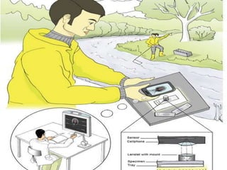
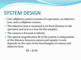
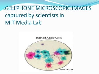
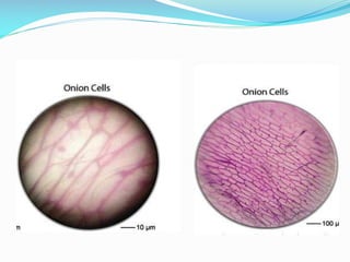
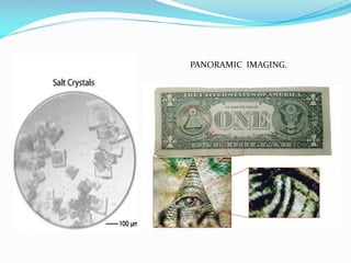
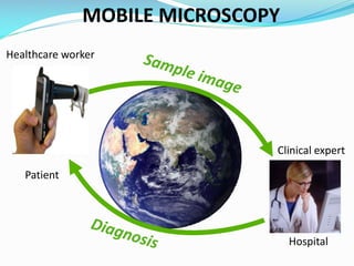
BUILDING A
CELLPHONE
MICROSCOPE
PRESENTED BY
 Priyanka Guleria

(B120030083)
 Pushpinder Singh Sidhu

(B120030085)
 Rajat Chandel

(B120030087)
 Rajbir

(B120030091)
CONTENTS











Introduction……
Overview of benefits and limits……
System design……
Construction……
Cellphone microscopic images……
Uses……
Different applications……
Future work……
Conclusion……
References……
INTRODUCTION
 Today, an estimated six billion cellphone subscriptions

exist worldwide with about 70% of those in developing
countries.
 Developing countries often suffer from a lack of access to
adequate healthcare, which is due to the cost and training
associated with high-tech scientific instruments required
for medical analysis.
 Thus, scientists present a low-cost portable microscope
that uses a cellphone camera and a simple, secondary lens
that is placed on top of the specimen.
 Cellphone microscopes provide a unique opportunity to
make disease diagnosis and healthcare accessible to
everyone, even in remote and undeveloped parts of the
world.
 Starting in 2008, mobile computational photography has

reached a tipping point and, largely due to the enabling
capabilities of cellphone cameras, various approaches to
cellphone microscopy have started to appear.
 Cellphone microscope enables visualization of samples,
followed by capture, organization, and transmission of
images critical for diagnosis.
OVERVIEW OF BENEFITS AND
LIMITATIONS
 Cost-effective: Only a single lens is required in addition
to a cellphone camera.So its cheap.
 Non-intrusive: setup does not require intrusive
modification of the phone.
 Flexibility: detached camera-lens configuration allows
any camera to be used for microscopy.
 Minimal computation: We do not require extensive postprocessing, as e.g. holographic approaches.
 Computational illumination: Using a second cellphone
display as the background illumination allows enhanced
microscopic images to be captured.
 Network connectivity.
 Potential to significantly contribute to the technology

available for global healthcare, particularly in the
developing world and rural areas.
SYSTEM DESIGN
 Our cellphone camera consists of a specimen, an objective

lens, and a cellphone camera.
 The objective lens is mounted at its focal distance to the
specimen and acts as a lens for flat samples.
 The camera is focused at infinity.
 The optical magnification M of the system is independent
of the distance between camera and sample; it only
depends on the ratio of the focal lengths of camera and
objective lens:

M = fc/fo
 Due to our unique configuration, the camera can be freely

moved around the objective lens so as to capture different
viewpoints.
 Our setup is much more legible by detaching the phone
camera from the objective lens, thereby allowing any
available cellphone to capture microscopic imagery.
 It does no require any additional optical elements to be
mounted on the camera itself.
CONSTRUCTION

 Insert your objective lens in the rubber tube and fix it.
 Use pieces of electrical tape to hold the lens in place.
 Create a stand to light and hold a microscope slide. The

stand will enable you to keep the cell phone microscope as
still as possible while testing it.
 Use scissors to make two notches directly across from each
other on the side and bottom of the paper tube.
 To create the light source, strip off 1 cm of the plastic
insulator with the wire stripper from both ends of the two
lengths of electrical wire.
 over the lighted specimen on the microscope stand.
 Insert the flashlight bulb into the base with the battery.
 Set the paper tube over the bulb so that the wires come

through the notches.
 Place the prepared microscope slide specimen on top of the
paper tube so that the light is shining through it.
 Turn on the cell phone microscope by activating the cell

phone's camera function and place the iris of the cell phone
microscope directly .
 Place the prepared microscope slide specimen on top of the
paper tube so that the light is shining through it.
 Turn on the cell phone microscope by activating the cell
phone's camera function and place the iris of the cell phone
microscope directly over the lighted specimen on the
microscope stand.
CELLPHONE MICROSCOPIC IMAGES
captured by scientists in
MIT Media Lab
PANORAMIC IMAGING.
MOBILE MICROSCOPY
Healthcare worker

Clinical expert
Patient

ABL

Hospital
WHY WE USED THE CONVEX
LENS
 The convex lenses can be used in magnifying glasses.
 They are thicker in the centre than in the edges.
 Convex mirrors are used as streetlight reflectors as they

spread light over greater area.
 The focal length is positive, and is the distance at which a
beam of light will be focused to a single spot.
USES AND APPLICATIONS
 Used to examine blood samples in the field and help spot






some of the world's deadliest diseases.
And uses the built-in camera to process the images,
allowing doctors to quickly screen for diseases including
TB and sickle-cell.
Doctors can perform complex high-resolution light
microscopy on a blood or sputum sample placed on a slide.
Used to see the more magnified images of the substance.
The microscope helps reveal intestinal worms in
schoolchildren.
 Scientists, engineers, and researchers use microscopes to

get more information about the specimens they study.
 Investigate the silt deposits in different regions of a stream
using your cell phone microscope.
 To measure the dimensions of small objects .
 Preparation of own microscope specimen slides.
FUTURE WORK
 Research is going on for making it useful for learning about

the more sophisticated illuminations in future.
 Planning for the diagnosis of other urine and blood
diseases and classifying them , using cell microscopy.
 Looking at other medical gadgets and trying to make them
entirely digital , like this cellphone microscopy technique.
CONCLUSION
 Cellphones are widely available, especially in developing

parts of the world.
 These devices can be converted into scientific instruments
for microscopic imaging.
 Equipped with wireless network connections, cellphones
also allow the transmission of recorded data for remote
analysis or statistical inference.
 It has the potential to make disease diagnosis and
screening accessible in parts of the world that have no
adequate access to healthcare.
REFERENCES
 http://www.wired.com/wiredscience/2011/03/diy-cellphone





microscope/
allafrica.com/stories/201304190125.html
www.scidev.net › Home › New Technologies › News
cellscope.berkeley.edu/
www.technologyreview.com
www.smartplanet.com/blog/pure-genius/new-in-telemedicine-thecell-phone-microscope/4141

 www.youtube.com/watch?v=5qcJySNLs84

 scibuddy@sciencebuddies.org.

cellphone microscope

  • 1.
  • 2.
    PRESENTED BY  PriyankaGuleria (B120030083)  Pushpinder Singh Sidhu (B120030085)  Rajat Chandel (B120030087)  Rajbir (B120030091)
  • 3.
    CONTENTS           Introduction…… Overview of benefitsand limits…… System design…… Construction…… Cellphone microscopic images…… Uses…… Different applications…… Future work…… Conclusion…… References……
  • 4.
    INTRODUCTION  Today, anestimated six billion cellphone subscriptions exist worldwide with about 70% of those in developing countries.  Developing countries often suffer from a lack of access to adequate healthcare, which is due to the cost and training associated with high-tech scientific instruments required for medical analysis.  Thus, scientists present a low-cost portable microscope that uses a cellphone camera and a simple, secondary lens that is placed on top of the specimen.  Cellphone microscopes provide a unique opportunity to make disease diagnosis and healthcare accessible to everyone, even in remote and undeveloped parts of the world.
  • 5.
     Starting in2008, mobile computational photography has reached a tipping point and, largely due to the enabling capabilities of cellphone cameras, various approaches to cellphone microscopy have started to appear.  Cellphone microscope enables visualization of samples, followed by capture, organization, and transmission of images critical for diagnosis.
  • 6.
    OVERVIEW OF BENEFITSAND LIMITATIONS  Cost-effective: Only a single lens is required in addition to a cellphone camera.So its cheap.  Non-intrusive: setup does not require intrusive modification of the phone.  Flexibility: detached camera-lens configuration allows any camera to be used for microscopy.  Minimal computation: We do not require extensive postprocessing, as e.g. holographic approaches.  Computational illumination: Using a second cellphone display as the background illumination allows enhanced microscopic images to be captured.
  • 7.
     Network connectivity. Potential to significantly contribute to the technology available for global healthcare, particularly in the developing world and rural areas.
  • 8.
    SYSTEM DESIGN  Ourcellphone camera consists of a specimen, an objective lens, and a cellphone camera.  The objective lens is mounted at its focal distance to the specimen and acts as a lens for flat samples.  The camera is focused at infinity.  The optical magnification M of the system is independent of the distance between camera and sample; it only depends on the ratio of the focal lengths of camera and objective lens: M = fc/fo
  • 9.
     Due toour unique configuration, the camera can be freely moved around the objective lens so as to capture different viewpoints.  Our setup is much more legible by detaching the phone camera from the objective lens, thereby allowing any available cellphone to capture microscopic imagery.  It does no require any additional optical elements to be mounted on the camera itself.
  • 10.
    CONSTRUCTION  Insert yourobjective lens in the rubber tube and fix it.  Use pieces of electrical tape to hold the lens in place.  Create a stand to light and hold a microscope slide. The stand will enable you to keep the cell phone microscope as still as possible while testing it.  Use scissors to make two notches directly across from each other on the side and bottom of the paper tube.  To create the light source, strip off 1 cm of the plastic insulator with the wire stripper from both ends of the two lengths of electrical wire.
  • 11.
     over thelighted specimen on the microscope stand.  Insert the flashlight bulb into the base with the battery.  Set the paper tube over the bulb so that the wires come through the notches.  Place the prepared microscope slide specimen on top of the paper tube so that the light is shining through it.
  • 12.
     Turn onthe cell phone microscope by activating the cell phone's camera function and place the iris of the cell phone microscope directly .  Place the prepared microscope slide specimen on top of the paper tube so that the light is shining through it.  Turn on the cell phone microscope by activating the cell phone's camera function and place the iris of the cell phone microscope directly over the lighted specimen on the microscope stand.
  • 13.
    CELLPHONE MICROSCOPIC IMAGES capturedby scientists in MIT Media Lab
  • 15.
  • 16.
  • 17.
    WHY WE USEDTHE CONVEX LENS  The convex lenses can be used in magnifying glasses.  They are thicker in the centre than in the edges.  Convex mirrors are used as streetlight reflectors as they spread light over greater area.  The focal length is positive, and is the distance at which a beam of light will be focused to a single spot.
  • 18.
    USES AND APPLICATIONS Used to examine blood samples in the field and help spot     some of the world's deadliest diseases. And uses the built-in camera to process the images, allowing doctors to quickly screen for diseases including TB and sickle-cell. Doctors can perform complex high-resolution light microscopy on a blood or sputum sample placed on a slide. Used to see the more magnified images of the substance. The microscope helps reveal intestinal worms in schoolchildren.
  • 19.
     Scientists, engineers,and researchers use microscopes to get more information about the specimens they study.  Investigate the silt deposits in different regions of a stream using your cell phone microscope.  To measure the dimensions of small objects .  Preparation of own microscope specimen slides.
  • 20.
    FUTURE WORK  Researchis going on for making it useful for learning about the more sophisticated illuminations in future.  Planning for the diagnosis of other urine and blood diseases and classifying them , using cell microscopy.  Looking at other medical gadgets and trying to make them entirely digital , like this cellphone microscopy technique.
  • 21.
    CONCLUSION  Cellphones arewidely available, especially in developing parts of the world.  These devices can be converted into scientific instruments for microscopic imaging.  Equipped with wireless network connections, cellphones also allow the transmission of recorded data for remote analysis or statistical inference.  It has the potential to make disease diagnosis and screening accessible in parts of the world that have no adequate access to healthcare.
  • 22.
    REFERENCES  http://www.wired.com/wiredscience/2011/03/diy-cellphone     microscope/ allafrica.com/stories/201304190125.html www.scidev.net ›Home › New Technologies › News cellscope.berkeley.edu/ www.technologyreview.com www.smartplanet.com/blog/pure-genius/new-in-telemedicine-thecell-phone-microscope/4141  www.youtube.com/watch?v=5qcJySNLs84   scibuddy@sciencebuddies.org.