
Recommended
More Related Content
What's hot
What's hot (20)
Similar to Blood film preparation and reporting
Similar to Blood film preparation and reporting (20)
More from jadcaesar
Recently uploaded
Recently uploaded (20)
Blood film preparation and reporting
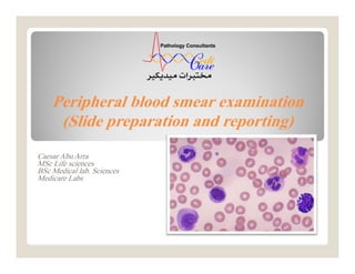
- 1. Peripheral blood smear examination (Slide preparation and reporting) Caesar Abu Arra MSc Life sciences BSc Medical lab. Sciences Medicare Labs
- 2. Aim of blood smear Evaluation of anemia Infections • bacteria, malaria, microfilaria..etc. Abnormal cells • blasts, inclusions..etc. Cells morphology and count Abu Jad Caesar
- 3. Making blood films Three basic steps to make blood film: Preparation of blood smear. Fixation of blood smear. Staining of blood smear. Abu Jad Caesar
- 4. Preparation of blood smear Different types of blood smears: The wedge smear slide to slide method. The spun smear. Two additional types of blood smears (used for specific purposes): o Buffy coat smear for WBCs < 1.0×10⁹/L. o Thick blood smears for blood parasites . Abu Jad Caesar
- 5. Wedge blood smear Smears should be made within 1 hour of blood collection from EDTA specimens stored at room temperature to avoid distortion of cell morphology. Small drop (10 µl) Hold the spreader slide at a 30°- 45° angle, and draw it back against the drop of blood. Air dry at room temperature. Abu Jad Caesar
- 6. Abu Jad Caesar Wedge blood smear
- 7. Procedure notes 1. The smear should be made without delay. Any delay results in an abnormal distribution of the white blood cells, with many of the large white cells accumulating at the thin edge of the smear. 2. HCT↑ ↓ The angle of the spreader slide HCT↓ ↑ The angle of the spreader slide Abu Jad Caesar
- 8. large angle low HCT small angle high HCT Abu Jad Caesar Procedure notes
- 9. Spin method Automated method Placeadrop of blood in thecenter of aglassslide. Spin atahighspeed in aspecialcentrifugecytospin. Blood spreads uniformly. Abu Jad Caesar
- 10. Characteristics of a Good Smear Thick at one end, thinning out to a smooth rounded feather edge. Should occupy 2/3 of the total slide area. Should not touch any edge of the slide. Abu Jad Caesar
- 11. tail body head Abu Jad Caesar Characteristics of a Good Smear
- 12. Common causes of a poor blood smear Drop of blood too large or too small. Spreader slide pushed across the slide in a jerky manner. Failure to keep the spreader slide at a 30° angle with the slide. Failure to push the spreader slide completely across the slide. Abu Jad Caesar
- 13. Common causes of a poor blood smear Holes in film: Slide contaminated with fat or grease Cellular degenerative changes: delay in fixing Inadequate fixing time Methanol contaminated with water. Abu Jad Caesar
- 14. Biologic causes of a poor smear Cold agglutinin - RBCs will clump together. Warm the blood at 37° C for 5 minutes. Lipemia - holes will appear in the smear. Nothing you can do to correct this. Rouleaux - RBC’s will form into stacks as coins. Nothing you can do to correct this. Abu Jad Caesar
- 15. Staining of blood smear Romanowsky stain: Any Combination of: Eosin Y (Acidic or anionic dye): o Binds to cationic sites (cytoplasm & Hb) Methylene blue (Basic, cationic or Azure B dye): o Binds to anionic sites (nucleus and RNA) Abu Jad Caesar
- 16. Staining of blood smear Buffer: Used to maintain an adequate pH. 0.05M Na2PO4 (pH 6.4) or dH2O (pH 6.4-6.8) Abu Jad Caesar
- 17. pH of the phosphate buffer If the pH is too acidic: RBC :Pinker WBC nuclei : very pale staining (very pale purple) If the pH is too basic: RBC :Grayish blue WBC nuclei :very deeply purple Abu Jad Caesar
- 18. pH of the phosphate buffer Abu Jad Caesar
- 19. Staining Troubleshooting Too Acid Stain: Insufficient staining time Prolonged buffering or washing Old stain Correction: Lengthen staining time Check stain and buffer pH Shorten buffering or wash time Abu Jad Caesar
- 20. Staining Troubleshooting Too Alkaline Stain: Thick blood smear Prolonged staining Insufficient washing Alkaline pH of stain components Correction : Check pH Shorten stain time Prolong buffering time Abu Jad Caesar
- 21. Abu Jad Caesar
- 22. Macroscopic view: Quality of the smear The microscopic view: Progressing from low power to high power: Blood film examination - preliminary Abu Jad Caesar
- 23. Blood film examination - preliminary Abu Jad Caesar
- 24. Low-Power (10x) Scan Determine the overall staining quality of the blood smear. Determine if there is a good distribution of the cells. o Discussed later Find an optimal area for the detailed examination and enumeration of cells Blood film examination - preliminary Abu Jad Caesar
- 25. High-Power (40x) Scan Determine the WBC estimate. o WBCs are counted in 10 fields and averaged X 2000 o The estimate could be reported according to the table below: Blood film examination - preliminary Abu Jad Caesar
- 26. • High-Power (40x) Scan Grading scale for WBC count (X103 cell/µl) Blood film examination - preliminary Low WBC High WBC Mild 3-4.5 Mild 10-15 Moderate 2-3 Moderate 15-30 Severe <2 Marked 30-100 Extreme >100 Abu Jad Caesar
- 27. Oil Immersion (100x) Examination Perform a 100 WBC differential count. Correct WBC count that has greater than 10 NRBCs per 100 WBCs. o Report as: WBC count was corrected due to presence of NRBCs. Blood film examination - preliminary Abu Jad Caesar
- 28. Oil Immersion (100x) Examination to correct a WBC count (/µl): Uncorrected WBC (/ ) X l Blood film examination - preliminary Abu Jad Caesar
- 29. Oil Immersion (100x) Examination Evaluate the RBCs for: o Anisocytosis o Poikilocytosis o Inclusions o Hypochromasia o Polychromasia. Blood film examination - preliminary Abu Jad Caesar
- 30. Oil Immersion (100x) Examination Perform a platelet estimate and evaluate platelet morphology: o Platelets are counted in 10 fields and averaged: • X 15 Automated smear. • X 20 Wedge smear is used. Blood film examination - preliminary Abu Jad Caesar
- 31. Oil Immersion (100x) Examination Grading scale for platelets count (X103 cell/µl) Blood film examination - preliminary Abu Jad Caesar Low Platelets High Platelets Mild 100-150 Mild 500-700 Moderate 50-100 Moderate 700-900 Severe <50 Severe 900-1000 Extreme >1000
- 32. Oil Immersion (100x) Examination The absolute WBC count (/µl) may be determined: Relative count (%) X Total WBC count (/µl) Blood film examination - preliminary Abu Jad Caesar
- 33. Relative vs. Absolute value: Relative value: o Measures the percentage of corresponding WBC type in peripheral blood. Blood film examination - preliminary Abu Jad Caesar
- 34. Relative vs. Absolute value: Absolute value: o Measures the total count of corresponding WBC type /µl in peripheral blood. o Gives more meaningful information than the percentage. o It is the preferred reporting method for the WBC differential. • Reported as absolute cytosis or absolute cytopenia. Blood film examination - preliminary Abu Jad Caesar
- 35. Relative vs. Absolute value: Example: leukopenic patient and increased susceptibility to infection and sepsis: WBC=5000 Relative Neutrophils=50% o Absolute= 2500 cell/µl Normal WBC=2000 Relative Neutrophils=85% o Absolute= 1700 cell/µl Low Blood film examination - preliminary Abu Jad Caesar
- 36. Abu Jad Caesar
- 37. Examination of blood films should include: RBC Size, Shape, color Hemoglobin distribution Arrangement and distribution Inclusions nucleated RBCs Examination of blood films Abu Jad Caesar
- 38. Examination of blood films should include: WBC Total counts Differential counts Abnormal and immature WBC Examination of blood films Abu Jad Caesar
- 39. Examination of blood films should include: Platelets Counts should be verified by estimation Size Clumping Examination of blood films Abu Jad Caesar
- 40. Examination of blood films should include: Parasites Examination of blood films Abu Jad Caesar
- 41. Pancytopenia Deficiency in all the three cell lines RBC, WBC, and platelets. Aplastic anemia When report, recommend bone marrow assessment. Bicytopenia Deficiency in two of the three cell lines Viruses and drugs Terms Abu Jad Caesar
- 42. PARASITES Abu Jad Caesar Thick film When parasites are scanty Thin film Identification of species
- 43. PARASITES Abu Jad CaesarAbu Jad Caesar Plasmodium falciparum Trypanosoma spp. Microfilaria
- 45. 1 to 4 μm in diameter and vary in shape. Reddish-purple granules Life span 9-12 days One megakaryocyte can release several thousand platelets (4000). Report as platelets are adequate with normal-sized ones OR platelets are normal Abu Jad Caesar Platelets
- 46. Large and Giant platelets Large platelets (4-7 µm) oITP Abu Jad Caesar Platelets
- 47. Large and Giant platelets Giant platelet (7-20 µm) oPlatelets seems to be size of RBC. oBernard Soulier syndrome Abu Jad Caesar Platelets
- 48. Large and Giant platelets NOTE: o When estimate, Large platelets and platelet aggregates NOT counted. Report as Occasional, Few, Many Abu Jad Caesar Platelets
- 49. Platelet anisocytosis: Small large giant platelets will be seen Report as: Platelets anisocytosis ranging in size from tiny to large giant platelets were seen. Abu Jad Caesar Platelets
- 50. Thrombocytosis Post infection and Inflammation Report as: o Thrombocytosis (with grading). o Platelets were estimated and approved to be about ( /µl) In case of microcytosis (RBC and PLT diameters are similar) report as : o Thrombocytosis due severe microcytosis. Abu Jad Caesar Platelets
- 51. Thrombocytopenia: Could be due to: o Decreased production Aplastic anemia o Increased destruction ITP Abu Jad Caesar Platelets
- 52. Thrombocytopenia: In case of no aggregates report as: o Thrombocytopenia (with grading). o No platelet clumps are noted OR Negative for microaggregates. o Platelets were estimated and approved to be about ( /µl) Abu Jad Caesar Platelets
- 53. Pseudothrombocytopenia: In vitro EDTA dependent phenomena oPlatelet clumping Abu Jad Caesar Platelets
- 54. Pseudothrombocytopenia: In vitro EDTA dependent phenomena oPlatelet satellitism • Platelets adhere to neutrophil surface Abu Jad Caesar Platelets
- 55. Pseudothrombocytopenia: Repeat on new samples (EDTA and sodium citrated) Scan for platelet clumping in thin and thick portion of smear Abu Jad Caesar Platelets
- 56. Pseudothrombocytopenia: Reporting criteria: Two samples were drawn to rule out EDTA- induced pseudothrombocytopenia: o EDTA smear revealed platelet clumps / Platelet satellitism o Na+ citrated smear revealed normal platelets count and distribution. Conclusion: Platelets are adequate Abu Jad Caesar Platelets
- 57. Morphology of Normal RBC Biconcave disc Diameter 7 ~ 8 μm Central pallor occupy 3rd of total size Approx same as nucleus of mature lymphocyte Stained with eosin component of Romanowsky dyes Abu Jad Caesar RBC
- 59. RBC Abnormality Examination of blood films for RBC’s should include: Anisocytosis Poikilocytosis Hemoglobin distribution Arrangement and Distribution Inclusions NRBCs Abu Jad Caesar
- 60. RBC Abnormality Abu Jad Caesar
- 61. Arrangement and Distribution Normal arrangement and distribution: The thin portion of the smear where RBC’s are slightly separated from one another or at most, barely touching, with no overlap. The thin area should represent at least 3rd of the entire film. Abu Jad Caesar
- 62. Arrangement and Distribution Normal arrangement and distribution: The reviewer should avoid the thicker portion of the slide where cells are overlapping and the edges of smear where cells may be distorted in size, shape, and color. o An exception is to be made when scanning for platelet clumping Abu Jad Caesar
- 63. Arrangement and Distribution Rouleaux RBC’s appear as stacks of coins (linear) Due to ↑ Globulins or fibrinogens seen in: o Multiple myeloma o Macroglobulinemia Abu Jad Caesar
- 64. Arrangement and Distribution Agglutination More irregular and round clumping than linear rouleaux Seen in: o Cold agglutinin o Anti RBC antibodies o Autoimmune HA o Macroglobulinemia Abu Jad Caesar
- 65. Inclusions Basophilic stippling (Punctate basophilia) Aggregates of Ribrosomes. Multiple blue black inclusions (coarse or punctate ) Seen in: o Lead and arsenic poisoning o Thalassemias o Alcoholism o Megaloblastic anemias Abu Jad Caesar
- 66. Inclusions Howell Jolly bodies Large round inclusions which are remnant of nuclear chromatin Appear singly or doubly in an eccentric position on the cell periphery Deep purple Abu Jad Caesar
- 67. Inclusions Howell Jolly bodies Seen in: o Postsplenectomy o Megalobalstic anaemia o Haemolytic anaemia Abu Jad Caesar
- 68. Inclusions Pappenheimer Bodies Ferric compound complexed with protein Small dark blue bodies of uniform size. Abu Jad Caesar
- 69. Inclusions Pappenheimer Bodies Unlike basophilic stippling: o Basophilic stippling appears homogeneously over the cell whereas Pappenheimers tend to appear as single or clusters in the cells periphery. Unlike Howell–Jolly bodies: o Howell–Jolly bodies appear to be larger Abu Jad Caesar
- 70. Inclusions Pappenheimer Bodies Seen in: o Sideroblastic erythropoiesis o Postsplenectomy o Hemolytic anemia o Thalassemia Abu Jad Caesar
- 71. Inclusions Heinz bodies Result of denatured or precipitated hemoglobin Purple, blue, large, single or multiple inclusions attached to the inner surface of the red blood cell. Abu Jad Caesar
- 72. Inclusions Heinz bodies Stained by supravital stains Not Stained by Romanowsky stain. Seen in: o Postsplenectomy o oxidant stress of drugs and chemicals Abu Jad Caesar
- 73. Inclusions Cabot rings Represent a part of the mitotic spindle, remnants of microtubules, or a fragment of the nuclear membrane. Have no internal structure Abu Jad Caesar
- 74. Inclusions Cabot rings Red or reddish purple Could be appear in a figure-of-eight conformation Abu Jad Caesar
- 75. Inclusions Cabot rings Seen rarely in o Megaloblastic anemias o Postsplenectomy Abu Jad Caesar
- 76. Poikilocytosis Shape variation Mention the abnormal shapes rather than the term poikilocytosis. Grading scale: Abu Jad Caesar
- 77. Poikilocytosis Spherocytes Loss of biconcavity o ↓ Surface-to-volume ratio o ↑ Osmotic fragility Smaller diameter Dense-staining o No central pallor area Abu Jad Caesar
- 78. Poikilocytosis Spherocytes Seen in: o Hereditary spherocytosis o Immune hemolytic anemia Abu Jad Caesar
- 79. Poikilocytosis Target cell Codocytes ↑ membrane cholesterol and phospholipid and ↓ cellular hemoglobin. ↑ surface-to-volume ratio Abu Jad Caesar
- 80. Poikilocytosis Target cell Seen in: o Obstructive liver disease o Iron deficiency anemia o Thalassemia o Post-transfusion Abu Jad Caesar
- 81. Poikilocytosis Leptocyte Synonymous with target cell, BUT: o Red cells with large unstained central area. Abu Jad Caesar
- 82. Poikilocytosis Leptocyte Seen in: o IDA o Thalassemia Abu Jad Caesar
- 83. Poikilocytosis Elliptocyte and ovalocyte Elliptocytes o Pencil, rod, or cigar shaped o Hb appears to be concentrated on both ends of the cell o Seen in IDA Abu Jad Caesar
- 84. Poikilocytosis Elliptocyte and ovalocyte Ovalocyte o More egg-shaped o Have a greater tendency to vary in their Hb content o Seen in megaloblastic anemia Abu Jad Caesar
- 85. Poikilocytosis Elliptocyte and ovalocyte Abu Jad Caesar
- 86. Poikilocytosis Teardrop cell Dacrocytes Physiologic mechanism is unknown Seen in: o IDA o Pernicious anemia Abu Jad Caesar
- 87. Poikilocytosis Stomatocyte RBC with narrow slit-like area of central pallor Normal size, but are not biconcave Seen in: o Acute alcoholism o Hemolytic anemia o Malignancies Abu Jad Caesar
- 88. Poikilocytosis Acanthocyte Thorn Cells, Spur Cells 3 to 12 irregular, uneven length spicules distributed along the periphery of the cell membrane Abu Jad Caesar
- 89. Poikilocytosis Acanthocyte Seen in: o Severe hepatic disease o Autoimmune HA o Postsplenectomy o Vitamin E deficiency Abu Jad Caesar
- 90. Poikilocytosis Echinocyte Burr Cells, crenated cells 10 to 30 regular spicules, evenly placed over the surface of RBC Considered pathologic and should be reported. Abu Jad Caesar
- 91. Poikilocytosis Echinocyte Seen in: o Liver disease o Renal disease o Severe burns Abu Jad Caesar
- 92. Poikilocytosis Fragmented cells Several types o Schistocytes o Helmet Cells (bite cell) usually 2 projections o Keratocytes Abu Jad Caesar
- 93. Poikilocytosis Fragmented cells All fragmented RBC reported as schistocytes Sometimes counted as platelets o Estimate platelets and report thrombocytosis due to presence of RBC fragments Abu Jad Caesar
- 94. Poikilocytosis Fragmented cells Seen in: o DIC o TTP o HUS Abu Jad Caesar
- 95. Anisocytosis Size variation Grading scale: Abu Jad Caesar
- 96. Anisocytosis Size variation Normocytic Microcytic Macrocytic Abu Jad Caesar
- 97. Anisocytosis Normocyte MCV 80-100 fl Diameter 7 to 8 μm Abu Jad Caesar
- 98. Anisocytosis Microcytes MCV <80 fl Diameter <7 μm Results from a defect in hemoglobin formation Seen in: o IDA o Thalassemia Abu Jad Caesar
- 99. Anisocytosis Macrocytes MCV >100 fl Diameter >9 μm Result from impaired DNA synthesis. Seen in: Megaloblastic anemia (↓ B12 and folic acid). Aplastic anaemia Abu Jad Caesar
- 100. Anisocytosis Anisocytosis is a feature of most anemias. o Report as anisocytosis (with Qual. or Quan. Grading): Abu Jad Caesar RDW: 16-18 Slight RDW: 18-22 Moderate RDW: >22 Marked or severe
- 101. Anisocytosis Mixture of large and small with normal MCV and high RDW usually due: Recent blood transfusion Anemia or recovery stages of anemia. o Report as Anisocytosis with dimorphic RBC population (with grading): Abu Jad Caesar RDW: 16-18 Slight RDW: 18-22 Moderate RDW: >22 Marked or severe
- 102. Hemoglobin Distribution MCHC used in conjunction with MCV used do describe anemias Normochromia Hypochromia Hyperchromia Polychromasia Anisochromia Abu Jad Caesar
- 103. Hemoglobin Distribution Normochromia: MCHC 32-36% Normal intensity of staining. The central pallor < 3µm (<3rd of total size) Abu Jad Caesar
- 104. Hemoglobin Distribution Hypochromia : MCHC < 32% Unusually palely RBC (↓Hb content) The central pallor > 3µm (>1/3 rd of total size) Seen in: o IDA o Thalassemia Abu Jad Caesar
- 105. Hemoglobin Distribution Hypochromia : MCHC < 32% Grading: Abu Jad Caesar
- 106. Hemoglobin Distribution Hyperchromia : MCHC > 36% Decreased or absent central pallor Seen in: o Sperocytosis (↓surface-to-volume ratio) o hemolytic anemias (hemolysis caused by burns) Abu Jad Caesar
- 107. Hemoglobin Distribution Hyperchromia : MCHC > 36% Even though true hyperchromia does exist it is not reported as such: o It is reported in terms of the cell abnormalities resulting from the increased volume of hemoglobin and the decreased surface area (e.g spherocytes). Abu Jad Caesar
- 108. Hemoglobin Distribution Polychromasia Premature RBC in circulation called Polychromatophilic cells actually reticulocytes. Gray-blue in color and usually larger than normal RBC o Result of the residual RNA involved in Hb synthesis. Abu Jad Caesar
- 109. Hemoglobin Distribution Polychromasia Seen in: o Acute and chronic blood loss. o Recovering from anemias Abu Jad Caesar
- 110. Hemoglobin Distribution Polychromasia Grading excellent indicator of therapeutic effectiveness when patient is given iron or vitamin therapy as treatment of anemia Abu Jad Caesar
- 111. Hemoglobin Distribution Anisochromia Hypochromic cells and normochromic cells in the same film Seen in: o Development or recovering from anemias o Post-transfusion Abu Jad Caesar
- 112. Hemoglobin Distribution MCHC used in conjunction with MCV used do describe anemias Abu Jad Caesar
- 113. White blood cells WBC categories: Granulocytes (PMN): o Neutrophils, Eosinophils and basophiles o Granules in their cell cytoplasm. o Multilobed nucleus Agranulocytes (MNC): o Lymphocytes and monocytes. o Do not have granules (contain non specific azurophilic granules) o Nonlobular nuclei. Abu Jad Caesar
- 115. Neutrophil Terms Shift to the left o Release of younger granulocytes (specifically bands and metamyelocytes from the bone marrow). Abu Jad Caesar
- 116. Neutrophil Terms Shift to the right o An abnormal cell maturation situation that occurs when hypersegmented cells are seen. • It is indicative of vitamin B12 and/or folate deficiency. Abu Jad Caesar
- 117. Neutrophil Cytoplasm o Pink Nucleus o Dark purple blue o Dense chromatin o 3-5 lobes Abu Jad Caesar
- 118. Neutrophil Report as: o Mature/normal appearing neutrophils. o In case of leukocytosis neutrophilic leukocytosis o Absolute / relative neutrophilia or neutropenia Abu Jad Caesar
- 119. Neutrophil Neutrophilia o Acute bacterial infection. o Many inflammatory processes. Neutropenia o Typhoid fever o Brucellosis o Viral diseases. Abu Jad Caesar
- 120. Abnormal neutrophil Abnormal neutrophil morphology: Band or stab forms Hypersegmentation (≥ 6 lobes) Vacuolization Toxic granulation Dohle bodies Abu Jad Caesar
- 121. Band or Stab Cytoplasm o Pink Nucleus o U shaped nucleus of uniform thickness. o Purplish red o Dense chromatin Abu Jad Caesar
- 122. Band or Stab Normally form 5-10% of WBC’s Increased bands: o Acute infection (usually bacterial) o Non infectious inflammatory disease Abu Jad Caesar
- 123. Band or Stab Include it when calculate absolute neutrophil Report as: o Many/Few neutrophilc bands OR with shift to the left Abu Jad Caesar
- 124. Hypersegmentation Neutropils with ≥ 6 lobes Normally < 3% of WBC’s Seen in megaloblastic anemia Abu Jad Caesar
- 125. Hypersegmentation Report as: o Few/many hypersegmented neutrophils OR with shift to the right. o Further hematological examinations are recommended (B12 & folic acid). Abu Jad Caesar
- 126. Vacuolization Vacuoles in neutrophil contain ingested microorganisms. Seen in: o Severe infection (leukemoid reaction) o Non infectious inflammation Abu Jad Caesar
- 127. Toxic granulation ↑ staining density and number of granules Seen in: o Bacterial infection and leukemoid reaction o Other inflammation Abu Jad Caesar
- 128. Döhle bodies Pale blue inclusions at the periphery of the cytoplasm Strands of RER that have aggregated o Bacterial infection and leukemoid reaction o Inflammation Abu Jad Caesar
- 130. Eosinophil Cytoplasm o Full of orange-red granules Nucleus o Blue purple o Coarsely granular chromatin o 2 Lobes (like a pair of glass) Abu Jad Caesar
- 131. Eosinophil Eosinophilia o Parasitic infection o Allergic Disorders o Eosinophilic leukemia Abu Jad Caesar
- 133. Basophil Cytoplasm o Pale blue with coarse violet-blue granules Nucleus o Blue purple o Coarsely granular chromatin o 2 Lobes Abu Jad Caesar
- 134. Basophil Basophilia o Allergic Reactions o Infections Smallpox Chicken pox Influenza Abu Jad Caesar
- 136. Monocyte Cytoplasm o Pale gray-blue o Vacuoles and granules may be observed Nucleus o Blue-purple o large irregularly, folded, kidney shaped, ….etc Abu Jad Caesar
- 137. Monocyte Often mistaken for a large lymphocyte Abu Jad Caesar
- 138. Monocyte Monocytosis o Tuberculosis o Brucellosis o Malaria Abu Jad Caesar
- 140. Lymphocyte Cytoplasm o Light sky blue Nucleus o Dark blue o Dense chromatin o Round Abu Jad Caesar
- 141. Lymphocyte Report as: o Mature-appearing lymphocytes with homogenous chromatin pattern (Resting lymphocytes). o In case of leukocytosis Lymphocytic leukocytosis Abu Jad Caesar
- 142. Lymphocyte Lymphocytosis o Lymphocytic leukemia o Hepatitis o Mumps o Syphilis Lymphocytopenia o Severe bacterial infection Abu Jad Caesar
- 144. Abnormal lymphocyte Abnormal lymphocyte morphology: Reactive lymphocytes Smudge cells (Basket cell) Turk cell (Transformed lymphocyte or immunoblast) Atypical Lymphocytes (Malignant-appearing cell) All should be reported Abu Jad Caesar
- 145. Reactive lymphocyte Normally < 10% of the total lymphocytes Nucleus: o Slightly large with more open chromatin Cytoplasm: o Abundant and irregular (↓N:C) Abu Jad Caesar
- 146. Reactive lymphocyte Seen in: o Infectious mononucleosis o Viral infections The term atypical should not be used interchangeably with reactive lymphocyte. Abu Jad Caesar
- 147. Smudge cell Normally < 1% Remnants of cells that lack: o Identifiable cytoplasmic membrane and Nuclear structure. Associated with abnormally fragile lymphocytes (mostly CLL) Should be reported. Abu Jad Caesar
- 148. Turk cell Reactive lymphocyte with large nucleolus, abundant and deeply basophilic cytoplasm Seen in bacterial and viral infection Round nucleus Abu Jad Caesar
- 149. Atypical Lymphocyte Malignant-appearing cells Discussed later Abu Jad Caesar
- 150. Leukemia Two major types according to cell maturity: Acute o The malignant cells are immature (stem cells, blasts, or other immature precursors. Chronic o The cells are predominantly mature. Abu Jad Caesar
- 151. Leukemia Two major types according to cell lineage: Myeloid (Myelogenous) o Granulocytic leukemia o Monocytic leukemia o Megakaryocytic leukemia o Erythrocytic leukemia Lymphoid (Lymphocytic) Abu Jad Caesar
- 152. Leukemia So, four major types of leukemias: Acute myeloid leukemia (AML) Chronic myeloid leukemia (CML) Acute lymphoblastic leukemia (ALL) Chronic lymphocytic leukemia (CLL) Abu Jad Caesar
- 153. Acute vs. Chronic Abu Jad Caesar
- 154. WBC Reporting WBC Reporting should includes: Leukocytosis/Leukocytopenia. Maturation stage Absolute value of major cell. Chromatin (fine, coarse, clumped) Nucleolus ?? Amount of cytoplasm ??. Abu Jad Caesar
- 155. WBC Reporting WBC Reporting should includes: For Leukemia, recommend flow cytometry for immunological markers For CML Recommend Philadelphia chromosome (BCR–ABL fusion genepositive >95%) Abu Jad Caesar
- 164. CML CML is characterized by three phases Chronic phase Accelerated phase Blast phase Abu Jad Caesar
- 165. CML Chronic phase Neutrophilic leukocytosis o From segmented neutrophils to occasional blasts (≤10%) But mainly mature Basophilia/eosinophilia Abu Jad Caesar
- 166. CML Chronic phase Thrombocytosis (~50% of cases). Normocytic anemia ↑ uric acid , LDH, Vitamin B12 Abu Jad Caesar
- 167. CML Accelerated phase Worsening of anemia Basophils (>20%). Shift to the more immature myeloid forms (blasts 10-19%). Abu Jad Caesar
- 168. CML Accelerated phase Persistent thrombocytopenia ( 100 X 109/L) Persistent thrombocytosis ( 1000 X 109/L) Abu Jad Caesar
- 169. CML Blast phase Blast crisis (≥20%) Conversion of CML to AML Abu Jad Caesar
- 170. CML Abu Jad Caesar
- 171. Leukemoid reaction Moderate or marked leukocytosis Usually o >30000 cell/µl Absolute neutrophilia Shift to the left o Mostly neutrophilic metamyelocytes and band forms. o Immaturity is similarly observed in the early stages of CML Abu Jad Caesar
- 172. Leukemoid reaction Not a result of a leukemic disease Infectious disease o Bacterial (most common) o Toxoplasma and viral (less common) Other inflammatory process o Uremia, acute gout and burns Drug and chemical intoxication Abu Jad Caesar
- 173. Leukemoid reaction Reactive changes of neutrophils Usually appear as follow arrangement 1- Toxic granulation o Nonspecific reactive changes 2- Döhle bodies o Nonspecific reactive changes 3- Cytoplasmic vacuolization o Strongly indicates a serious bacterial infection Abu Jad Caesar
- 174. Leukemoid reaction Reactive changes of neutrophils Usually appear as follow arrangement 1- Toxic granulation o Nonspecific reactive changes 2- Döhle bodies o Nonspecific reactive changes 3- Cytoplasmic vacuolization o Strongly indicates a serious bacterial infection Abu Jad Caesar
- 175. Leukemoid reaction Abu Jad Caesar
- 176. Report as: WBC immaturity. Neutrophils reactive changes Features in keeping with marked response to infectious or inflammatory processes (Leukemoid reaction). Abu Jad Caesar Leukemoid reaction
- 177. Slight leukocytosis with neutrophilia With or without the shift to the left. Toxic granulation Report as: Features in keeping with mild response to infection or inflammatory process. Abu Jad Caesar Mild infection
- 178. Mild to severe normocytic normochromic anemia ↓ platelet counts WBC count is variable (↓ to ↑↑↑) Auer rods in blasts cytoplasm (for AML) The blood smear usually reveals blasts or other immature cells Circulating NRBC are occasionally seen Abu Jad Caesar AML & ALL
- 179. Abu Jad Caesar AML & ALL
- 180. Abu Jad Caesar AML & ALL
- 181. Abu Jad Caesar For AML AML & ALL
- 182. Abu Jad Caesar AML & ALL For ALL
- 183. Abu Jad Caesar AML & ALL
- 184. Abu Jad Caesar AML & ALL
- 185. Abu Jad Caesar AML & ALL
- 186. Abu Jad Caesar AML & ALL
- 187. Abu Jad Caesar AML & ALL
- 188. Abu Jad Caesar AML & ALL
- 189. Abu Jad Caesar AML & ALL
- 190. Abu Jad Caesar AML & ALL
- 191. Abu Jad Caesar AML & ALL
- 192. Abu Jad Caesar AML & ALL
- 193. Abu Jad Caesar AML & ALL
- 194. Normochromic normocytic anemia ↑ Indirect serum bilirubin level Thrombocytopenia is not common. Abu Jad Caesar CLL
- 195. Lymphocytic leukocytosis From mature appearing lymphocytes to prolymphocytes (10%) o Small lymphocytes or slightly larger than a normal lymphocyte and have a hypercondensed nuclear chromatin pattern Smudge cells are common Abu Jad Caesar CLL
- 196. Abu Jad Caesar CLL Smudge cells
- 197. WBC Reporting o Leukocytosis/Leukocytopenia. o Maturation stage o Smudge cell o Chromatin (fine, coarse, clumped) o Nucleolus ?? (for prolymphocyte) o Amount of cytoplasm ??. o For definitive diagnosis, recommend flow cytometry for immunological markers. Abu Jad Caesar WBC Reporting for CLL
- 198. WBC Reporting o Mostly reported as: Mature-appearing lymphocyte with hypercondensed nuclear chromatin pattern and few/many prolymphocytes Abu Jad Caesar WBC Reporting for CLL
- 199. Abu Jad Caesar CLL A lymphoblasts B lymphocytes C smudge cells
- 200. THANK YOU A B U J A D C A E S A R