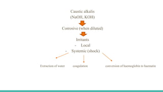
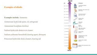
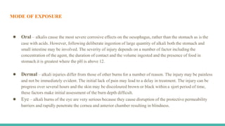
3. INTRODUCTION
Alkali toxicity, though rare, can lead to significant medical problems. In developed nations, alkaline ingestions are
more predominant when compared to acidic ingestions.
Worldwide caustic ingestions occurring amongst children are usually accidental and therefore benign given the
small amounts ingested. In contrast, adult caustic ingestions are usually intentional, involving larger amounts, and
have more significant sequelae. Patients with intentional ingestions require more involved medical management and
long term treatment.
4. CAUSTICS
caustics / corossives are the substances which fixes, damage, erode the surface with which it comes in contact.
They destroyes the body tissues with which they come in contact.
They are -
● Acids
● Alkalis
● Others
5. Caustic alkalis
(NaOH, KOH)
Corrosive (when diluted)
Irritants
- Local
- Systemic (shock)
Extraction of water coagulation conversion of haemoglobin to haematin
6. ALKALI
Alkalis are the substances that neutralise the action of acids
Alkalis in concentrated form act as corrosive poision
In dilute form they act as irritant poison
Most common cause of toxicity than acids.
It can be accidental, homicidal, suicidal
9. EPIDEMIOLOGY
● In developed countries, there is less morbidity and mortality related to caustic ingestion secondary to better
product regulations and education about alkali products. Exposure to caustics is still a problem in
underdeveloped nations.
● Caustic ingestions are either intentional usually amongst adolescents and adults with suicidal ideation,
unintentional mostly toddlers and children that gain access to the substance and are curious about it, or
incidental often associated with occupational or industrial exposures.
● The majority of reported exposures are unintentional; however, the majority of serious injuries occur after
intentional ingestions.
10. MODE OF EXPOSURE
● Oral – alkalis cause the most severe corrosive effects on the oesophagus, rather than the stomach as is the
case with acids. However, following deliberate ingestion of large quantity of alkali both the stomach and
small intestine may be involved. The severity of injury depends on a number of factor including the
concentration of the agent, the duration of contact and the volume ingested and the presence of food in
stomach.it is greatest where the pH is above 12.
● Dermal – alkali injuries differ from those of other burns for a number of reason. The injury may be painless
and not be immediately evident. The initial lack of pain may lead to a delay in treatment. The injury can be
progress over several hours and the skin may be discoloured brown or black within a sjort period of time,
these factors make initial assessment of the burn depth difficult.
● Eye – alkali burns of the eye are very serious because they cause disruption of the protective permeability
barriers and rapidly penetrate the cornea and anterior chamber resulting in blindness.
11. CAUSES
● Accidental poisoning occurs by mistaking an alkali solution for water, lemonade, beet etc.. because of careless
storage of these chemicals in inadequately labelled, ordinary – looking bottles or jar.
● Industrial accidents involving these substances are reported from time to time.
● Suicidal cases are occasionally encountered. Homicides are quite rare.
● Ammonia may sometimes be spayed or thrown on a victim to facilitate robbery.
12. SYMPTOMS
● Corrosion of GI mucosa with greyish pseudo membrane formation. Oesophagus is severity affected resulting
in dysphagia, vomiting, drooling, and haematemesis.
● Abdominal pain, diarrhoea.
● When strong alkalis is ingested, abrosins, blisters, and brownish discolouration are seen on lips and the skin
over the mouth.
● Haemorrhage intotissue is also seen.
● Skin involvement results in greyish, soapy necrotic area without charring.
● Eye involvement can produce severe complications of opthalmological emergency.
● Inhalation of Ammonia respiratory symptoms can be seen.
13. ● Oesophageal stricture formation is a major long term complication
● Contact with skin causes greyish, soapy, necrotic area.
● vomit matters are alkaline and do not effervesce on contact with ground
● It is at first thick and slimy but later contains of altered blood and shreds of mucosa.
● The motion consist of mucus and blood
14. MODE OF ACTION
● Alkali produces liquefaction necrosis which results in extensive penetrating damage because of
saponification of fat and solubilisation of protein.
● Production of ulcers is coomon which may persist for several weeks.
● Oesophagus is more severely affected than the stomach which is in contrast to acids.
15. ● Further injury is caused by thrombosis of the blood vessels.
● Alkalis most severely affects the squamous epithelium of the oesophagus but the stomach only 20% of cases.
- liquid alkalis multiple long strictures
- solid alkalis short dense, often localised at the levels of the carina or the aortic arch, an
anatomically narrow part of the oesophagus where impaction of solid occurs.
16. Alkalis produce liquefaction necrosis
Extensive penetrating damage because of saponification of fats and solubilisation of protein
Production of ulcers persist for several weeks
Oesophagus is more severely affected
17. COMPLICATIONS
Alkali ingestions may lead to stricture formation. Esophageal strictures can cause odynophagia and
dysphagia with subsequent malnutrition. Squamous cell carcinoma of the esophagus is a complication
of grade 3 esophageal caustic injuries.
Cancer can present decades after the initial exposure. Hence total removal of the esophagus is
recommended if reconstructive surgery for strictures is necessary.
18. DIAGNOSIS
1. Test the pH of the saliva. Neutral pH does NOT mean caustic ingestion did not occur.
2. Labs -CBC -ABG –Urine
3. X ray neck- esophageal perforation
4. Lesion in the gastric antrum (arrows) demonstrated by x-ray Scintigraphy - Note retention in area of the lesion on
both 1-hr and 2- hr images. Uptake in fundus of stomach is also persistent although no pathology existed in this area.
5. Stomach contents- white, solid, slimy lumps, freaks or granules litmus paper blue. If exposed to air, becomes
moist and gets dissolved.
19. 6. Platinum wire flame test – touch platinum wire to the unknown substances and then place it in a flame. Sodium
gives an intense persistent yellow flame, potassium gives a deep purple flame.
7. Endoscopy
8. CT
9. Fume test for ammonia
Place an open bottle of concentrate HCl near a sample of stomach contents, aspirate or vomitus. Copious white
fumes of ammonium chloride will emanate if ammonia is presence of ammonia in the atmosphere.
23. NON PHARMACOLOGICAL
● Diluents like milk or water may be given as a first aid measure for alkali ingestion.
● Assess fluid and electrolyte balance
● IV fluids
24. MANAGEMENT
▪ Respiratory distress may require endotracheal intubation, cricothyrotomy, or tracheostomy
depending on severity. Oxygen must be administered as necessary.
▪ Neutralisation with acidic solutions is contraindicated since the resultant exothermic reaction
can enhance the risk of perforation. However, if the action can be taken within 5 minutes of
ingestion, neutralisation may help.
▪ The followings are contraindicated – emesis ( leads to new exposure and risk of aspiration),
gastric lavage ( risk of perforation), catharsis, activated charcoal (obscures endoscopic view).
▪ Prophylactic AB’s
▪ H2 blockers
▪ Sucralfate – 1g/6 hrs
▪ Corticosteroids
▪ proton pump inhibitor
25. Skin contact -
Wash with running water until the skin no longer feels soapy. Relieve pain and treat shock.
Eye contact -
Anesthetize the conjunctival and corneal surfaces with topical anesthetic (eg, proparacaine). Irrigate with water or
saline continuously for 20–30 minutes, holding the lids open. Amphoteric solutions may be more effective than
water or saline and some are available in Europe (Diphoterine, Prevor). Check pH with pH test paper and repeat
irrigation for additional 30-minute periods until the pH is near 7.0. Check for corneal damage with fluorescein and
slit-lamp examination; consult an ophthalmologist for further treatment.
26. If circumferential 2nd degree or 3rd degree burns of the oesophagus are seen. Exploratory laparotomy should be
undertaken followed by gastric resection and esophagectomy. In case gastric necrosis is evident at
laparoscopy.
Patients who have suffered from stricture formation require long term endoscopy follow up for the presence of
neoplastic changes of the oesophagus which may occur with a delay of several years or decades.