2. 12/7/23, 20:52 Causes of upper GI bleeding - UpToDate
https://www.uptodate.com/contents/image/print?imageKey=GAST%2F103148&source=graphics_gallery&topicKey=2644&search=sangrado gastrointestinal 2/11
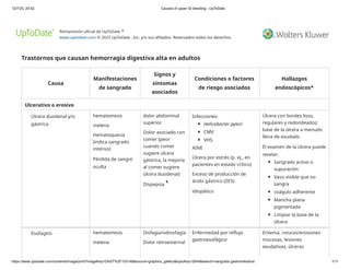
Pérdida de sangre
oculta
impactación de
alimentos
Medicamentos que pueden
causar "esofagitis por
píldoras":
Eritromicina
tetraciclina
doxiciclina
clindamicina
Trimetoprim-
sulfametoxazol
AINE
Bisfosfonatos orales
Cloruro de potasio
quinidina
suplementos de hierro
Infecciones:
VHS
CMV
Candida albicans
VIH
superficiales o profundas,
estenosis, cicatrización
Esofagitis péptica:
Las ulceraciones suelen
ser de forma irregular o
lineales, múltiples y
distales; puede
acompañarse de esófago
de Barrett
Inducido por píldoras:
Las ulceraciones suelen
ser únicas y profundas, y
se producen en puntos de
estasis (especialmente
cerca de la carina), sin
afectar el esófago distal.
Esofagitis infecciosa:
HSV – Úlceras
superficiales, discretas,
con bordes bien
delimitados que
tienden a involucrar el
esófago superior o
medio; se pueden ver
vesículas
CMV: las úlceras varían
de pequeñas y
superficiales a grandes
(>1 cm) y profundas; la
mayoría de los
pacientes tienen
múltiples lesiones
3. 12/7/23, 20:52 Causes of upper GI bleeding - UpToDate
https://www.uptodate.com/contents/image/print?imageKey=GAST%2F103148&source=graphics_gallery&topicKey=2644&search=sangrado gastrointestinal 3/11
Candida – Placas
blancas difusas
VIH: tiende a afectar el
esófago medio o distal,
las úlceras pueden ser
superficiales o
profundas y pueden ser
grandes
Gastritis/gastropatía
Duodenitis/duodenopatía
Pérdida de sangre
oculta
hematemesis
melena
Dispepsia Factores de riesgo:
H. pylori
AINE
Consumo excesivo de
alcohol
lesión por radiación
Estrés fisiológico
Cirugía de pérdida de
peso
reflujo biliar
Factores de riesgo de
sangrado:
Uso de anticoagulantes
Erythematous mucosa
Superficial erosions
Nodularity
Diffuse oozing
Complications of portal hypertension
Esophagogastric varices Hematemesis
Melena
Hematochezia
(indicates brisk
bleeding)
Stigmata of chronic
liver disease , in
particular, signs of
portal
hypertension
(splenomegaly,
ascites,
thrombocytopenia)
Portal hypertension from:
Cirrhosis
Portal vein thrombosis
Noncirrhotic portal
hypertension
Vascular structures that
protrude into the
esophageal and/or gastric
lumen
Findings associated with an
increased risk of
hemorrhage:
Longitudinal red
streaks on the varices
Δ
4. 12/7/23, 20:52 Causes of upper GI bleeding - UpToDate
https://www.uptodate.com/contents/image/print?imageKey=GAST%2F103148&source=graphics_gallery&topicKey=2644&search=sangrado gastrointestinal 4/11
(red wale marks)
Cherry-colored spots
that are flat and overlie
varices
Raised, discrete red
spots (hematocystic
spots)
Esophageal varices:
F1: Small, straight
varices
F2: Enlarged, tortuous
varices that occupy less
than one-third of the
lumen
F3: Large, coil-shaped
varices that occupy
more than one-third of
the lumen
Gastric varices:
GOV1:
Gastroesophageal
varices along the lesser
curvature of the
stomach
GOV2:
Gastroesophageal
varices along the
greater curvature of
the stomach
IGV1: Isolated gastric
varices in the fundus
IGV2: Isolated gastric
varices at other loci in
5. 12/7/23, 20:52 Causes of upper GI bleeding - UpToDate
https://www.uptodate.com/contents/image/print?imageKey=GAST%2F103148&source=graphics_gallery&topicKey=2644&search=sangrado gastrointestinal 5/11
the stomach
Ectopic varices Hematemesis
Melena
Hematochezia
(indicates brisk
bleeding)
Stigmata of chronic
liver disease , in
particular, signs of
portal
hypertension
(splenomegaly,
ascites,
thrombocytopenia)
Portal hypertension from:
Cirrhosis
Portal vein thrombosis
Noncirrhotic portal
hypertension
Vascular structures that
protrude into areas of the
gastrointestinal tract lumen
other than the esophagus or
stomach (eg, small bowel,
rectum)
Portal hypertensive
gastropathy
Occult blood loss
Hematemesis
Melena
Hematochezia
(indicates brisk
bleeding)
Stigmata of chronic
liver disease , in
particular, signs of
portal
hypertension
(splenomegaly,
ascites,
thrombocytopenia)
Portal hypertension from:
Cirrhosis
Portal vein thrombosis
Noncirrhotic portal
hypertension
Mosaic-like pattern that
gives the gastric mucosa a
"snakeskin" appearance
Mucosal changes are usually
most evident in the fundus
and body; in more severe
cases, oozing, bleeding,
subepithelial hemorrhages,
and increased vascularity
similar to angiomas are
evident, often involving the
gastric fundus, gastric body,
and antrum
Vascular lesions
Angiodysplasia Hematemesis
Melena
Hematochezia
Occult blood loss
May have brisk
bleeding
Cutaneous
angiodysplasia in
patients with
hereditary
hemorrhagic
telangiectasia
(Osler-Weber-
Rendu syndrome)
End-stage kidney disease
Aortic stenosis
Left ventricular assist device
Hereditary hemorrhagic
telangiectasia
von Willebrand disease
Radiation therapy
Small (5 to 10 mm), flat,
cherry-red lesions, often with
a fern-like pattern of
arborizing, ectatic blood
vessels radiating from a
central vessel
Δ
Δ
6. 12/7/23, 20:52 Causes of upper GI bleeding - UpToDate
https://www.uptodate.com/contents/image/print?imageKey=GAST%2F103148&source=graphics_gallery&topicKey=2644&search=sangrado gastrointestinal 6/11
Idiopathic
Dieulafoy's lesion Hematemesis
Melena
Hematochezia
(indicates brisk
bleeding; bleeding
is often particularly
brisk)
Etiology unknown
Bleeding may be associated
with NSAIDs, cardiovascular
disease, hypertension,
chronic kidney disease,
diabetes, or alcohol abuse
Usually located in the
proximal stomach (within 6
cm of the esophagogastric
junction) along the lesser
curvature (although can be
found anywhere in the GI
tract)
May have active arterial
spurting from the mucosa
without an associated ulcer
or mass
If the bleeding has stopped,
there may be a raised nipple
or visible vessel without an
associated ulcer
Endoscopic ultrasound may
help confirm the diagnosis
Gastric antral vascular
ectasia (GAVE)
Hematemesis
Melena
Hematochezia
(indicates brisk
bleeding)
Occult blood loss
In patients with
cirrhosis, there
may be stigmata of
chronic liver
disease , in
particular, signs of
portal
hypertension
(splenomegaly,
ascites,
thrombocytopenia)
Idiopathic
Cirrhosis with portal
hypertension
Kidney
disease/transplantation
Diabetes mellitus
Systemic sclerosis
(scleroderma)
Bone marrow transplantation
Longitudinal rows of flat,
reddish stripes radiating
from the pylorus into the
antrum that resemble the
stripes on a watermelon
Blue rubber bleb nevus
syndrome (Bean
syndrome)
Hematemesis
Melena
Venous
malformations and
hemangiomas of
Blue or purple nodules,
round or multilobular; may
Δ
7. 12/7/23, 20:52 Causes of upper GI bleeding - UpToDate
https://www.uptodate.com/contents/image/print?imageKey=GAST%2F103148&source=graphics_gallery&topicKey=2644&search=sangrado gastrointestinal 7/11
Hematochezia
(indicates brisk
bleeding)
Occult blood loss
any organ,
including:
Skin
Central
nervous
system
Liver
Muscles
Lymphatics
Intussusception
occur anywhere in the
gastrointestinal tract
Traumatic or iatrogenic
Mallory-Weiss syndrome Hematemesis
following an
increase in intra-
abdominal pressure
Melena
Hematochezia
(indicates brisk
bleeding)
Epigastric pain
Back pain
Vomiting/retching (often
related to alcohol
consumption)
Straining at stool or lifting
Coughing
Seizures
Blunt abdominal trauma
Hiatal hernia may increase
the risk of developing a tear
Tear in the esophagogastric
junction
Usually singular and
longitudinal, but may be
multiple
Visualization may require
retroflexion of the
gastroscope in the cardia of
the stomach
The tear may be covered by
an adherent clot
Foreign body ingestion Hematemesis
Melena
Hematochezia
(indicates brisk
bleeding)
Occult blood loss
Dysphagia
Odynophagia
Neck or abdominal
pain
Choking
Hypersalivation
Psychiatric disorders
Altered mental status (toxin
induced, dementia, etc)
Loose dentures
Visualization of the foreign
body endoscopically (plain
radiographs of the neck,
chest, and abdomen may
reveal a radiopaque foreign
body or signs of perforation)
8. 12/7/23, 20:52 Causes of upper GI bleeding - UpToDate
https://www.uptodate.com/contents/image/print?imageKey=GAST%2F103148&source=graphics_gallery&topicKey=2644&search=sangrado gastrointestinal 8/11
Retrosternal
fullness
Post-surgical
anastomotic bleeding
("marginal ulcers")
Occult blood loss
Hematemesis
Melena
Hematochezia
(indicates brisk
bleeding)
Epigastric pain
Nausea
Billroth II surgery
Gastric bypass surgery
NSAID use
H. pylori infection
Smoking
Ulceration/friable mucosa at
an anastomotic site
Post-
polypectomy/endoscopic
resection/endoscopic
sphincterotomy
Hematemesis
Melena
Hematochezia
(indicates brisk
bleeding)
Past history of
instrumentation
(may be as long as
three weeks prior
to presentation)
Large lesions Bleeding at resection site;
ulceration at the site may be
seen
Cameron lesions Occult blood loss
Hematemesis
Melena
Hematochezia
(indicates brisk
bleeding)
Hiatal hernia
Reflux esophagitis
Linear ulcers or erosions on
the mucosal folds of a hiatal
hernia at the diaphragmatic
impression
Aortoenteric fistula Hematemesis
Melena
Hematochezia
(indicates brisk
bleeding)
May have a "herald"
bleed followed by
massive bleeding
Back pain
Fever
Signs of sepsis
Pulsatile abdominal
mass
Abdominal bruit
Infectious aortitis (syphilis,
tuberculosis)
Prosthetic aortic graft
Atherosclerotic aortic
aneurysm
Penetrating ulcers
Tumor invasion
Trauma
Radiation injury
Endoscopy is important,
primarily to exclude other,
more common causes of
acute upper GI bleeding
Endoscopy with an
enteroscope or side-viewing
duodenoscope may reveal a
graft, an ulcer or erosion at
the site of an adherent clot,
or an extrinsic pulsatile mass
9. 12/7/23, 20:52 Causes of upper GI bleeding - UpToDate
https://www.uptodate.com/contents/image/print?imageKey=GAST%2F103148&source=graphics_gallery&topicKey=2644&search=sangrado gastrointestinal 9/11
Foreign body perforation in the distal duodenum or
esophagus
Tumors
Upper GI tumors Hematemesis
Melena
Hematochezia
(indicates brisk
bleeding)
Occult blood loss
Weight loss
Anorexia
Nausea/vomiting
Early satiety
Epigastric pain
Dysphagia (for
tumors in the
esophagus or
proximal stomach)
Gastric outlet
obstruction
Palpable mass
Paraneoplastic
manifestations:
Diffuse
seborrheic
keratoses
Acanthosis
nigricans
Membranous
nephropathy
Coagulopathy
Virtually any tumor type may
bleed
Benign tumors:
Leiomyoma
Lipoma
Polyp (hyperplastic,
adenomatous,
hamartomatous,
inflammatory)
Malignant tumors:
Adenocarcinoma
GI stromal tumors
Lymphoma
Kaposi sarcoma
Carcinoid
Melanoma
Metastatic tumors
Ulcerated mass in the
esophagus, stomach, or
duodenum
In gastric malignancies, the
folds surrounding the ulcer
crater may be nodular,
clubbed, fused, or stop short
of the ulcer margin; the
margins may be
overhanging, irregular, or
thickened
Bleeding lymphoma may
appear as an ulcerated mass
or polypoid lesion or as a
gastric ulcer
Miscellaneous
Hemobilia Hematemesis
Melena
Biliary colic
Jaundice
(obstructive)
Past history of liver or biliary
tract instrumentation and/or
Blood or clot emanating
from the ampulla (a side-
viewing duodenoscope may
10. 12/7/23, 20:52 Causes of upper GI bleeding - UpToDate
https://www.uptodate.com/contents/image/print?imageKey=GAST%2F103148&source=graphics_gallery&topicKey=2644&search=sangrado gastrointestinal 10/11
Hematochezia
(indicates brisk
bleeding)
Sepsis (biliary) injury, including the
following:
Liver biopsy
Cholecystectomy
Endoscopic biliary
biopsies or stenting
TIPS placement
Angioembolization
Blunt or penetrating
abdominal trauma
Gallstones
Cholecystitis
Hepatic or bile duct
tumors
Intrahepatic stents
Hepatic artery
aneurysms
Hepatic abscesses
be required to visualize the
ampulla)
If a clot has formed in the
bile duct, bleeding may not
be appreciated until the clot
is removed
ERCP may reveal a filling
defect in the bile duct
Hemosuccus
pancreaticus
Hematemesis
Melena
Hematochezia
(indicates brisk
bleeding)
Abdominal pain
Past evidence of
symptoms/signs of
pancreatitis
Imaging evidence
of pancreatitis
(current or in the
past)
Elevated amylase
and lipase (current
or in the past)
Chronic pancreatitis
Pancreatic pseudocysts
Pancreatic tumors
Pancreatic pseudoaneurysm
Therapeutic endoscopy of
the pancreas or pancreatic
duct:
Pancreatic stone
removal
Pancreatic duct
sphincterotomy
Pseudocyst drainage
Pancreatic duct stenting
Blood or clot emanating
from the ampulla (a side-
viewing duodenoscope may
be required to visualize the
ampulla)
Cross-sectional imaging or
angiography is often
required to confirm the
diagnosis
11. 12/7/23, 20:52 Causes of upper GI bleeding - UpToDate
https://www.uptodate.com/contents/image/print?imageKey=GAST%2F103148&source=graphics_gallery&topicKey=2644&search=sangrado gastrointestinal 11/11
CMV: citomegalovirus; HSV: virus del herpes simple; ZES: síndrome de Zollinger-Ellison; AINE: fármaco antiinflamatorio no esteroideo;
VIH: virus de la inmunodeficiencia humana; GI: gastrointestinal; TIPS: derivación portosistémica intrahepática transyugular; CPRE:
colangiopancreatografía retrógrada endoscópica.
* Si hay sangrado activo o grandes cantidades de sangre residual, los hallazgos endoscópicos característicos pueden oscurecerse.
¶ Plenitud posprandial, saciedad temprana, dolor epigástrico o ardor.
Δ La evidencia de enfermedad hepática crónica incluye ictericia, esplenomegalia, ascitis, trombocitopenia, eritema palmar, angioma en
araña, ginecomastia, atrofia testicular y contractura de Dupuytren.
Gráfico 103148 Versión 6.0