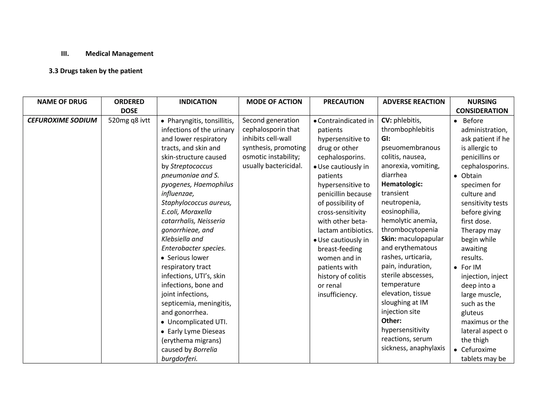
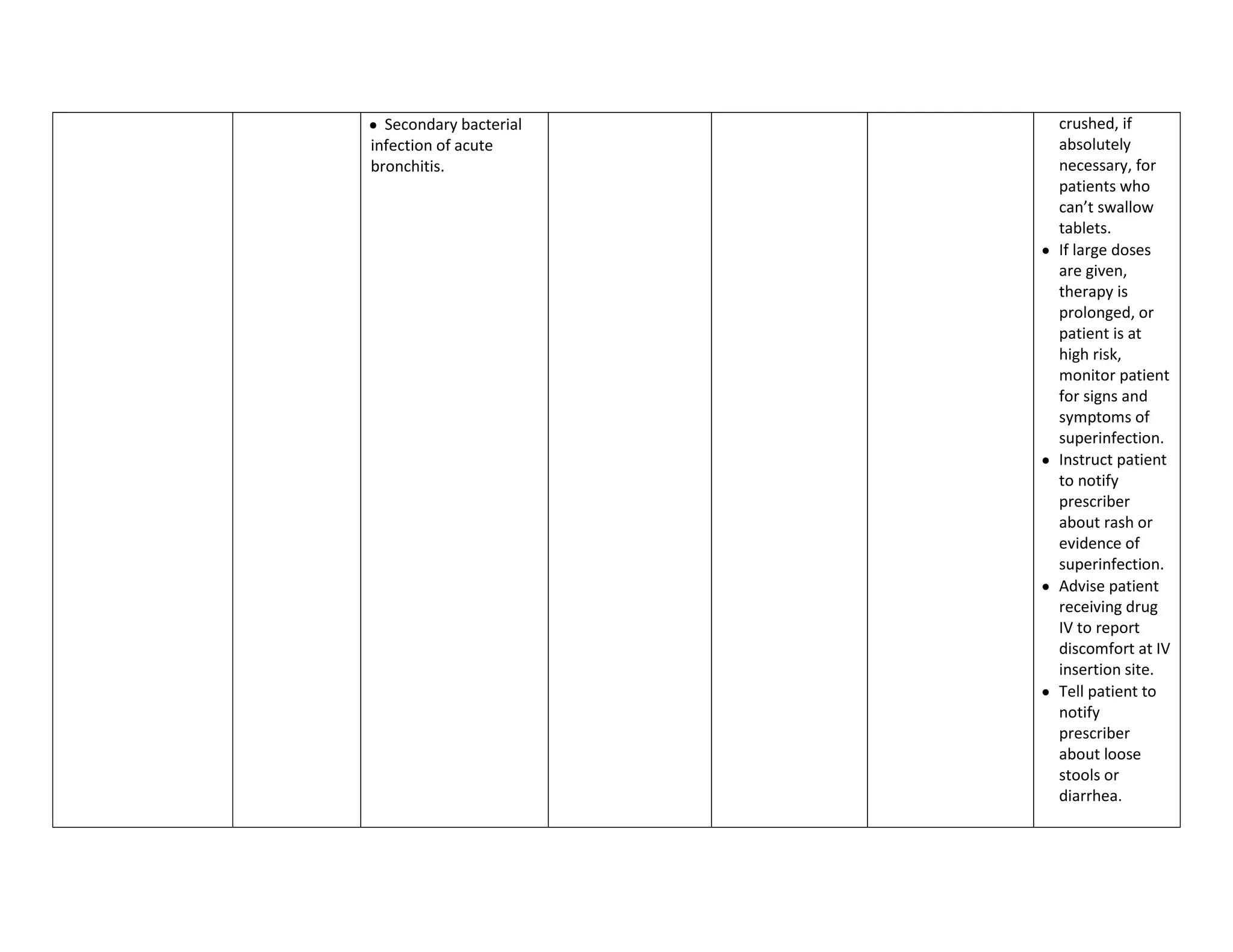
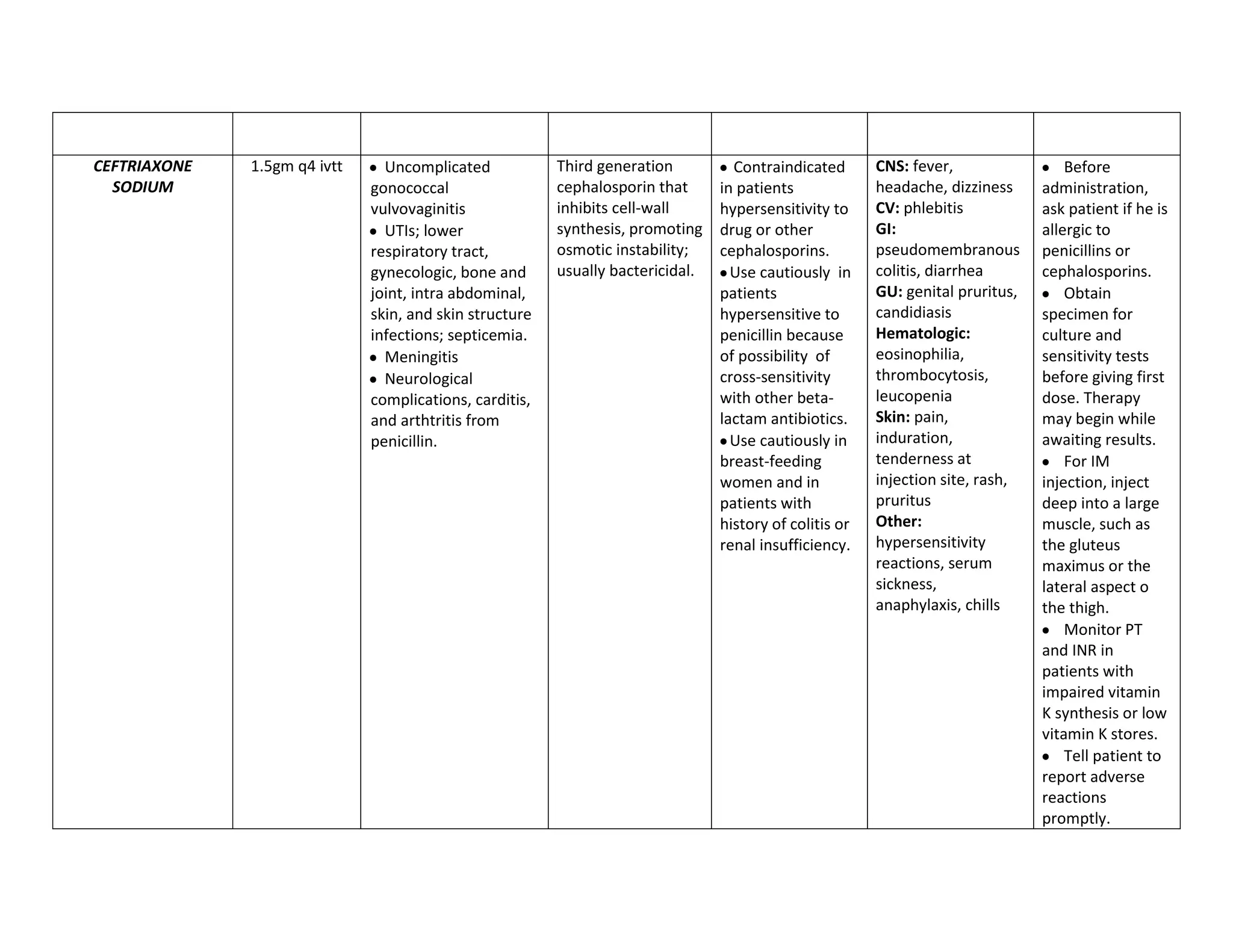
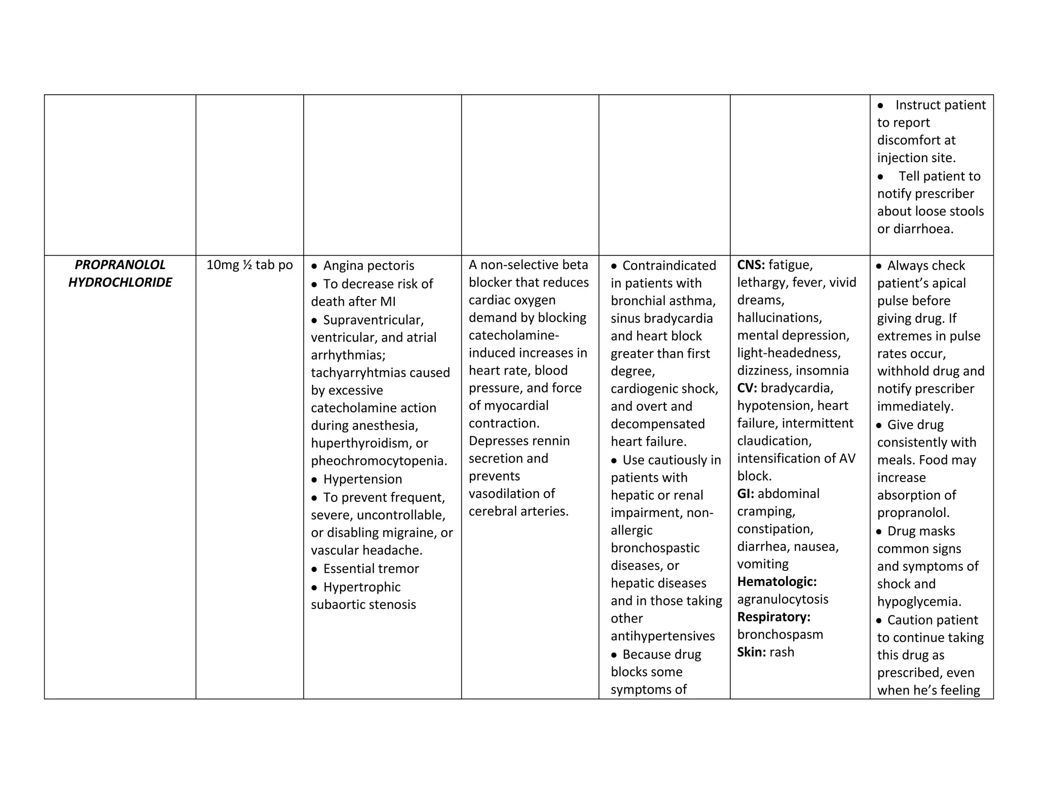
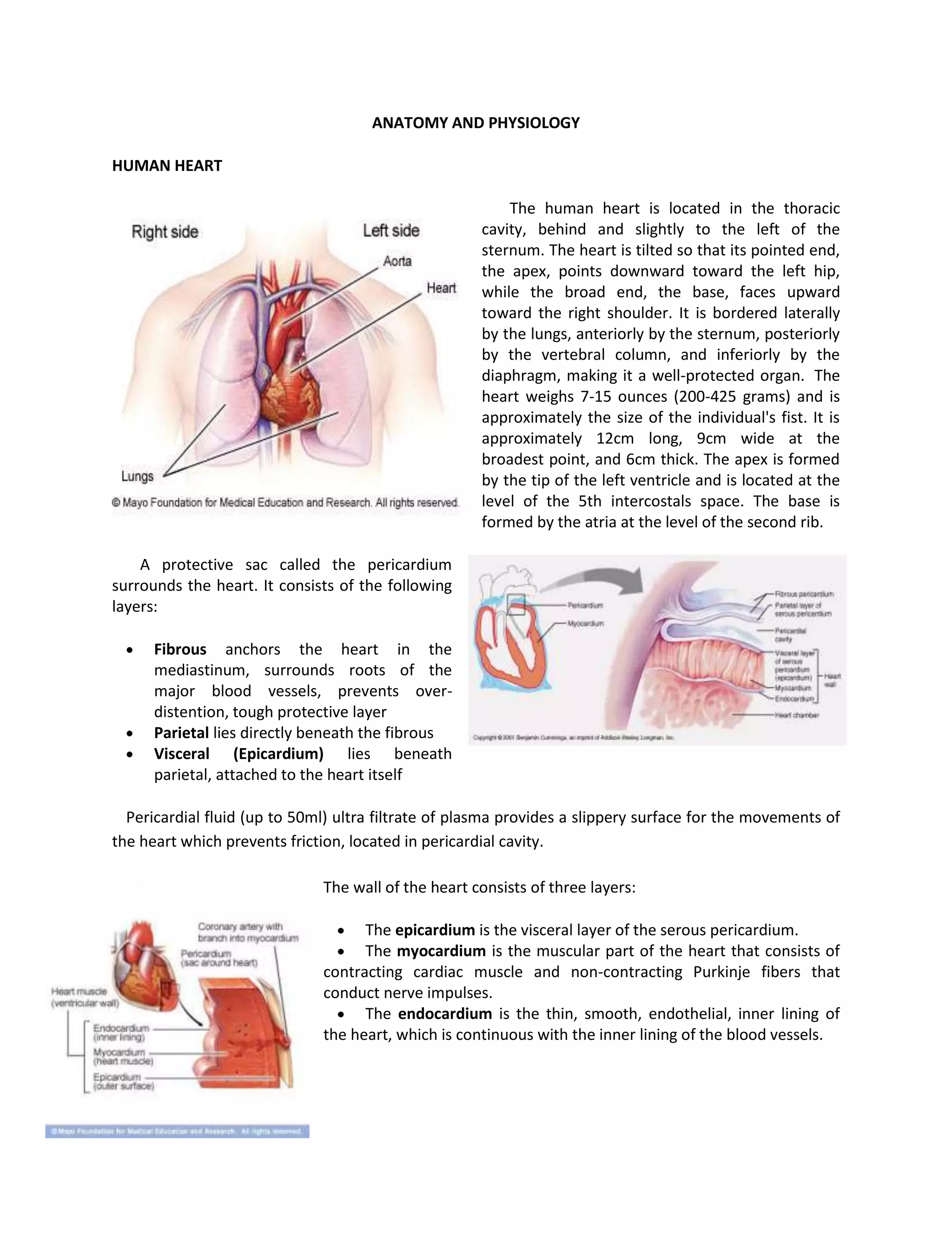
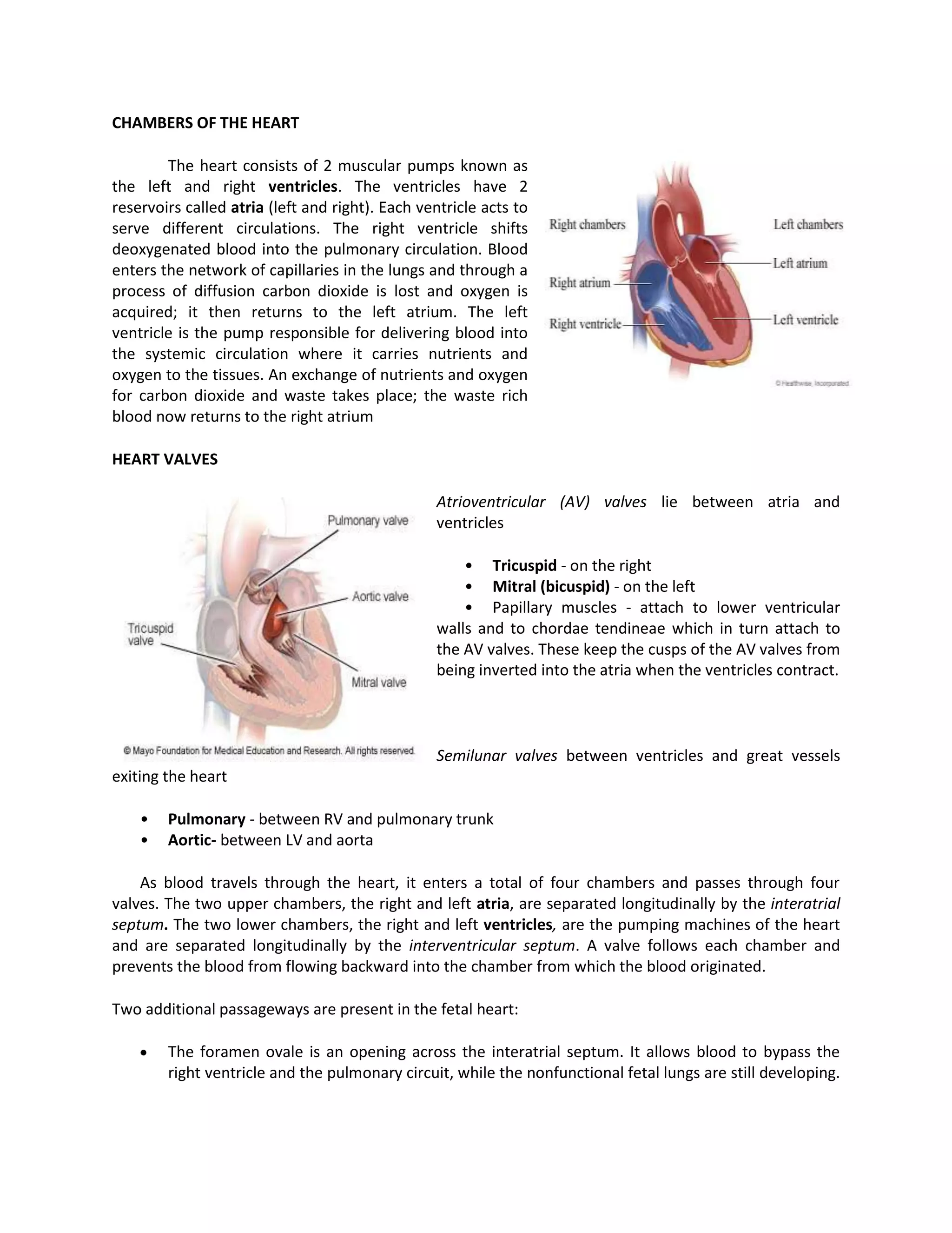
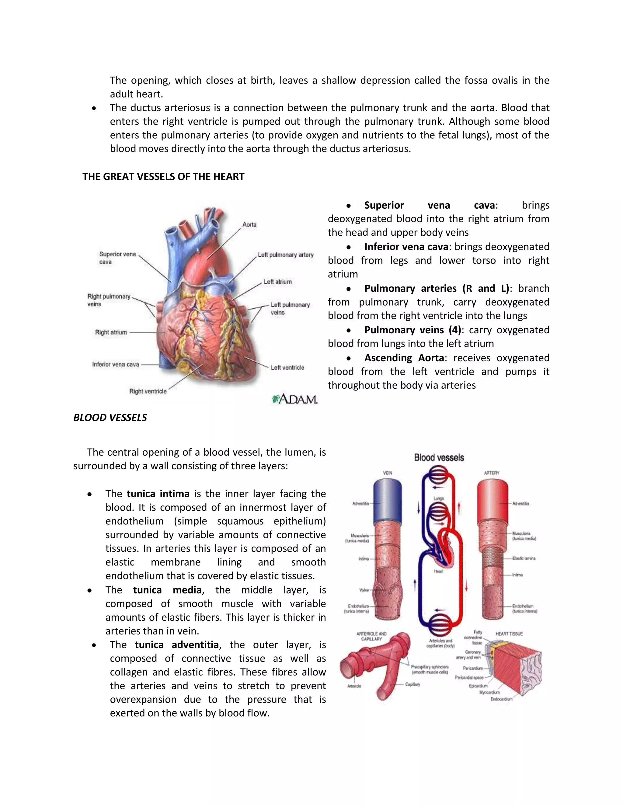
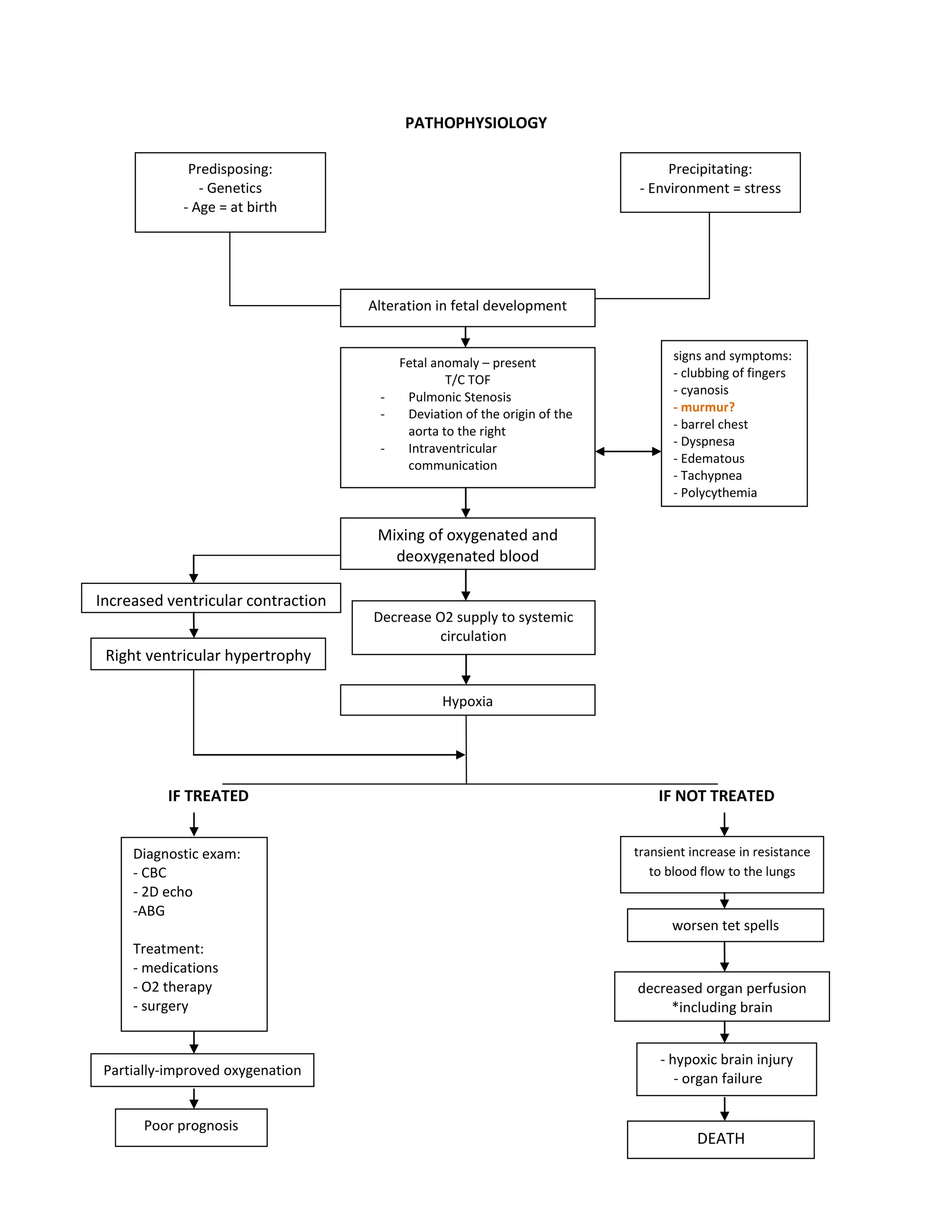
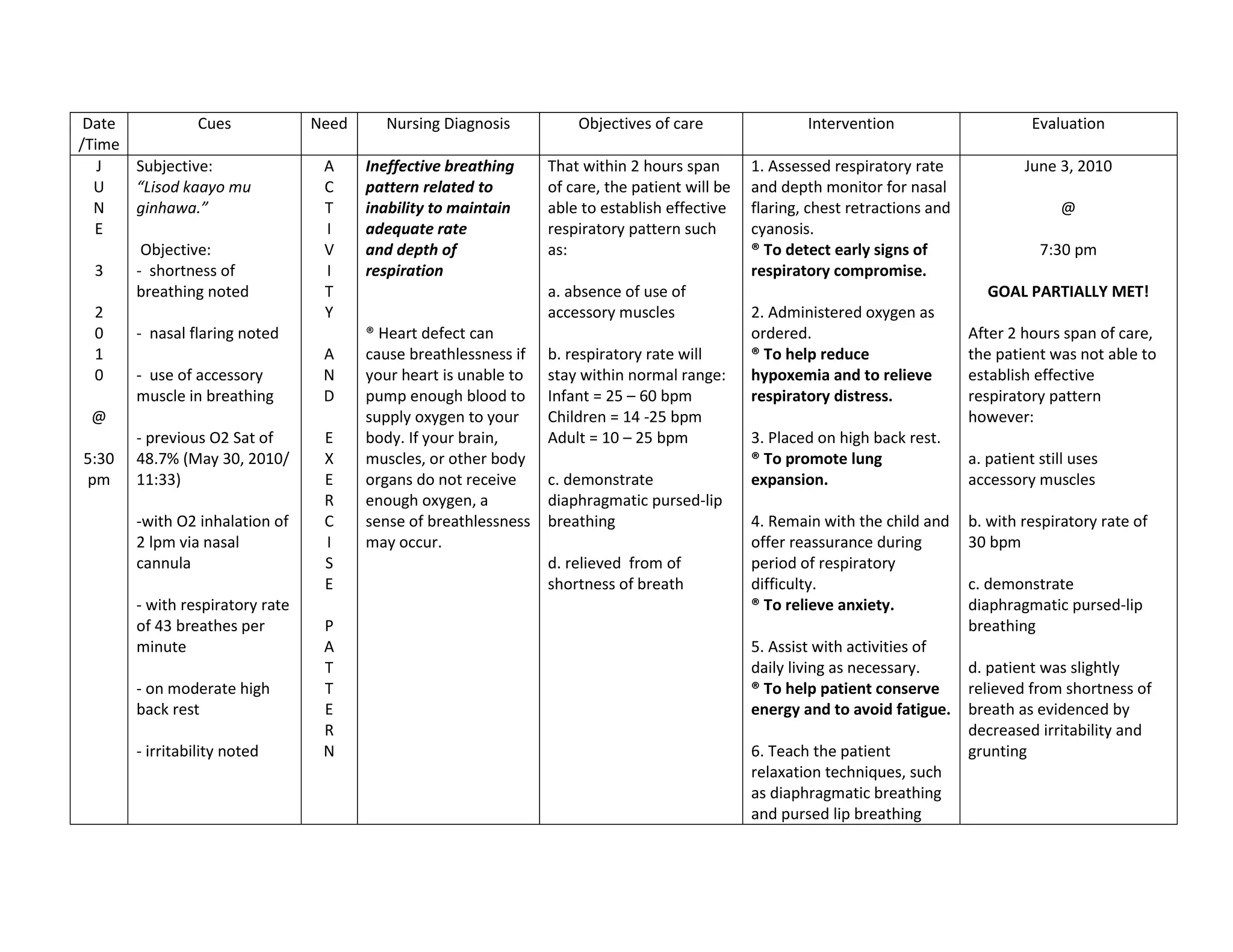
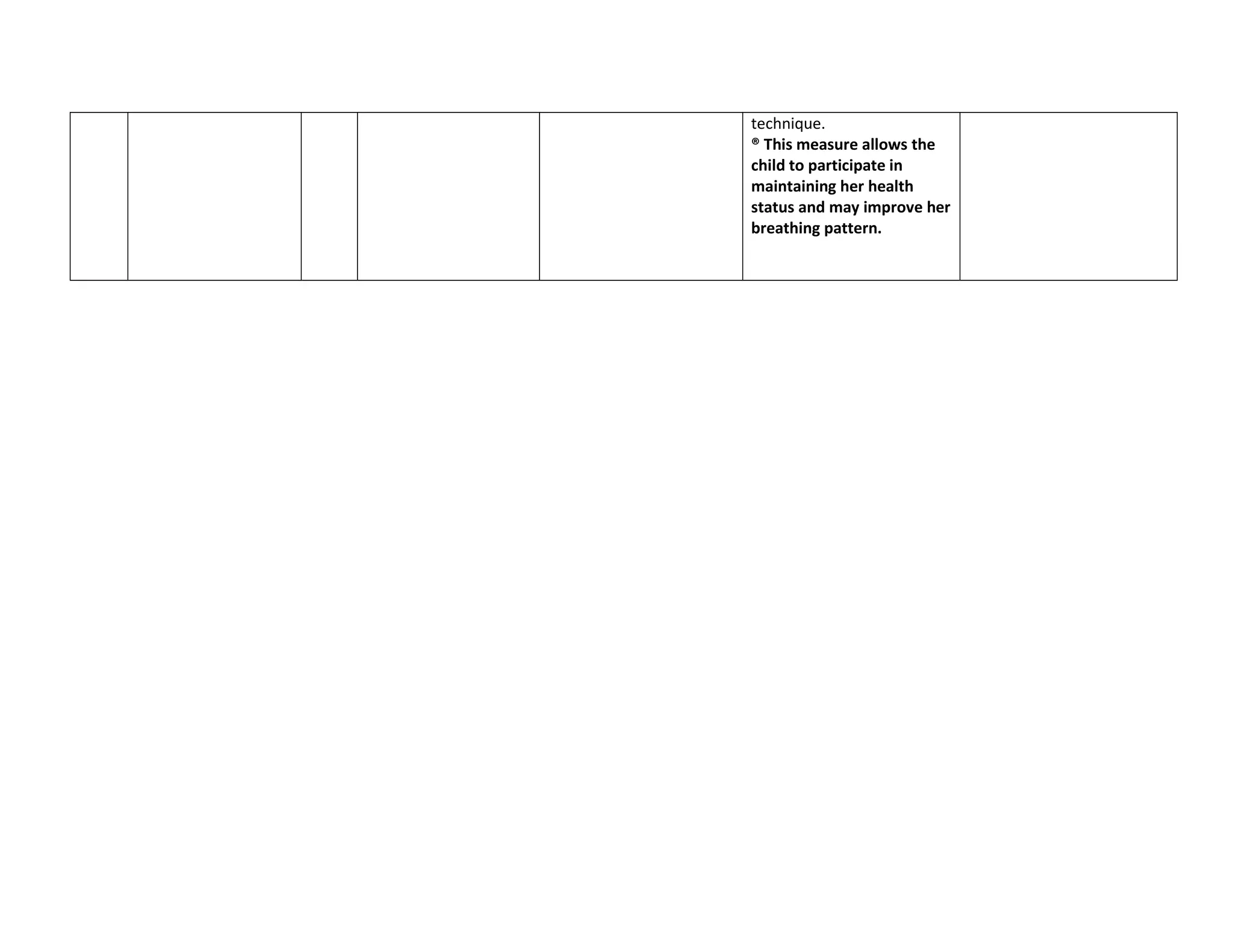
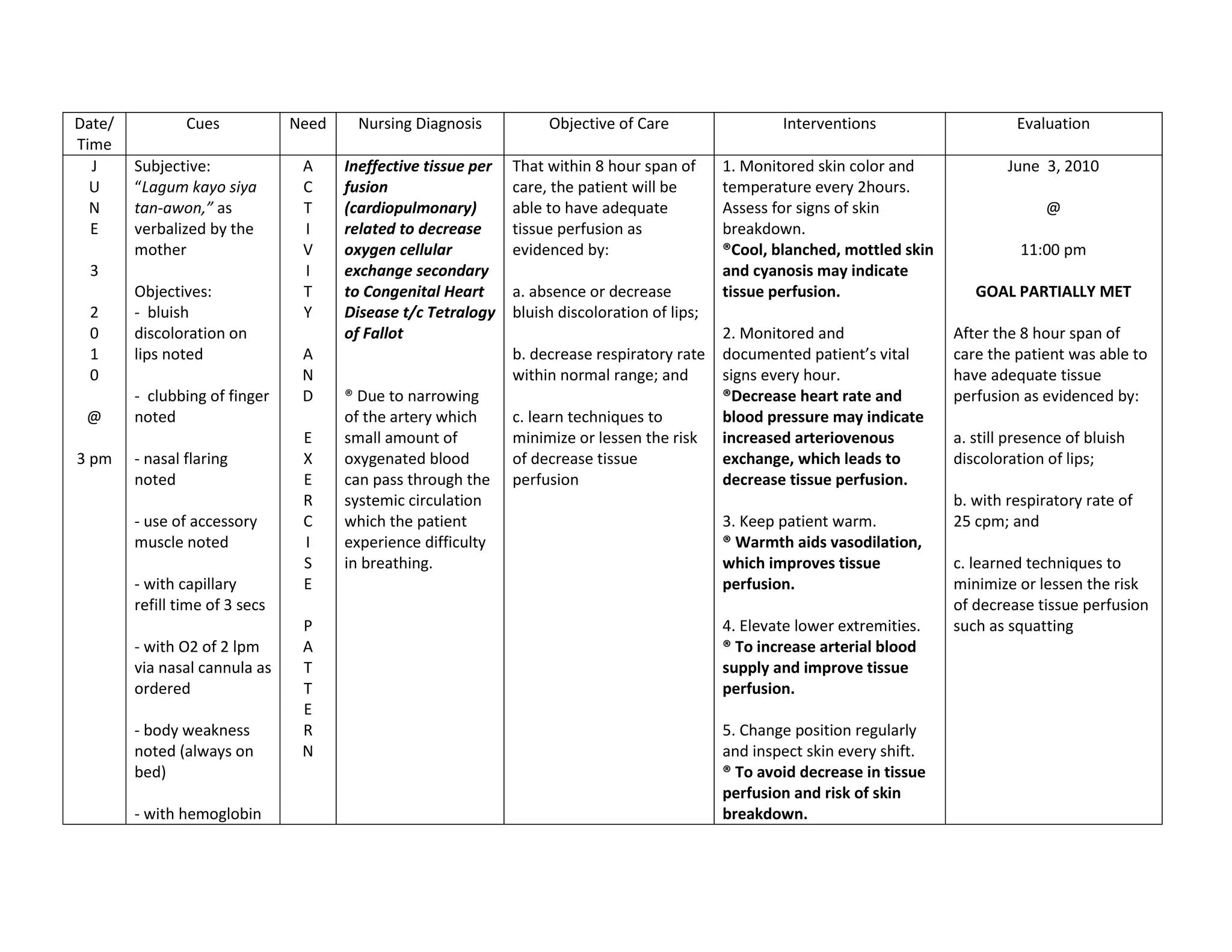
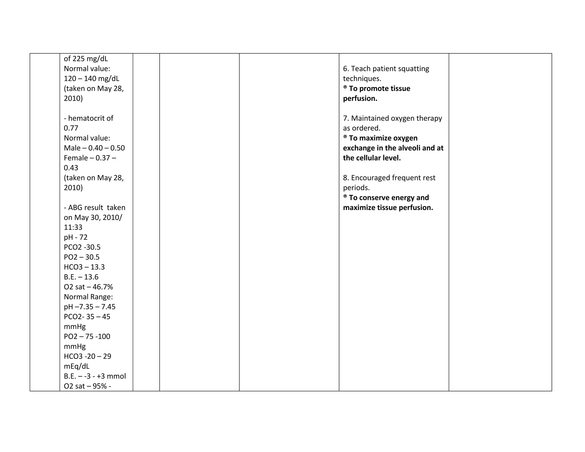
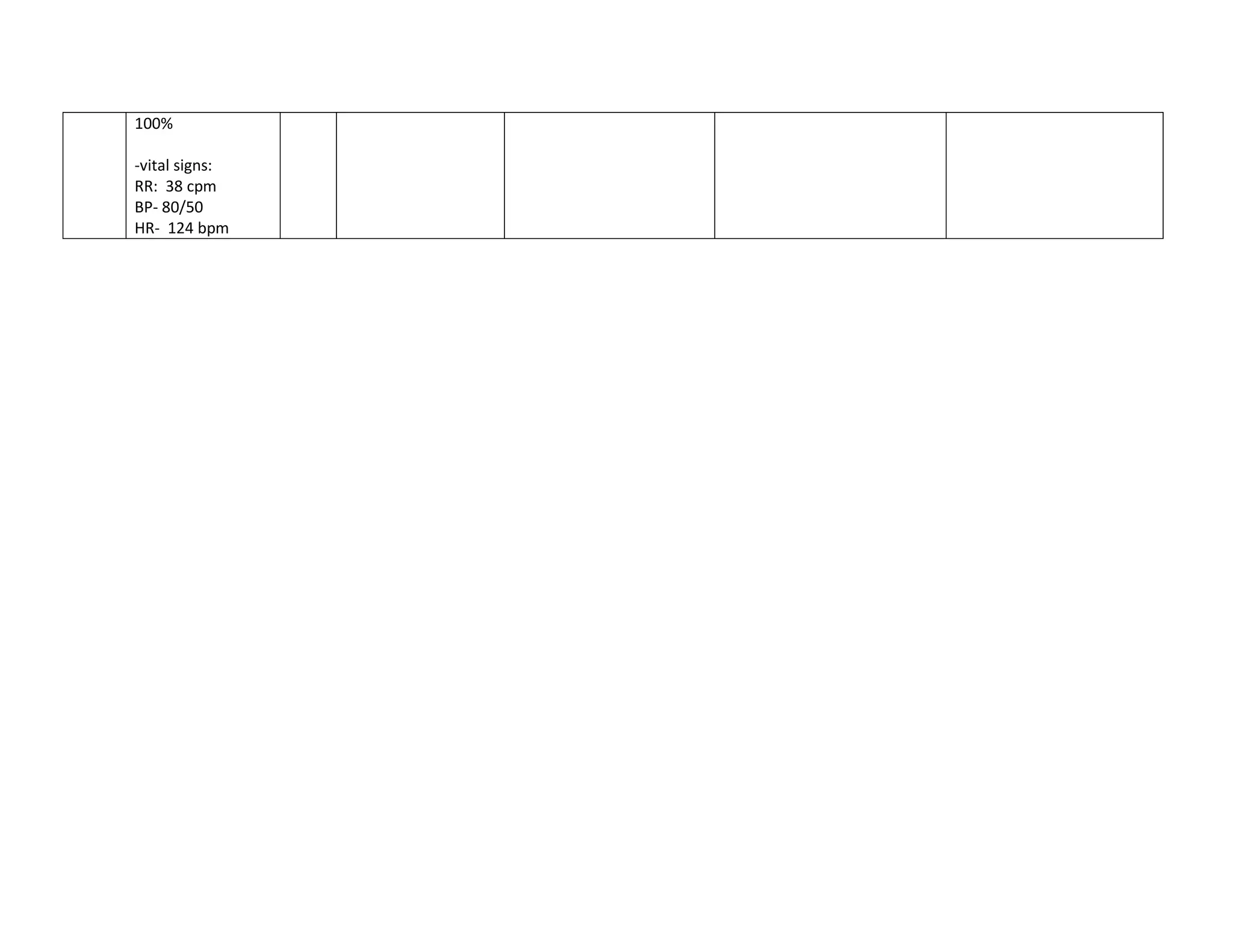
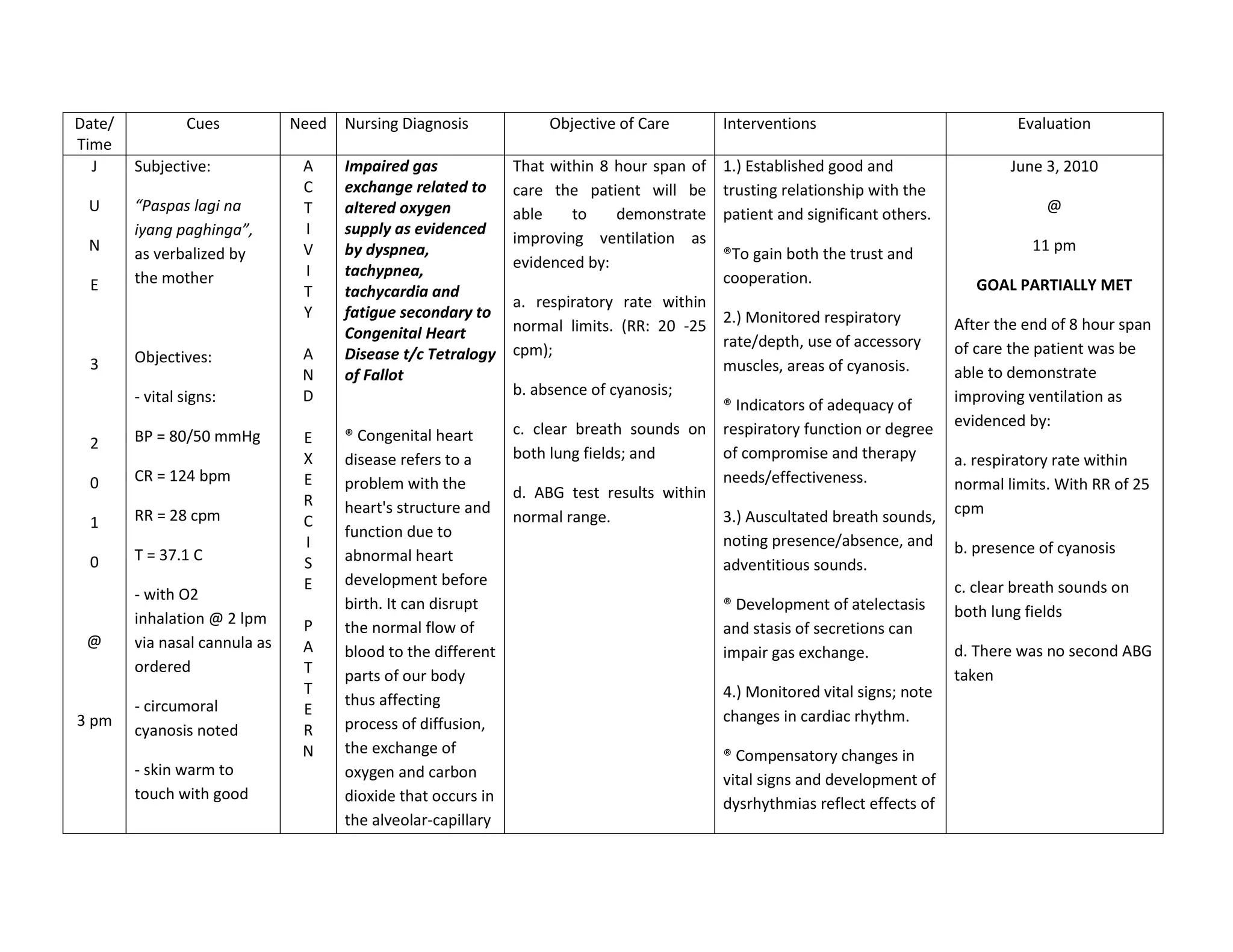
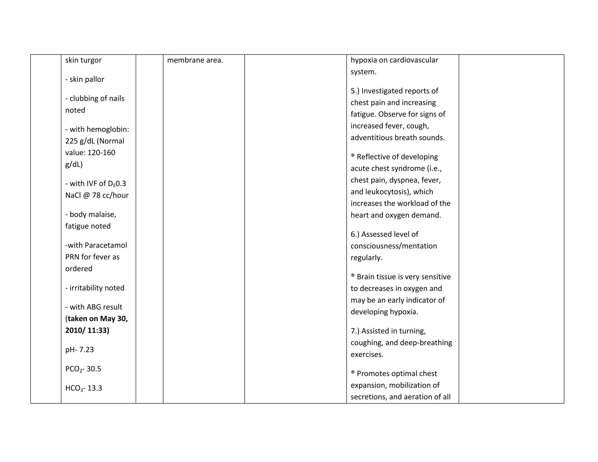
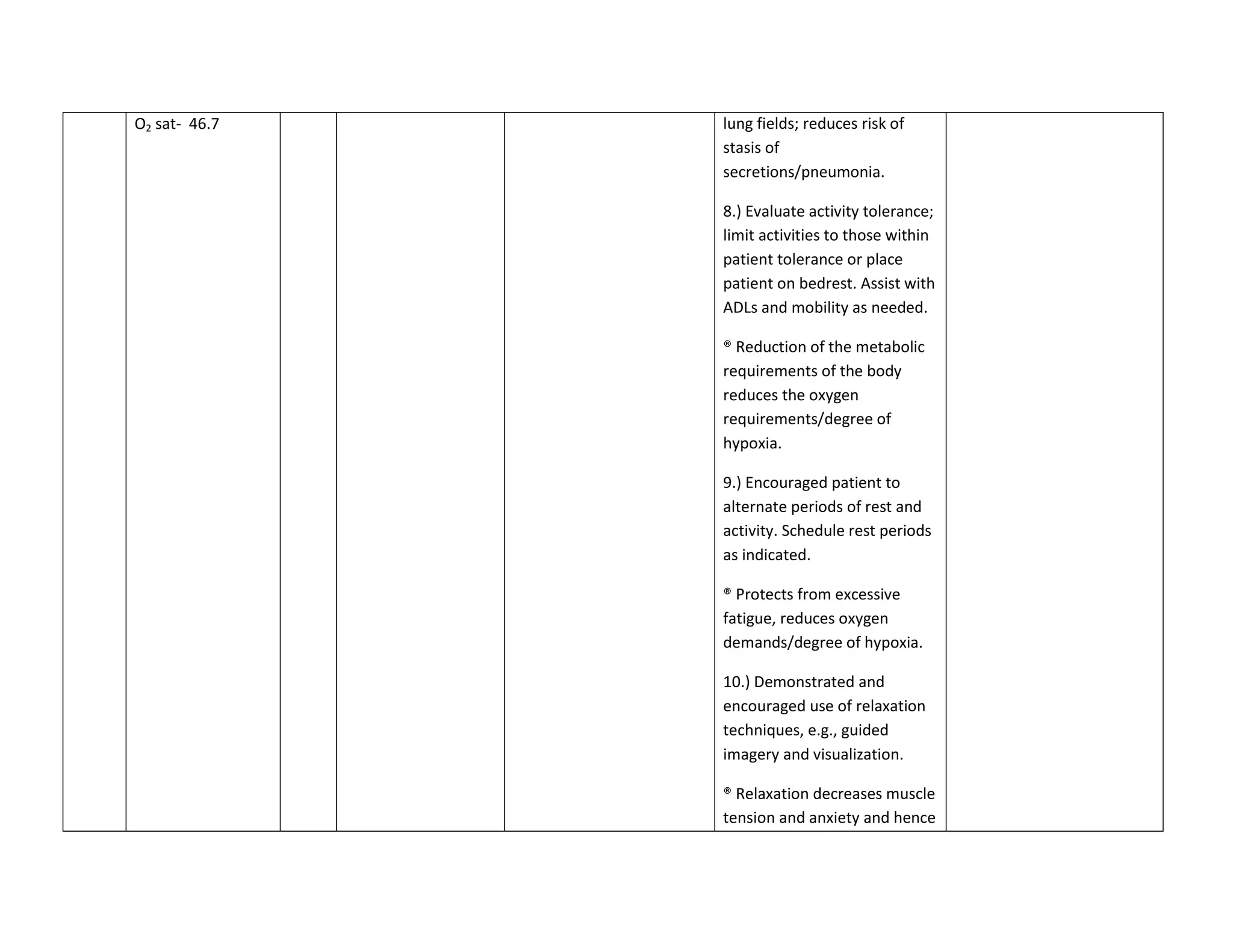
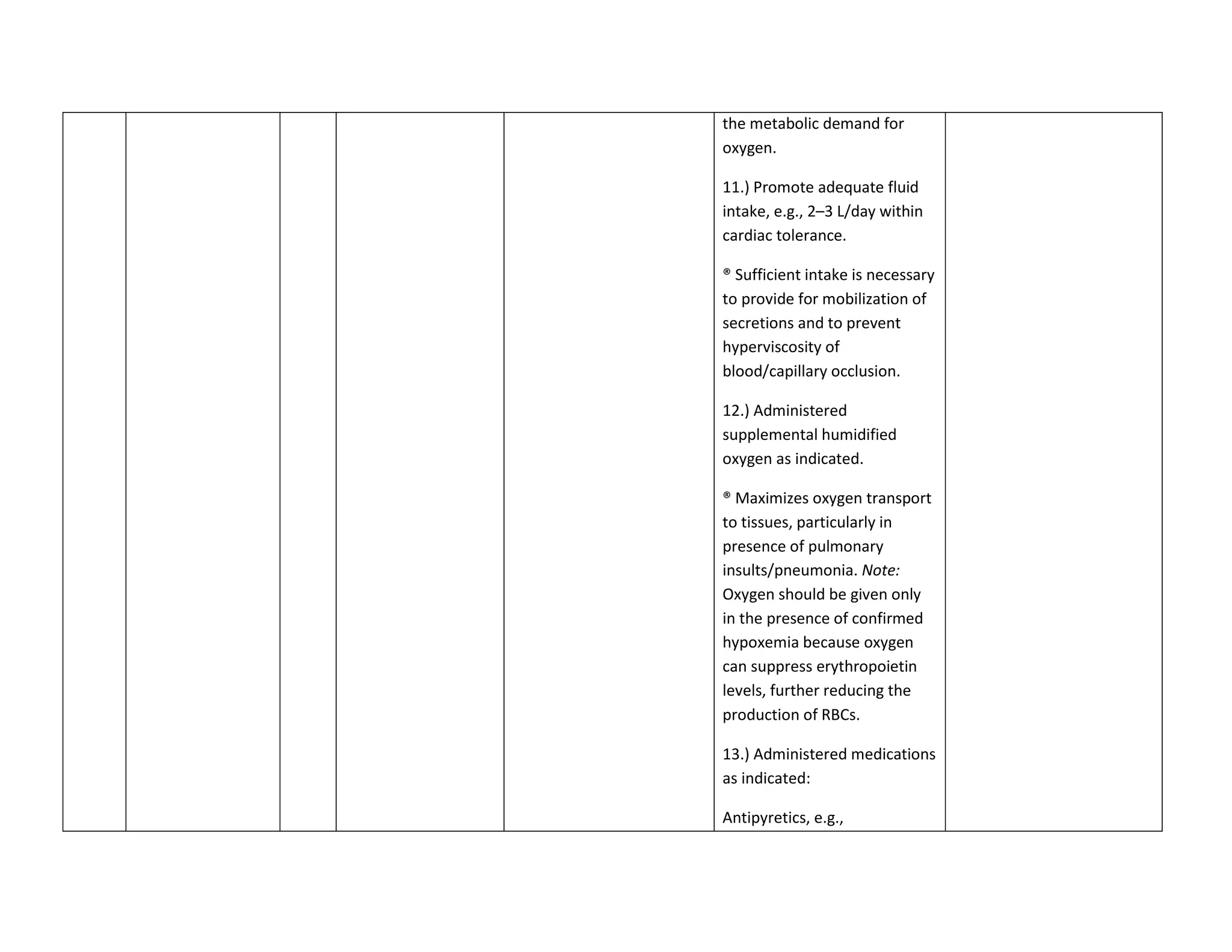
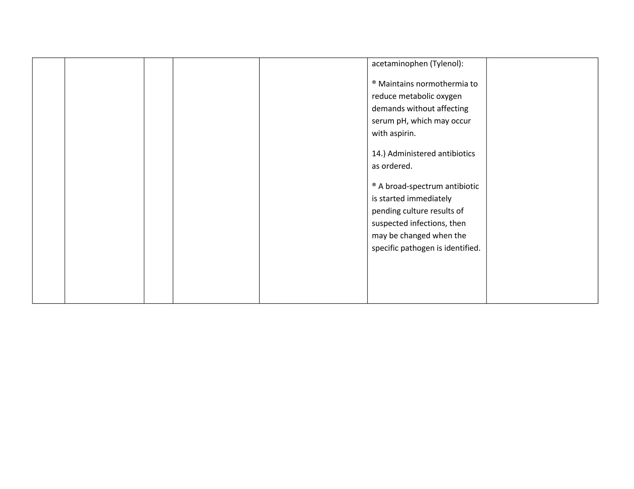
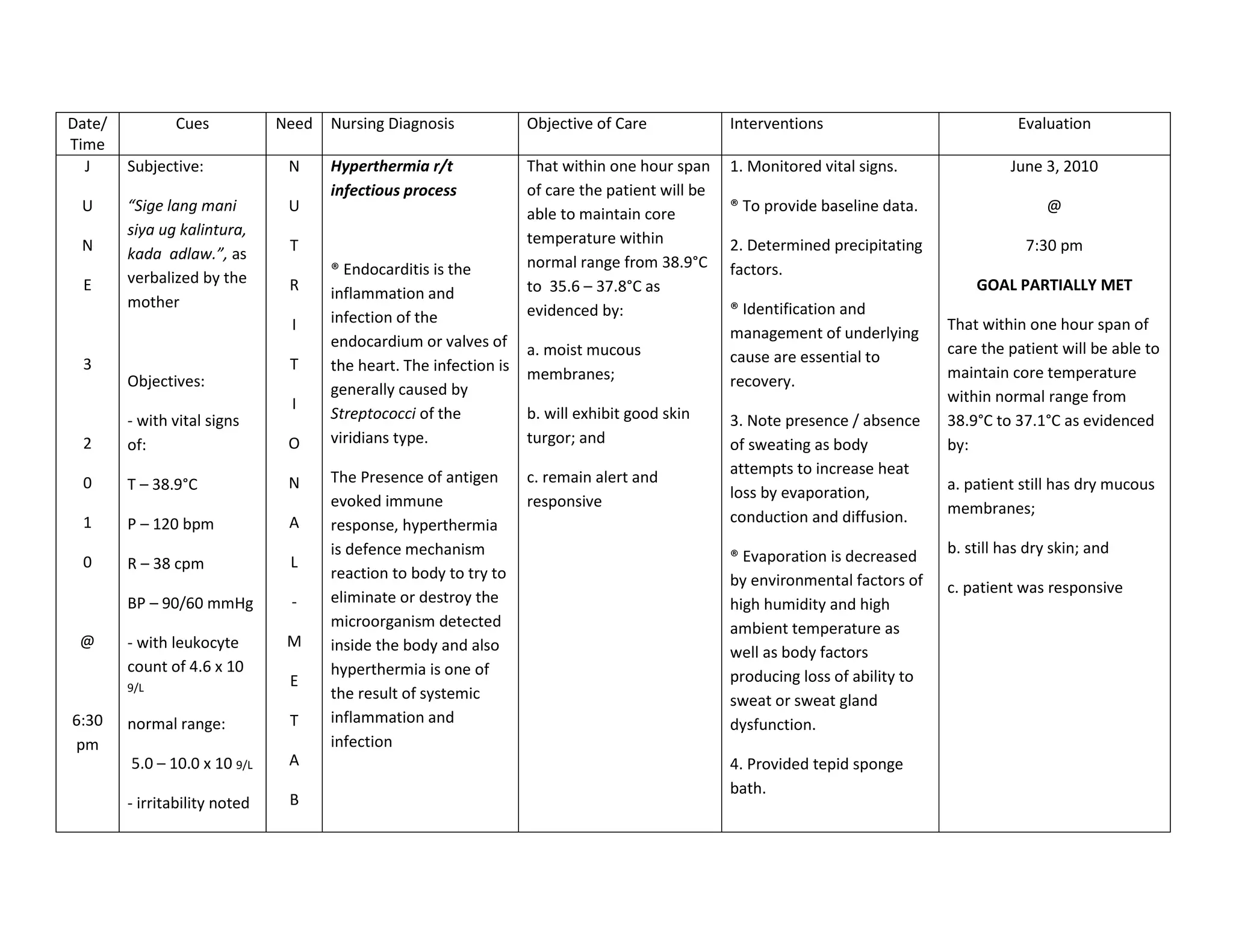
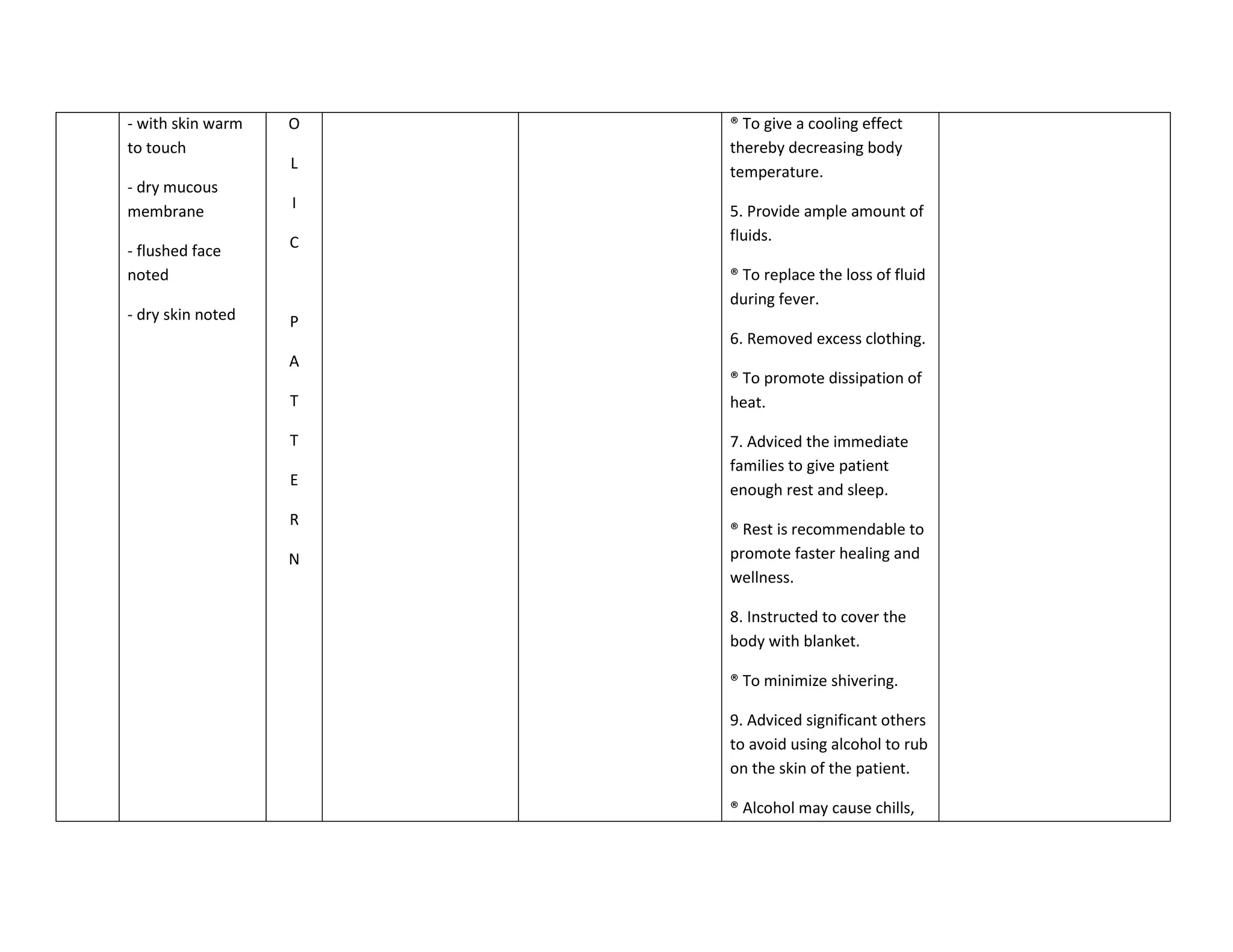
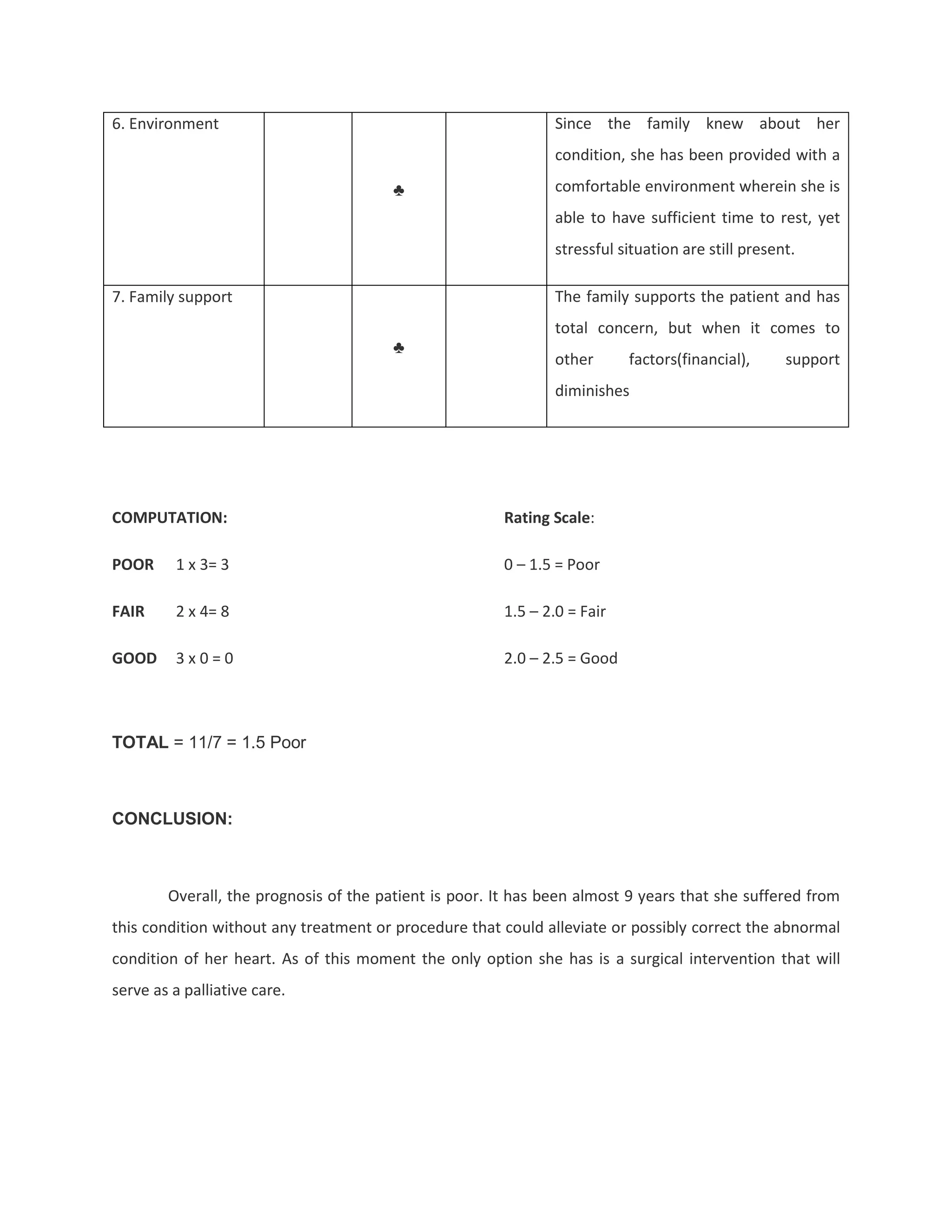
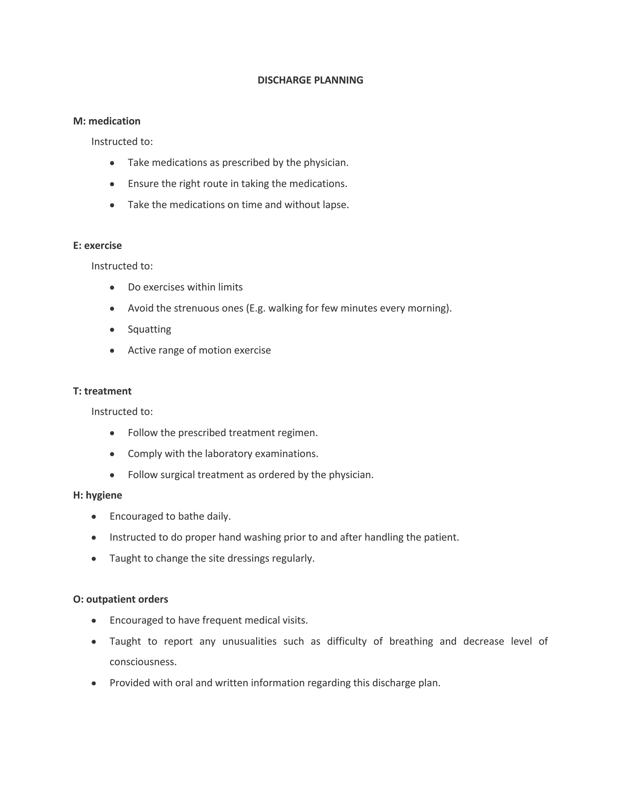
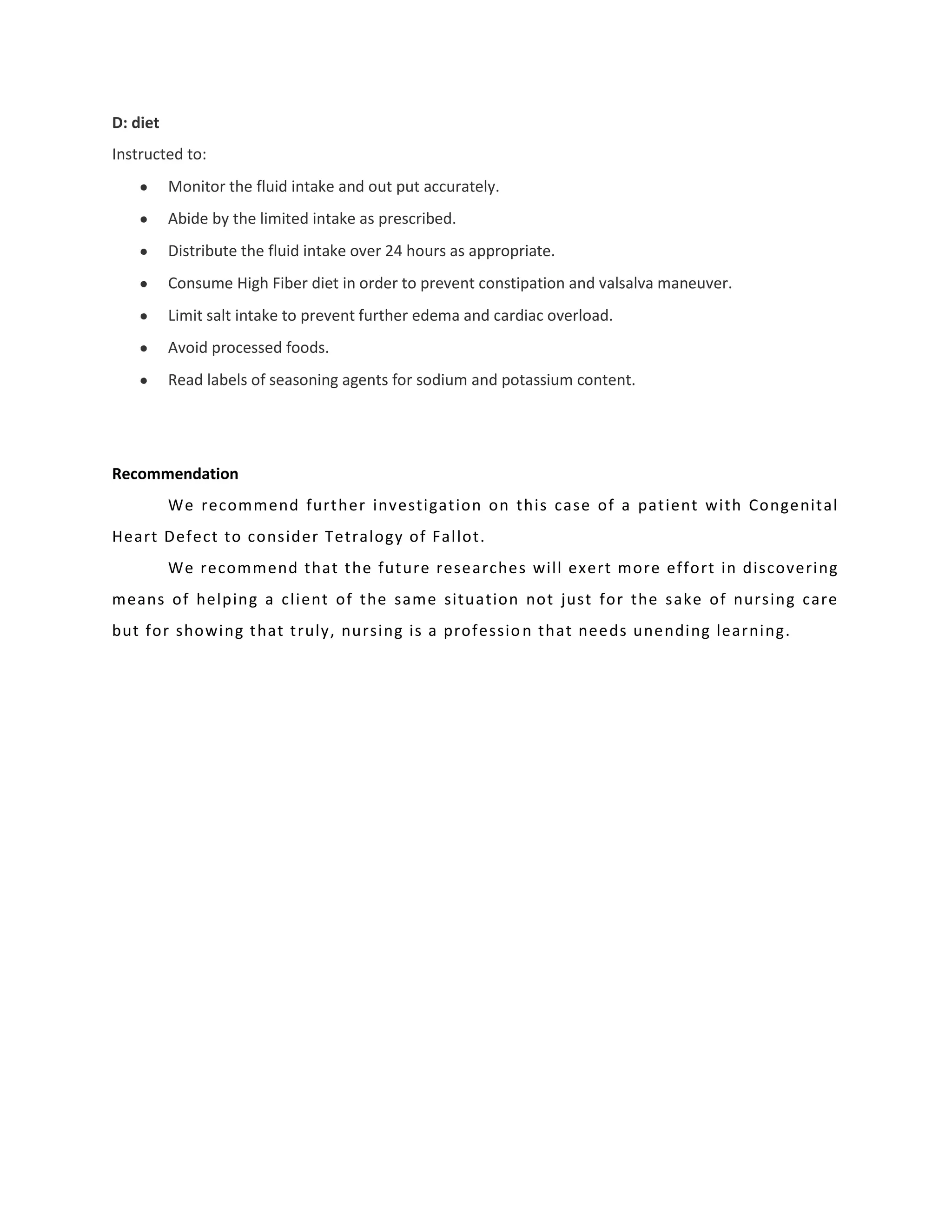
INTRODUCTION


                       “You can never be wrong when you think of your patient”
                                               -Rabj


        Our heart plays an important role in our body. It has four chambers, muscular organ that
functions as a powerful pump. It continuously pumps blood through the body’s extensive network of
arteries and veins. This circulation of blood delivers oxygen and nutrients to the body while removing
waste products.

        Congenital heart disease is a type of defect or malformation in one or more structures of the
heart or blood vessels that occurs before birth. Many heart defects and common health condition such
as heart disease occur as a result of interactions of multiple gene mutations and environmental
influences.

        These defects occur while the fetus is developing in the uterus and affect 8-10 out of every
1,000 children. Congenital heart defects may produce symptoms at birth, during childhood, and
sometimes not until adulthood. About 500,000 adults in the U.S. have congenital heart disease
(http://www.webmd.com/heart-disease/guide/congenital-heart-disease?page=2).

        According to the Mortality Country Fact Sheet of the year 2006, the top 4 causes of death in
children are congenital anomalies. It covers about 8% out of 100% total percentage of deaths among
children in both national and local areas. (www.who.international.mortality.philippines.com)

         Patient X is a 9 year old, female, who was diagnosed with congenital heart defect, cyanotic to
consider tetralogy of fallot. It defined as defects with decrease pulmonary blood flow. It is a form of
congenital heart defect which includes four abnormalities – narrowing of the pulmonary artery, a septal
defect between the ventricles, hypertrophy of the right ventricles and displacement of the aorta to the
right. The condition results in deficient oxygenation of the blood with cyanosis, dyspnea, polycythemia,
clubbing of the fingers.

        We choose this as the prime proponent of this case presentation primarily because the content
of this presentation tackles about the condition of the patient whose the course of the disease, its
management of both medical and nursing interventions and as well as pertinent health teachings which
the group find the case as medium for continuing professional learning. Nevertheless it serves as a tool
not only for the betterment of the patient and her family but also for us nurses to be able to learn more
about the realms of medical-surgical nursing its importance and implications.

        As a member of the health team- we should be the one that drives the family and with the
patient itself to empower them to become more self reliant with their own health.
OBJECTIVES


Discuss congenital heart defect;
present the medical interventions done to the patient;
identify needs of the patient and formulate a nursing care plan for each;
itemize the various drugs under the patient’s treatment regimen with information relevant to
the patient’s condition;
determine the prognosis of the patient with the following criteria: duration of illness, onset,
precipitating factors, willingness to take medications, age, environment, and family support;
and, state the general prognosis of the patient and;
formulate health teachings to the mother of our client regarding the continuity of treatment.
PATIENT’S PROFILE

Name:                  Heart

Age:                   9 years old

Address:               Florida, Kapalong, Davao del Norte

Civil Status:          Single

Father’s Name:         Superior Vena Cava

Mother’s Name:         Inferior Vena Cava

Occupation:            Father: Farmer

                       Mother: Housewife

Date of Birth:         February 23, 2001

Admission Date/Time:   May 28, 2010/ 3:45 pm

Attending Physician:   Dr. Gemma Maranian

Diagnosis:             Congenital heart defect, cyanotic type t/c TOF
PHYSICAL ASSESSMENT


General Survey

          Upon seeing the patient, she has an ectomorphic body built. She has an IVF of D5LR 1 liter at
108 cc/hr infusing well at right metalcarpal vein at 650 level and hooked to oxygen inhalation at 2 lpm
via nasal cannula and was placed on a high back rest, she has difficulty in breathing and has a capillary
refill time of 3 seconds. She has a foley catheter attached to uro bag draining to 100cc level with amber
yellow color urine. The patient is on diet as tolerated with strict aspiration precaution.


Vital Signs

Temp – 38.9°C

PR- 120 bpm

RR- 38 cpm

CR- 120 bpm

BP – 90/60 mmHg


Skin

       She has a darker skin complexion and is evenly distributed. Skin is dry and warm to touch.
Clubbing of finger nails noted with bluish discoloration of nailbeds on both upper and lower
extremitiesm with untimmed and dirty finger nails. She has some round scars on both legs.


Head

      Hair is black, long and evenly distributed, no infestations noted upon inspection. Head is
normocephalic with no abnormalities noted.


Eyes

        Eyes are symmetrical and are aligned at the upper pinnea of the ear. Iris is color brown and
pupils are equally rounded and are reactive to light accommodation with a diameter of 2 mm. No
lesions or discharges noted upon assessment.
Ears

      Ears are symmetrical and are aligned at the outer canthus of the eye. Eardrums are intact with
cerumen noted upon inspection. No lesions, discharges or abnormalities noted.


Nose

        Nasal flaring noted. No lesions or discharges noted upon inspection.


Neck and Throat

        Patient was able to swallow without any difficulty. Use of accessory muscle was noted. Neck was
able to flex from front to back and side to side without any discomfort. Upon palpation of the neck, no
mass was noted.


Mouth

      Lips have bluish discoloration with dry mucous membranes and dry tongue. No lesions or
abnormalities noted.


Chest

        Chest is slightly barrel chest with right side of the chest enlarged, with AP diameter of 2:1. Apex
of the heart is at the 4-5th midclavicular line. With clear breath sounds noted with use of accessory
muscles noted.


Abdomen

       The abdomen is flat and brown in skin color. Bowel sounds were heard upon auscultation with 5
bowel sounds per minute. No lesions noted upon inspection.


Genito-Urinary

         Patient was on folely catheter attached to urobag at 100 cc level with amber yellow colored
urine. No lesions or abnormalities noted.
MEDICAL HEALTH HISTORY


PAST MEDICAL HISTORY


Perinatal
        Prenatal check – up was started at the 5th month of pregnancy. Tetanus toxoid 1 was given. The
mother was febrile during the 3rd month of pregnancy. The mother did not consult a physician and there
were no medications taken. Her mother gave birth at home full term via normal spontaneous vaginal
delivery assisted by a hilot on the moonlit night of February 23, 2001. The baby presented poor and
delayed crying with cyanosis.


Past Medical Illness
        Her mother noted that at 1 year of age, the patient had an episode of syncope, (-) colds and (+)
cough for one month which led to her admission to a local hospital and then was referred to Davao
Regional Hospital where she was diagnosed with Congenital Heart Disease. 2D Echo was done where it
was found out that the patient had a hole in the heart. Surgical management was advised but not
heeded due to lack of financial resources. She was then discharged with a home medication of
spironolactone (Aldactone) but the patient did not comply. Due to poor compliance to the medication
and advised surgical management, her condition did not alleviate and she experienced on and off
dyspnea, orthopnea and occasional congestion.


Growth and Development
        During infancy, she was exclusively breastfed for six months and afterwards she was introduced
to solid foods like cereals and other table foods. Their source of water is from a water pump. The patient
has a good appetite.


Developmental Milestone
        The patient was able to attend school from nursery up to kindergarten. Unfortunately, she was
not able to continue her schooling because she finds it difficult to travel by walking.


PRESENT ILLNESS
Heart was doing well on the onset of her present illness. Unexpectedly, after defecating and
performing the Valsalva maneuver, she felt restless, irritable and short of breath. She even uttered
irritably the words, “Unsa man ning sakita ni uy!”. Despite of Heart’s complaint, she wasn’t rushed to
the hospital because of financial constraints. After two days of continually feeling short of breath and
restless, her mother decided to bring her to the Center of Child Health and Development (CCHD) here at
Davao Regional Hospital. Dr. dela Cruz was her attending physician then and she advised Heart’s parents
to admit their child at the Pediatric unit to be able to give the needed interventions for Heart. Thus,
after two days of restlessness, irritability and shortness of breath she was admitted as an in patient at
the Pediatric Unit of Davao Regional Hospital.


FAMILY HEALTH HISTORY
        Based on the diagram presented it reveals that on the Paternal side, Rodolfo the grandfather
has a heart problem, while her wife Dolores the Grandmother has no other hereditary disease. Together
they have five children namely: Beth, the eldest who has hypertension, Fe the second child who was
diagnosed with Diabetes Milletus, Bobby the third child and is the father of our patient do not have any
hereditary diseases, Bernie the fourth child has Heart Problem and is said to have inherited it to their
father and Flor as the youngest do not have any other hereditary diseases aside from mentioned above.
        On the Maternal side the grandfather of our patient Domingo was diagnosed with hypertension
while her wife Primitiva had no other hereditary diseases. They have 3 children namely: Cipriano as the
eldest and has Hypertension, the second child Miraluna and is the mother of our patient, and the
youngest is Felipe and was diagnosed with bronchial asthma.
        Generally, other conditions experienced both by the maternal and paternal side are common
colds and cough.
GENOGRAM


        PATERNAL SIDE                                                             MATERNAL SIDE


                                Dolores
       Rodolfo                                                                                                 Primitiva
                                                                                   Domingo


                        Bobby                        Flor
Beth         FE                           Bernie
32 ╬                                                                    Cipriano                  Miraluna                 Felipe ╬


                                                                                                             = Hypertension
                                             Heart                     Kristine
                                                                                                         = Diabetes Mellitus

                                                                                                      ╬ = Bronchial Asthma

                                                                                                             = Heart Problem
COURSE IN THE WARD


I.   Ongoing Diagnostic Work-ups

     1.1 Complete Blood Count


         Definition:
         The CBC is used as a broad screening test to check for such disorders as anemia,
         infection, and many other diseases. It is actually a panel of tests that examines different
         parts of the blood and includes the following:

        White blood cell (WBC) count is a count of the actual number of white blood cells per
         volume of blood. Both increases and decreases can be significant.

        White blood cell types (WBC differential). There are five different types of white blood
         cells, each with its own function in protecting us from infection. The differential
         classifies a person's white blood cells into each type: neutrophils (also known as segs,
         PMNs, granulocytes, grans), lymphocytes, monocytes, eosinophils, and basophils.

        Red blood cell (RBC) count is a count of the actual number of red blood cells per volume
         of blood. Both increases and decreases can point to abnormal conditions.

        Hemoglobin (Hgb). The hemoglobin molecule fills up the red blood cells. It carries
         oxygen and gives the blood cell its red color. The hemoglobin test measures the amount
         of hemoglobin in blood and is a good measure of the blood's ability to carry oxygen
         throughout the body.
   Hematocrit (HCT, packed cell volume, PCV). This test measures the amount of space
    (volume) red blood cells take up in the blood. The value is given as a percentage of red
    blood cells in a volume of blood. For example, a hematocrit of 38 means that 38% of the
    blood's volume is made of red blood cells. Hematocrit and hemoglobin values are the
    two major tests that show if anemia or polycythemia is present.

   Platelet (thrombocyte) count is the number of platelets in a given volume of blood. Both
    increases and decreases can point to abnormal conditions of excess bleeding or clotting.
    Mean platelet volume (MPV) is a machine-calculated measurement of the average size
    of your platelets. New platelets are larger, and an increased MPV occurs when increased
    numbers of platelets are being produced. MPV gives your doctor information about
    platelet production in your bone marrow.

   Mean corpuscular volume (MCV) is a measurement of the average size of your RBCs.
    The MCV is elevated when your RBCs are larger than normal (macrocytic), for example
    in anemia caused by vitamin B12 deficiency. When the MCV is decreased, your RBCs are
    smaller than normal (microcytic) as is seen in iron deficiency anemia or thalassemias.

   Mean corpuscular hemoglobin (MCH) is a calculation of the average amount of oxygen-
    carrying hemoglobin inside a red blood cell. Macrocytic RBCs are large so tend to have a
    higher MCH, while microcytic red cells would have a lower value.

   Mean corpuscular hemoglobin concentration (MCHC) is a calculation of the average
    concentration of hemoglobin inside a red cell. Decreased MCHC values (hypochromia)
    are seen in conditions where the hemoglobin is abnormally diluted inside the red cells,
    such as in iron deficiency anemia and in thalassemia. Increased MCHC values
    (hyperchromia) are seen in conditions where the hemoglobin is abnormally
    concentrated inside the red cells, such as in burn patients and hereditary spherocytosis,
    a relatively rare congenital disorder.

   Red cell distribution width (RDW) is a calculation of the variation in the size of your
    RBCs. In some anemias, such as pernicious anemia, the amount of variation
    (anisocytosis) in RBC size (along with variation in shape – poikilocytosis) causes an
    increase in the RDW.

    Nursing Care Prior to Procedure:
       1. Explain test procedure. Explain that slight discomfort may be felt when skin is
           punctured.
       2. Avoid stress if possible because altered physiologic status influences and
           changes normal values.
3. Dehydration or over hydration can dramatically alter values; for example, large
                  volumes of IV fluids can dilute the blood and values will appear as lower counts.
                  The presence of either these states should be communicated to the laboratory.
               4. Fasting is not necessary. However, fat-laden meals may alter some test results
                  as a result of lipidemia.

         Nursing Care During and After the Procedure:
            1. Apply manual pressure and dressings to the puncture site on removal of the
                needle.
            2. Monitor of the puncture site for oozing or hematoma formation. Maintain
                pressure dressings if necessary. Notify physician for unusual problems with
                bleeding.
            3. Resume normal activities and diet.


         Result with indications:
         Hematology/CBC
Date            Test                      Result     Normal Values         Indication

May 28, 2010      Hemoglobin (Hgb):       225        120 –140 mg/dl        May be increased when
                                          mg/dl                            the body perceives low
                                                                           blood oxygenation that
                                                                           can be due to decreased
                                                                           cardiac output

                  Leukocyte number        4.6        4.5 – 11.0 x 10⁹ /L   May be increased with
                  concentration:                     or 4,500 -            infections, inflammation,
                                                     11,000/mcL3           cancer, leukemia;
                                                                           decreased with some
                                                                           medications (such as
                                                                           methotrexate), some
                                                                           autoimmune conditions,
                                                                           some severe infections,
                                                                           bone marrow failure, and
                                                                           congenital marrow
                                                                           aplasia (marrow doesn't
                                                                           develop normally).

                  Segmenters:             0.56       2.5-7.5 x 10⁹/L       High count may indicate
                                                                           bacterial infection, burns,
                                                                           stress, and inflammation.
                                                                           Low count may indicate
                                                                           radiation exposure, drug
                                                                           toxicity, vitamin B12
deficiency, and
                                                               systematic lupus
                                                               erythematosus (SLE).

               Eosinophils:         0.02                       High Eosinophil count
                                                               may indicate an allergic
                                                               reactions, parasitic
                                                               infections, autoimmune
                                                               diseases. While a low
                                                               count may indicate drug
                                                               toxicity or stress.

               Thrombocyte:         84     150.0– 400.0 x      Decreased or increased
                                           109 /L              with conditions that
                                           or                  affect platelet
                                           150,000 -           production; decreased
                                           400,000/mcL3        when greater numbers
                                                               used, as with bleeding;
                                                               decreased with some
                                                               inherited disorders (such
                                                               as Wiskott-Aldrich,
                                                               Bernard-Soulier), with
                                                               Systemic lupus
                                                               erythematosus,
                                                               pernicious anemia,
                                                               hypersplenism (spleen
                                                               takes too many out of
                                                               circulation), leukemia,
                                                               and chemotherapy

               Erythrocyte:         0.77                       Decreased with anemia;
                                                               increased when too many
                                                               made and with fluid loss
                                                               due to diarrhea,
                                                               dehydration, and burns.

               Hematocrit           0.77   Male: 0.40 – 0.50   Increased with
                                           Female: 0.37-       dehydration, fluid loss
                                           0.43
May 30, 2010   Erythrocyte Volume   0.83   Male: 0.40 – 0.50   Increased with
               Fraction:                   Female: 0.37-       dehydration, fluid loss
                                           0.43
June 4, 2010     Hematocrit              0.52       Male: 0.40 – 0.50   Increased with
                                                    Female: 0.37-       dehydration, fluid loss
                                                    0.43


          Benefits:
          A complete blood count may be done to:
               Find the cause of symptoms such as fatigue, weakness, fever, bruising, or weight
                  loss.
               Find anemia.
               See how much blood has been lost if there is bleeding.
               Diagnose polycythemia and leukemia.
               Find an infection.
               Check how the body is dealing with some types of drug or radiation treatment.
               Check how abnormal bleeding is affecting the blood cells and counts.
               Screen for high and low values before a surgery.
               See if there are too many of too few of certain types of cells. This may help find
                  other conditions, such as too many eosinophils may mean an allergy or asthma
                  is present.
               As a part of regular physical examination. A blood count can give valuable
                  information about the general state of your health.

          Risk:
          There is a little chance of a problem from having a blood sample taken from a vein.
               You may get a small bruise at the site. You can lower the chace of bruising by
                  keeping pressure on the site for several minutes.
               In rare cases, the vein may become swollen after the blood sample is taken. This
                  problem is called phlebitis. A warm compress can be used several times a day to
                  treat this.
               Ongoing bleeding can be a problem for people with bleeding disorders. Aspirin,
                  warfarin (Coumadin), and other blood-thinning medicines can make bleeding
                  more likely. If you have bleeding or clotting problems, or if you take blood-
                  thinning medicine, tell your doctor before your blood sample is taken.


      1.2 Chest Radiography
          APL (Apicolordotic) View
Definition:
An x-ray (radiograph) is a noninvasive medical test that helps physicians diagnose and treat
medical conditions. Imaging with x-rays involves exposing a part of the body to a small dose
of ionizing radiation to produce pictures of the inside of the body. X-rays are the oldest and
most frequently used form of medical imaging.

Nursing Care Prior to Procedure:
   1. Explain test purpose, procedure and interfering factors. Emphasize that chest x-ray
       is painless.
   2. Have your client to remove some or all of clothes and wear a gown during the exam.
   3. Have your client remove jewelry, dentures, eye glasses and any metal objects or
       clothing that might interfere with the x-ray images.
   4. Follow guidelines for safe, effective, informed pretest care.

Nursing care during and after the procedure:
   1. Due to the cool temperature in the examination room and the coldness of the
       recording plate, some client may experience slight discomfort. Individuals with
       arthritis or injuries to the chest wall, shoulders or arms may have discomfort trying
       to stay still. Assist them in finding the most comfortable position possible that still
       ensures diagnostic image quality.
   2. After the procedure, return client’s jewelry, dentures, eye glasses and other
       belongings.
3. Follow safe, effective, informed post test care.

Results with indication:
        Date: May 28, 2010

        Chest APL: Peripheral pulmonary vessels are accentuated. Heart is slightly enlarged.
        Diaphragm and bony thorax are unremarkable. Soft tissue does not appear unusual.

        Impression: Mild Cardiomegaly with Pulmonary Congestion. Congenital Heart
        Disease is entertained.

Benefits
    No radiation remains in a patient's body after an x-ray examination.
    X-rays usually have no side effects in the diagnostic range.
    X-ray equipment is relatively inexpensive and widely available in emergency rooms,
        physician offices, ambulatory care centers, nursing homes and other locations,
        making it convenient for both patients and physicians.
    Because x-ray imaging is fast and easy, it is particularly useful in emergency
        diagnosis and treatment.


Risk
       There is always a slight chance of cancer from excessive exposure to radiation.
        However, the benefit of an accurate diagnosis far outweighs the risk.
       The chest x-ray is one of the lowest radiation exposure medical examinations
        performed today. The effective radiation dose from this procedure is about 0.1 mSv,
        which is about the same as the average person receives from background radiation
        in 10 days.
       Women should always inform their physician or x-ray technologist if there is any
        possibility that they are pregnant.


1.3 Arterial Blood Gas
Definition:
Arterial Blood Gases is a means of assessing the adequacy of oxygenation and ventilation, to
evaluate acid base status by measuring the respiratory and non respiratory components and
to monitor effectiveness of therapy. They are also used to monitor critically ill patients, to
establish baseline values in the perioperative and postoperative period, to detect and treat
electrolyte imbalances, to titrate appropriate oxygen flow rates, etc.


An ABG measures:
     Partial pressure of oxygen (PaO2). This measure the pressure of oxygen dissolved in
       the blood and how well oxygen is able to move from the airspace of the lungs into
       the blood.
     Partial pressure of carbon dioxide (PaCO2). This measures how much carbon
       dioxide is dissolved in the blood and how well carbon dioxide is able to move out of
       the body.
     pH. The pH measures hydrogen ions (H+) in blood. The pH of blood is usually
       between 7.35 and 7.45. A pH of less than 7.0 is called acid and a pH greater than 7.0
       is called basic (alkaline). So blood is slightly basic.
     Bicarbonate (HCO3). Bicarbonate is a chemical (buffer) that keeps the pH of blood
       from becoming too acidic or too basic.
     Oxygen content (O2CT) and oxygen saturation (O2Sat) values. O2 content
       measures the amount of oxygen in the blood. Oxygen saturation measures how
       much of the hemoglobin in the red blood cells is carrying oxygen (O2).
Nursing Care Prior to Procedure:
                  1. Explain the purpose and procedure for obtaining arterial blood sample.
                  2. If the patient is apprehensive, explain that a local anesthetic can be used.
                  3. Follow safe, effective, informed care.

           Nursing care during and after the procedure:
              1. Evaluate color, motion, sensation, degree of warmth, capillary refill time, and
                  quality of pulse in the affected extremity or at the puncture site.
              2. Monitor puncture site and dressing for arterial bleeding for several hours. No
                  vigorous activity of the extremity should be undertaken for 24 hours.
              3. Follow safe, effective, informed post test care.


           Result with indications:
Date/Time     Test                  Test      Normal Values           Indication
                                    Result
May 30,       pH:                   7.231     7.35 – 7.45             The pH or H+ indicates if a patient is
2010/ 11:33                                                           acidotic (pH < 7.35; H+ >45) or
                                                                      alkalemic (pH > 7.45; H+ < 35). In
                                                                      this situation, the patient is
                                                                      acidotic.

               Partial pressure    30.5       35-45 mm Hg             The carbon dioxide partial pressure
               of carbon dioxide              (4.6 – 5.9 k Pa)        (PaCO2) indicates a respiratory
               (PaCO2):                                               problem: for a constant metabolic
                                                                      rate, the PaCO2 is determined
                                                                      entirely by ventilation. A high
                                                                      PaCO2 (respiratory acidosis)
                                                                      indicates underventilation, a low
                                                                      PaCO2 (respiratory alkalosis) hyper-
                                                                      or overventilation. PaCO2 levels can
                                                                      also become abnormal when the
                                                                      respiratory system is working to
                                                                      compensate for a metabolic issue
                                                                      so as to normalize the blood pH. An
                                                                      elevated PaCO2 level is desired in
                                                                      some disorders associated with
                                                                      respiratory failure; this is known as
                                                                      permissive hypercapnia.

               Partial pressure    30.5       75-100 mm Hg (9.9       A low O2 indicates that the patient
               of oxygen                      – 13.2 k Pa)            is not respiring properly, and is
(PaO2):                                            hypoxemic. At a PaO2 of less than
                                                       60 mm Hg, supplemental oxygen
                                                       should be administered. At a PaO2
                                                       of less than 26 mm Hg, the patient
                                                       is at risk of death and must be
                                                       oxygenated immediately.

    Bicarbonate          13.3       20-29 mEq/L        The HCO3− ion indicates whether a
    (HCO3):                         (20 – 29 mmol/L)   metabolic problem is present (such
                                                       as ketoacidosis). A low HCO3−
                                                       indicates metabolic acidosis, a high
                                                       HCO3− indicates metabolic alkalosis.
                                                       HCO3− levels can also become
                                                       abnormal when the kidneys are
                                                       working to compensate for a
                                                       respiratory issue so as to normalize
                                                       the blood pH.

    Oxygen               48.7       95%-100%           This is the sum of oxygen dissolved
    saturation                      (0.95- 1.00)       in plasma and chemically bound to
    (O2Sat):                                           hemoglobin. Low percentage of O2
                                                       sat indicates low levels of
                                                       hemoglobin in the red blood cell,
                                                       carrying oxygen.

    Base Excess          -13.6      -3 to +3 mmol/l    The base excess is used for the
    (B.E.):                                            assessment of the metabolic
                                                       component of acid-base disorders,
                                                       and indicates whether the patient
                                                       has metabolic acidosis or metabolic
                                                       alkalosis. A negative base excess
                                                       indicates that the patient has
                                                       metabolic acidosis (primary or
                                                       secondary to respiratory alkalosis).
                                                       A positive base excess indicates
                                                       that the patient has metabolic
                                                       alkalosis (primary or secondary to
                                                       respiratory acidosis).


Benefits
An arterial blood gas (ABG) test is done to:
   Check for severe breathing problems and lung diseases, such as asthma, cystic
        fibrosis, or chronic obstructive pulmonary disease (COPD).
       See how well treatment for lung diseases is working.
       Find out if you need extra oxygen or help with breathing (mechanical ventilation).
       Find out if you are receiving the right amount of oxygen when you are using oxygen
        in the hospital.
       Measure the acid-base level in the blood of people who have heart failure, kidney
        failure, uncontrolled diabetes, sleep disorders, severe infections, or after a drug
        overdose.

Risk
There is little chance of a problem from having blood sample taken from an artery.
     You may get a small bruise at the site. You can lower the chance of bruising by
        keeping pressure on the site for at least 10 minutes after the needle is removed
        (longer if you have bleeding problems or take blood thinners).
     You may feel lightheaded, faint, dizzy, or nauseated while the blood is being drawn
        from your artery.
     Ongoing bleeding can be a problem for people with bleeding disorders. Aspirin,
        warfarin (Coumadin), and other blood-thinning medicines can make bleeding more
        likely. If you have bleeding or clotting problems, or if you take blood-thinning
        medicine, tell your doctor before your blood sample is taken.
     On rare occasions, the needle may damage a nerve or the artery, causing the artery
        to become blocked.
     Though problems are rare, be careful with the arm or leg that had the blood draw.
        Do not lift or carry objects for about 24 hours after you have had blood drawn from
        an artery.


1.4 ECG
Definition:
Electrocardiogram (ECG or EKG) is a transthoracic interpretation of the electrical activity of
the heart over time captured and externally recorded by skin electrodes. Electrical impulses
in the heart originate in the sinoatrial node and travel through the intrinsic conducting
system to the heart muscle. The impulses stimulate the myocardial muscle fibres to contract
and thus induce systole. The electrical waves can be measured at selectively placed
electrodes (electrical contacts) on the skin. Electrodes on different sides of the heart
measure the activity of different parts of the heart muscle. It is the best way to measure and
diagnose abnormal rhythms of the heart, particularly abnormal rhythms caused by damage
to the conductive tissue that carries electrical signals, or abnormal rhythms caused by levels
of dissolved alts (electrolytes), such as potassium, that are too high or low. In myocardial
infarction (MI), the ECG can identify damaged heart muscle. But it can only identify damage
to muscle in certain areas, so it can’t rule out damage in other areas. The ECG cannot
reliably measure the pumping ability of the heart; for with ultrasound-based
(echocardiography) or nuclear medicine test are used.

Procedure:
    1. Have the patient assume a supine position; however, recordings can be taken during
       exercise.
    2. Prepare the skin sites and, if necessary, shave and place electrodes on the four
       extremities and on specific chest sites. Ensure that the right leg is the ground.
    3. Remember that all 12 leads can be recorded simultaneously by newer ECG
       machines.
    4. Remember that a rhythm strip is a 2-minute recording from a single lead, usually
       lead II. It is frequently used to evaluate dysrhythmias.
    5. Follow safe, effective and informed intra-test care.


Nursing Care Prior to Procedure:
1. Explain test purpose, procedure and interfering factors. Emphasize that ECG is
      painless and does not deliver electrical current to the body. A resting ECG is no
      more than 1-minute record of the heart’s electrical activity.
   2. Have the patient completely relax to ensure a satisfactory tracing.
   3. Be aware that ideally, the person should rest for 15 minutes before ECG, and longer
      if possible.
   4. Follow guidelines for safe, effective, informed pretest care.

Nursing care during and after the procedure:
   1. Recognize the limitations of an ECG. A normal ECG does not rule out coronary artery
       disease or areas of cardiac ischemia. Conversely, an abnormal ECG in and of itself
       does not always signify heart disease.
   2. Interpret test results and counsel and monitor the patient appropriately. A resting
       ECG is usually normal in those patients who experience only angina. It can provide
       evidence of prior heart damage. The ECG is one diagnostic tool within a repertoire
       of diagnostic modalities and should be viewed as such. The presence or absence of
       heart disease should not be presumed solely on basis of ECG.
   3. Follow guidelines for safe, effective, informed post-test care.


1.5 Two Dimensional Echocardiography (2 D Echo)


Definition:
An echocardiogram (also called echo, transthoracic echocardiogram or TTE, exercise or
stress echocardiogram, dobutamine stress echocardiogram or DSE, or transesophageal
echocardiogram or TEE) is a noninvasive (the skin is not pierced) procedure used to assess
the heart's function and structures. A transducer (like a microphone) sends out ultrasonic
sound waves at a frequency too high to be heard. When the transducer is placed on your
chest at certain locations and angles, the ultrasonic sound waves move through the skin and
other body tissues to the heart tissues, where the waves echo off of the heart structures.
The transducer picks up the reflected waves and sends them to a computer. The computer
interprets the echoes into images of the heart walls and valves.
An echocardiogram can utilize one or more of four special types of echocardiography:
     M-Mode echocardiography: This is the simplest type of echocardiography. It
       produces an image that is similar to a line tracing rather than an actual picture of
       heart structures. M-mode echo is useful for measuring heart structures, such as the
       heart's pumping chambers, the size of the heart itself, and the thickness of the heart
       walls.
     Doppler echocardiography: This Doppler technique is used to measure and assess
       the flow of blood through the heart's chambers and valves. The amount of blood
       pumped out with each beat is an indication of the heart's functioning. Also, Doppler
       can detect abnormal blood flow within the heart, which can indicate a problem with
       one or more of the heart's four valves or with the heart's walls.
     Color Doppler: Color Doppler is an enhanced form of Doppler echocardiography.
       With color Doppler, different colors are used to designate the direction of blood
       flow. This simplifies the interpretation of the Doppler technique.
     2-D (2-dimensional) echocardiography: This technique is used to "see" the actual
       structures and motion of the heart structures. A 2-D echo view appears cone-
       shaped on the monitor, and the real-time motion of the heart's structures can be
       observed. This enables the physician to see the various heart structures at work and
       evaluate them.
     3-D (3-dimensional) echocardiography: 3-D echo is a relatively new type of echo
       that captures 3-dimensional views of the heart structures with greater depth than
       the 2-D echo. The live or ("real time") images allow for a more accurate assessment
       of heart function by using measurements taken while the heart is beating. 3-D echo
       shows enhanced views of the heart's anatomy and can be used to determine the
       appropriate plan of treatment for a person with heart disease.


Purpose:
Echocardiography is used to diagnose cardiovascular diseases. In fact, it is one of the most
widely used diagnostic tests for heart disease. It can provide a wealth of helpful information,
including the size and shape of the heart, its pumping capacity and the location and extent
of any damage to its tissues. It is especially useful for assessing diseases of the heart valves.
It not only allows doctors to evaluate the heart valves, but it can detect abnormalities in the
pattern of blood flow, such as the backward flow of blood through partly closed heart
valves, known as regurgitation. By assessing the motion of the heart wall, echocardiography
can help detect the presence and assess the severity of coronary artery disease, as well as
help determine whether any chest pain is related to heart disease. Echocardiography can
also help detect hypertrophic cardiomyopathy. The biggest advantage to echocardiography
is that it is noninvasive (doesn't involve breaking the skin or entering body cavities) and has
no known risks or side effects.
Nursing Care Prior to Procedure:
         1. Explain test purpose, procedure and interfering factors. Emphasize that 2 D echo
             imaging is painless and does not deliver electrical current or whatsoever to the
             body.
         2. Have the patient completely relax to ensure a satisfactory imaging.
         3. Follow guidelines for safe, effective, informed pre-test care.

      Nursing care during and after the procedure:
         1. For the procedure, the client may have to remove clothing from waist up. Ensure
             privacy by using drapes across the chest and limiting access into the procedure
             room during the test.
         2. The echocardiography technician will apply warm gel to the client’s chest. Aware
             the client beforehand.
         3. The client can resume normal activities, unless the physician instructs differently.

II.   Medical Management

      2.1 Phlebotomy
          Definition:
          Phlebotomy (A.K.A. venesection and venotomy) is the removal of a certain amount of
          blood under controlled conditions. Patients with elevated hematocrits (eg, those with
          polycythemia vera) or excessive iron absorption (eg, hemochromatosis) can usually be
          managed by periodically (once or twice weekly) removing 1 unit (about 500 ml) of whole
          blood. Eventually this depletes patient’s iron stores, thereby rendering the patient iron
          deficient and consequently unable to manufacture RBC’s excessively. The actual
          procedure for the therapeutic phlebotomy is similar to that for blood donation.

          Nursing Care Prior to Procedure:
             1. Explain test procedure. Explain that slight discomfort may be felt when skin is
                 punctured.
             2. Follow safe, effective, informed care.

          Nursing care during and after the procedure:
          Phlebotomy consists of venipuncture and blood withdrawal. Standard precautions are
          used.
              1. Patients are placed in a semi recumbent position. The skin over the antecubital
                 fossa is carefully cleansed with an antiseptic preparation, a tourniquet is applied
                 and venipuncture is performed. Withdrawal of 450 ml of blood usually takes less
                 than 15 minutes.
              2. After the needle is removed, patient is asked to hold the involved arm straight
                 up, and firm pressure is applied with strerile gauze for 2 or 3 minutes or until
                 bleeding stops. A firm bandage is then applied.
3. Patient should remain recumbent until they feel able to sit up. Patients who
          experience weakness or faintness should rest a longer period.
       4. Instruct patient to leave the dressing on and to avoid heavy lifting for several
          hours, to avoid smoking for 1 hour, to avoid drinking alcoholic beverages for 3
          hours, to avoid iron supplements (for those with Polycythemia vera), to increase
          fluid intake for 2 days, and to eat healthy meals for 2 weeks.

       Benefits
           Decrease over production of RBCs.
           Medical management for patients with polycythemia vera and
               hemochromatosis.

       Risk
              Fatigue
              Dizzines
              Phlebitis, or puncture site/vein becomes swollen (on rare cases).


2.2 Oxygen therapy

   Definition:
   Oxygen therapy is the administration of oxygen at a concentration greater than that
   found in the environmental atmosphere. At sea level, the concentration of oxygen in
   room air is 21%. The goal of oxygen therapy is to provide adequate transport of oxygen
   in the blood while decreasing the work of breathing and reducing stress on the
   myocardium.
   Oxygen transport to the tissue depends on actors such as cardiac output, arterial oxygen
   content concentration of hemoglobin, and metabolic requirements. These factors must
   be kept in mind when oxygen therapy is considered.

   Nursing care prior to procedure:
      1. The nurse instructs the patient or family in the methods for administering
          oxygen and informs the patient and family that the oxygen is available in gas,
          liquid, and concentrated form.
      2. Follow safe, effective, informed care.

   Nursing care during and after procedure:
      1. Humidity must be provided while oxygen is used to counteract the dry, irritating
          effects of the compressed oxygen on the airway.
III.    Medical Management

     3.3 Drugs taken by the patient


  NAME OF DRUG            ORDERED             INDICATION               MODE OF ACTION          PRECAUTION           ADVERSE REACTION            NURSING
                           DOSE                                                                                                             CONSIDERATION
CEFUROXIME SODIUM       520mg q8 ivtt     Pharyngitis, tonsillitis,   Second generation       Contraindicated in    CV: phlebitis,            Before
                                        infections of the urinary     cephalosporin that      patients              thrombophlebitis         administration,
                                        and lower respiratory         inhibits cell-wall      hypersensitive to     GI:                      ask patient if he
                                        tracts, and skin and          synthesis, promoting    drug or other         pseuomembranous          is allergic to
                                        skin-structure caused         osmotic instability;    cephalosporins.       colitis, nausea,         penicillins or
                                        by Streptococcus              usually bactericidal.   Use cautiously in     anorexia, vomiting,      cephalosporins.
                                        pneumoniae and S.                                     patients              diarrhea                 Obtain
                                        pyogenes, Haemophilus                                 hypersensitive to     Hematologic:             specimen for
                                        influenzae,                                           penicillin because    transient                culture and
                                        Staphylococcus aureus,                                of possibility of     neutropenia,             sensitivity tests
                                        E.coli, Moraxella                                     cross-sensitivity     eosinophilia,            before giving
                                        catarrhalis, Neisseria                                with other beta-      hemolytic anemia,        first dose.
                                        gonorrhieae, and                                      lactam antibiotics.   thrombocytopenia         Therapy may
                                        Klebsiella and                                        Use cautiously in     Skin: maculopapular      begin while
                                        Enterobacter species.                                 breast-feeding        and erythematous         awaiting
                                          Serious lower                                       women and in          rashes, urticaria,       results.
                                        respiratory tract                                     patients with         pain, induration,        For IM
                                        infections, UTI’s, skin                               history of colitis    sterile abscesses,       injection, inject
                                        infections, bone and                                  or renal              temperature              deep into a
                                        joint infections,                                     insufficiency.        elevation, tissue        large muscle,
                                        septicemia, meningitis,                                                     sloughing at IM          such as the
                                        and gonorrhea.                                                              injection site           gluteus
                                          Uncomplicated UTI.                                                        Other:                   maximus or the
                                          Early Lyme Dieseas                                                        hypersensitivity         lateral aspect o
                                        (erythema migrans)                                                          reactions, serum         the thigh
                                        caused by Borrelia                                                          sickness, anaphylaxis    Cefuroxime
                                        burgdorferi.                                                                                         tablets may be
Secondary bacterial   crushed, if
infection of acute      absolutely
bronchitis.             necessary, for
                        patients who
                        can’t swallow
                        tablets.
                        If large doses
                        are given,
                        therapy is
                        prolonged, or
                        patient is at
                        high risk,
                        monitor patient
                        for signs and
                        symptoms of
                        superinfection.
                        Instruct patient
                        to notify
                        prescriber
                        about rash or
                        evidence of
                        superinfection.
                        Advise patient
                        receiving drug
                        IV to report
                        discomfort at IV
                        insertion site.
                        Tell patient to
                        notify
                        prescriber
                        about loose
                        stools or
                        diarrhea.
CEFTRIAXONE   1.5gm q4 ivtt     Uncomplicated            Third generation          Contraindicated       CNS: fever,                 Before
  SODIUM                      gonococcal                 cephalosporin that      in patients             headache, dizziness     administration,
                              vulvovaginitis             inhibits cell-wall      hypersensitivity to     CV: phlebitis           ask patient if he is
                                UTIs; lower              synthesis, promoting    drug or other           GI:                     allergic to
                              respiratory tract,         osmotic instability;    cephalosporins.         pseudomembranous        penicillins or
                              gynecologic, bone and      usually bactericidal.     Use cautiously in     colitis, diarrhea       cephalosporins.
                              joint, intra abdominal,                            patients                GU: genital pruritus,       Obtain
                              skin, and skin structure                           hypersensitive to       candidiasis             specimen for
                              infections; septicemia.                            penicillin because      Hematologic:            culture and
                                Meningitis                                       of possibility of       eosinophilia,           sensitivity tests
                                Neurological                                     cross-sensitivity       thrombocytosis,         before giving first
                              complications, carditis,                           with other beta-        leucopenia              dose. Therapy
                              and arthtritis from                                lactam antibiotics.     Skin: pain,             may begin while
                              penicillin.                                          Use cautiously in     induration,             awaiting results.
                                                                                 breast-feeding          tenderness at               For IM
                                                                                 women and in            injection site, rash,   injection, inject
                                                                                 patients with           pruritus                deep into a large
                                                                                 history of colitis or   Other:                  muscle, such as
                                                                                 renal insufficiency.    hypersensitivity        the gluteus
                                                                                                         reactions, serum        maximus or the
                                                                                                         sickness,               lateral aspect o
                                                                                                         anaphylaxis, chills     the thigh.
                                                                                                                                     Monitor PT
                                                                                                                                 and INR in
                                                                                                                                 patients with
                                                                                                                                 impaired vitamin
                                                                                                                                 K synthesis or low
                                                                                                                                 vitamin K stores.
                                                                                                                                     Tell patient to
                                                                                                                                 report adverse
                                                                                                                                 reactions
                                                                                                                                 promptly.
Instruct patient
                                                                                                                                  to report
                                                                                                                                  discomfort at
                                                                                                                                  injection site.
                                                                                                                                       Tell patient to
                                                                                                                                  notify prescriber
                                                                                                                                  about loose stools
                                                                                                                                  or diarrhoea.

 PROPRANOLOL    10mg ½ tab po     Angina pectoris           A non-selective beta     Contraindicated     CNS: fatigue,              Always check
HYDROCHLORIDE                     To decrease risk of       blocker that reduces   in patients with      lethargy, fever, vivid   patient’s apical
                                death after MI              cardiac oxygen         bronchial asthma,     dreams,                  pulse before
                                  Supraventricular,         demand by blocking     sinus bradycardia     hallucinations,          giving drug. If
                                ventricular, and atrial     catecholamine-         and heart block       mental depression,       extremes in pulse
                                arrhythmias;                induced increases in   greater than first    light-headedness,        rates occur,
                                tachyarryhtmias caused      heart rate, blood      degree,               dizziness, insomnia      withhold drug and
                                by excessive                pressure, and force    cardiogenic shock,    CV: bradycardia,         notify prescriber
                                catecholamine action        of myocardial          and overt and         hypotension, heart       immediately.
                                during anesthesia,          contraction.           decompensated         failure, intermittent      Give drug
                                huperthyroidism, or         Depresses rennin       heart failure.        claudication,            consistently with
                                pheochromocytopenia.        secretion and            Use cautiously in   intensification of AV    meals. Food may
                                  Hypertension              prevents               patients with         block.                   increase
                                  To prevent frequent,      vasodilation of        hepatic or renal      GI: abdominal            absorption of
                                severe, uncontrollable,     cerebral arteries.     impairment, non-      cramping,                propranolol.
                                or disabling migraine, or                          allergic              constipation,              Drug masks
                                vascular headache.                                 bronchospastic        diarrhea, nausea,        common signs
                                  Essential tremor                                 diseases, or          vomiting                 and symptoms of
                                  Hypertrophic                                     hepatic diseases      Hematologic:             shock and
                                subaortic stenosis                                 and in those taking   agranulocytosis          hypoglycemia.
                                                                                   other                 Respiratory:               Caution patient
                                                                                   antihypertensives     bronchospasm             to continue taking
                                                                                     Because drug        Skin: rash               this drug as
                                                                                   blocks some                                    prescribed, even
                                                                                   symptoms of                                    when he’s feeling
hypoglycemia, use                              well.
                                                                         cautiously in                                    Instruct patient
                                                                         patients who have                              to take drug with
                                                                         diabetes mellitus.                             food.
                                                                            Use cautiously in                             Tell patient not
                                                                         patients with                                  to stop drug
                                                                         throtoxicosis                                  suddenly because
                                                                         becaude drug may                               this can worsen
                                                                         mask some signs                                chest pain and
                                                                         and symptoms of                                trigger a heart
                                                                         the disorder.                                  attack.
                                                                            Elderly patients
                                                                         may experience
                                                                         enhanced adverse
                                                                         reactions and may
                                                                         need dosage
                                                                         adjustment.
DIAZEPAM   1ml ivtt      Anxiety                   Unknown. A             Contraindicated       CNS: drowsiness,          Use Diastat
                         Acute          alcohol    benzodiazepine that in patients              dysarthia, slurred      rectal gel to treat
                      withdrawal                   probably potentiates hypersensitive to       speech, tremor,         no more than five
                         Muscle spasm              the effects of GABA, drug or soy protein;    transient amnesia,      episodes per
                          Cardioversion            depresses the CNS,   in patients             fatigue,ataxia,         month and no
                         Adjunct     treatment     and suppresses the   experiencing shock,     headache, insomnia,     more than one
                      for seizure disorders        spread of seizure    coma, or acute          paradoxical anxiety,    episode every 5
                         Status     epilepticus,   activity.            alcohol                 hallucinations, minor   days because
                      severe          recurrent                         intoxication; in        changes at EEG          tolerance may
                      seizures                                          pregnant women,         patterns.               develop.
                         Patients on stable                             especially in first     CV: hypotension, CV       When using oral
                      regimens of antiseptic                            trimester; and in       collapse, bradycardia   concentrate
                      drugs      who      need                          children younger        EENT: diplopia,         solution, dilute
                      diazepam intermittently                           than 6 months.          blurred vision,         dose just before
                      to control bouts of                                 Diastat rectal gel    nystagmus               giving.
                      increase seizure activity.                        is contraindicated      GI: nausea,               Monitor
                                                                        in patients with        constipation,           periodic hepatic,
acute angle-closure   diarrhea with rectal    renal, and
                                                                           glaucoma.             form                    hematopoietic
                                                                             Use cautiously in   GU: incontinence,       function studies in
                                                                           patients with liver   urine retention         patients receiving
                                                                           or renal              Hematologic:            repeated or
                                                                           impairment,           neutropenia             prolonged
                                                                           depression, or        Hepatic: jaundice       therapy.
                                                                           chronic open-angle    Respiratory:              Use of this drug
                                                                           glaucoma. Use         respiratory             may lead to abuse
                                                                           cautiously in         depression, apnea       and addiction.
                                                                           elderly and           Skin: rash              Don’t withdraw
                                                                           debilitated                                   drug abruptly
                                                                           patients.                                     after long-term
                                                                                                                         use; withdrawal
                                                                                                                         symptoms may
                                                                                                                         occur.
                                                                                                                           Warn woman to
                                                                                                                         avoid use during
                                                                                                                         pregnancy.
PARACETAMOL   325mg/tab q4     Fever / mild pain   Unknown. Thought          Contraindicated     Hematologic:               Use liquid form
               prn for fever                       to produce analgesia      in patients         hemolytic anemia,       for children and
                                                   by bocking pain           hypersensitive      neutropenia,            patients who have
                                                   impulses by               to drug.            leucopenia,             difficulty
                                                   inhibiting synthesis      Use cautiously in   pancytopenia            swallowing.
                                                   of prostaglandin in       patients with       Hepatic: jaundice          In children
                                                   the CNS or other          long term           Metabolic:              don’t exceed five
                                                   substances that           alcohol use         hypoglycemia            doses in 24 hours.
                                                   sensitize pain            because             Skin: rash, urticaria
                                                   receptors to              therapeutic
                                                   stimulation. The          doses cause
                                                   drug may relieve          hepatotoxicity in
                                                   fever through             these patients.
                                                   central action in the
                                                   hypothalamic heat-
regulating center.

MUPIROCIN   Treating impetigo, a      Mupirocin Ointment        Tell your doctor or    Burning; pain;           Inform pt. that:
OINTMENT    type of skin infection.   is an antibacterial. It   pharmacist if you      stinging.
                                      kills sensitive           have any medical                                    If you have
                                      bacteria by stopping      conditions,            Severe allergic              any questions
                                      the production of         especially if any of   reactions (rash;             about
                                      essential proteins        the following apply    hives; itching;              Mupirocin
                                      needed by the             to you:                difficulty breathing;        Ointment,
                                      bacteria to survive.                             tightness in the             please talk
                                                                    if you are         chest; swelling of the       with your
                                                                    pregnant,          mouth, face, lips, or        doctor,
                                                                    planning to        tongue); severe              pharmacist, or
                                                                    become             irritation; severe           other health
                                                                    pregnant, or       itching.                     care provider.
                                                                    are breast-                                     Mupirocin
                                                                    feeding                                         Ointment is to
                                                                    if you are                                      be used only
                                                                    taking any                                      by the patient
                                                                    prescription or                                 for whom it is
                                                                    nonprescriptio                                  prescribed. Do
                                                                    n medicine,                                     not share it
                                                                    herbal                                          with other
                                                                    preparation, or                                 people.
                                                                    dietary                                         If your
                                                                    supplement                                      symptoms do
                                                                    if you have                                     not improve
                                                                    allergies to                                    or if they
                                                                    medicines,                                      become
                                                                    foods, or other                                 worse, check
                                                                    substances                                      with your
                                                                    if you are                                      doctor.
                                                                    treating a burn
                                                                    or open wound
if you have
                                                                      kidney
                                                                      problems

CLOXACILLIN   Cloxacillin is used to    Disrupt the synthesis     If you have ever       an allergic           Take cloxacillin
              treat many different      of the peptidoglycan      had an allergic        reaction              exactly as directed
              types of infections       layer of bacterial cell   reaction to another    (shortness of         by your doctor. If
              caused by                 walls. The                penicillin or to a     breath; closing of    you do not
              staphylococcus bacteria   peptidoglycan layer       cephalosporin, do      your throat;          understand these
              ("staph" infections).     is important for cell     not take cloxacillin   hives; swelling of    instructions, ask
                                        wall structural           unless your doctor     your lips, face, or   your pharmacist,
                                        integrity.                is aware of your       tongue; rash; or      nurse, or doctor
                                                                  allergy and            fainting);            to explain them to
                                                                  monitors your          seizures;             you.
                                                                  therapy.               severe watery
                                                                                         diarrhea and          Take each dose
                                                                  Before taking this     abdominal             with a full glass of
                                                                  medication, tell       cramps; or            water. Take
                                                                  your doctor if you     unusual bleeding      cloxacillin on an
                                                                  have kidney            or bruising.          empty stomach 1
                                                                  disease, stomach or                          hour before or 2
                                                                  intestinal disease,                          hours after meals.
                                                                  or infectious
                                                                  mononucleosis.                               Do not drink juice
                                                                  You may not be                               or carbonated
                                                                  able to take                                 beverages (soda)
                                                                  cloxacillin because                          with your dose of
                                                                  of an increased risk                         cloxacillin. These
                                                                  of side effects.                             beverages will
                                                                                                               decrease the
                                                                  If you are a                                 effectiveness of
                                                                  diabetic, some                               the drug.
                                                                  glucose urine tests
                                                                  may give false                               Cloxacillin should
positive results        be taken at evenly
while you are           spaced intervals
taking cloxacillin.     throughout the
                        day and night to
Cloxacillin is in the   keep the level in
FDA pregnancy           your blood high
category B. This        enough to treat
means that it is        the infection.
unlikely to harm an
unborn baby. Do         Do not crush,
not, however, take      chew, or open the
cloxacillin without     capsules. Swallow
first talking to your   them whole.
doctor if you are       Shake the
pregnant. It is not     suspension well
known whether           before measuring
cloxacillin passes      a dose. To ensure
into breast milk. Do    that you get a
not take this           correct dose,
medication without      measure the
first talking to your   liquid form of
doctor if you are       cloxacillin with a
breast-feeding a        dose-measuring
baby.                   spoon or cup, not
                        a regular
                        tablespoon. If you
                        do not have a
                        dose-measuring
                        device, ask your
                        pharmacist where
                        you can get one.
                        Take all of the
                        cloxacillin that has
                        been prescribed
for you even if
you begin to feel
better. Your
symptoms may
start to improve
before the
infection is
completely
treated. Store the
capsules at room
temperature and
store the
suspension in the
refrigerator for
longer use. The
suspension is
good for 14 days if
it is stored in the
refrigerator.
Throw away any
unused liquid
after this amount
of time.
ANATOMY AND PHYSIOLOGY

HUMAN HEART

                                                         The human heart is located in the thoracic
                                                     cavity, behind and slightly to the left of the
                                                     sternum. The heart is tilted so that its pointed end,
                                                     the apex, points downward toward the left hip,
                                                     while the broad end, the base, faces upward
                                                     toward the right shoulder. It is bordered laterally
                                                     by the lungs, anteriorly by the sternum, posteriorly
                                                     by the vertebral column, and inferiorly by the
                                                     diaphragm, making it a well-protected organ. The
                                                     heart weighs 7-15 ounces (200-425 grams) and is
                                                     approximately the size of the individual's fist. It is
                                                     approximately 12cm long, 9cm wide at the
                                                     broadest point, and 6cm thick. The apex is formed
                                                     by the tip of the left ventricle and is located at the
                                                     level of the 5th intercostals space. The base is
                                                     formed by the atria at the level of the second rib.

    A protective sac called the pericardium
surrounds the heart. It consists of the following
layers:

      Fibrous anchors the heart in the
      mediastinum, surrounds roots of the
      major blood vessels, prevents over-
      distention, tough protective layer
      Parietal lies directly beneath the fibrous
      Visceral (Epicardium) lies beneath
      parietal, attached to the heart itself

  Pericardial fluid (up to 50ml) ultra filtrate of plasma provides a slippery surface for the movements of
the heart which prevents friction, located in pericardial cavity.

                              The wall of the heart consists of three layers:

                                    The epicardium is the visceral layer of the serous pericardium.
                                    The myocardium is the muscular part of the heart that consists of
                              contracting cardiac muscle and non-contracting Purkinje fibers that
                              conduct nerve impulses.
                                    The endocardium is the thin, smooth, endothelial, inner lining of
                              the heart, which is continuous with the inner lining of the blood vessels.
CHAMBERS OF THE HEART

        The heart consists of 2 muscular pumps known as
the left and right ventricles. The ventricles have 2
reservoirs called atria (left and right). Each ventricle acts to
serve different circulations. The right ventricle shifts
deoxygenated blood into the pulmonary circulation. Blood
enters the network of capillaries in the lungs and through a
process of diffusion carbon dioxide is lost and oxygen is
acquired; it then returns to the left atrium. The left
ventricle is the pump responsible for delivering blood into
the systemic circulation where it carries nutrients and
oxygen to the tissues. An exchange of nutrients and oxygen
for carbon dioxide and waste takes place; the waste rich
blood now returns to the right atrium

HEART VALVES

                                                 Atrioventricular (AV) valves lie between atria and
                                                 ventricles

                                                     • Tricuspid - on the right
                                                     • Mitral (bicuspid) - on the left
                                                     • Papillary muscles - attach to lower ventricular
                                                 walls and to chordae tendineae which in turn attach to
                                                 the AV valves. These keep the cusps of the AV valves from
                                                 being inverted into the atria when the ventricles contract.


                                                 Semilunar valves between ventricles and great vessels
exiting the heart

    •   Pulmonary - between RV and pulmonary trunk
    •   Aortic- between LV and aorta

    As blood travels through the heart, it enters a total of four chambers and passes through four
valves. The two upper chambers, the right and left atria, are separated longitudinally by the interatrial
septum. The two lower chambers, the right and left ventricles, are the pumping machines of the heart
and are separated longitudinally by the interventricular septum. A valve follows each chamber and
prevents the blood from flowing backward into the chamber from which the blood originated.

Two additional passageways are present in the fetal heart:

        The foramen ovale is an opening across the interatrial septum. It allows blood to bypass the
        right ventricle and the pulmonary circuit, while the nonfunctional fetal lungs are still developing.
The opening, which closes at birth, leaves a shallow depression called the fossa ovalis in the
       adult heart.
       The ductus arteriosus is a connection between the pulmonary trunk and the aorta. Blood that
       enters the right ventricle is pumped out through the pulmonary trunk. Although some blood
       enters the pulmonary arteries (to provide oxygen and nutrients to the fetal lungs), most of the
       blood moves directly into the aorta through the ductus arteriosus.

 THE GREAT VESSELS OF THE HEART

                                                                    Superior      vena       cava:      brings
                                                            deoxygenated blood into the right atrium from
                                                            the head and upper body veins
                                                                    Inferior vena cava: brings deoxygenated
                                                            blood from legs and lower torso into right
                                                            atrium
                                                                    Pulmonary arteries (R and L): branch
                                                            from pulmonary trunk, carry deoxygenated
                                                            blood from the right ventricle into the lungs
                                                                    Pulmonary veins (4): carry oxygenated
                                                            blood from lungs into the left atrium
                                                                    Ascending Aorta: receives oxygenated
                                                            blood from the left ventricle and pumps it
                                                            throughout the body via arteries

BLOOD VESSELS


   The central opening of a blood vessel, the lumen, is
surrounded by a wall consisting of three layers:

      The tunica intima is the inner layer facing the
      blood. It is composed of an innermost layer of
      endothelium (simple squamous epithelium)
      surrounded by variable amounts of connective
      tissues. In arteries this layer is composed of an
      elastic membrane lining and smooth
      endothelium that is covered by elastic tissues.
      The tunica media, the middle layer, is
      composed of smooth muscle with variable
      amounts of elastic fibers. This layer is thicker in
      arteries than in vein.
       The tunica adventitia, the outer layer, is
       composed of connective tissue as well as
       collagen and elastic fibres. These fibres allow
       the arteries and veins to stretch to prevent
       overexpansion due to the pressure that is
       exerted on the walls by blood flow.
Blood vessels have three kinds that form a closed system of passageways:

       Arteries carry blood away from the heart. The three
       kinds of arteries are categorized by size and
       function:
           o Elastic arteries (conducting arteries) are the
                largest arteries and include the aorta and
                other nearby branches. The tunica media of
                elastic arteries contains a large amount of
                elastic connective tissue, which enables the
                artery to expand as blood enters the lumen
                from the contracting heart. During
                relaxation of the heart, the elastic wall of
                the artery recoils to its original position,
                forcing blood forward and smoothing the
                jerky discharge of blood from the heart.
           o Muscular arteries (conducting arteries)
                branch from elastic arteries and distribute blood the various body regions. Abundant
                smooth muscle in the thick tunica media allows these arteries to regulate blood flow by
                vasoconstriction (narrowing of the lumen) or vasodilation (widening of the lumen). Most
                named arteries of the body are muscular arteries.
           o Arterioles are small, nearly microscopic, blood vessels that branch from muscular
                arteries. Most arterioles have all three tunics present in their walls, with considerable
                smooth muscle in the tunica media. The smallest arterioles consist of endothelium
                surrounded by a single layer of smooth muscle. Arterioles regulate the flow of blood
                into capillaries by vasoconstriction and vasodilation.
       Capillaries are microscopic blood vessels with extremely thin walls. Only the tunica intima is
       present in these walls, and some walls consist exclusively of a single layer of endothelium.
       Capillaries penetrate most body tissues with dense interweaving networks called capillary beds.
       The thing walls of capillaries allow the diffusion of oxygen and nutrients out of the capillaries,
       while allowing carbon dioxide and wastes into the capillaries.
           o Metarterioles (precapillaries) are the blood vessels between arterioles and venules.
                Although metarterioles pass through capillary beds with capillaries, they are not true
                capillaries because metarterioles, like arterioles, have smooth muscle present in the
                tunica media. The smooth muscle of a metarteriole allows it to acts as a shunt to
                regulate blood flow into the true capillaries that branch from it. The thoroughfare
                channel, the tail end of the metarteriole that connects to the venule, lacks smooth
                muscle.
           o True capillaries form the bulk of the capillary bed. They branch away from a
                metarteriole at its arteriole end and return to merge with the metarteriole at its venule
                end (thoroughfare channel).
           o Some true capillaries connect directly from an arteriole to a metarteriole or venule.
                Although the walls of true capillaries lack muscle fibers, they possess a ring of smooth
                muscle called a precapillary sphincter where they emerge from the metarteriole. The
                precapillary sphincter regulates blood flow through the capillary. There are three types
                of true capillaries:
   Continuous capillaries have continuous, unbroken walls consisting of cells that
                        are connected by tight junctions. Most capillaries are of this type.
                     Fenestrated capillaries have continuous walls between endothelial cells, but the
                        cells have numerous pores (fenestrations) that increase their permeability.
                        These capillaries are found in the kidneys, lining the small intestine, and in other
                        areas where a high transfer rate of substances into or out of the capillary is
                        required.
                     Sinusoidal capillaries (sinusoids) have large gaps between endothelial cells that
                        permit the passage of blood cells. These capillaries are found in the bone
                        marrow, spleen, and liver.
        Veins carry blood toward the heart. The three kinds of veins are listed here in the order that
        they merge to form increasingly larger blood vessels:
            o Postcapillary venules, the smallest veins, form when capillaries merge as they exit a
               capillary bed. Much like capillaries, they are very porous, but with scattered smooth
               muscle fibers in the tunica media.
            o Venules form when postcapillary venules join. Although the walls of larger venules
               contain all three layers, they are still porous enough to allow white blood cells to pass.
            o Veins have walls with all three layers, but the tunica intima and tunica media are much
               thinner than in similarly sized arteries. Few elastic or muscle fibers are present. The wall
               consists of primarily of a well-developed tunica adventitia. Many veins, especially those
               in the limbs, have valves, formed from folds of the tunica intima, which prevent the
               backflow of blood.

       Many regions of the body receive blood supplies from two or more arteries. The points where
these arteries merge are called arterial anastomoses. Arterial anastomoses allow tissues to receive
blood even after one of the arteries supplying blood has been blocked.

BLOOD FLOW THROUGH THE HEART

         The heart has four chambers that pump
blood. The chambers are called the right atrium,
right ventricle, left atrium, and left ventricle. The
right and left sides of the heart are separated by
muscular wall called the septum. This prevents
blood without oxygen from mixing with
oxygenated blood. The heart also has valves that
separate the chambers and connect to major blood
vessels.

        Blood flows from the body into the right
atrium. The blood on the right side of the heart
had been used by the body and is deoxygenated or
oxygen-poor.
The deoxygenated blood flows from the
                                                       right atrium through the tricuspid valve to the
                                                       right ventricle.

                                                              From the right ventricle, blood is pumped
                                                       through the pulmonary valve into the blood vessel
                                                       that goes to the lungs. This blood then picks up
                                                       oxygen becomes oxygenated or oxygen-rich.

                                                               Oxygenated blood flows from the lungs
                                                       through blood vessels back to the hearts left
                                                       atrium.

        From the left atrium, blood goes through the mitral valve and into the left ventricle.

         The left ventricle pumps blood through the aortic valve to a major blood vessel called aorta and
into systemic flow.

        The blood delivers oxygen to the body, the returns through veins to the right atrium and repeats
the blood flow cycle.

BLOOD SUPPLY TO THE HEART

                                               The coronary circulation consists of blood vessels, called
                                          coronary arteries, which supply oxygen and nutrients to the
                                          tissues of the heart. Blood entering the chambers of the heart
                                          cannot provide this service because the endocardium is too
                                          thick for effective diffusion (and only the left side of the heart
                                          contains oxygenated blood). Instead, the following two arteries
                                          that arise from the aorta and encircle the heart in the
                                          artioventricular groove provide this function:

                                                 The left coronary artery has the following two
                                          branches: The anterior interventricular artery (left anterior
                                          descending, or LAD, artery) and the circumflex artery.
                                                 The right coronary artery has the following two
                                          branches: The posterior interventricular artery and the
                                          marginal artery.

    The blood from the coronary circulation returns to the right atrium by way of an enlarged blood
vessel, the coronary sinus. Three veins, the great cardiac vein, the middle cardiac vein, and the small
cardiac vein, feed the coronary sinus.
CONDUCTION SYSTEM

1.      Sinoatrial (SA)node - pacemaker, located at the
surface of the right atrium, impulse causes both atria to
contract (rate 60-100 BPM); a conduction pathway called
Bachman's bundle runs from right to left atrium allowing for
simultaneous atrial contractions. SA node sends impulse to
the AV node via the anterior, medial and posterior
internodal pathways.

2.       Atrioventricular (AV) node - (rate 40-60 BPM) last
part of atria to be depolarized, sends impulse down the:

3.      Bundle of His- distributes action potential over
medial surfaces of the ventricles (rate 20-40 BPM) runs to
the top of the interventricular septum to the:

4.      Right and left bundle branches - Actual contraction
stimulated by conductive myofibers called Purkinje fibers
that pass into the fibers of the myocardium of the ventricles.
PATHOPHYSIOLOGY

              Predisposing:                                                      Precipitating:
                - Genetics                                                  - Environment = stress
             - Age = at birth


                                    Alteration in fetal development


                                                                                  signs and symptoms:
                                         Fetal anomaly – present
                                                 T/C TOF                          - clubbing of fingers
                                                                                  - cyanosis
                                     -    Pulmonic Stenosis
                                     -    Deviation of the origin of the          - murmur?
                                          aorta to the right                      - barrel chest
                                                                                  - Dyspnesa
                                     -    Intraventricular
                                          communication                           - Edematous
                                                                                  - Tachypnea
                                                                                  - Polycythemia


                                     Mixing of oxygenated and
                                       deoxygenated blood

Increased ventricular contraction
                                    Decrease O2 supply to systemic
                                             circulation
 Right ventricular hypertrophy

                                                Hypoxia


          IF TREATED                                                           IF NOT TREATED


     Diagnostic exam:                                                      transient increase in resistance
     - CBC                                                                    to blood flow to the lungs
     - 2D echo
     -ABG
                                                                                 worsen tet spells
     Treatment:
     - medications
     - O2 therapy                                                          decreased organ perfusion
     - surgery                                                                  *including brain


                                                                               - hypoxic brain injury
 Partially-improved oxygenation
                                                                                  - organ failure


       Poor prognosis
                                                                                      DEATH
A. Pulmonary Stenosis
        A narrowing of the right ventricular outflow tract and can occur at the pulmonary valve (valvular
stenosis) or just below the pulmonary valve (infundibular stenosis). Infundibular stenosis os most caused
by overgrowth of the heart muscle wall (hypertrophy of the septoparietal trabeculae), however the
events leading to the formation of the overriding aorta are also believed to be a cause. The pulmonic
stenosis is the major cause of the malformations, with the other associated malformations acting as
compensatory mechanisms to the pulmonic stenosis. The degree of the stenosis varies between
individuals with TOF, and is the primary determinant of symptoms and severity. This infrequently
described as subpulmonary stenosis or subpulmonary obstruction.

B. Overriding Aorta
        An aortic valve with biventricular connection, that is, it is situated above the ventricular septal
defect and connected to both the right and the left ventricle. The degree to which the aorta is attached
to the right ventricle is referred to as its degree of “override.” The aortic root can be displaced toward
the front (anteriorly) or directly above the septal defect, but it is always abnormally located to the root
of the pulmonary artery. The degree of the override is quite variable, with 5-95% of the valve being
connected to the right ventricle.

C. Ventricular Septal Defect (VSD)
        It is a hole between the two bottom chambers (ventricles) of the heart. The defect is centred
around the most superior aspect of the ventricular septum (the outlet septum), and in the majority of
cases is single and large. In some cases thickening of the septum (septal hypertrophy) can narrow the
margins of the defect.

D. Right Ventrical Hypertrophy
        The right ventricle is more muscular than normal, causing a characteristic boot-shaped (Coeur-
en-sabot) appearance as seen by chest X-ray. Due to the missarangement of the external ventricular
septum, the right ventricular wall increases in size to deal with the increases obstruction to the right
outflow tract. This feature is now generally agreed to be a secondary anomaly, as the level of
hypertrophy generally increases with age.

Etiology: The causes of most CHDs are unknown, although genetic studies suggest a multifactorial
etiology. Prenatal factors associated with a higher incidence of TOF include maternal rubella (or other
viral illnesses) during pregnancy, poor prenatal nutrition, maternal alcohol use, maternal age older than
40 years, and diabetes. Children with Down syndrome have a higher incidence of TOF.
PATHOPHYSIOLOGY:

        Tetralogy of fallot results in low oxygenation of blood due to the mixing of oxygenated and
deoxygenated blood in the left ventricle via the VSD and the preferential flow of the mixed blood from
both ventricles through the aorta because of the obstruction to flow through the pulmonary valve. This
is known as right-to-left shunt. The primary symptom is low blood oxygen saturation with or without
cyanosis from birth or developing in the first year of life. If the baby is not cyanotic then it is sometimes
referred to as the “pink tet”. Other symptoms include a heart murmur which may range from almost
imperceptible to very loud, difficulty in feeding, failure to gain weight, retarded growth and physical
development, dyspnea on exertion, clubbing of the fingers and toes, and polycythemia.

        Children with tetralogy of fallot may develop “tet spells”. The precise mechanism of these
episodes in doubt, but presumably results from a transient increase in resistance to blood flow to the
lungs with increased preferential flow of desaturated blood to the body. Tet spells are characterized by
a sudden, marked increase in cyanosis followed by syncope, and may result on hypoxic brain injury and
death. Older children will often squat during tet spell, which increases systemic vascular resistance and
allows for a temporary reversal of the shunt.

        A small percentage of children with tetralogy of fallot may also have additional ventricular septal
defects, an atrial septal defect/ASD or abnormalities in the branching pattern of their coronary artieries.
Some patients with tetralogy of fallot have complete obstruction to flow from the right ventricle, or
pulmonary atresia. Tetralogy of fallot may be associated with chromosomal abnormalities, such as
22q11 deletion syndrome.

         The pulmonary stenosis and right ventricular outflow tract obstruction seen with tetralogy of
fallot usually limits blood flow to the lungs. When blood flow to the lungs is restricted, the combination
of the ventricular septal defect and overriding aorta allows oxygen-poor blood (“blue”) returning to the
right atrium and right ventricle to be pumped out the aorta to the body.

         The “shunting” of oxygen-poor from the right ventricle to the body results in a reduction in the
arterial oxygen saturation so that babies appear cyanotic, or blue. The cyanosis occurs because oxygen-
poor blood is darker and has a blue color, so that the lips and skin appear blue.

        The extent of cyanosis is dependent on the amount of narrowing of the pulmonary valve and
right ventricular outflow tract. A narrower outflow tract from the right ventricle is more restrictive to
blood flow to the lungs, which in turn lowers the arterial oxygen level since more oxygen-poor blood is
shunted form the right ventricle to the aorta.

        Tetralogy of fallot is most often diagnosed in the first few weeks of life due to either a loud
murmur or cyanosis. Babies with tetralogy of fallot usually have a patent ductus arteriousus at birth that
provides additional blood flow to the lungs, so severe cyanosis is rare early after birth. As the ductus
artesiosus closes, which is typically, will in the first days of life, cyanosis can develop or become more
severe. The degree of cyanosis is proportional to lung blood flow and thus depends upon the degree of
narrowing of the outflow tract to the pulmonary arteries. Rapid breathing in response to low oxygen
level and reduced pulmonary blood flow can occur. The heart murmur, which is commonly loud and
harsh, is often absent in the first few days of life. The arterial oxygen saturation of babies with tetralogy
of fallot can suddenly drop markedly. This phenomenon, called a “tetralogy spell,” usually results from a
sudden increased constriction of the outflow tract to the lungs so that pulmonary blood flow is further
restricted. The lips and skin of babies who have a sudden decrease in arterial oxygen level will appear
acutely bluer.

          Children having a tetralogy spell will initially become extremely irritable in response to the
critically low oxygen levels, and they may become sleepy or unresponsive if the severe cyanosis persists.

       A tetralogy spell can be treated by comforting the infant and flexing the knees forward and
upward. Most often, however, immediate medical attention is necessary.

        When a newborn baby with significant cyanosis is first seen, they are often placed in
supplemental oxygen. The increased oxygen improves the child’s oxygen levels in cases of lung disease,
but breathing extra oxygen will have little effect on the oxygen levels of a child with tetralogy of fallot.

        Failure to respond to this “hyperoxia test” is often the first clue to suspect a cyanotic cardiac
defect. Infants with tetralogy of fallot can have normal oxygen levels if the pulmonary stenosis is mild
(refered to as “pink” tetralogy of fallot). In these children, the first clue to suggest a cardiac defect is
detection of a loud murmur when the infant is examined.

      Once congenital heart disease is suspected, echocardiography can rapidly and accurately
demonstrate the four related defects characteristics of tetralogy of fallot.

       Cardiac catheterization is occasionally required to evaluate the size and distribution of the
pulmonary arteries and to clarify the branching patterns of the coronary arteries. Catheterization can
also demonstrate whether patients have pulmonary blood flow supplied by an abnormal blood vessel
from the aorta (aortopulmonary collateral).

      Once tetralogy of fallot is diagnosed, the immediate management focuses on determining
whether the child’s oxygen levels are in a safe range.

         If oxygen levels are critically low soon after birth, a prostaglandin infusion is usually initiated to
keep the ductus arteriosus open which will provide additional pulmonary blood flow and increase the
child’s oxygen levels.

       These infants will usually require surgical intervention in the neonatal period. Infants with
normal oxygen levels are only mild cyanosis are usually able to go home in the first week of life.

        Complete repair is usually done electively when the children are about six months of age, as
long as the oxygen levels remain adequate. Progressive or sudden decrease in oxygen saturation may
prompt earlier corrective repair.
Surgical correction of the defect is always necessary. Occasionally, patients will require a surgical
palliative prior to the final correction.

        Corrrective repair of tetralogy of fallot involves closure of the ventricular septal defect with a
synthetic Dacron patch so that the blood can flow normally from the left ventricle to the aorta.

        The narrowing of the pulmonary valve and right ventricular outflow tract is then augmented
(enlarged) by a combination of cutting away (resecting) obstructive muscle tissue in the right ventricle
and by enlarging the outflow pathway with a patch.

        In some babies, however, the coronary arteries will branch across the right ventricular outflow
tract where the patch would normally be placed. In these babies, an incision in this area to place the
patch would damage the coronary artery so this cannot be safely done.

        When this occurs, a hole is made in the front surface of the right ventricle to the bifurcation of
the pulmonary arteries to provide unobstructed blood flow from the right ventricle to the lungs.
Date           Cues             Need     Nursing Diagnosis           Objectives of care                 Intervention                       Evaluation
/Time
   J  Subjective:                 A     Ineffective breathing     That within 2 hours span       1. Assessed respiratory rate              June 3, 2010
  U   “Lisod kaayo mu             C     pattern related to        of care, the patient will be   and depth monitor for nasal
  N   ginhawa.”                   T     inability to maintain     able to establish effective    flaring, chest retractions and                 @
   E                              I     adequate rate             respiratory pattern such       cyanosis.
       Objective:                 V     and depth of              as:                            ® To detect early signs of                  7:30 pm
   3  - shortness of              I     respiration                                              respiratory compromise.
      breathing noted             T                               a. absence of use of                                               GOAL PARTIALLY MET!
   2                              Y                               accessory muscles              2. Administered oxygen as
   0  - nasal flaring noted             ® Heart defect can                                       ordered.                         After 2 hours span of care,
   1                              A     cause breathlessness if   b. respiratory rate will       ® To help reduce                 the patient was not able to
   0  - use of accessory          N     your heart is unable to   stay within normal range:      hypoxemia and to relieve         establish effective
      muscle in breathing         D     pump enough blood to      Infant = 25 – 60 bpm           respiratory distress.            respiratory pattern
  @                                     supply oxygen to your     Children = 14 -25 bpm                                           however:
      - previous O2 Sat of        E     body. If your brain,      Adult = 10 – 25 bpm            3. Placed on high back rest.
 5:30 48.7% (May 30, 2010/        X     muscles, or other body                                   ® To promote lung                a. patient still uses
 pm   11:33)                      E     organs do not receive     c. demonstrate                 expansion.                       accessory muscles
                                  R     enough oxygen, a          diaphragmatic pursed-lip
       -with O2 inhalation of     C     sense of breathlessness   breathing                      4. Remain with the child and     b. with respiratory rate of
       2 lpm via nasal            I     may occur.                                               offer reassurance during         30 bpm
       cannula                    S                               d. relieved from of            period of respiratory
                                  E                               shortness of breath            difficulty.                      c. demonstrate
       - with respiratory rate                                                                   ® To relieve anxiety.            diaphragmatic pursed-lip
       of 43 breathes per         P                                                                                               breathing
       minute                     A                                                              5. Assist with activities of
                                  T                                                              daily living as necessary.       d. patient was slightly
       - on moderate high         T                                                              ® To help patient conserve       relieved from shortness of
       back rest                  E                                                              energy and to avoid fatigue.     breath as evidenced by
                                  R                                                                                               decreased irritability and
       - irritability noted       N                                                              6. Teach the patient             grunting
                                                                                                 relaxation techniques, such
                                                                                                 as diaphragmatic breathing
                                                                                                 and pursed lip breathing
technique.
® This measure allows the
child to participate in
maintaining her health
status and may improve her
breathing pattern.
Date/           Cues            Need     Nursing Diagnosis           Objective of Care                  Interventions                    Evaluation
Time
  J     Subjective:              A     Ineffective tissue per   That within 8 hour span of      1. Monitored skin color and             June 3, 2010
  U     “Lagum kayo siya         C     fusion                   care, the patient will be       temperature every 2hours.
  N     tan-awon,” as            T     (cardiopulmonary)        able to have adequate           Assess for signs of skin                      @
  E     verbalized by the        I     related to decrease      tissue perfusion as             breakdown.
        mother                   V     oxygen cellular          evidenced by:                   ®Cool, blanched, mottled skin             11:00 pm
 3                               I     exchange secondary                                       and cyanosis may indicate
        Objectives:              T     to Congenital Heart      a. absence or decrease          tissue perfusion.                  GOAL PARTIALLY MET
 2      - bluish                 Y     Disease t/c Tetralogy    bluish discoloration of lips;
 0      discoloration on               of Fallot                                                2. Monitored and                After the 8 hour span of
 1      lips noted               A                              b. decrease respiratory rate    documented patient’s vital      care the patient was able to
 0                               N                              within normal range; and        signs every hour.               have adequate tissue
        - clubbing of finger     D     ® Due to narrowing                                       ®Decrease heart rate and        perfusion as evidenced by:
 @      noted                          of the artery which      c. learn techniques to          blood pressure may indicate
                                 E     small amount of          minimize or lessen the risk     increased arteriovenous         a. still presence of bluish
3 pm    - nasal flaring          X     oxygenated blood         of decrease tissue              exchange, which leads to        discoloration of lips;
        noted                    E     can pass through the     perfusion                       decrease tissue perfusion.
                                 R     systemic circulation                                                                     b. with respiratory rate of
        - use of accessory       C     which the patient                                        3. Keep patient warm.           25 cpm; and
        muscle noted             I     experience difficulty                                    ® Warmth aids vasodilation,
                                 S     in breathing.                                            which improves tissue           c. learned techniques to
        - with capillary         E                                                              perfusion.                      minimize or lessen the risk
        refill time of 3 secs                                                                                                   of decrease tissue perfusion
                                 P                                                              4. Elevate lower extremities.   such as squatting
        - with O2 of 2 lpm       A                                                              ® To increase arterial blood
        via nasal cannula as     T                                                              supply and improve tissue
        ordered                  T                                                              perfusion.
                                 E
        - body weakness          R                                                              5. Change position regularly
        noted (always on         N                                                              and inspect skin every shift.
        bed)                                                                                    ® To avoid decrease in tissue
                                                                                                perfusion and risk of skin
        - with hemoglobin                                                                       breakdown.
of 225 mg/dL
Normal value:         6. Teach patient squatting
120 – 140 mg/dL       techniques.
(taken on May 28,     ® To promote tissue
2010)                 perfusion.

- hematocrit of       7. Maintained oxygen therapy
0.77                  as ordered.
Normal value:         ® To maximize oxygen
Male – 0.40 – 0.50    exchange in the alveoli and at
Female – 0.37 –       the cellular level.
0.43
(taken on May 28,     8. Encouraged frequent rest
2010)                 periods.
                      ® To conserve energy and
- ABG result taken    maximize tissue perfusion.
on May 30, 2010/
11:33
pH - 72
PCO2 -30.5
PO2 – 30.5
HCO3 – 13.3
B.E. – 13.6
O2 sat – 46.7%
Normal Range:
pH –7.35 – 7.45
PCO2- 35 – 45
mmHg
PO2 – 75 -100
mmHg
HCO3 -20 – 29
mEq/dL
B.E. – -3 - +3 mmol
O2 sat – 95% -
100%

-vital signs:
RR: 38 cpm
BP- 80/50
HR- 124 bpm
Date/           Cues           Need   Nursing Diagnosis             Objective of Care        Interventions                              Evaluation
Time
  J     Subjective:             A     Impaired gas              That within 8 hour span of   1.) Established good and                  June 3, 2010
                                C     exchange related to       care the patient will be     trusting relationship with the
 U      “Paspas lagi na         T     altered oxygen            able    to    demonstrate    patient and significant others.                @
        iyang paghinga”,        I     supply as evidenced
 N                                                              improving ventilation as                                                  11 pm
        as verbalized by        V     by dyspnea,                                            ®To gain both the trust and
                                I     tachypnea,                evidenced by:
 E      the mother                                                                           cooperation.                         GOAL PARTIALLY MET
                                T     tachycardia and
                                                                a. respiratory rate within
                                Y     fatigue secondary to
                                      Congenital Heart          normal limits. (RR: 20 -25 2.) Monitored respiratory           After the end of 8 hour span
                                                                                           rate/depth, use of accessory        of care the patient was be
        Objectives:             A     Disease t/c Tetralogy     cpm);
 3                                                                                         muscles, areas of cyanosis.         able to demonstrate
                                N     of Fallot
        - vital signs:          D                               b. absence of cyanosis;                                        improving ventilation as
                                                                                           ® Indicators of adequacy of
                                                                c. clear breath sounds on respiratory function or degree       evidenced by:
 2      BP = 80/50 mmHg         E     ® Congenital heart
                                X     disease refers to a       both lung fields; and      of compromise and therapy           a. respiratory rate within
 0      CR = 124 bpm            E     problem with the                                     needs/effectiveness.                normal limits. With RR of 25
                                R                               d. ABG test results within
        RR = 28 cpm                   heart's structure and                                                                    cpm
 1                              C                               normal range.              3.) Auscultated breath sounds,
                                      function due to
                                I                                                          noting presence/absence, and        b. presence of cyanosis
 0      T = 37.1 C              S     abnormal heart
                                                                                           adventitious sounds.
                                E     development before                                                                       c. clear breath sounds on
        - with O2
                                      birth. It can disrupt I                                ® Development of atelectasis      both lung fields
        inhalation @ 2 lpm      P     the normal flow of                                     and stasis of secretions can
 @      via nasal cannula as    A     blood to the different                                 impair gas exchange.              d. There was no second ABG
        ordered                 T     parts of our body                                                                        taken
                                T                                                            4.) Monitored vital signs; note
        - circumoral                  thus affecting
                                E                                                            changes in cardiac rhythm.
3 pm                                  process of diffusion,
        cyanosis noted          R
                                N     the exchange of                                        ® Compensatory changes in
        - skin warm to                oxygen and carbon                                      vital signs and development of
        touch with good               dioxide that occurs in                                 dysrhythmias reflect effects of
                                      the alveolar-capillary
skin turgor            membrane area.   hypoxia on cardiovascular
                                        system.
- skin pallor
                                        5.) Investigated reports of
- clubbing of nails
                                        chest pain and increasing
noted                                   fatigue. Observe for signs of
- with hemoglobin:                      increased fever, cough,
225 g/dL (Normal                        adventitious breath sounds.
value: 120-160
                                        ® Reflective of developing
g/dL)                                   acute chest syndrome (i.e.,
- with IVF of D50.3                     chest pain, dyspnea, fever,
NaCl @ 78 cc/hour                       and leukocytosis), which
                                        increases the workload of the
- body malaise,                         heart and oxygen demand.
fatigue noted
                                        6.) Assessed level of
-with Paracetamol                       consciousness/mentation
PRN for fever as                        regularly.
ordered
                                        ® Brain tissue is very sensitive
- irritability noted                    to decreases in oxygen and
                                        may be an early indicator of
- with ABG result
                                        developing hypoxia.
(taken on May 30,
2010/ 11:33)                            7.) Assisted in turning,
                                        coughing, and deep-breathing
pH- 7.23
                                        exercises.
PCO2- 30.5
                                        ® Promotes optimal chest
HCO3- 13.3                              expansion, mobilization of
                                        secretions, and aeration of all
O2 sat- 46.7   lung fields; reduces risk of
               stasis of
               secretions/pneumonia.

               8.) Evaluate activity tolerance;
               limit activities to those within
               patient tolerance or place
               patient on bedrest. Assist with
               ADLs and mobility as needed.

               ® Reduction of the metabolic
               requirements of the body
               reduces the oxygen
               requirements/degree of
               hypoxia.

               9.) Encouraged patient to
               alternate periods of rest and
               activity. Schedule rest periods
               as indicated.

               ® Protects from excessive
               fatigue, reduces oxygen
               demands/degree of hypoxia.

               10.) Demonstrated and
               encouraged use of relaxation
               techniques, e.g., guided
               imagery and visualization.

               ® Relaxation decreases muscle
               tension and anxiety and hence
the metabolic demand for
oxygen.

11.) Promote adequate fluid
intake, e.g., 2–3 L/day within
cardiac tolerance.

® Sufficient intake is necessary
to provide for mobilization of
secretions and to prevent
hyperviscosity of
blood/capillary occlusion.

12.) Administered
supplemental humidified
oxygen as indicated.

® Maximizes oxygen transport
to tissues, particularly in
presence of pulmonary
insults/pneumonia. Note:
Oxygen should be given only
in the presence of confirmed
hypoxemia because oxygen
can suppress erythropoietin
levels, further reducing the
production of RBCs.

13.) Administered medications
as indicated:

Antipyretics, e.g.,
acetaminophen (Tylenol):

® Maintains normothermia to
reduce metabolic oxygen
demands without affecting
serum pH, which may occur
with aspirin.

14.) Administered antibiotics
as ordered.

® A broad-spectrum antibiotic
is started immediately
pending culture results of
suspected infections, then
may be changed when the
specific pathogen is identified.
Date/          Cues            Need   Nursing Diagnosis             Objective of Care             Interventions                             Evaluation
Time
  J     Subjective:             N     Hyperthermia r/t              That within one hour span     1. Monitored vital signs.                June 3, 2010
                                      infectious process            of care the patient will be
 U      “Sige lang mani         U                                   able to maintain core         ® To provide baseline data.                   @
        siya ug kalintura,                                          temperature within
 N                              T                                                                 2. Determined precipitating                7:30 pm
        kada adlaw.”, as                                            normal range from 38.9°C
        verbalized by the             ® Endocarditis is the                                       factors.
 E                              R     inflammation and              to 35.6 – 37.8°C as                                              GOAL PARTIALLY MET
        mother                                                      evidenced by:                 ® Identification and
                                I     infection of the                                                                           That within one hour span of
                                      endocardium or valves of                                    management of underlying
                                                                    a. moist mucous               cause are essential to         care the patient will be able to
 3                              T     the heart. The infection is                                                                maintain core temperature
        Objectives:                                                 membranes;                    recovery.
                                      generally caused by                                                                        within normal range from
                                I
        - with vital signs            Streptococci of the           b. will exhibit good skin     3. Note presence / absence     38.9°C to 37.1°C as evidenced
 2      of:                     O     viridians type.               turgor; and                   of sweating as body            by:
                                      The Presence of antigen       c. remain alert and           attempts to increase heat
 0      T – 38.9°C              N                                                                                                a. patient still has dry mucous
                                      evoked immune                                               loss by evaporation,
                                                                    responsive                                                   membranes;
 1      P – 120 bpm             A     response, hyperthermia                                      conduction and diffusion.
                                      is defence mechanism                                        ® Evaporation is decreased     b. still has dry skin; and
 0      R – 38 cpm              L
                                      reaction to body to try to                                  by environmental factors of    c. patient was responsive
        BP – 90/60 mmHg         -     eliminate or destroy the                                    high humidity and high
                                      microorganism detected                                      ambient temperature as
 @      - with leukocyte        M     inside the body and also                                    well as body factors
        count of 4.6 x 10             hyperthermia is one of
                                E                                                                 producing loss of ability to
        9/L
                                      the result of systemic                                      sweat or sweat gland
6:30    normal range:           T     inflammation and                                            dysfunction.
pm                                    infection
        5.0 – 10.0 x 10 9/L     A                                                                 4. Provided tepid sponge
                                                                                                  bath.
        - irritability noted    B
- with skin warm   O   ® To give a cooling effect
to touch               thereby decreasing body
                   L   temperature.
- dry mucous
membrane           I   5. Provide ample amount of
                   C   fluids.
- flushed face
noted                  ® To replace the loss of fluid
                       during fever.
- dry skin noted   P
                       6. Removed excess clothing.
                   A
                       ® To promote dissipation of
                   T   heat.

                   T   7. Adviced the immediate
                       families to give patient
                   E
                       enough rest and sleep.
                   R
                       ® Rest is recommendable to
                   N   promote faster healing and
                       wellness.

                       8. Instructed to cover the
                       body with blanket.

                       ® To minimize shivering.

                       9. Adviced significant others
                       to avoid using alcohol to rub
                       on the skin of the patient.

                       ® Alcohol may cause chills,
actually elevating
temperature and alcohol is
very drying to skin.

10. Administered
antipyretics.

® used to reduce fever by its
central action on the
hypothalamus which is to
decrease the temperature.
Date/          Cues           Need       Nursing Diagnosis               Objective of Care            Interventions                    Evaluation
Time
                                                                                                                                      June 4, 2010
        Subjective:            P     Activity Intolerance         Within a day span of care,    1. Established rapport.
  J     “Dili kaayo ko         H     related to imbalance         the patient will be able to         To promote trust and                 @
        nagalihok ug mag       Y     between oxygen supply        demonstrate tolerable            communication.
 U      lakaw-lakaw kay        S     and demand secondary         activities as evidenced:      2. Monitored vital signs.               5:00 pm
        dali ra kaayo ko       I     to Congenital Heart                                              To provide baseline
 N      hanguson.”, as         O     Disease t/c Tetralogy of     a. get up and sit in bed;        data.                         GOAL PARTIALLY MET
        verbalized by the      L     Fallot                       b. get out of bed without     3. Monitored physiologic
 E                                                                                                                           Within a day span of care, the
        patient                O                                     assistance; and               responses to increased
                                                                                                                             patient was able to
                               G        During each heartbeat,    c. vital signs with normal       activity level.
                                                                                                                             demonstrate tolerable
        Objectives:            I     typically about 60 to 90        range:                           To ensure return to
                                                                                                                             activities as evidenced;
 3      - vital signs:         C     ml (about 2 to 3 oz) of                                       normal within the span
          RR - 42 cpm                blood are pumped out of       i.     BP will range from       of care.
                                                                                                                              a. “Makalingkod-lingkod
          BP – 80/50           -     the heart. The blood                 80/50mmHg        to   4. Discussed the need for
                                                                                                                                 naku         sa     akong
          CR – 120 bpm               contains oxygen-bearing              90/60mmHg                activity.
                                                                                                                                 higdaanan, Ma’am.” as
 2      - unable to sit in     O     red blood cells which        ii.     CR will range from          To improve physical        verbalized;
          bed or get up        X     supplies blood to muscles            120 bpm to 100           and psychological well-    b. BP        ranged     from
 0        and walk             Y     and tissues in the body.             bpm                      being.                        80/50mmHg               to
        - always lie in bed    G                                  iii.    RR will range from    5. Taught on how to
 1                                                                                                                               90/60mmHg;
        - use of accessory     E     During congenital heart              42 cpm to 38 cpm         conserve energy while      c. However, the patient has
          muscles in           N     disease, particularly the                                     performing ADL (sitting
 0                                                                                                                               difficulty getting out of
          breathing            A     tetralogy of fallot, there                                    while dressing).              bed         and     needs
        - nasal flaring        T     would be low                                                     This measure               assistance.
          noted                I     oxygenation of blood due                                      reduces cellular
        - with O2 @ 2 lpm      O     to mixing of oxygenated                                       metabolism and oxygen
 @        via nasal            N     and deoxygenated blood                                        demand.
          cannula as                 in the left ventricle via                                  6. Taught on exercises for
          ordered              N     Ventricular Septal Defect.                                    increasing strength and
        - cyanotic             E     The primary symptom is                                        endurance (Active and
5:00
        - poor capillary       E     low blood oxygen                                              passive ROM).
pm
          refill; more than    D     saturation. With or                                              To improve
          3 sec                      without cyanosis from                                         breathing and gradually
- clubbing of        birth. Due to such           increase activity level.
  fingers noted      circumstances,            7. Supported and
- previous O2 Sat    symptoms will include        encouraged activity to
  of 48.7% (May      dyspnea on exertion,         patient’s level of
  30, 2010/ 11:33)   clubbing of the fingers      tolerance.
- ABG analysis –     and toes and
  respiratory        polycytemia.
  acidosis
PROGNOSIS


                         POOR   FAIR       GOOD

                          1      2          3                   JUSTIFICATION

1. Duration of illness                             Since 2002, she was advised to undergo
                                                   surgical management but her family
                                                   opted not to follow the physician’s
                          ♣
                                                   medical advice due to lack of financial
                                                   resources and they believed that the
                                                   condition of their daughter is just mild

2. Onset                                           It was rated as poor because her family
                                                   did   not    seek    medical     attention
                                                   immediately due to lack of knowledge
                          ♣
                                                   and later when they knew about the
                                                   condition, still they refused and was not
                                                   able to comply with the treatment

3. Precipitating                                   During      pregnancy,     her    mother
factors                                            experienced fever but did not take
                                 ♣
                                                   medicines and was under stress due to
                                                   some family problems.

4. Willingness to                                  They are willing to follow the physician’s
take medicines/                                    advice but due to lack of financial
compliance to
                                 ♣                 resources they can’t comply with the
treatment regimen
                                                   ideal treatment that their daughter
                                                   should undergo

5. Age                                             Since the condition is congenital, it only

                          ♣                        means that it cannot be avoided in such
                                                   ways and has no choice at all.
6. Environment                                                 Since the family knew about her
                                                               condition, she has been provided with a

                                        ♣                      comfortable environment wherein she is
                                                               able to have sufficient time to rest, yet
                                                               stressful situation are still present.

7. Family support                                              The family supports the patient and has
                                                               total concern, but when it comes to
                                        ♣
                                                               other       factors(financial),     support
                                                               diminishes


COMPUTATION:                                            Rating Scale:

POOR     1 x 3= 3                                       0 – 1.5 = Poor

FAIR     2 x 4= 8                                       1.5 – 2.0 = Fair

GOOD     3x0=0                                          2.0 – 2.5 = Good


TOTAL = 11/7 = 1.5 Poor


CONCLUSION:


        Overall, the prognosis of the patient is poor. It has been almost 9 years that she suffered from
this condition without any treatment or procedure that could alleviate or possibly correct the abnormal
condition of her heart. As of this moment the only option she has is a surgical intervention that will
serve as a palliative care.
DISCHARGE PLANNING


M: medication
    Instructed to:
              Take medications as prescribed by the physician.
              Ensure the right route in taking the medications.
              Take the medications on time and without lapse.


E: exercise
    Instructed to:
              Do exercises within limits
              Avoid the strenuous ones (E.g. walking for few minutes every morning).
              Squatting
              Active range of motion exercise


T: treatment
    Instructed to:
              Follow the prescribed treatment regimen.
              Comply with the laboratory examinations.
              Follow surgical treatment as ordered by the physician.
H: hygiene
        Encouraged to bathe daily.
        Instructed to do proper hand washing prior to and after handling the patient.
        Taught to change the site dressings regularly.


O: outpatient orders
        Encouraged to have frequent medical visits.
        Taught to report any unusualities such as difficulty of breathing and decrease level of
        consciousness.
        Provided with oral and written information regarding this discharge plan.
D: diet
Instructed to:
          Monitor the fluid intake and out put accurately.
          Abide by the limited intake as prescribed.
          Distribute the fluid intake over 24 hours as appropriate.
          Consume High Fiber diet in order to prevent constipation and valsalva maneuver.
          Limit salt intake to prevent further edema and cardiac overload.
          Avoid processed foods.
          Read labels of seasoning agents for sodium and potassium content.


Recommendation
          We recommend further investigation on this case of a patient with Congenital
Heart Defect to consider Tetralogy of Fallot.
          We recommend that the future researches will exert more effort in discovering
means of helping a client of the same situation not just for the sake of nursing care
but for showing that truly, nursing is a professio n that needs unending learning.

Case study of TOF

  • 1.
    INTRODUCTION “You can never be wrong when you think of your patient” -Rabj Our heart plays an important role in our body. It has four chambers, muscular organ that functions as a powerful pump. It continuously pumps blood through the body’s extensive network of arteries and veins. This circulation of blood delivers oxygen and nutrients to the body while removing waste products. Congenital heart disease is a type of defect or malformation in one or more structures of the heart or blood vessels that occurs before birth. Many heart defects and common health condition such as heart disease occur as a result of interactions of multiple gene mutations and environmental influences. These defects occur while the fetus is developing in the uterus and affect 8-10 out of every 1,000 children. Congenital heart defects may produce symptoms at birth, during childhood, and sometimes not until adulthood. About 500,000 adults in the U.S. have congenital heart disease (http://www.webmd.com/heart-disease/guide/congenital-heart-disease?page=2). According to the Mortality Country Fact Sheet of the year 2006, the top 4 causes of death in children are congenital anomalies. It covers about 8% out of 100% total percentage of deaths among children in both national and local areas. (www.who.international.mortality.philippines.com) Patient X is a 9 year old, female, who was diagnosed with congenital heart defect, cyanotic to consider tetralogy of fallot. It defined as defects with decrease pulmonary blood flow. It is a form of congenital heart defect which includes four abnormalities – narrowing of the pulmonary artery, a septal defect between the ventricles, hypertrophy of the right ventricles and displacement of the aorta to the right. The condition results in deficient oxygenation of the blood with cyanosis, dyspnea, polycythemia, clubbing of the fingers. We choose this as the prime proponent of this case presentation primarily because the content of this presentation tackles about the condition of the patient whose the course of the disease, its management of both medical and nursing interventions and as well as pertinent health teachings which the group find the case as medium for continuing professional learning. Nevertheless it serves as a tool not only for the betterment of the patient and her family but also for us nurses to be able to learn more about the realms of medical-surgical nursing its importance and implications. As a member of the health team- we should be the one that drives the family and with the patient itself to empower them to become more self reliant with their own health.
  • 2.
    OBJECTIVES Discuss congenital heartdefect; present the medical interventions done to the patient; identify needs of the patient and formulate a nursing care plan for each; itemize the various drugs under the patient’s treatment regimen with information relevant to the patient’s condition; determine the prognosis of the patient with the following criteria: duration of illness, onset, precipitating factors, willingness to take medications, age, environment, and family support; and, state the general prognosis of the patient and; formulate health teachings to the mother of our client regarding the continuity of treatment.
  • 3.
    PATIENT’S PROFILE Name: Heart Age: 9 years old Address: Florida, Kapalong, Davao del Norte Civil Status: Single Father’s Name: Superior Vena Cava Mother’s Name: Inferior Vena Cava Occupation: Father: Farmer Mother: Housewife Date of Birth: February 23, 2001 Admission Date/Time: May 28, 2010/ 3:45 pm Attending Physician: Dr. Gemma Maranian Diagnosis: Congenital heart defect, cyanotic type t/c TOF
  • 4.
    PHYSICAL ASSESSMENT General Survey Upon seeing the patient, she has an ectomorphic body built. She has an IVF of D5LR 1 liter at 108 cc/hr infusing well at right metalcarpal vein at 650 level and hooked to oxygen inhalation at 2 lpm via nasal cannula and was placed on a high back rest, she has difficulty in breathing and has a capillary refill time of 3 seconds. She has a foley catheter attached to uro bag draining to 100cc level with amber yellow color urine. The patient is on diet as tolerated with strict aspiration precaution. Vital Signs Temp – 38.9°C PR- 120 bpm RR- 38 cpm CR- 120 bpm BP – 90/60 mmHg Skin She has a darker skin complexion and is evenly distributed. Skin is dry and warm to touch. Clubbing of finger nails noted with bluish discoloration of nailbeds on both upper and lower extremitiesm with untimmed and dirty finger nails. She has some round scars on both legs. Head Hair is black, long and evenly distributed, no infestations noted upon inspection. Head is normocephalic with no abnormalities noted. Eyes Eyes are symmetrical and are aligned at the upper pinnea of the ear. Iris is color brown and pupils are equally rounded and are reactive to light accommodation with a diameter of 2 mm. No lesions or discharges noted upon assessment.
  • 5.
    Ears Ears are symmetrical and are aligned at the outer canthus of the eye. Eardrums are intact with cerumen noted upon inspection. No lesions, discharges or abnormalities noted. Nose Nasal flaring noted. No lesions or discharges noted upon inspection. Neck and Throat Patient was able to swallow without any difficulty. Use of accessory muscle was noted. Neck was able to flex from front to back and side to side without any discomfort. Upon palpation of the neck, no mass was noted. Mouth Lips have bluish discoloration with dry mucous membranes and dry tongue. No lesions or abnormalities noted. Chest Chest is slightly barrel chest with right side of the chest enlarged, with AP diameter of 2:1. Apex of the heart is at the 4-5th midclavicular line. With clear breath sounds noted with use of accessory muscles noted. Abdomen The abdomen is flat and brown in skin color. Bowel sounds were heard upon auscultation with 5 bowel sounds per minute. No lesions noted upon inspection. Genito-Urinary Patient was on folely catheter attached to urobag at 100 cc level with amber yellow colored urine. No lesions or abnormalities noted.
  • 6.
    MEDICAL HEALTH HISTORY PASTMEDICAL HISTORY Perinatal Prenatal check – up was started at the 5th month of pregnancy. Tetanus toxoid 1 was given. The mother was febrile during the 3rd month of pregnancy. The mother did not consult a physician and there were no medications taken. Her mother gave birth at home full term via normal spontaneous vaginal delivery assisted by a hilot on the moonlit night of February 23, 2001. The baby presented poor and delayed crying with cyanosis. Past Medical Illness Her mother noted that at 1 year of age, the patient had an episode of syncope, (-) colds and (+) cough for one month which led to her admission to a local hospital and then was referred to Davao Regional Hospital where she was diagnosed with Congenital Heart Disease. 2D Echo was done where it was found out that the patient had a hole in the heart. Surgical management was advised but not heeded due to lack of financial resources. She was then discharged with a home medication of spironolactone (Aldactone) but the patient did not comply. Due to poor compliance to the medication and advised surgical management, her condition did not alleviate and she experienced on and off dyspnea, orthopnea and occasional congestion. Growth and Development During infancy, she was exclusively breastfed for six months and afterwards she was introduced to solid foods like cereals and other table foods. Their source of water is from a water pump. The patient has a good appetite. Developmental Milestone The patient was able to attend school from nursery up to kindergarten. Unfortunately, she was not able to continue her schooling because she finds it difficult to travel by walking. PRESENT ILLNESS
  • 7.
    Heart was doingwell on the onset of her present illness. Unexpectedly, after defecating and performing the Valsalva maneuver, she felt restless, irritable and short of breath. She even uttered irritably the words, “Unsa man ning sakita ni uy!”. Despite of Heart’s complaint, she wasn’t rushed to the hospital because of financial constraints. After two days of continually feeling short of breath and restless, her mother decided to bring her to the Center of Child Health and Development (CCHD) here at Davao Regional Hospital. Dr. dela Cruz was her attending physician then and she advised Heart’s parents to admit their child at the Pediatric unit to be able to give the needed interventions for Heart. Thus, after two days of restlessness, irritability and shortness of breath she was admitted as an in patient at the Pediatric Unit of Davao Regional Hospital. FAMILY HEALTH HISTORY Based on the diagram presented it reveals that on the Paternal side, Rodolfo the grandfather has a heart problem, while her wife Dolores the Grandmother has no other hereditary disease. Together they have five children namely: Beth, the eldest who has hypertension, Fe the second child who was diagnosed with Diabetes Milletus, Bobby the third child and is the father of our patient do not have any hereditary diseases, Bernie the fourth child has Heart Problem and is said to have inherited it to their father and Flor as the youngest do not have any other hereditary diseases aside from mentioned above. On the Maternal side the grandfather of our patient Domingo was diagnosed with hypertension while her wife Primitiva had no other hereditary diseases. They have 3 children namely: Cipriano as the eldest and has Hypertension, the second child Miraluna and is the mother of our patient, and the youngest is Felipe and was diagnosed with bronchial asthma. Generally, other conditions experienced both by the maternal and paternal side are common colds and cough.
  • 8.
    GENOGRAM PATERNAL SIDE MATERNAL SIDE Dolores Rodolfo Primitiva Domingo Bobby Flor Beth FE Bernie 32 ╬ Cipriano Miraluna Felipe ╬ = Hypertension Heart Kristine = Diabetes Mellitus ╬ = Bronchial Asthma = Heart Problem
  • 10.
    COURSE IN THEWARD I. Ongoing Diagnostic Work-ups 1.1 Complete Blood Count Definition: The CBC is used as a broad screening test to check for such disorders as anemia, infection, and many other diseases. It is actually a panel of tests that examines different parts of the blood and includes the following:  White blood cell (WBC) count is a count of the actual number of white blood cells per volume of blood. Both increases and decreases can be significant.  White blood cell types (WBC differential). There are five different types of white blood cells, each with its own function in protecting us from infection. The differential classifies a person's white blood cells into each type: neutrophils (also known as segs, PMNs, granulocytes, grans), lymphocytes, monocytes, eosinophils, and basophils.  Red blood cell (RBC) count is a count of the actual number of red blood cells per volume of blood. Both increases and decreases can point to abnormal conditions.  Hemoglobin (Hgb). The hemoglobin molecule fills up the red blood cells. It carries oxygen and gives the blood cell its red color. The hemoglobin test measures the amount of hemoglobin in blood and is a good measure of the blood's ability to carry oxygen throughout the body.
  • 11.
    Hematocrit (HCT, packed cell volume, PCV). This test measures the amount of space (volume) red blood cells take up in the blood. The value is given as a percentage of red blood cells in a volume of blood. For example, a hematocrit of 38 means that 38% of the blood's volume is made of red blood cells. Hematocrit and hemoglobin values are the two major tests that show if anemia or polycythemia is present.  Platelet (thrombocyte) count is the number of platelets in a given volume of blood. Both increases and decreases can point to abnormal conditions of excess bleeding or clotting. Mean platelet volume (MPV) is a machine-calculated measurement of the average size of your platelets. New platelets are larger, and an increased MPV occurs when increased numbers of platelets are being produced. MPV gives your doctor information about platelet production in your bone marrow.  Mean corpuscular volume (MCV) is a measurement of the average size of your RBCs. The MCV is elevated when your RBCs are larger than normal (macrocytic), for example in anemia caused by vitamin B12 deficiency. When the MCV is decreased, your RBCs are smaller than normal (microcytic) as is seen in iron deficiency anemia or thalassemias.  Mean corpuscular hemoglobin (MCH) is a calculation of the average amount of oxygen- carrying hemoglobin inside a red blood cell. Macrocytic RBCs are large so tend to have a higher MCH, while microcytic red cells would have a lower value.  Mean corpuscular hemoglobin concentration (MCHC) is a calculation of the average concentration of hemoglobin inside a red cell. Decreased MCHC values (hypochromia) are seen in conditions where the hemoglobin is abnormally diluted inside the red cells, such as in iron deficiency anemia and in thalassemia. Increased MCHC values (hyperchromia) are seen in conditions where the hemoglobin is abnormally concentrated inside the red cells, such as in burn patients and hereditary spherocytosis, a relatively rare congenital disorder.  Red cell distribution width (RDW) is a calculation of the variation in the size of your RBCs. In some anemias, such as pernicious anemia, the amount of variation (anisocytosis) in RBC size (along with variation in shape – poikilocytosis) causes an increase in the RDW. Nursing Care Prior to Procedure: 1. Explain test procedure. Explain that slight discomfort may be felt when skin is punctured. 2. Avoid stress if possible because altered physiologic status influences and changes normal values.
  • 12.
    3. Dehydration orover hydration can dramatically alter values; for example, large volumes of IV fluids can dilute the blood and values will appear as lower counts. The presence of either these states should be communicated to the laboratory. 4. Fasting is not necessary. However, fat-laden meals may alter some test results as a result of lipidemia. Nursing Care During and After the Procedure: 1. Apply manual pressure and dressings to the puncture site on removal of the needle. 2. Monitor of the puncture site for oozing or hematoma formation. Maintain pressure dressings if necessary. Notify physician for unusual problems with bleeding. 3. Resume normal activities and diet. Result with indications: Hematology/CBC Date Test Result Normal Values Indication May 28, 2010 Hemoglobin (Hgb): 225 120 –140 mg/dl May be increased when mg/dl the body perceives low blood oxygenation that can be due to decreased cardiac output Leukocyte number 4.6 4.5 – 11.0 x 10⁹ /L May be increased with concentration: or 4,500 - infections, inflammation, 11,000/mcL3 cancer, leukemia; decreased with some medications (such as methotrexate), some autoimmune conditions, some severe infections, bone marrow failure, and congenital marrow aplasia (marrow doesn't develop normally). Segmenters: 0.56 2.5-7.5 x 10⁹/L High count may indicate bacterial infection, burns, stress, and inflammation. Low count may indicate radiation exposure, drug toxicity, vitamin B12
  • 13.
    deficiency, and systematic lupus erythematosus (SLE). Eosinophils: 0.02 High Eosinophil count may indicate an allergic reactions, parasitic infections, autoimmune diseases. While a low count may indicate drug toxicity or stress. Thrombocyte: 84 150.0– 400.0 x Decreased or increased 109 /L with conditions that or affect platelet 150,000 - production; decreased 400,000/mcL3 when greater numbers used, as with bleeding; decreased with some inherited disorders (such as Wiskott-Aldrich, Bernard-Soulier), with Systemic lupus erythematosus, pernicious anemia, hypersplenism (spleen takes too many out of circulation), leukemia, and chemotherapy Erythrocyte: 0.77 Decreased with anemia; increased when too many made and with fluid loss due to diarrhea, dehydration, and burns. Hematocrit 0.77 Male: 0.40 – 0.50 Increased with Female: 0.37- dehydration, fluid loss 0.43 May 30, 2010 Erythrocyte Volume 0.83 Male: 0.40 – 0.50 Increased with Fraction: Female: 0.37- dehydration, fluid loss 0.43
  • 14.
    June 4, 2010 Hematocrit 0.52 Male: 0.40 – 0.50 Increased with Female: 0.37- dehydration, fluid loss 0.43 Benefits: A complete blood count may be done to:  Find the cause of symptoms such as fatigue, weakness, fever, bruising, or weight loss.  Find anemia.  See how much blood has been lost if there is bleeding.  Diagnose polycythemia and leukemia.  Find an infection.  Check how the body is dealing with some types of drug or radiation treatment.  Check how abnormal bleeding is affecting the blood cells and counts.  Screen for high and low values before a surgery.  See if there are too many of too few of certain types of cells. This may help find other conditions, such as too many eosinophils may mean an allergy or asthma is present.  As a part of regular physical examination. A blood count can give valuable information about the general state of your health. Risk: There is a little chance of a problem from having a blood sample taken from a vein.  You may get a small bruise at the site. You can lower the chace of bruising by keeping pressure on the site for several minutes.  In rare cases, the vein may become swollen after the blood sample is taken. This problem is called phlebitis. A warm compress can be used several times a day to treat this.  Ongoing bleeding can be a problem for people with bleeding disorders. Aspirin, warfarin (Coumadin), and other blood-thinning medicines can make bleeding more likely. If you have bleeding or clotting problems, or if you take blood- thinning medicine, tell your doctor before your blood sample is taken. 1.2 Chest Radiography APL (Apicolordotic) View
  • 15.
    Definition: An x-ray (radiograph)is a noninvasive medical test that helps physicians diagnose and treat medical conditions. Imaging with x-rays involves exposing a part of the body to a small dose of ionizing radiation to produce pictures of the inside of the body. X-rays are the oldest and most frequently used form of medical imaging. Nursing Care Prior to Procedure: 1. Explain test purpose, procedure and interfering factors. Emphasize that chest x-ray is painless. 2. Have your client to remove some or all of clothes and wear a gown during the exam. 3. Have your client remove jewelry, dentures, eye glasses and any metal objects or clothing that might interfere with the x-ray images. 4. Follow guidelines for safe, effective, informed pretest care. Nursing care during and after the procedure: 1. Due to the cool temperature in the examination room and the coldness of the recording plate, some client may experience slight discomfort. Individuals with arthritis or injuries to the chest wall, shoulders or arms may have discomfort trying to stay still. Assist them in finding the most comfortable position possible that still ensures diagnostic image quality. 2. After the procedure, return client’s jewelry, dentures, eye glasses and other belongings.
  • 16.
    3. Follow safe,effective, informed post test care. Results with indication: Date: May 28, 2010 Chest APL: Peripheral pulmonary vessels are accentuated. Heart is slightly enlarged. Diaphragm and bony thorax are unremarkable. Soft tissue does not appear unusual. Impression: Mild Cardiomegaly with Pulmonary Congestion. Congenital Heart Disease is entertained. Benefits  No radiation remains in a patient's body after an x-ray examination.  X-rays usually have no side effects in the diagnostic range.  X-ray equipment is relatively inexpensive and widely available in emergency rooms, physician offices, ambulatory care centers, nursing homes and other locations, making it convenient for both patients and physicians.  Because x-ray imaging is fast and easy, it is particularly useful in emergency diagnosis and treatment. Risk  There is always a slight chance of cancer from excessive exposure to radiation. However, the benefit of an accurate diagnosis far outweighs the risk.  The chest x-ray is one of the lowest radiation exposure medical examinations performed today. The effective radiation dose from this procedure is about 0.1 mSv, which is about the same as the average person receives from background radiation in 10 days.  Women should always inform their physician or x-ray technologist if there is any possibility that they are pregnant. 1.3 Arterial Blood Gas
  • 17.
    Definition: Arterial Blood Gasesis a means of assessing the adequacy of oxygenation and ventilation, to evaluate acid base status by measuring the respiratory and non respiratory components and to monitor effectiveness of therapy. They are also used to monitor critically ill patients, to establish baseline values in the perioperative and postoperative period, to detect and treat electrolyte imbalances, to titrate appropriate oxygen flow rates, etc. An ABG measures:  Partial pressure of oxygen (PaO2). This measure the pressure of oxygen dissolved in the blood and how well oxygen is able to move from the airspace of the lungs into the blood.  Partial pressure of carbon dioxide (PaCO2). This measures how much carbon dioxide is dissolved in the blood and how well carbon dioxide is able to move out of the body.  pH. The pH measures hydrogen ions (H+) in blood. The pH of blood is usually between 7.35 and 7.45. A pH of less than 7.0 is called acid and a pH greater than 7.0 is called basic (alkaline). So blood is slightly basic.  Bicarbonate (HCO3). Bicarbonate is a chemical (buffer) that keeps the pH of blood from becoming too acidic or too basic.  Oxygen content (O2CT) and oxygen saturation (O2Sat) values. O2 content measures the amount of oxygen in the blood. Oxygen saturation measures how much of the hemoglobin in the red blood cells is carrying oxygen (O2).
  • 18.
    Nursing Care Priorto Procedure: 1. Explain the purpose and procedure for obtaining arterial blood sample. 2. If the patient is apprehensive, explain that a local anesthetic can be used. 3. Follow safe, effective, informed care. Nursing care during and after the procedure: 1. Evaluate color, motion, sensation, degree of warmth, capillary refill time, and quality of pulse in the affected extremity or at the puncture site. 2. Monitor puncture site and dressing for arterial bleeding for several hours. No vigorous activity of the extremity should be undertaken for 24 hours. 3. Follow safe, effective, informed post test care. Result with indications: Date/Time Test Test Normal Values Indication Result May 30, pH: 7.231 7.35 – 7.45 The pH or H+ indicates if a patient is 2010/ 11:33 acidotic (pH < 7.35; H+ >45) or alkalemic (pH > 7.45; H+ < 35). In this situation, the patient is acidotic. Partial pressure 30.5 35-45 mm Hg The carbon dioxide partial pressure of carbon dioxide (4.6 – 5.9 k Pa) (PaCO2) indicates a respiratory (PaCO2): problem: for a constant metabolic rate, the PaCO2 is determined entirely by ventilation. A high PaCO2 (respiratory acidosis) indicates underventilation, a low PaCO2 (respiratory alkalosis) hyper- or overventilation. PaCO2 levels can also become abnormal when the respiratory system is working to compensate for a metabolic issue so as to normalize the blood pH. An elevated PaCO2 level is desired in some disorders associated with respiratory failure; this is known as permissive hypercapnia. Partial pressure 30.5 75-100 mm Hg (9.9 A low O2 indicates that the patient of oxygen – 13.2 k Pa) is not respiring properly, and is
  • 19.
    (PaO2): hypoxemic. At a PaO2 of less than 60 mm Hg, supplemental oxygen should be administered. At a PaO2 of less than 26 mm Hg, the patient is at risk of death and must be oxygenated immediately. Bicarbonate 13.3 20-29 mEq/L The HCO3− ion indicates whether a (HCO3): (20 – 29 mmol/L) metabolic problem is present (such as ketoacidosis). A low HCO3− indicates metabolic acidosis, a high HCO3− indicates metabolic alkalosis. HCO3− levels can also become abnormal when the kidneys are working to compensate for a respiratory issue so as to normalize the blood pH. Oxygen 48.7 95%-100% This is the sum of oxygen dissolved saturation (0.95- 1.00) in plasma and chemically bound to (O2Sat): hemoglobin. Low percentage of O2 sat indicates low levels of hemoglobin in the red blood cell, carrying oxygen. Base Excess -13.6 -3 to +3 mmol/l The base excess is used for the (B.E.): assessment of the metabolic component of acid-base disorders, and indicates whether the patient has metabolic acidosis or metabolic alkalosis. A negative base excess indicates that the patient has metabolic acidosis (primary or secondary to respiratory alkalosis). A positive base excess indicates that the patient has metabolic alkalosis (primary or secondary to respiratory acidosis). Benefits An arterial blood gas (ABG) test is done to:
  • 20.
    Check for severe breathing problems and lung diseases, such as asthma, cystic fibrosis, or chronic obstructive pulmonary disease (COPD).  See how well treatment for lung diseases is working.  Find out if you need extra oxygen or help with breathing (mechanical ventilation).  Find out if you are receiving the right amount of oxygen when you are using oxygen in the hospital.  Measure the acid-base level in the blood of people who have heart failure, kidney failure, uncontrolled diabetes, sleep disorders, severe infections, or after a drug overdose. Risk There is little chance of a problem from having blood sample taken from an artery.  You may get a small bruise at the site. You can lower the chance of bruising by keeping pressure on the site for at least 10 minutes after the needle is removed (longer if you have bleeding problems or take blood thinners).  You may feel lightheaded, faint, dizzy, or nauseated while the blood is being drawn from your artery.  Ongoing bleeding can be a problem for people with bleeding disorders. Aspirin, warfarin (Coumadin), and other blood-thinning medicines can make bleeding more likely. If you have bleeding or clotting problems, or if you take blood-thinning medicine, tell your doctor before your blood sample is taken.  On rare occasions, the needle may damage a nerve or the artery, causing the artery to become blocked.  Though problems are rare, be careful with the arm or leg that had the blood draw. Do not lift or carry objects for about 24 hours after you have had blood drawn from an artery. 1.4 ECG
  • 21.
    Definition: Electrocardiogram (ECG orEKG) is a transthoracic interpretation of the electrical activity of the heart over time captured and externally recorded by skin electrodes. Electrical impulses in the heart originate in the sinoatrial node and travel through the intrinsic conducting system to the heart muscle. The impulses stimulate the myocardial muscle fibres to contract and thus induce systole. The electrical waves can be measured at selectively placed electrodes (electrical contacts) on the skin. Electrodes on different sides of the heart measure the activity of different parts of the heart muscle. It is the best way to measure and diagnose abnormal rhythms of the heart, particularly abnormal rhythms caused by damage to the conductive tissue that carries electrical signals, or abnormal rhythms caused by levels of dissolved alts (electrolytes), such as potassium, that are too high or low. In myocardial infarction (MI), the ECG can identify damaged heart muscle. But it can only identify damage to muscle in certain areas, so it can’t rule out damage in other areas. The ECG cannot reliably measure the pumping ability of the heart; for with ultrasound-based (echocardiography) or nuclear medicine test are used. Procedure: 1. Have the patient assume a supine position; however, recordings can be taken during exercise. 2. Prepare the skin sites and, if necessary, shave and place electrodes on the four extremities and on specific chest sites. Ensure that the right leg is the ground. 3. Remember that all 12 leads can be recorded simultaneously by newer ECG machines. 4. Remember that a rhythm strip is a 2-minute recording from a single lead, usually lead II. It is frequently used to evaluate dysrhythmias. 5. Follow safe, effective and informed intra-test care. Nursing Care Prior to Procedure:
  • 22.
    1. Explain testpurpose, procedure and interfering factors. Emphasize that ECG is painless and does not deliver electrical current to the body. A resting ECG is no more than 1-minute record of the heart’s electrical activity. 2. Have the patient completely relax to ensure a satisfactory tracing. 3. Be aware that ideally, the person should rest for 15 minutes before ECG, and longer if possible. 4. Follow guidelines for safe, effective, informed pretest care. Nursing care during and after the procedure: 1. Recognize the limitations of an ECG. A normal ECG does not rule out coronary artery disease or areas of cardiac ischemia. Conversely, an abnormal ECG in and of itself does not always signify heart disease. 2. Interpret test results and counsel and monitor the patient appropriately. A resting ECG is usually normal in those patients who experience only angina. It can provide evidence of prior heart damage. The ECG is one diagnostic tool within a repertoire of diagnostic modalities and should be viewed as such. The presence or absence of heart disease should not be presumed solely on basis of ECG. 3. Follow guidelines for safe, effective, informed post-test care. 1.5 Two Dimensional Echocardiography (2 D Echo) Definition: An echocardiogram (also called echo, transthoracic echocardiogram or TTE, exercise or stress echocardiogram, dobutamine stress echocardiogram or DSE, or transesophageal echocardiogram or TEE) is a noninvasive (the skin is not pierced) procedure used to assess the heart's function and structures. A transducer (like a microphone) sends out ultrasonic sound waves at a frequency too high to be heard. When the transducer is placed on your chest at certain locations and angles, the ultrasonic sound waves move through the skin and other body tissues to the heart tissues, where the waves echo off of the heart structures. The transducer picks up the reflected waves and sends them to a computer. The computer interprets the echoes into images of the heart walls and valves.
  • 23.
    An echocardiogram canutilize one or more of four special types of echocardiography:  M-Mode echocardiography: This is the simplest type of echocardiography. It produces an image that is similar to a line tracing rather than an actual picture of heart structures. M-mode echo is useful for measuring heart structures, such as the heart's pumping chambers, the size of the heart itself, and the thickness of the heart walls.  Doppler echocardiography: This Doppler technique is used to measure and assess the flow of blood through the heart's chambers and valves. The amount of blood pumped out with each beat is an indication of the heart's functioning. Also, Doppler can detect abnormal blood flow within the heart, which can indicate a problem with one or more of the heart's four valves or with the heart's walls.  Color Doppler: Color Doppler is an enhanced form of Doppler echocardiography. With color Doppler, different colors are used to designate the direction of blood flow. This simplifies the interpretation of the Doppler technique.  2-D (2-dimensional) echocardiography: This technique is used to "see" the actual structures and motion of the heart structures. A 2-D echo view appears cone- shaped on the monitor, and the real-time motion of the heart's structures can be observed. This enables the physician to see the various heart structures at work and evaluate them.  3-D (3-dimensional) echocardiography: 3-D echo is a relatively new type of echo that captures 3-dimensional views of the heart structures with greater depth than the 2-D echo. The live or ("real time") images allow for a more accurate assessment of heart function by using measurements taken while the heart is beating. 3-D echo shows enhanced views of the heart's anatomy and can be used to determine the appropriate plan of treatment for a person with heart disease. Purpose: Echocardiography is used to diagnose cardiovascular diseases. In fact, it is one of the most widely used diagnostic tests for heart disease. It can provide a wealth of helpful information, including the size and shape of the heart, its pumping capacity and the location and extent of any damage to its tissues. It is especially useful for assessing diseases of the heart valves. It not only allows doctors to evaluate the heart valves, but it can detect abnormalities in the pattern of blood flow, such as the backward flow of blood through partly closed heart valves, known as regurgitation. By assessing the motion of the heart wall, echocardiography can help detect the presence and assess the severity of coronary artery disease, as well as help determine whether any chest pain is related to heart disease. Echocardiography can also help detect hypertrophic cardiomyopathy. The biggest advantage to echocardiography is that it is noninvasive (doesn't involve breaking the skin or entering body cavities) and has no known risks or side effects.
  • 24.
    Nursing Care Priorto Procedure: 1. Explain test purpose, procedure and interfering factors. Emphasize that 2 D echo imaging is painless and does not deliver electrical current or whatsoever to the body. 2. Have the patient completely relax to ensure a satisfactory imaging. 3. Follow guidelines for safe, effective, informed pre-test care. Nursing care during and after the procedure: 1. For the procedure, the client may have to remove clothing from waist up. Ensure privacy by using drapes across the chest and limiting access into the procedure room during the test. 2. The echocardiography technician will apply warm gel to the client’s chest. Aware the client beforehand. 3. The client can resume normal activities, unless the physician instructs differently. II. Medical Management 2.1 Phlebotomy Definition: Phlebotomy (A.K.A. venesection and venotomy) is the removal of a certain amount of blood under controlled conditions. Patients with elevated hematocrits (eg, those with polycythemia vera) or excessive iron absorption (eg, hemochromatosis) can usually be managed by periodically (once or twice weekly) removing 1 unit (about 500 ml) of whole blood. Eventually this depletes patient’s iron stores, thereby rendering the patient iron deficient and consequently unable to manufacture RBC’s excessively. The actual procedure for the therapeutic phlebotomy is similar to that for blood donation. Nursing Care Prior to Procedure: 1. Explain test procedure. Explain that slight discomfort may be felt when skin is punctured. 2. Follow safe, effective, informed care. Nursing care during and after the procedure: Phlebotomy consists of venipuncture and blood withdrawal. Standard precautions are used. 1. Patients are placed in a semi recumbent position. The skin over the antecubital fossa is carefully cleansed with an antiseptic preparation, a tourniquet is applied and venipuncture is performed. Withdrawal of 450 ml of blood usually takes less than 15 minutes. 2. After the needle is removed, patient is asked to hold the involved arm straight up, and firm pressure is applied with strerile gauze for 2 or 3 minutes or until bleeding stops. A firm bandage is then applied.
  • 25.
    3. Patient shouldremain recumbent until they feel able to sit up. Patients who experience weakness or faintness should rest a longer period. 4. Instruct patient to leave the dressing on and to avoid heavy lifting for several hours, to avoid smoking for 1 hour, to avoid drinking alcoholic beverages for 3 hours, to avoid iron supplements (for those with Polycythemia vera), to increase fluid intake for 2 days, and to eat healthy meals for 2 weeks. Benefits  Decrease over production of RBCs.  Medical management for patients with polycythemia vera and hemochromatosis. Risk  Fatigue  Dizzines  Phlebitis, or puncture site/vein becomes swollen (on rare cases). 2.2 Oxygen therapy Definition: Oxygen therapy is the administration of oxygen at a concentration greater than that found in the environmental atmosphere. At sea level, the concentration of oxygen in room air is 21%. The goal of oxygen therapy is to provide adequate transport of oxygen in the blood while decreasing the work of breathing and reducing stress on the myocardium. Oxygen transport to the tissue depends on actors such as cardiac output, arterial oxygen content concentration of hemoglobin, and metabolic requirements. These factors must be kept in mind when oxygen therapy is considered. Nursing care prior to procedure: 1. The nurse instructs the patient or family in the methods for administering oxygen and informs the patient and family that the oxygen is available in gas, liquid, and concentrated form. 2. Follow safe, effective, informed care. Nursing care during and after procedure: 1. Humidity must be provided while oxygen is used to counteract the dry, irritating effects of the compressed oxygen on the airway.
  • 26.
    III. Medical Management 3.3 Drugs taken by the patient NAME OF DRUG ORDERED INDICATION MODE OF ACTION PRECAUTION ADVERSE REACTION NURSING DOSE CONSIDERATION CEFUROXIME SODIUM 520mg q8 ivtt Pharyngitis, tonsillitis, Second generation Contraindicated in CV: phlebitis, Before infections of the urinary cephalosporin that patients thrombophlebitis administration, and lower respiratory inhibits cell-wall hypersensitive to GI: ask patient if he tracts, and skin and synthesis, promoting drug or other pseuomembranous is allergic to skin-structure caused osmotic instability; cephalosporins. colitis, nausea, penicillins or by Streptococcus usually bactericidal. Use cautiously in anorexia, vomiting, cephalosporins. pneumoniae and S. patients diarrhea Obtain pyogenes, Haemophilus hypersensitive to Hematologic: specimen for influenzae, penicillin because transient culture and Staphylococcus aureus, of possibility of neutropenia, sensitivity tests E.coli, Moraxella cross-sensitivity eosinophilia, before giving catarrhalis, Neisseria with other beta- hemolytic anemia, first dose. gonorrhieae, and lactam antibiotics. thrombocytopenia Therapy may Klebsiella and Use cautiously in Skin: maculopapular begin while Enterobacter species. breast-feeding and erythematous awaiting Serious lower women and in rashes, urticaria, results. respiratory tract patients with pain, induration, For IM infections, UTI’s, skin history of colitis sterile abscesses, injection, inject infections, bone and or renal temperature deep into a joint infections, insufficiency. elevation, tissue large muscle, septicemia, meningitis, sloughing at IM such as the and gonorrhea. injection site gluteus Uncomplicated UTI. Other: maximus or the Early Lyme Dieseas hypersensitivity lateral aspect o (erythema migrans) reactions, serum the thigh caused by Borrelia sickness, anaphylaxis Cefuroxime burgdorferi. tablets may be
  • 27.
    Secondary bacterial crushed, if infection of acute absolutely bronchitis. necessary, for patients who can’t swallow tablets. If large doses are given, therapy is prolonged, or patient is at high risk, monitor patient for signs and symptoms of superinfection. Instruct patient to notify prescriber about rash or evidence of superinfection. Advise patient receiving drug IV to report discomfort at IV insertion site. Tell patient to notify prescriber about loose stools or diarrhea.
  • 28.
    CEFTRIAXONE 1.5gm q4 ivtt Uncomplicated Third generation Contraindicated CNS: fever, Before SODIUM gonococcal cephalosporin that in patients headache, dizziness administration, vulvovaginitis inhibits cell-wall hypersensitivity to CV: phlebitis ask patient if he is UTIs; lower synthesis, promoting drug or other GI: allergic to respiratory tract, osmotic instability; cephalosporins. pseudomembranous penicillins or gynecologic, bone and usually bactericidal. Use cautiously in colitis, diarrhea cephalosporins. joint, intra abdominal, patients GU: genital pruritus, Obtain skin, and skin structure hypersensitive to candidiasis specimen for infections; septicemia. penicillin because Hematologic: culture and Meningitis of possibility of eosinophilia, sensitivity tests Neurological cross-sensitivity thrombocytosis, before giving first complications, carditis, with other beta- leucopenia dose. Therapy and arthtritis from lactam antibiotics. Skin: pain, may begin while penicillin. Use cautiously in induration, awaiting results. breast-feeding tenderness at For IM women and in injection site, rash, injection, inject patients with pruritus deep into a large history of colitis or Other: muscle, such as renal insufficiency. hypersensitivity the gluteus reactions, serum maximus or the sickness, lateral aspect o anaphylaxis, chills the thigh. Monitor PT and INR in patients with impaired vitamin K synthesis or low vitamin K stores. Tell patient to report adverse reactions promptly.
  • 29.
    Instruct patient to report discomfort at injection site. Tell patient to notify prescriber about loose stools or diarrhoea. PROPRANOLOL 10mg ½ tab po Angina pectoris A non-selective beta Contraindicated CNS: fatigue, Always check HYDROCHLORIDE To decrease risk of blocker that reduces in patients with lethargy, fever, vivid patient’s apical death after MI cardiac oxygen bronchial asthma, dreams, pulse before Supraventricular, demand by blocking sinus bradycardia hallucinations, giving drug. If ventricular, and atrial catecholamine- and heart block mental depression, extremes in pulse arrhythmias; induced increases in greater than first light-headedness, rates occur, tachyarryhtmias caused heart rate, blood degree, dizziness, insomnia withhold drug and by excessive pressure, and force cardiogenic shock, CV: bradycardia, notify prescriber catecholamine action of myocardial and overt and hypotension, heart immediately. during anesthesia, contraction. decompensated failure, intermittent Give drug huperthyroidism, or Depresses rennin heart failure. claudication, consistently with pheochromocytopenia. secretion and Use cautiously in intensification of AV meals. Food may Hypertension prevents patients with block. increase To prevent frequent, vasodilation of hepatic or renal GI: abdominal absorption of severe, uncontrollable, cerebral arteries. impairment, non- cramping, propranolol. or disabling migraine, or allergic constipation, Drug masks vascular headache. bronchospastic diarrhea, nausea, common signs Essential tremor diseases, or vomiting and symptoms of Hypertrophic hepatic diseases Hematologic: shock and subaortic stenosis and in those taking agranulocytosis hypoglycemia. other Respiratory: Caution patient antihypertensives bronchospasm to continue taking Because drug Skin: rash this drug as blocks some prescribed, even symptoms of when he’s feeling
  • 30.
    hypoglycemia, use well. cautiously in Instruct patient patients who have to take drug with diabetes mellitus. food. Use cautiously in Tell patient not patients with to stop drug throtoxicosis suddenly because becaude drug may this can worsen mask some signs chest pain and and symptoms of trigger a heart the disorder. attack. Elderly patients may experience enhanced adverse reactions and may need dosage adjustment. DIAZEPAM 1ml ivtt Anxiety Unknown. A Contraindicated CNS: drowsiness, Use Diastat Acute alcohol benzodiazepine that in patients dysarthia, slurred rectal gel to treat withdrawal probably potentiates hypersensitive to speech, tremor, no more than five Muscle spasm the effects of GABA, drug or soy protein; transient amnesia, episodes per Cardioversion depresses the CNS, in patients fatigue,ataxia, month and no Adjunct treatment and suppresses the experiencing shock, headache, insomnia, more than one for seizure disorders spread of seizure coma, or acute paradoxical anxiety, episode every 5 Status epilepticus, activity. alcohol hallucinations, minor days because severe recurrent intoxication; in changes at EEG tolerance may seizures pregnant women, patterns. develop. Patients on stable especially in first CV: hypotension, CV When using oral regimens of antiseptic trimester; and in collapse, bradycardia concentrate drugs who need children younger EENT: diplopia, solution, dilute diazepam intermittently than 6 months. blurred vision, dose just before to control bouts of Diastat rectal gel nystagmus giving. increase seizure activity. is contraindicated GI: nausea, Monitor in patients with constipation, periodic hepatic,
  • 31.
    acute angle-closure diarrhea with rectal renal, and glaucoma. form hematopoietic Use cautiously in GU: incontinence, function studies in patients with liver urine retention patients receiving or renal Hematologic: repeated or impairment, neutropenia prolonged depression, or Hepatic: jaundice therapy. chronic open-angle Respiratory: Use of this drug glaucoma. Use respiratory may lead to abuse cautiously in depression, apnea and addiction. elderly and Skin: rash Don’t withdraw debilitated drug abruptly patients. after long-term use; withdrawal symptoms may occur. Warn woman to avoid use during pregnancy. PARACETAMOL 325mg/tab q4 Fever / mild pain Unknown. Thought Contraindicated Hematologic: Use liquid form prn for fever to produce analgesia in patients hemolytic anemia, for children and by bocking pain hypersensitive neutropenia, patients who have impulses by to drug. leucopenia, difficulty inhibiting synthesis Use cautiously in pancytopenia swallowing. of prostaglandin in patients with Hepatic: jaundice In children the CNS or other long term Metabolic: don’t exceed five substances that alcohol use hypoglycemia doses in 24 hours. sensitize pain because Skin: rash, urticaria receptors to therapeutic stimulation. The doses cause drug may relieve hepatotoxicity in fever through these patients. central action in the hypothalamic heat-
  • 32.
    regulating center. MUPIROCIN Treating impetigo, a Mupirocin Ointment Tell your doctor or Burning; pain; Inform pt. that: OINTMENT type of skin infection. is an antibacterial. It pharmacist if you stinging. kills sensitive have any medical If you have bacteria by stopping conditions, Severe allergic any questions the production of especially if any of reactions (rash; about essential proteins the following apply hives; itching; Mupirocin needed by the to you: difficulty breathing; Ointment, bacteria to survive. tightness in the please talk if you are chest; swelling of the with your pregnant, mouth, face, lips, or doctor, planning to tongue); severe pharmacist, or become irritation; severe other health pregnant, or itching. care provider. are breast- Mupirocin feeding Ointment is to if you are be used only taking any by the patient prescription or for whom it is nonprescriptio prescribed. Do n medicine, not share it herbal with other preparation, or people. dietary If your supplement symptoms do if you have not improve allergies to or if they medicines, become foods, or other worse, check substances with your if you are doctor. treating a burn or open wound
  • 33.
    if you have kidney problems CLOXACILLIN Cloxacillin is used to Disrupt the synthesis If you have ever an allergic Take cloxacillin treat many different of the peptidoglycan had an allergic reaction exactly as directed types of infections layer of bacterial cell reaction to another (shortness of by your doctor. If caused by walls. The penicillin or to a breath; closing of you do not staphylococcus bacteria peptidoglycan layer cephalosporin, do your throat; understand these ("staph" infections). is important for cell not take cloxacillin hives; swelling of instructions, ask wall structural unless your doctor your lips, face, or your pharmacist, integrity. is aware of your tongue; rash; or nurse, or doctor allergy and fainting); to explain them to monitors your seizures; you. therapy. severe watery diarrhea and Take each dose Before taking this abdominal with a full glass of medication, tell cramps; or water. Take your doctor if you unusual bleeding cloxacillin on an have kidney or bruising. empty stomach 1 disease, stomach or hour before or 2 intestinal disease, hours after meals. or infectious mononucleosis. Do not drink juice You may not be or carbonated able to take beverages (soda) cloxacillin because with your dose of of an increased risk cloxacillin. These of side effects. beverages will decrease the If you are a effectiveness of diabetic, some the drug. glucose urine tests may give false Cloxacillin should
  • 34.
    positive results be taken at evenly while you are spaced intervals taking cloxacillin. throughout the day and night to Cloxacillin is in the keep the level in FDA pregnancy your blood high category B. This enough to treat means that it is the infection. unlikely to harm an unborn baby. Do Do not crush, not, however, take chew, or open the cloxacillin without capsules. Swallow first talking to your them whole. doctor if you are Shake the pregnant. It is not suspension well known whether before measuring cloxacillin passes a dose. To ensure into breast milk. Do that you get a not take this correct dose, medication without measure the first talking to your liquid form of doctor if you are cloxacillin with a breast-feeding a dose-measuring baby. spoon or cup, not a regular tablespoon. If you do not have a dose-measuring device, ask your pharmacist where you can get one. Take all of the cloxacillin that has been prescribed
  • 35.
    for you evenif you begin to feel better. Your symptoms may start to improve before the infection is completely treated. Store the capsules at room temperature and store the suspension in the refrigerator for longer use. The suspension is good for 14 days if it is stored in the refrigerator. Throw away any unused liquid after this amount of time.
  • 36.
    ANATOMY AND PHYSIOLOGY HUMANHEART The human heart is located in the thoracic cavity, behind and slightly to the left of the sternum. The heart is tilted so that its pointed end, the apex, points downward toward the left hip, while the broad end, the base, faces upward toward the right shoulder. It is bordered laterally by the lungs, anteriorly by the sternum, posteriorly by the vertebral column, and inferiorly by the diaphragm, making it a well-protected organ. The heart weighs 7-15 ounces (200-425 grams) and is approximately the size of the individual's fist. It is approximately 12cm long, 9cm wide at the broadest point, and 6cm thick. The apex is formed by the tip of the left ventricle and is located at the level of the 5th intercostals space. The base is formed by the atria at the level of the second rib. A protective sac called the pericardium surrounds the heart. It consists of the following layers: Fibrous anchors the heart in the mediastinum, surrounds roots of the major blood vessels, prevents over- distention, tough protective layer Parietal lies directly beneath the fibrous Visceral (Epicardium) lies beneath parietal, attached to the heart itself Pericardial fluid (up to 50ml) ultra filtrate of plasma provides a slippery surface for the movements of the heart which prevents friction, located in pericardial cavity. The wall of the heart consists of three layers: The epicardium is the visceral layer of the serous pericardium. The myocardium is the muscular part of the heart that consists of contracting cardiac muscle and non-contracting Purkinje fibers that conduct nerve impulses. The endocardium is the thin, smooth, endothelial, inner lining of the heart, which is continuous with the inner lining of the blood vessels.
  • 37.
    CHAMBERS OF THEHEART The heart consists of 2 muscular pumps known as the left and right ventricles. The ventricles have 2 reservoirs called atria (left and right). Each ventricle acts to serve different circulations. The right ventricle shifts deoxygenated blood into the pulmonary circulation. Blood enters the network of capillaries in the lungs and through a process of diffusion carbon dioxide is lost and oxygen is acquired; it then returns to the left atrium. The left ventricle is the pump responsible for delivering blood into the systemic circulation where it carries nutrients and oxygen to the tissues. An exchange of nutrients and oxygen for carbon dioxide and waste takes place; the waste rich blood now returns to the right atrium HEART VALVES Atrioventricular (AV) valves lie between atria and ventricles • Tricuspid - on the right • Mitral (bicuspid) - on the left • Papillary muscles - attach to lower ventricular walls and to chordae tendineae which in turn attach to the AV valves. These keep the cusps of the AV valves from being inverted into the atria when the ventricles contract. Semilunar valves between ventricles and great vessels exiting the heart • Pulmonary - between RV and pulmonary trunk • Aortic- between LV and aorta As blood travels through the heart, it enters a total of four chambers and passes through four valves. The two upper chambers, the right and left atria, are separated longitudinally by the interatrial septum. The two lower chambers, the right and left ventricles, are the pumping machines of the heart and are separated longitudinally by the interventricular septum. A valve follows each chamber and prevents the blood from flowing backward into the chamber from which the blood originated. Two additional passageways are present in the fetal heart: The foramen ovale is an opening across the interatrial septum. It allows blood to bypass the right ventricle and the pulmonary circuit, while the nonfunctional fetal lungs are still developing.
  • 38.
    The opening, whichcloses at birth, leaves a shallow depression called the fossa ovalis in the adult heart. The ductus arteriosus is a connection between the pulmonary trunk and the aorta. Blood that enters the right ventricle is pumped out through the pulmonary trunk. Although some blood enters the pulmonary arteries (to provide oxygen and nutrients to the fetal lungs), most of the blood moves directly into the aorta through the ductus arteriosus. THE GREAT VESSELS OF THE HEART Superior vena cava: brings deoxygenated blood into the right atrium from the head and upper body veins Inferior vena cava: brings deoxygenated blood from legs and lower torso into right atrium Pulmonary arteries (R and L): branch from pulmonary trunk, carry deoxygenated blood from the right ventricle into the lungs Pulmonary veins (4): carry oxygenated blood from lungs into the left atrium Ascending Aorta: receives oxygenated blood from the left ventricle and pumps it throughout the body via arteries BLOOD VESSELS The central opening of a blood vessel, the lumen, is surrounded by a wall consisting of three layers: The tunica intima is the inner layer facing the blood. It is composed of an innermost layer of endothelium (simple squamous epithelium) surrounded by variable amounts of connective tissues. In arteries this layer is composed of an elastic membrane lining and smooth endothelium that is covered by elastic tissues. The tunica media, the middle layer, is composed of smooth muscle with variable amounts of elastic fibers. This layer is thicker in arteries than in vein. The tunica adventitia, the outer layer, is composed of connective tissue as well as collagen and elastic fibres. These fibres allow the arteries and veins to stretch to prevent overexpansion due to the pressure that is exerted on the walls by blood flow.
  • 39.
    Blood vessels havethree kinds that form a closed system of passageways: Arteries carry blood away from the heart. The three kinds of arteries are categorized by size and function: o Elastic arteries (conducting arteries) are the largest arteries and include the aorta and other nearby branches. The tunica media of elastic arteries contains a large amount of elastic connective tissue, which enables the artery to expand as blood enters the lumen from the contracting heart. During relaxation of the heart, the elastic wall of the artery recoils to its original position, forcing blood forward and smoothing the jerky discharge of blood from the heart. o Muscular arteries (conducting arteries) branch from elastic arteries and distribute blood the various body regions. Abundant smooth muscle in the thick tunica media allows these arteries to regulate blood flow by vasoconstriction (narrowing of the lumen) or vasodilation (widening of the lumen). Most named arteries of the body are muscular arteries. o Arterioles are small, nearly microscopic, blood vessels that branch from muscular arteries. Most arterioles have all three tunics present in their walls, with considerable smooth muscle in the tunica media. The smallest arterioles consist of endothelium surrounded by a single layer of smooth muscle. Arterioles regulate the flow of blood into capillaries by vasoconstriction and vasodilation. Capillaries are microscopic blood vessels with extremely thin walls. Only the tunica intima is present in these walls, and some walls consist exclusively of a single layer of endothelium. Capillaries penetrate most body tissues with dense interweaving networks called capillary beds. The thing walls of capillaries allow the diffusion of oxygen and nutrients out of the capillaries, while allowing carbon dioxide and wastes into the capillaries. o Metarterioles (precapillaries) are the blood vessels between arterioles and venules. Although metarterioles pass through capillary beds with capillaries, they are not true capillaries because metarterioles, like arterioles, have smooth muscle present in the tunica media. The smooth muscle of a metarteriole allows it to acts as a shunt to regulate blood flow into the true capillaries that branch from it. The thoroughfare channel, the tail end of the metarteriole that connects to the venule, lacks smooth muscle. o True capillaries form the bulk of the capillary bed. They branch away from a metarteriole at its arteriole end and return to merge with the metarteriole at its venule end (thoroughfare channel). o Some true capillaries connect directly from an arteriole to a metarteriole or venule. Although the walls of true capillaries lack muscle fibers, they possess a ring of smooth muscle called a precapillary sphincter where they emerge from the metarteriole. The precapillary sphincter regulates blood flow through the capillary. There are three types of true capillaries:
  • 40.
    Continuous capillaries have continuous, unbroken walls consisting of cells that are connected by tight junctions. Most capillaries are of this type.  Fenestrated capillaries have continuous walls between endothelial cells, but the cells have numerous pores (fenestrations) that increase their permeability. These capillaries are found in the kidneys, lining the small intestine, and in other areas where a high transfer rate of substances into or out of the capillary is required.  Sinusoidal capillaries (sinusoids) have large gaps between endothelial cells that permit the passage of blood cells. These capillaries are found in the bone marrow, spleen, and liver. Veins carry blood toward the heart. The three kinds of veins are listed here in the order that they merge to form increasingly larger blood vessels: o Postcapillary venules, the smallest veins, form when capillaries merge as they exit a capillary bed. Much like capillaries, they are very porous, but with scattered smooth muscle fibers in the tunica media. o Venules form when postcapillary venules join. Although the walls of larger venules contain all three layers, they are still porous enough to allow white blood cells to pass. o Veins have walls with all three layers, but the tunica intima and tunica media are much thinner than in similarly sized arteries. Few elastic or muscle fibers are present. The wall consists of primarily of a well-developed tunica adventitia. Many veins, especially those in the limbs, have valves, formed from folds of the tunica intima, which prevent the backflow of blood. Many regions of the body receive blood supplies from two or more arteries. The points where these arteries merge are called arterial anastomoses. Arterial anastomoses allow tissues to receive blood even after one of the arteries supplying blood has been blocked. BLOOD FLOW THROUGH THE HEART The heart has four chambers that pump blood. The chambers are called the right atrium, right ventricle, left atrium, and left ventricle. The right and left sides of the heart are separated by muscular wall called the septum. This prevents blood without oxygen from mixing with oxygenated blood. The heart also has valves that separate the chambers and connect to major blood vessels. Blood flows from the body into the right atrium. The blood on the right side of the heart had been used by the body and is deoxygenated or oxygen-poor.
  • 41.
    The deoxygenated bloodflows from the right atrium through the tricuspid valve to the right ventricle. From the right ventricle, blood is pumped through the pulmonary valve into the blood vessel that goes to the lungs. This blood then picks up oxygen becomes oxygenated or oxygen-rich. Oxygenated blood flows from the lungs through blood vessels back to the hearts left atrium. From the left atrium, blood goes through the mitral valve and into the left ventricle. The left ventricle pumps blood through the aortic valve to a major blood vessel called aorta and into systemic flow. The blood delivers oxygen to the body, the returns through veins to the right atrium and repeats the blood flow cycle. BLOOD SUPPLY TO THE HEART The coronary circulation consists of blood vessels, called coronary arteries, which supply oxygen and nutrients to the tissues of the heart. Blood entering the chambers of the heart cannot provide this service because the endocardium is too thick for effective diffusion (and only the left side of the heart contains oxygenated blood). Instead, the following two arteries that arise from the aorta and encircle the heart in the artioventricular groove provide this function: The left coronary artery has the following two branches: The anterior interventricular artery (left anterior descending, or LAD, artery) and the circumflex artery. The right coronary artery has the following two branches: The posterior interventricular artery and the marginal artery. The blood from the coronary circulation returns to the right atrium by way of an enlarged blood vessel, the coronary sinus. Three veins, the great cardiac vein, the middle cardiac vein, and the small cardiac vein, feed the coronary sinus.
  • 42.
    CONDUCTION SYSTEM 1. Sinoatrial (SA)node - pacemaker, located at the surface of the right atrium, impulse causes both atria to contract (rate 60-100 BPM); a conduction pathway called Bachman's bundle runs from right to left atrium allowing for simultaneous atrial contractions. SA node sends impulse to the AV node via the anterior, medial and posterior internodal pathways. 2. Atrioventricular (AV) node - (rate 40-60 BPM) last part of atria to be depolarized, sends impulse down the: 3. Bundle of His- distributes action potential over medial surfaces of the ventricles (rate 20-40 BPM) runs to the top of the interventricular septum to the: 4. Right and left bundle branches - Actual contraction stimulated by conductive myofibers called Purkinje fibers that pass into the fibers of the myocardium of the ventricles.
  • 43.
    PATHOPHYSIOLOGY Predisposing: Precipitating: - Genetics - Environment = stress - Age = at birth Alteration in fetal development signs and symptoms: Fetal anomaly – present T/C TOF - clubbing of fingers - cyanosis - Pulmonic Stenosis - Deviation of the origin of the - murmur? aorta to the right - barrel chest - Dyspnesa - Intraventricular communication - Edematous - Tachypnea - Polycythemia Mixing of oxygenated and deoxygenated blood Increased ventricular contraction Decrease O2 supply to systemic circulation Right ventricular hypertrophy Hypoxia IF TREATED IF NOT TREATED Diagnostic exam: transient increase in resistance - CBC to blood flow to the lungs - 2D echo -ABG worsen tet spells Treatment: - medications - O2 therapy decreased organ perfusion - surgery *including brain - hypoxic brain injury Partially-improved oxygenation - organ failure Poor prognosis DEATH
  • 44.
    A. Pulmonary Stenosis A narrowing of the right ventricular outflow tract and can occur at the pulmonary valve (valvular stenosis) or just below the pulmonary valve (infundibular stenosis). Infundibular stenosis os most caused by overgrowth of the heart muscle wall (hypertrophy of the septoparietal trabeculae), however the events leading to the formation of the overriding aorta are also believed to be a cause. The pulmonic stenosis is the major cause of the malformations, with the other associated malformations acting as compensatory mechanisms to the pulmonic stenosis. The degree of the stenosis varies between individuals with TOF, and is the primary determinant of symptoms and severity. This infrequently described as subpulmonary stenosis or subpulmonary obstruction. B. Overriding Aorta An aortic valve with biventricular connection, that is, it is situated above the ventricular septal defect and connected to both the right and the left ventricle. The degree to which the aorta is attached to the right ventricle is referred to as its degree of “override.” The aortic root can be displaced toward the front (anteriorly) or directly above the septal defect, but it is always abnormally located to the root of the pulmonary artery. The degree of the override is quite variable, with 5-95% of the valve being connected to the right ventricle. C. Ventricular Septal Defect (VSD) It is a hole between the two bottom chambers (ventricles) of the heart. The defect is centred around the most superior aspect of the ventricular septum (the outlet septum), and in the majority of cases is single and large. In some cases thickening of the septum (septal hypertrophy) can narrow the margins of the defect. D. Right Ventrical Hypertrophy The right ventricle is more muscular than normal, causing a characteristic boot-shaped (Coeur- en-sabot) appearance as seen by chest X-ray. Due to the missarangement of the external ventricular septum, the right ventricular wall increases in size to deal with the increases obstruction to the right outflow tract. This feature is now generally agreed to be a secondary anomaly, as the level of hypertrophy generally increases with age. Etiology: The causes of most CHDs are unknown, although genetic studies suggest a multifactorial etiology. Prenatal factors associated with a higher incidence of TOF include maternal rubella (or other viral illnesses) during pregnancy, poor prenatal nutrition, maternal alcohol use, maternal age older than 40 years, and diabetes. Children with Down syndrome have a higher incidence of TOF.
  • 45.
    PATHOPHYSIOLOGY: Tetralogy of fallot results in low oxygenation of blood due to the mixing of oxygenated and deoxygenated blood in the left ventricle via the VSD and the preferential flow of the mixed blood from both ventricles through the aorta because of the obstruction to flow through the pulmonary valve. This is known as right-to-left shunt. The primary symptom is low blood oxygen saturation with or without cyanosis from birth or developing in the first year of life. If the baby is not cyanotic then it is sometimes referred to as the “pink tet”. Other symptoms include a heart murmur which may range from almost imperceptible to very loud, difficulty in feeding, failure to gain weight, retarded growth and physical development, dyspnea on exertion, clubbing of the fingers and toes, and polycythemia. Children with tetralogy of fallot may develop “tet spells”. The precise mechanism of these episodes in doubt, but presumably results from a transient increase in resistance to blood flow to the lungs with increased preferential flow of desaturated blood to the body. Tet spells are characterized by a sudden, marked increase in cyanosis followed by syncope, and may result on hypoxic brain injury and death. Older children will often squat during tet spell, which increases systemic vascular resistance and allows for a temporary reversal of the shunt. A small percentage of children with tetralogy of fallot may also have additional ventricular septal defects, an atrial septal defect/ASD or abnormalities in the branching pattern of their coronary artieries. Some patients with tetralogy of fallot have complete obstruction to flow from the right ventricle, or pulmonary atresia. Tetralogy of fallot may be associated with chromosomal abnormalities, such as 22q11 deletion syndrome. The pulmonary stenosis and right ventricular outflow tract obstruction seen with tetralogy of fallot usually limits blood flow to the lungs. When blood flow to the lungs is restricted, the combination of the ventricular septal defect and overriding aorta allows oxygen-poor blood (“blue”) returning to the right atrium and right ventricle to be pumped out the aorta to the body. The “shunting” of oxygen-poor from the right ventricle to the body results in a reduction in the arterial oxygen saturation so that babies appear cyanotic, or blue. The cyanosis occurs because oxygen- poor blood is darker and has a blue color, so that the lips and skin appear blue. The extent of cyanosis is dependent on the amount of narrowing of the pulmonary valve and right ventricular outflow tract. A narrower outflow tract from the right ventricle is more restrictive to blood flow to the lungs, which in turn lowers the arterial oxygen level since more oxygen-poor blood is shunted form the right ventricle to the aorta. Tetralogy of fallot is most often diagnosed in the first few weeks of life due to either a loud murmur or cyanosis. Babies with tetralogy of fallot usually have a patent ductus arteriousus at birth that provides additional blood flow to the lungs, so severe cyanosis is rare early after birth. As the ductus artesiosus closes, which is typically, will in the first days of life, cyanosis can develop or become more
  • 46.
    severe. The degreeof cyanosis is proportional to lung blood flow and thus depends upon the degree of narrowing of the outflow tract to the pulmonary arteries. Rapid breathing in response to low oxygen level and reduced pulmonary blood flow can occur. The heart murmur, which is commonly loud and harsh, is often absent in the first few days of life. The arterial oxygen saturation of babies with tetralogy of fallot can suddenly drop markedly. This phenomenon, called a “tetralogy spell,” usually results from a sudden increased constriction of the outflow tract to the lungs so that pulmonary blood flow is further restricted. The lips and skin of babies who have a sudden decrease in arterial oxygen level will appear acutely bluer. Children having a tetralogy spell will initially become extremely irritable in response to the critically low oxygen levels, and they may become sleepy or unresponsive if the severe cyanosis persists. A tetralogy spell can be treated by comforting the infant and flexing the knees forward and upward. Most often, however, immediate medical attention is necessary. When a newborn baby with significant cyanosis is first seen, they are often placed in supplemental oxygen. The increased oxygen improves the child’s oxygen levels in cases of lung disease, but breathing extra oxygen will have little effect on the oxygen levels of a child with tetralogy of fallot. Failure to respond to this “hyperoxia test” is often the first clue to suspect a cyanotic cardiac defect. Infants with tetralogy of fallot can have normal oxygen levels if the pulmonary stenosis is mild (refered to as “pink” tetralogy of fallot). In these children, the first clue to suggest a cardiac defect is detection of a loud murmur when the infant is examined. Once congenital heart disease is suspected, echocardiography can rapidly and accurately demonstrate the four related defects characteristics of tetralogy of fallot. Cardiac catheterization is occasionally required to evaluate the size and distribution of the pulmonary arteries and to clarify the branching patterns of the coronary arteries. Catheterization can also demonstrate whether patients have pulmonary blood flow supplied by an abnormal blood vessel from the aorta (aortopulmonary collateral). Once tetralogy of fallot is diagnosed, the immediate management focuses on determining whether the child’s oxygen levels are in a safe range. If oxygen levels are critically low soon after birth, a prostaglandin infusion is usually initiated to keep the ductus arteriosus open which will provide additional pulmonary blood flow and increase the child’s oxygen levels. These infants will usually require surgical intervention in the neonatal period. Infants with normal oxygen levels are only mild cyanosis are usually able to go home in the first week of life. Complete repair is usually done electively when the children are about six months of age, as long as the oxygen levels remain adequate. Progressive or sudden decrease in oxygen saturation may prompt earlier corrective repair.
  • 47.
    Surgical correction ofthe defect is always necessary. Occasionally, patients will require a surgical palliative prior to the final correction. Corrrective repair of tetralogy of fallot involves closure of the ventricular septal defect with a synthetic Dacron patch so that the blood can flow normally from the left ventricle to the aorta. The narrowing of the pulmonary valve and right ventricular outflow tract is then augmented (enlarged) by a combination of cutting away (resecting) obstructive muscle tissue in the right ventricle and by enlarging the outflow pathway with a patch. In some babies, however, the coronary arteries will branch across the right ventricular outflow tract where the patch would normally be placed. In these babies, an incision in this area to place the patch would damage the coronary artery so this cannot be safely done. When this occurs, a hole is made in the front surface of the right ventricle to the bifurcation of the pulmonary arteries to provide unobstructed blood flow from the right ventricle to the lungs.
  • 48.
    Date Cues Need Nursing Diagnosis Objectives of care Intervention Evaluation /Time J Subjective: A Ineffective breathing That within 2 hours span 1. Assessed respiratory rate June 3, 2010 U “Lisod kaayo mu C pattern related to of care, the patient will be and depth monitor for nasal N ginhawa.” T inability to maintain able to establish effective flaring, chest retractions and @ E I adequate rate respiratory pattern such cyanosis. Objective: V and depth of as: ® To detect early signs of 7:30 pm 3 - shortness of I respiration respiratory compromise. breathing noted T a. absence of use of GOAL PARTIALLY MET! 2 Y accessory muscles 2. Administered oxygen as 0 - nasal flaring noted ® Heart defect can ordered. After 2 hours span of care, 1 A cause breathlessness if b. respiratory rate will ® To help reduce the patient was not able to 0 - use of accessory N your heart is unable to stay within normal range: hypoxemia and to relieve establish effective muscle in breathing D pump enough blood to Infant = 25 – 60 bpm respiratory distress. respiratory pattern @ supply oxygen to your Children = 14 -25 bpm however: - previous O2 Sat of E body. If your brain, Adult = 10 – 25 bpm 3. Placed on high back rest. 5:30 48.7% (May 30, 2010/ X muscles, or other body ® To promote lung a. patient still uses pm 11:33) E organs do not receive c. demonstrate expansion. accessory muscles R enough oxygen, a diaphragmatic pursed-lip -with O2 inhalation of C sense of breathlessness breathing 4. Remain with the child and b. with respiratory rate of 2 lpm via nasal I may occur. offer reassurance during 30 bpm cannula S d. relieved from of period of respiratory E shortness of breath difficulty. c. demonstrate - with respiratory rate ® To relieve anxiety. diaphragmatic pursed-lip of 43 breathes per P breathing minute A 5. Assist with activities of T daily living as necessary. d. patient was slightly - on moderate high T ® To help patient conserve relieved from shortness of back rest E energy and to avoid fatigue. breath as evidenced by R decreased irritability and - irritability noted N 6. Teach the patient grunting relaxation techniques, such as diaphragmatic breathing and pursed lip breathing
  • 49.
    technique. ® This measureallows the child to participate in maintaining her health status and may improve her breathing pattern.
  • 50.
    Date/ Cues Need Nursing Diagnosis Objective of Care Interventions Evaluation Time J Subjective: A Ineffective tissue per That within 8 hour span of 1. Monitored skin color and June 3, 2010 U “Lagum kayo siya C fusion care, the patient will be temperature every 2hours. N tan-awon,” as T (cardiopulmonary) able to have adequate Assess for signs of skin @ E verbalized by the I related to decrease tissue perfusion as breakdown. mother V oxygen cellular evidenced by: ®Cool, blanched, mottled skin 11:00 pm 3 I exchange secondary and cyanosis may indicate Objectives: T to Congenital Heart a. absence or decrease tissue perfusion. GOAL PARTIALLY MET 2 - bluish Y Disease t/c Tetralogy bluish discoloration of lips; 0 discoloration on of Fallot 2. Monitored and After the 8 hour span of 1 lips noted A b. decrease respiratory rate documented patient’s vital care the patient was able to 0 N within normal range; and signs every hour. have adequate tissue - clubbing of finger D ® Due to narrowing ®Decrease heart rate and perfusion as evidenced by: @ noted of the artery which c. learn techniques to blood pressure may indicate E small amount of minimize or lessen the risk increased arteriovenous a. still presence of bluish 3 pm - nasal flaring X oxygenated blood of decrease tissue exchange, which leads to discoloration of lips; noted E can pass through the perfusion decrease tissue perfusion. R systemic circulation b. with respiratory rate of - use of accessory C which the patient 3. Keep patient warm. 25 cpm; and muscle noted I experience difficulty ® Warmth aids vasodilation, S in breathing. which improves tissue c. learned techniques to - with capillary E perfusion. minimize or lessen the risk refill time of 3 secs of decrease tissue perfusion P 4. Elevate lower extremities. such as squatting - with O2 of 2 lpm A ® To increase arterial blood via nasal cannula as T supply and improve tissue ordered T perfusion. E - body weakness R 5. Change position regularly noted (always on N and inspect skin every shift. bed) ® To avoid decrease in tissue perfusion and risk of skin - with hemoglobin breakdown.
  • 51.
    of 225 mg/dL Normalvalue: 6. Teach patient squatting 120 – 140 mg/dL techniques. (taken on May 28, ® To promote tissue 2010) perfusion. - hematocrit of 7. Maintained oxygen therapy 0.77 as ordered. Normal value: ® To maximize oxygen Male – 0.40 – 0.50 exchange in the alveoli and at Female – 0.37 – the cellular level. 0.43 (taken on May 28, 8. Encouraged frequent rest 2010) periods. ® To conserve energy and - ABG result taken maximize tissue perfusion. on May 30, 2010/ 11:33 pH - 72 PCO2 -30.5 PO2 – 30.5 HCO3 – 13.3 B.E. – 13.6 O2 sat – 46.7% Normal Range: pH –7.35 – 7.45 PCO2- 35 – 45 mmHg PO2 – 75 -100 mmHg HCO3 -20 – 29 mEq/dL B.E. – -3 - +3 mmol O2 sat – 95% -
  • 52.
    100% -vital signs: RR: 38cpm BP- 80/50 HR- 124 bpm
  • 53.
    Date/ Cues Need Nursing Diagnosis Objective of Care Interventions Evaluation Time J Subjective: A Impaired gas That within 8 hour span of 1.) Established good and June 3, 2010 C exchange related to care the patient will be trusting relationship with the U “Paspas lagi na T altered oxygen able to demonstrate patient and significant others. @ iyang paghinga”, I supply as evidenced N improving ventilation as 11 pm as verbalized by V by dyspnea, ®To gain both the trust and I tachypnea, evidenced by: E the mother cooperation. GOAL PARTIALLY MET T tachycardia and a. respiratory rate within Y fatigue secondary to Congenital Heart normal limits. (RR: 20 -25 2.) Monitored respiratory After the end of 8 hour span rate/depth, use of accessory of care the patient was be Objectives: A Disease t/c Tetralogy cpm); 3 muscles, areas of cyanosis. able to demonstrate N of Fallot - vital signs: D b. absence of cyanosis; improving ventilation as ® Indicators of adequacy of c. clear breath sounds on respiratory function or degree evidenced by: 2 BP = 80/50 mmHg E ® Congenital heart X disease refers to a both lung fields; and of compromise and therapy a. respiratory rate within 0 CR = 124 bpm E problem with the needs/effectiveness. normal limits. With RR of 25 R d. ABG test results within RR = 28 cpm heart's structure and cpm 1 C normal range. 3.) Auscultated breath sounds, function due to I noting presence/absence, and b. presence of cyanosis 0 T = 37.1 C S abnormal heart adventitious sounds. E development before c. clear breath sounds on - with O2 birth. It can disrupt I ® Development of atelectasis both lung fields inhalation @ 2 lpm P the normal flow of and stasis of secretions can @ via nasal cannula as A blood to the different impair gas exchange. d. There was no second ABG ordered T parts of our body taken T 4.) Monitored vital signs; note - circumoral thus affecting E changes in cardiac rhythm. 3 pm process of diffusion, cyanosis noted R N the exchange of ® Compensatory changes in - skin warm to oxygen and carbon vital signs and development of touch with good dioxide that occurs in dysrhythmias reflect effects of the alveolar-capillary
  • 54.
    skin turgor membrane area. hypoxia on cardiovascular system. - skin pallor 5.) Investigated reports of - clubbing of nails chest pain and increasing noted fatigue. Observe for signs of - with hemoglobin: increased fever, cough, 225 g/dL (Normal adventitious breath sounds. value: 120-160 ® Reflective of developing g/dL) acute chest syndrome (i.e., - with IVF of D50.3 chest pain, dyspnea, fever, NaCl @ 78 cc/hour and leukocytosis), which increases the workload of the - body malaise, heart and oxygen demand. fatigue noted 6.) Assessed level of -with Paracetamol consciousness/mentation PRN for fever as regularly. ordered ® Brain tissue is very sensitive - irritability noted to decreases in oxygen and may be an early indicator of - with ABG result developing hypoxia. (taken on May 30, 2010/ 11:33) 7.) Assisted in turning, coughing, and deep-breathing pH- 7.23 exercises. PCO2- 30.5 ® Promotes optimal chest HCO3- 13.3 expansion, mobilization of secretions, and aeration of all
  • 55.
    O2 sat- 46.7 lung fields; reduces risk of stasis of secretions/pneumonia. 8.) Evaluate activity tolerance; limit activities to those within patient tolerance or place patient on bedrest. Assist with ADLs and mobility as needed. ® Reduction of the metabolic requirements of the body reduces the oxygen requirements/degree of hypoxia. 9.) Encouraged patient to alternate periods of rest and activity. Schedule rest periods as indicated. ® Protects from excessive fatigue, reduces oxygen demands/degree of hypoxia. 10.) Demonstrated and encouraged use of relaxation techniques, e.g., guided imagery and visualization. ® Relaxation decreases muscle tension and anxiety and hence
  • 56.
    the metabolic demandfor oxygen. 11.) Promote adequate fluid intake, e.g., 2–3 L/day within cardiac tolerance. ® Sufficient intake is necessary to provide for mobilization of secretions and to prevent hyperviscosity of blood/capillary occlusion. 12.) Administered supplemental humidified oxygen as indicated. ® Maximizes oxygen transport to tissues, particularly in presence of pulmonary insults/pneumonia. Note: Oxygen should be given only in the presence of confirmed hypoxemia because oxygen can suppress erythropoietin levels, further reducing the production of RBCs. 13.) Administered medications as indicated: Antipyretics, e.g.,
  • 57.
    acetaminophen (Tylenol): ® Maintainsnormothermia to reduce metabolic oxygen demands without affecting serum pH, which may occur with aspirin. 14.) Administered antibiotics as ordered. ® A broad-spectrum antibiotic is started immediately pending culture results of suspected infections, then may be changed when the specific pathogen is identified.
  • 58.
    Date/ Cues Need Nursing Diagnosis Objective of Care Interventions Evaluation Time J Subjective: N Hyperthermia r/t That within one hour span 1. Monitored vital signs. June 3, 2010 infectious process of care the patient will be U “Sige lang mani U able to maintain core ® To provide baseline data. @ siya ug kalintura, temperature within N T 2. Determined precipitating 7:30 pm kada adlaw.”, as normal range from 38.9°C verbalized by the ® Endocarditis is the factors. E R inflammation and to 35.6 – 37.8°C as GOAL PARTIALLY MET mother evidenced by: ® Identification and I infection of the That within one hour span of endocardium or valves of management of underlying a. moist mucous cause are essential to care the patient will be able to 3 T the heart. The infection is maintain core temperature Objectives: membranes; recovery. generally caused by within normal range from I - with vital signs Streptococci of the b. will exhibit good skin 3. Note presence / absence 38.9°C to 37.1°C as evidenced 2 of: O viridians type. turgor; and of sweating as body by: The Presence of antigen c. remain alert and attempts to increase heat 0 T – 38.9°C N a. patient still has dry mucous evoked immune loss by evaporation, responsive membranes; 1 P – 120 bpm A response, hyperthermia conduction and diffusion. is defence mechanism ® Evaporation is decreased b. still has dry skin; and 0 R – 38 cpm L reaction to body to try to by environmental factors of c. patient was responsive BP – 90/60 mmHg - eliminate or destroy the high humidity and high microorganism detected ambient temperature as @ - with leukocyte M inside the body and also well as body factors count of 4.6 x 10 hyperthermia is one of E producing loss of ability to 9/L the result of systemic sweat or sweat gland 6:30 normal range: T inflammation and dysfunction. pm infection 5.0 – 10.0 x 10 9/L A 4. Provided tepid sponge bath. - irritability noted B
  • 59.
    - with skinwarm O ® To give a cooling effect to touch thereby decreasing body L temperature. - dry mucous membrane I 5. Provide ample amount of C fluids. - flushed face noted ® To replace the loss of fluid during fever. - dry skin noted P 6. Removed excess clothing. A ® To promote dissipation of T heat. T 7. Adviced the immediate families to give patient E enough rest and sleep. R ® Rest is recommendable to N promote faster healing and wellness. 8. Instructed to cover the body with blanket. ® To minimize shivering. 9. Adviced significant others to avoid using alcohol to rub on the skin of the patient. ® Alcohol may cause chills,
  • 60.
    actually elevating temperature andalcohol is very drying to skin. 10. Administered antipyretics. ® used to reduce fever by its central action on the hypothalamus which is to decrease the temperature.
  • 61.
    Date/ Cues Need Nursing Diagnosis Objective of Care Interventions Evaluation Time June 4, 2010 Subjective: P Activity Intolerance Within a day span of care, 1. Established rapport. J “Dili kaayo ko H related to imbalance the patient will be able to To promote trust and @ nagalihok ug mag Y between oxygen supply demonstrate tolerable communication. U lakaw-lakaw kay S and demand secondary activities as evidenced: 2. Monitored vital signs. 5:00 pm dali ra kaayo ko I to Congenital Heart To provide baseline N hanguson.”, as O Disease t/c Tetralogy of a. get up and sit in bed; data. GOAL PARTIALLY MET verbalized by the L Fallot b. get out of bed without 3. Monitored physiologic E Within a day span of care, the patient O assistance; and responses to increased patient was able to G During each heartbeat, c. vital signs with normal activity level. demonstrate tolerable Objectives: I typically about 60 to 90 range: To ensure return to activities as evidenced; 3 - vital signs: C ml (about 2 to 3 oz) of normal within the span RR - 42 cpm blood are pumped out of i. BP will range from of care. a. “Makalingkod-lingkod BP – 80/50 - the heart. The blood 80/50mmHg to 4. Discussed the need for naku sa akong CR – 120 bpm contains oxygen-bearing 90/60mmHg activity. higdaanan, Ma’am.” as 2 - unable to sit in O red blood cells which ii. CR will range from To improve physical verbalized; bed or get up X supplies blood to muscles 120 bpm to 100 and psychological well- b. BP ranged from 0 and walk Y and tissues in the body. bpm being. 80/50mmHg to - always lie in bed G iii. RR will range from 5. Taught on how to 1 90/60mmHg; - use of accessory E During congenital heart 42 cpm to 38 cpm conserve energy while c. However, the patient has muscles in N disease, particularly the performing ADL (sitting 0 difficulty getting out of breathing A tetralogy of fallot, there while dressing). bed and needs - nasal flaring T would be low This measure assistance. noted I oxygenation of blood due reduces cellular - with O2 @ 2 lpm O to mixing of oxygenated metabolism and oxygen @ via nasal N and deoxygenated blood demand. cannula as in the left ventricle via 6. Taught on exercises for ordered N Ventricular Septal Defect. increasing strength and - cyanotic E The primary symptom is endurance (Active and 5:00 - poor capillary E low blood oxygen passive ROM). pm refill; more than D saturation. With or To improve 3 sec without cyanosis from breathing and gradually
  • 62.
    - clubbing of birth. Due to such increase activity level. fingers noted circumstances, 7. Supported and - previous O2 Sat symptoms will include encouraged activity to of 48.7% (May dyspnea on exertion, patient’s level of 30, 2010/ 11:33) clubbing of the fingers tolerance. - ABG analysis – and toes and respiratory polycytemia. acidosis
  • 64.
    PROGNOSIS POOR FAIR GOOD 1 2 3 JUSTIFICATION 1. Duration of illness Since 2002, she was advised to undergo surgical management but her family opted not to follow the physician’s ♣ medical advice due to lack of financial resources and they believed that the condition of their daughter is just mild 2. Onset It was rated as poor because her family did not seek medical attention immediately due to lack of knowledge ♣ and later when they knew about the condition, still they refused and was not able to comply with the treatment 3. Precipitating During pregnancy, her mother factors experienced fever but did not take ♣ medicines and was under stress due to some family problems. 4. Willingness to They are willing to follow the physician’s take medicines/ advice but due to lack of financial compliance to ♣ resources they can’t comply with the treatment regimen ideal treatment that their daughter should undergo 5. Age Since the condition is congenital, it only ♣ means that it cannot be avoided in such ways and has no choice at all.
  • 65.
    6. Environment Since the family knew about her condition, she has been provided with a ♣ comfortable environment wherein she is able to have sufficient time to rest, yet stressful situation are still present. 7. Family support The family supports the patient and has total concern, but when it comes to ♣ other factors(financial), support diminishes COMPUTATION: Rating Scale: POOR 1 x 3= 3 0 – 1.5 = Poor FAIR 2 x 4= 8 1.5 – 2.0 = Fair GOOD 3x0=0 2.0 – 2.5 = Good TOTAL = 11/7 = 1.5 Poor CONCLUSION: Overall, the prognosis of the patient is poor. It has been almost 9 years that she suffered from this condition without any treatment or procedure that could alleviate or possibly correct the abnormal condition of her heart. As of this moment the only option she has is a surgical intervention that will serve as a palliative care.
  • 66.
    DISCHARGE PLANNING M: medication Instructed to: Take medications as prescribed by the physician. Ensure the right route in taking the medications. Take the medications on time and without lapse. E: exercise Instructed to: Do exercises within limits Avoid the strenuous ones (E.g. walking for few minutes every morning). Squatting Active range of motion exercise T: treatment Instructed to: Follow the prescribed treatment regimen. Comply with the laboratory examinations. Follow surgical treatment as ordered by the physician. H: hygiene Encouraged to bathe daily. Instructed to do proper hand washing prior to and after handling the patient. Taught to change the site dressings regularly. O: outpatient orders Encouraged to have frequent medical visits. Taught to report any unusualities such as difficulty of breathing and decrease level of consciousness. Provided with oral and written information regarding this discharge plan.
  • 67.
    D: diet Instructed to: Monitor the fluid intake and out put accurately. Abide by the limited intake as prescribed. Distribute the fluid intake over 24 hours as appropriate. Consume High Fiber diet in order to prevent constipation and valsalva maneuver. Limit salt intake to prevent further edema and cardiac overload. Avoid processed foods. Read labels of seasoning agents for sodium and potassium content. Recommendation We recommend further investigation on this case of a patient with Congenital Heart Defect to consider Tetralogy of Fallot. We recommend that the future researches will exert more effort in discovering means of helping a client of the same situation not just for the sake of nursing care but for showing that truly, nursing is a professio n that needs unending learning.