SlideShare a Scribd company logo
Learning Objectives
•   Review anatomy of the heart
•   Formation of the Heart
•   Congenital heart defects
•   Clinical Anatomy - Cardiac Disorders
Blood Flow
 Heart  Lungs 
 Heart  Body
Vena Cava -   AORTA -
   vein        artery


                Atria


Ventricles
UNoxygenated
 blood enters the
atrium on the right
side of the heart.
  Unoxygenated
  blood comes in
from the top of the
 body through the
   superior vena
       cava.
  Unoxygenated
  blood comes in
  from the lower
 body though the
inferior vena cava.
While the
unoxygenated
blood is in the
  right atrium,
  the tricuspid
valve is closed
   to keep the
   blood from
 flowing down
      to the
    ventricle.
The
   atrium
contracts
  and the
 tricuspid
   valve
  opens,
  forcing
the blood
down into
     the
ventricle.
The tricuspid
  valve closes
 again so that
 blood cannot
 move back up
into the atrium.
The ventricle
  contracts.
 This forces
      the
unoxygenated
blood through
      the
  pulmonary
valve and into
      the
  pulmonary
   arteries.
The right pulmonary
  artery takes the
unoxygenated blood
  to the right lung.
 The left pulmonary
  artery takes the
unoxygenated blood
  to the left lung.
 THE PULMONARY
ARTERIES ARE THE
  ONLY ARTERIES
   THAT CARRY
UNOXYGENEATED
     BLOOD.
In the lungs,
 the carbon
dioxide in the
    blood
diffuses into
 the alveoli.
The oxygen
in the lungs
diffuses into
 the blood.
   This is
 called gas      http://www.webmd.com/hw/health_guide_atoz/tp10237.asp
 exchange.
Oxygenated
 blood from the
lungs enters the
heart through the
   left atrium.
 The mitral valve
is closed to keep
  the blood from
   going into the
     ventricle.
Oxygenated blood
 from the right lung
returns to the heart
  through the right
   pulmonary vein.
 Oxygenated blood
  from the left lung
returns to the heart
   through the left
   pulmonary vein.
THE PULMONARY
 VEINS ARE THE
ONLY VEINS THAT
     CARRY
  OXYGENATED
    BLOOD.
The left atrium
  contracts.
 This forces
      the
 oxygenated
blood through
  the mitral
valve into the
Left ventricle.
The mitral
valve closes
again. This
 keeps the
oxygenated
 blood from
moving back
 up into the
   atrium.
Oxygenated
blood is forced
 into the aorta
  to be carried
  to the rest of
    the body.
Oxygenated
 blood is carried
to all body cells
  where oxygen
diffuses into the
cells and carbon
dioxide diffuses
 into the blood.
Blood carrying
carbon dioxide
then returns to
   the heart.
And the
 cycle
begins
 again.
Meanwhile…
  While the blood is
  moving oxygen and
    carbon dioxide
   around, it is also
   moving nutrients,
     other wastes,
    hormones, and
   antibodies at the
      same time.
How does
the Heart
Form?
Formation of the Heart
• Mesoderm divides into two layers
   • Mesoderm = one of the primary germ
     cell layers in the early embryo
• Heart precursor cells come from one of
  those two mesoderm layers
  (cardiogenic mesoderm)
• Heart precursor cells form a single
  heart tube by day 22 of embryogenesis
Formation of the Heart
• These cells differentiate into the
  endocardium and myocardium
   • Endocardium = innermost layer that
     lines the heart chambers and valves
     valves
   • Myocardium = the muscular layer of
     the atria and ventricles
• The heart tube grows and elongates
• Primitive heart begins to form around
  day 22‐ 23
• The heart tube begins to bulge into
  primitive heart chambers and
  undergoes right ward looping
• Followed by proper valve positioning
  and chamber formation
What is
Cyanosis?
CYANOSIS
 a physical sign causing bluish
  discoloration of the skin and mucous
  membranes.
 caused by a lack of oxygen in the blood.
 associated with
   cold temperatures,
   heart failure,
   lung diseases, and smothering.
   It is seen in infants at birth as
    a result of heart defects,
                           defects
    respiratory distress syndrome,
    or lung and breathing problems.
• The blue discoloration of cyanosis is seen
  most readily in the beds of the fingernails
  and toenails, and on the lips and tongue.
• It often appears transiently as a result of
  slowed blood flow through the skin due to
  the cold. As such, it is not a serious
  symptom.
• However, in other cases cyanosis is a
  serious symptom of underlying disease.
Congenital Heart Defects
• Abnormalities in heart present at birth
• Affects 8:1000 live births
• Examples:
   • Ventricular Septal Defect
   • Atrial Septal Defect
   • Coarctation of the Aorta
   • Tetralogy of Fallot
   • Transposition of the Great Arteries
Ventricular Septal Defect (VSD)
• Most common congenital cardiac
  anomaly
• There is a hole between the two
  ventricles
• Hole can vary in size and location
• Oxygenated blood forced through hole
  from left ventricle to right ventricle then
  returns to the lungs even though it
  already carries oxygen
• Consequences
   • Volume load causes enlargement of
     both ventricles and the pulmonary
     artery and exposes right ventricle
     and pulmonary arteries to high
     pressures
Remember:
o Small VSDs may close on their own and usually
  does not cause major problems.
   o These are the kinds that can close at any time
     during childhood.
   o A membranous VSD is found in the upper
     portion of the interventricular septum.
   o A muscular VSD is found in the lower part of
     the septum.
o Medium and large sized VSDs are unlikely to
  close spontaneously.
   o Surgery or other interventional procedures
     may be required to close these defects.
   Inlet and Outlet VSDs are less common types
     and are present where the blood enters or
     leaves the ventricles.
VSD Signs and Symptoms
•    Heart murmur
•    Difficulty maintaining weight
•    Increased breathing rate
•    Lower energy and easy tiring


    Ventricular Septal Defect - Animation

    http://www.medindia.net/animation/Ventricular_Septal_Defec
• Some of the infants may show poor
  weight gain, shortness of breath or even
  bluish discoloration of the lips, nails or
  skin.
• Most of the small VSDs may go
  unnoticed.
• A murmur can be heard with a
  stethoscope when the baby is a few
  weeks old.
•   Untreated moderate to large VSDs may lead to
    severe complications in a child.
•   Heart failure may result from the constant
    overload of the right ventricles.
•   Arrhythmias and Pulmonary hypertension can
    also be an outcome of the high volume of blood
    flowing through the right ventricle.
•   It is rare that these defects can go unnoticed
    and so complications are rather rare.

More Related Content

What's hot

Fetal circulation
Fetal circulationFetal circulation
Fetal circulation
DrChintansinh Parmar
 
Aortic regurgitation and stenosis sushila
Aortic regurgitation and stenosis sushilaAortic regurgitation and stenosis sushila
Aortic regurgitation and stenosis sushila
SushilaHamal
 
section 2, chapter 15: conduction of the heart
section 2, chapter 15: conduction of the heartsection 2, chapter 15: conduction of the heart
section 2, chapter 15: conduction of the heart
Michael Walls
 
Blood vessels physiology
Blood vessels   physiologyBlood vessels   physiology
Blood vessels physiology
Asha damodar
 
Atrial septal defect raj
Atrial  septal defect rajAtrial  septal defect raj
Atrial septal defect raj
raj shekar
 
06 circulatory disorders
06   circulatory disorders06   circulatory disorders
06 circulatory disordersmrtangextrahelp
 
Congenital heartdiseasekazucardioembryology
Congenital heartdiseasekazucardioembryologyCongenital heartdiseasekazucardioembryology
Congenital heartdiseasekazucardioembryologyKazuhito Mori
 
Acquired valvular heart disease
Acquired valvular heart diseaseAcquired valvular heart disease
Acquired valvular heart disease
KamalAdhikari13
 
14.structure & functions of capillaries, venules and veins
14.structure & functions of capillaries, venules and veins 14.structure & functions of capillaries, venules and veins
14.structure & functions of capillaries, venules and veins Kashif Khokhar
 
Complication of myocardial_infarction
Complication of myocardial_infarctionComplication of myocardial_infarction
Complication of myocardial_infarction
garest nurse
 
Heart related problems
Heart related problemsHeart related problems
Heart related problems
Umkal Hospital
 
Pathological look on Heart diseases
Pathological look on Heart diseasesPathological look on Heart diseases
Pathological look on Heart diseases
Raja Mohamed
 
Common diseases of the circulatory system (Lesson 4)
Common diseases of the circulatory system (Lesson 4)Common diseases of the circulatory system (Lesson 4)
Common diseases of the circulatory system (Lesson 4)
Jenil Urianza-Moises
 
Valves of heart
Valves of heartValves of heart
Valves of heart
Nikhil Vaishnav
 
hemodynamics
hemodynamicshemodynamics
hemodynamics
Lubna Abu Alrub,DDS
 
Anomalies of heart
Anomalies of heartAnomalies of heart
Anomalies of heart
ALAUF JALALUDEEN
 
Cardiovascular Pathology (part 1)
Cardiovascular Pathology (part 1)Cardiovascular Pathology (part 1)
Cardiovascular Pathology (part 1)
Louie Ray
 
Circulatory disorders
Circulatory disordersCirculatory disorders
Circulatory disorders
RASHA
 
Heart anatomy written copy
Heart anatomy written  copyHeart anatomy written  copy
Heart anatomy written copypuneet mahajan
 
Anatomy of the Cardiovascular system
Anatomy of the Cardiovascular systemAnatomy of the Cardiovascular system
Anatomy of the Cardiovascular system
romainsperera
 

What's hot (20)

Fetal circulation
Fetal circulationFetal circulation
Fetal circulation
 
Aortic regurgitation and stenosis sushila
Aortic regurgitation and stenosis sushilaAortic regurgitation and stenosis sushila
Aortic regurgitation and stenosis sushila
 
section 2, chapter 15: conduction of the heart
section 2, chapter 15: conduction of the heartsection 2, chapter 15: conduction of the heart
section 2, chapter 15: conduction of the heart
 
Blood vessels physiology
Blood vessels   physiologyBlood vessels   physiology
Blood vessels physiology
 
Atrial septal defect raj
Atrial  septal defect rajAtrial  septal defect raj
Atrial septal defect raj
 
06 circulatory disorders
06   circulatory disorders06   circulatory disorders
06 circulatory disorders
 
Congenital heartdiseasekazucardioembryology
Congenital heartdiseasekazucardioembryologyCongenital heartdiseasekazucardioembryology
Congenital heartdiseasekazucardioembryology
 
Acquired valvular heart disease
Acquired valvular heart diseaseAcquired valvular heart disease
Acquired valvular heart disease
 
14.structure & functions of capillaries, venules and veins
14.structure & functions of capillaries, venules and veins 14.structure & functions of capillaries, venules and veins
14.structure & functions of capillaries, venules and veins
 
Complication of myocardial_infarction
Complication of myocardial_infarctionComplication of myocardial_infarction
Complication of myocardial_infarction
 
Heart related problems
Heart related problemsHeart related problems
Heart related problems
 
Pathological look on Heart diseases
Pathological look on Heart diseasesPathological look on Heart diseases
Pathological look on Heart diseases
 
Common diseases of the circulatory system (Lesson 4)
Common diseases of the circulatory system (Lesson 4)Common diseases of the circulatory system (Lesson 4)
Common diseases of the circulatory system (Lesson 4)
 
Valves of heart
Valves of heartValves of heart
Valves of heart
 
hemodynamics
hemodynamicshemodynamics
hemodynamics
 
Anomalies of heart
Anomalies of heartAnomalies of heart
Anomalies of heart
 
Cardiovascular Pathology (part 1)
Cardiovascular Pathology (part 1)Cardiovascular Pathology (part 1)
Cardiovascular Pathology (part 1)
 
Circulatory disorders
Circulatory disordersCirculatory disorders
Circulatory disorders
 
Heart anatomy written copy
Heart anatomy written  copyHeart anatomy written  copy
Heart anatomy written copy
 
Anatomy of the Cardiovascular system
Anatomy of the Cardiovascular systemAnatomy of the Cardiovascular system
Anatomy of the Cardiovascular system
 

Similar to Heart lecture 1

Acyanoticcongenitalheartdisease 150417031927-conversion-gate01
Acyanoticcongenitalheartdisease 150417031927-conversion-gate01Acyanoticcongenitalheartdisease 150417031927-conversion-gate01
Acyanoticcongenitalheartdisease 150417031927-conversion-gate01
Manju Mulamootll Abraham
 
Cardiacpathologies
CardiacpathologiesCardiacpathologies
Cardiacpathologiescqpate
 
heart newborn.pptx
heart newborn.pptxheart newborn.pptx
heart newborn.pptx
ShambelNegese
 
HEART VALVULAR DISORDERS.pptx
HEART VALVULAR DISORDERS.pptxHEART VALVULAR DISORDERS.pptx
HEART VALVULAR DISORDERS.pptx
SUDHAGAUTAM6
 
Heart Article Exam Clinicals .pptx
Heart Article Exam Clinicals .pptxHeart Article Exam Clinicals .pptx
Heart Article Exam Clinicals .pptx
ssuser08e01f
 
Congenital Heart Disease.ppt
Congenital Heart Disease.pptCongenital Heart Disease.ppt
Congenital Heart Disease.ppt
supriya sharma
 
Congenital Heart Disease.ppt
Congenital Heart Disease.pptCongenital Heart Disease.ppt
Congenital Heart Disease.ppt
supriya sharma
 
Developmental Anomalies of Cardiac Valves
Developmental Anomalies of Cardiac Valves Developmental Anomalies of Cardiac Valves
Developmental Anomalies of Cardiac Valves
bhabajyoti
 
Congenital heart diseases
Congenital heart diseasesCongenital heart diseases
Congenital heart diseases
Zaid Ansari
 
Congenital heart failures in children
Congenital heart failures in childrenCongenital heart failures in children
Congenital heart failures in children
Eneutron
 
Congenital cardiac ...... lecture 61 18 4-2016
Congenital cardiac ...... lecture 61 18 4-2016Congenital cardiac ...... lecture 61 18 4-2016
Congenital cardiac ...... lecture 61 18 4-2016
pathologydept
 
CHD.pptx
CHD.pptxCHD.pptx
CHD.pptx
poojaasokan1
 
congintalanomalies oh heart and blood vessels
congintalanomalies oh heart  and blood vesselscongintalanomalies oh heart  and blood vessels
congintalanomalies oh heart and blood vessels
IshikaKakani
 
Valvular Heart Disease, Esther
Valvular Heart Disease, EstherValvular Heart Disease, Esther
Valvular Heart Disease, Esther
Esther Mary Mathew
 
Cardiovascular Embryology and Congenital abnormalities
Cardiovascular Embryology and Congenital abnormalitiesCardiovascular Embryology and Congenital abnormalities
Cardiovascular Embryology and Congenital abnormalitiesmeducationdotnet
 
CONGENITAL HEART DISEASES
CONGENITAL HEART DISEASESCONGENITAL HEART DISEASES
CONGENITAL HEART DISEASES
Dr Samir Jadav
 
Cyanotic heart diseases
Cyanotic heart diseasesCyanotic heart diseases
Cyanotic heart diseases
OrujulHassan
 
Congenital & Acquired valvular Heart diseases
Congenital & Acquired valvular Heart diseasesCongenital & Acquired valvular Heart diseases
Congenital & Acquired valvular Heart diseases
Abhishek Yadav
 
Congenital heart diseases (acyanotic)
Congenital heart diseases (acyanotic)Congenital heart diseases (acyanotic)
Congenital heart diseases (acyanotic)
Ashish Mankar
 

Similar to Heart lecture 1 (20)

Acyanoticcongenitalheartdisease 150417031927-conversion-gate01
Acyanoticcongenitalheartdisease 150417031927-conversion-gate01Acyanoticcongenitalheartdisease 150417031927-conversion-gate01
Acyanoticcongenitalheartdisease 150417031927-conversion-gate01
 
Cardiacpathologies
CardiacpathologiesCardiacpathologies
Cardiacpathologies
 
heart newborn.pptx
heart newborn.pptxheart newborn.pptx
heart newborn.pptx
 
HEART VALVULAR DISORDERS.pptx
HEART VALVULAR DISORDERS.pptxHEART VALVULAR DISORDERS.pptx
HEART VALVULAR DISORDERS.pptx
 
Heart Article Exam Clinicals .pptx
Heart Article Exam Clinicals .pptxHeart Article Exam Clinicals .pptx
Heart Article Exam Clinicals .pptx
 
Congenital Heart Disease.ppt
Congenital Heart Disease.pptCongenital Heart Disease.ppt
Congenital Heart Disease.ppt
 
Congenital Heart Disease.ppt
Congenital Heart Disease.pptCongenital Heart Disease.ppt
Congenital Heart Disease.ppt
 
Developmental Anomalies of Cardiac Valves
Developmental Anomalies of Cardiac Valves Developmental Anomalies of Cardiac Valves
Developmental Anomalies of Cardiac Valves
 
Congenital heart diseases
Congenital heart diseasesCongenital heart diseases
Congenital heart diseases
 
Congenital heart failures in children
Congenital heart failures in childrenCongenital heart failures in children
Congenital heart failures in children
 
Congenital cardiac ...... lecture 61 18 4-2016
Congenital cardiac ...... lecture 61 18 4-2016Congenital cardiac ...... lecture 61 18 4-2016
Congenital cardiac ...... lecture 61 18 4-2016
 
CHD.pptx
CHD.pptxCHD.pptx
CHD.pptx
 
congintalanomalies oh heart and blood vessels
congintalanomalies oh heart  and blood vesselscongintalanomalies oh heart  and blood vessels
congintalanomalies oh heart and blood vessels
 
Heart lecture 2
Heart lecture 2Heart lecture 2
Heart lecture 2
 
Valvular Heart Disease, Esther
Valvular Heart Disease, EstherValvular Heart Disease, Esther
Valvular Heart Disease, Esther
 
Cardiovascular Embryology and Congenital abnormalities
Cardiovascular Embryology and Congenital abnormalitiesCardiovascular Embryology and Congenital abnormalities
Cardiovascular Embryology and Congenital abnormalities
 
CONGENITAL HEART DISEASES
CONGENITAL HEART DISEASESCONGENITAL HEART DISEASES
CONGENITAL HEART DISEASES
 
Cyanotic heart diseases
Cyanotic heart diseasesCyanotic heart diseases
Cyanotic heart diseases
 
Congenital & Acquired valvular Heart diseases
Congenital & Acquired valvular Heart diseasesCongenital & Acquired valvular Heart diseases
Congenital & Acquired valvular Heart diseases
 
Congenital heart diseases (acyanotic)
Congenital heart diseases (acyanotic)Congenital heart diseases (acyanotic)
Congenital heart diseases (acyanotic)
 

Heart lecture 1

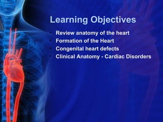
  • 1. Learning Objectives • Review anatomy of the heart • Formation of the Heart • Congenital heart defects • Clinical Anatomy - Cardiac Disorders
  • 2. Blood Flow Heart  Lungs  Heart  Body
  • 3. Vena Cava - AORTA - vein artery Atria Ventricles
  • 4.
  • 5. UNoxygenated blood enters the atrium on the right side of the heart. Unoxygenated blood comes in from the top of the body through the superior vena cava. Unoxygenated blood comes in from the lower body though the inferior vena cava.
  • 6. While the unoxygenated blood is in the right atrium, the tricuspid valve is closed to keep the blood from flowing down to the ventricle.
  • 7. The atrium contracts and the tricuspid valve opens, forcing the blood down into the ventricle.
  • 8. The tricuspid valve closes again so that blood cannot move back up into the atrium.
  • 9. The ventricle contracts. This forces the unoxygenated blood through the pulmonary valve and into the pulmonary arteries.
  • 10. The right pulmonary artery takes the unoxygenated blood to the right lung. The left pulmonary artery takes the unoxygenated blood to the left lung. THE PULMONARY ARTERIES ARE THE ONLY ARTERIES THAT CARRY UNOXYGENEATED BLOOD.
  • 11. In the lungs, the carbon dioxide in the blood diffuses into the alveoli. The oxygen in the lungs diffuses into the blood. This is called gas http://www.webmd.com/hw/health_guide_atoz/tp10237.asp exchange.
  • 12. Oxygenated blood from the lungs enters the heart through the left atrium. The mitral valve is closed to keep the blood from going into the ventricle.
  • 13. Oxygenated blood from the right lung returns to the heart through the right pulmonary vein. Oxygenated blood from the left lung returns to the heart through the left pulmonary vein. THE PULMONARY VEINS ARE THE ONLY VEINS THAT CARRY OXYGENATED BLOOD.
  • 14. The left atrium contracts. This forces the oxygenated blood through the mitral valve into the Left ventricle.
  • 15. The mitral valve closes again. This keeps the oxygenated blood from moving back up into the atrium.
  • 16. Oxygenated blood is forced into the aorta to be carried to the rest of the body.
  • 17. Oxygenated blood is carried to all body cells where oxygen diffuses into the cells and carbon dioxide diffuses into the blood. Blood carrying carbon dioxide then returns to the heart.
  • 19. Meanwhile… While the blood is moving oxygen and carbon dioxide around, it is also moving nutrients, other wastes, hormones, and antibodies at the same time.
  • 21. Formation of the Heart • Mesoderm divides into two layers • Mesoderm = one of the primary germ cell layers in the early embryo • Heart precursor cells come from one of those two mesoderm layers (cardiogenic mesoderm) • Heart precursor cells form a single heart tube by day 22 of embryogenesis
  • 22. Formation of the Heart • These cells differentiate into the endocardium and myocardium • Endocardium = innermost layer that lines the heart chambers and valves valves • Myocardium = the muscular layer of the atria and ventricles • The heart tube grows and elongates • Primitive heart begins to form around day 22‐ 23
  • 23. • The heart tube begins to bulge into primitive heart chambers and undergoes right ward looping • Followed by proper valve positioning and chamber formation
  • 25. CYANOSIS  a physical sign causing bluish discoloration of the skin and mucous membranes.  caused by a lack of oxygen in the blood.  associated with cold temperatures, heart failure, lung diseases, and smothering. It is seen in infants at birth as  a result of heart defects, defects  respiratory distress syndrome,  or lung and breathing problems.
  • 26. • The blue discoloration of cyanosis is seen most readily in the beds of the fingernails and toenails, and on the lips and tongue. • It often appears transiently as a result of slowed blood flow through the skin due to the cold. As such, it is not a serious symptom. • However, in other cases cyanosis is a serious symptom of underlying disease.
  • 27. Congenital Heart Defects • Abnormalities in heart present at birth • Affects 8:1000 live births • Examples: • Ventricular Septal Defect • Atrial Septal Defect • Coarctation of the Aorta • Tetralogy of Fallot • Transposition of the Great Arteries
  • 28.
  • 29. Ventricular Septal Defect (VSD) • Most common congenital cardiac anomaly • There is a hole between the two ventricles • Hole can vary in size and location • Oxygenated blood forced through hole from left ventricle to right ventricle then returns to the lungs even though it already carries oxygen • Consequences • Volume load causes enlargement of both ventricles and the pulmonary artery and exposes right ventricle and pulmonary arteries to high pressures
  • 31.
  • 32. o Small VSDs may close on their own and usually does not cause major problems. o These are the kinds that can close at any time during childhood. o A membranous VSD is found in the upper portion of the interventricular septum. o A muscular VSD is found in the lower part of the septum. o Medium and large sized VSDs are unlikely to close spontaneously. o Surgery or other interventional procedures may be required to close these defects. Inlet and Outlet VSDs are less common types and are present where the blood enters or leaves the ventricles.
  • 33. VSD Signs and Symptoms • Heart murmur • Difficulty maintaining weight • Increased breathing rate • Lower energy and easy tiring Ventricular Septal Defect - Animation http://www.medindia.net/animation/Ventricular_Septal_Defec
  • 34. • Some of the infants may show poor weight gain, shortness of breath or even bluish discoloration of the lips, nails or skin. • Most of the small VSDs may go unnoticed. • A murmur can be heard with a stethoscope when the baby is a few weeks old. • Untreated moderate to large VSDs may lead to severe complications in a child. • Heart failure may result from the constant overload of the right ventricles. • Arrhythmias and Pulmonary hypertension can also be an outcome of the high volume of blood flowing through the right ventricle. • It is rare that these defects can go unnoticed and so complications are rather rare.