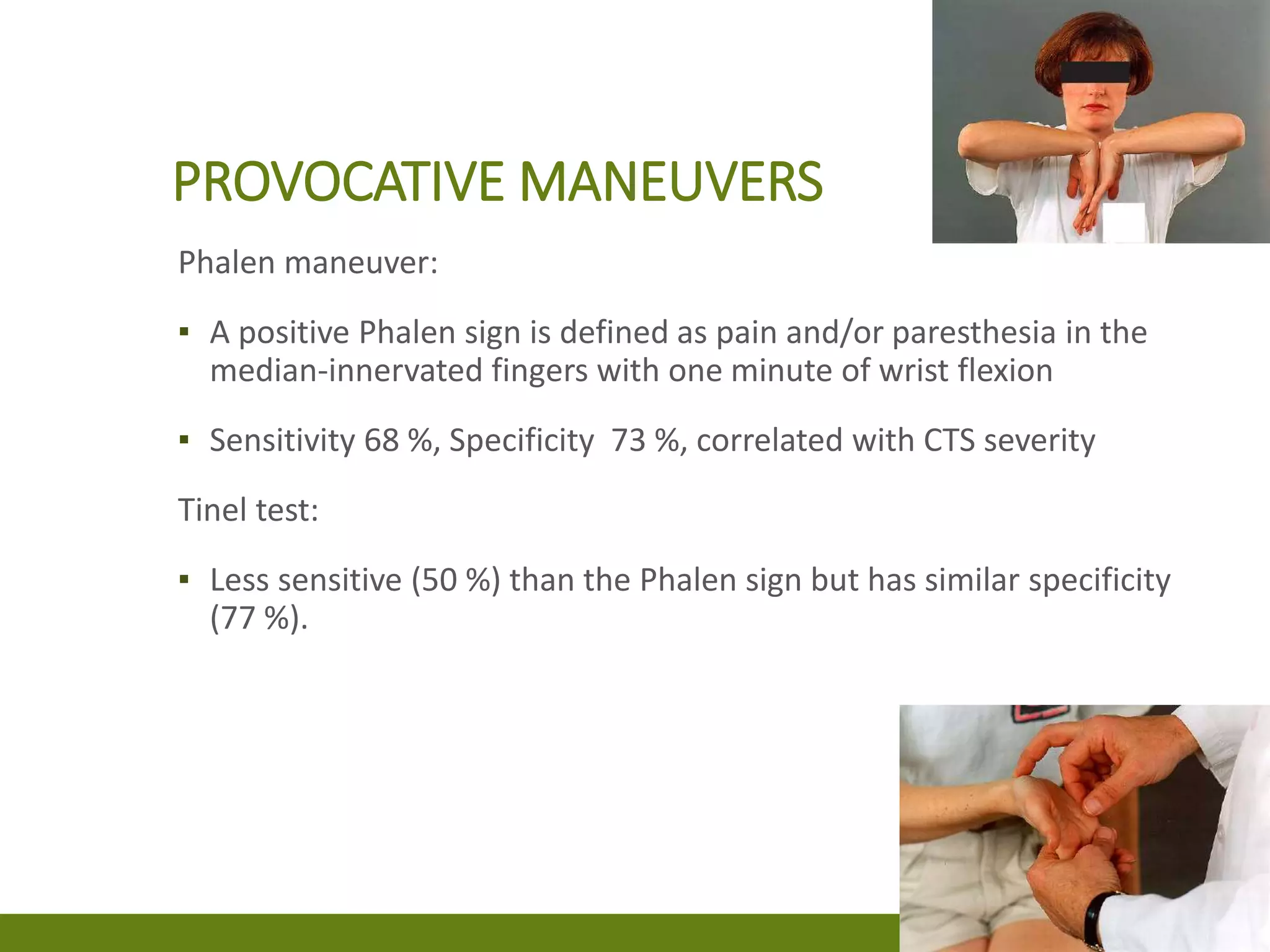
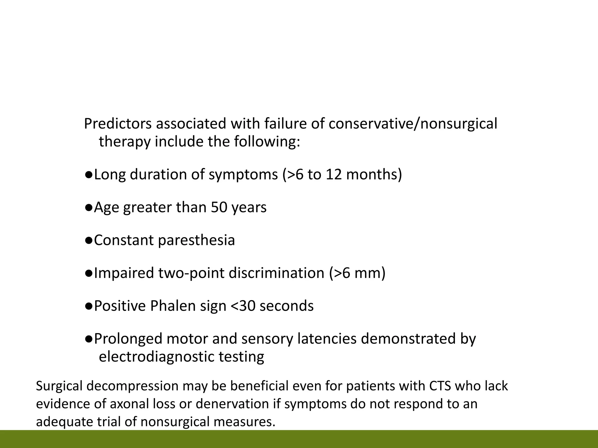
CARPAL TUNNEL SYNDROME
▪ Carpal tunnel syndrome (CTS) refers to the complex of
symptoms and signs brought on by compression of the
median nerve as it travels through the carpal tunnel.
▪ Patients commonly experience pain, paresthesia, and, less
commonly, weakness in the median nerve distribution.
▪ CTS is the most common compressive focal mononeuropathy
seen in clinical practice.
▪ When compression of the median nerve occurs, ischemia and
mechanical disruption of the nerve follow.
▪ The median nerve provides motor and sensory innervation to
the hand.
▪ The median nerve emerges with contributions from the C6,
C7, C8, and T1 nerve roots.
▪ Roots C6 and C7 supply the median sensory fibers that
provide sensation to the thenar eminence and the first three
and a half digits of the hand.
▪ Roots C8 and T1 supply the motor fibers to the muscles of the
forearm and hand that are innervated by the median nerve.
▪ Inflammation and compression of the median nerve most
commonly occurs within the carpal tunnel.
▪ Rarely, median entrapment can also occur more proximally in
the region of the elbow or just distal to the elbow.
▪ The muscles of the hand that are innervated by the median
nerve are the abductor pollicis brevis, the flexor pollicis brevis
(superficial head), the opponens pollicis, and the first and
second lumbricals.
▪ Although these muscles can be affected in patients with
severe or long-standing CTS, the lumbrical is sometimes
spared.
PATHOPHYSIOLOGY
▪ The pathophysiology of CTS is multifactorial.
▪ Increased pressure in the intracarpal canal plays a key role in
the development of clinical CTS .
▪ Anatomic compression and/or inflammation are possible
mechanisms.
▪ Increased pressure in the carpal tunnel can injure the nerve
directly, impair axonal transport, or compress vessels in the
perineurium and cause median nerve ischemia.
▪ Anatomic compression may result from a noninflammatory
fibrosis affecting the subsynovial connective tissue that
surrounds the flexor tendons.
▪ Other possible causes of compression include congenitally
small anatomic space, mass lesions (such as a cyst, neoplasm,
or persistent median artery) and edema or inflammatory
conditions that result from systemic illness such as
rheumatoid arthritis.
▪ Upper extremity posture also influences carpal tunnel
pressure.
▪ The lowest carpal tunnel pressure is seen when the wrist is in
a neutral or slightly flexed position, and it increases
proportionately with deviation from this posture
▪ Prevalence of CTS in the general population is 1 to 5 percent.
▪ CTS is more frequent in women (0.7 to 9.2 percent) than in
men (0.4 to 2.1 percent).
▪ The prevalence of CTS appears to be highest in obese women
and lowest in thin and normal-sized men.
▪ The female-to-male ratio for CTS prevalence is approximately
3:1
RISK FACTORS
▪ Obesity
▪ Female gender (smaller CT)
▪ Pregnancy (Accumulation of
fluid)
▪ Diabetes
▪ Rheumatoid arthritis
▪ Osteoarthritis of the hand
▪ Acromegaly
▪ Hypothyroidism (moderate)
▪ Connective tissue diseases
▪ Preexisting median
mononeuropathy
▪ Genetic predisposition (B/L)
▪ Aromatase inhibitor use
▪ Workplace factors
▪ Trauma
CLINICAL FEATURES
▪ The hallmark of classic CTS is pain or paresthesia (numbness and
tingling) in a distribution that includes the median nerve
territory, with involvement of the first three digits and the radial
half of the fourth digit.
▪ The symptoms of CTS are typically worse at night and often
awaken patients from sleep.
▪ Some patients react to these symptoms by shaking or wringing
their hands or by placing them under warm running water.
▪ Fixed sensory loss is usually a late finding characterized by a
distinctive clinical pattern that involves the median-innervated
fingers and spares the thenar eminence.
▪ This pattern occurs because the palmar sensory cutaneous
nerve arises proximal to the wrist and passes over, rather than
through, the carpal tunnel
▪ Although the sensory symptoms of CTS are
▪ Usually limited to the median-innervated fingers.
▪ Localized to the wrist or involve the entire hand.
▪ Rarely radiate proximally into the forearm, and
▪ Less frequently to radiate above the elbow to the shoulder,
▪ But the neck is not affected
PROVOCATING FACTORS
▪ Activities that involve flexing or extending the wrist or raising
the arms,
▪ Such as driving, reading, typing, and holding a telephone
▪ Bilateral CTS is common at first presentation with 65%
incidence.
▪ Clinical experience suggests that unilateral CTS is
encountered more frequently.
▪ In some cases, involvement is subclinical on one side and
symptomatic on the other.
▪ The clinical course of CTS may follow an alternating pattern
with periods of remission and exacerbation .
▪ In some cases, there is progression from intermittent to
persistent sensory complaints in the hand as CTS worsens,
and later to the development of motor symptoms in the
hand.
▪ Motor involvement leads to complaints of weakness or
clumsiness when using the hands, such as difficulty holding
objects, turning keys or doorknobs, buttoning clothing, or
opening jar lids.
▪ O/E: weakness of thumb abduction and opposition, and
atrophy of the thenar eminence.
EVALUATION AND DIAGNOSIS
▪ CTS is a clinical diagnosis.
▪ The diagnosis is suspected when the characteristic symptoms and
signs are present.
▪ The most important of these are nocturnal pain or paresthesia in
the distribution of the median nerve.
Standard symptoms: Dull, aching discomfort in the hand,
forearm, or upper arm, Paresthesia in the hand,
Weakness or clumsiness of the hand , Occurrence of any
of these symptoms in the median distribution
Provocative factors: Sleep, Sustained hand or arm
positions, Repetitive actions of the hand or wrist
Mitigating factors: Changes in hand posture, Shaking the
hand
PHYSICAL EXAMINATION
▪ Objective sensory and motor deficits corresponding to the
median nerve-innervated regions of the hand may be
present, but their absence does not rule out the diagnosis of
CTS.
▪ Sensation should be tested in all regions of the hand,
forearm, and upper arm.
▪ Objective weakness can occur in advanced CTS and is limited
to muscles of the thenar eminence.
▪ This manifests principally as weakness of thumb abduction
and thumb opposition.
▪ Atrophy of the thenar eminence may be present.
PROVOCATIVE MANEUVERS
Phalen maneuver:
▪ A positive Phalen sign is defined as pain and/or paresthesia in the
median-innervated fingers with one minute of wrist flexion
▪ Sensitivity 68 %, Specificity 73 %, correlated with CTS severity
Tinel test:
▪ Less sensitive (50 %) than the Phalen sign but has similar specificity
(77 %).
▪ The manual carpal compression test is performed by applying
pressure over the transverse carpal ligament, and it is
deemed positive if paresthesia occur within 30 seconds of
applying pressure.
▪ The average sensitivity (64%) and specificity (83%).
▪ The hand elevation test involves raising the hands above the
head for one minute.
▪ The test is positive if it reproduces the symptoms of CTS.
▪ The sensitivity and specificity appear to be similar to or
slightly better than those reported for Tinel and Phalen
maneuvers.
ELECTRODIAGNOSTIC TESTING
▪ Electro diagnostic studies are essential if surgical treatment
for CTS is being considered in order
1. To guide appropriate selection of patients for carpal tunnel
release (by confirming the diagnosis of CTS and the presence of
moderate to severe median nerve injury) and
2. To inform prognosis (severe nerve injury increases the risk of a
prolonged or incomplete recovery after surgery, but also
predicts the failure of non operative management).
NERVE CONDUCTION STUDIES
▪ Impaired median nerve conduction across the carpal tunnel in the
context of normal conduction elsewhere.
▪ Measure of conduction velocity across the carpal tunnel, as well as
determination of the amplitude of sensory and motor responses.
▪ Mild CTS may not produce any nerve conduction abnormalities.
▪ With increased compression of the median nerve, focal demyelination 
local conduction block and/or slowing of motor and sensory conduction
across the wrist.
▪ With even greater compression, the axonal loss  reduced amplitudes.
▪ Sensory fibers seem to be more sensitive to compression than motor
fibers.
▪ As a result, sensory fibers typically demonstrate changes on NCS earlier
than do motor fibers.
▪ Nerve compression results in damage to the myelin sheath
and manifests as DELAYED DISTAL LATENCIES and SLOWED
CONDUCTION VELOCITIES.
▪ With sustained or more severe compression, axon loss may
also occur, resulting in a reduction of the median nerve
compound motor or sensory nerve action potential
amplitude.
▪ Results obtained are compared with age-dependent normal
values, and other nerves of the same hand or the C/L hand.
▪ In particular, the ulnar nerve and /or radial nerve are also
evaluated to ensure that any abnormalities seen in the
median nerve are specific to that nerve and not part of a
more widespread disorder, such as a peripheral neuropathy.
ROUTINE NCS FOR THE DIAGNOSIS OF CTS
TYPICALLY INCLUDE THE FOLLOWING STUDIES
▪ Median motor conduction study recording from the abductor
pollicis brevis while stimulating at the wrist and elbow
▪ Ulnar motor conduction study recording from the abductor
digiti minimi while stimulating at the wrist and at the elbow
above and below the ulnar groove
▪ Median sensory response recording from digit two or three
while stimulating the wrist
▪ Ulnar sensory response recording from digit five while
stimulating the wrist
▪ Radial sensory response recording from the snuffbox while
stimulating over the lateral radius
IF ROUTINE NCS WAS NORMAL
ADDITIONAL COMPARISON STUDIES:
▪ Palmar mixed-nerve study, comparing palm-to-wrist peak
latencies of median and ulnar nerves, each recorded 8 cm
from the stimulating electrodes
▪ Second lumbrical (median) versus interossei (ulnar) distal
motor latencies
▪ Digit four sensory latencies stimulating the median and ulnar
nerves at the wrist individually at identical distances.
▪ SENSITIVITY: 56 to 85 %, SPECIFICITY: 94 to 99 %
MARTIN-GRUBER ANASTOMOSIS
▪ A subgroup of motor fibers splits from the median nerve in the forearm and
anastomose with the ulnar nerve as it travels through the forearm into the
hand.
▪ The median-to-ulnar motor fibers that make up this anastomosis innervate
the intrinsic muscles of the hand.
▪ Most often identified during ulnar nerve testing.
▪ During median nerve motor studies, amplitude of the CMAP is higher with
stimulation at the proximal elbow site than with stimulation at the wrist.
▪ In the setting of median nerve entrapment at the wrist (CTS), a surprisingly
fast median nerve conduction velocity in the forearm can be seen.
▪ Bypassing the site of entrapment by taking this circuitous route with the
ulnar nerve.
ELECTROMYOGRAPHY
▪ Patients who have classic signs and symptoms of
CTS and confirmatory findings on NCS, EMG is not
necessary.
▪ EMG is most useful
1. To exclude other conditions, such as polyneuropathy,
plexopathy, and radiculopathy, and
2. To assess severity if surgical decompression is being
considered
▪ Looks for evidence of pathologic changes in the
muscles innervated by the median nerve, typically
assessing the abductor pollicis brevis muscle.
▪ When secondary axonal loss is present, EMG may reveal
either
▪ Active denervation (eg, spontaneous activity such as fibrillation
potentials, positive sharp waves, and fasciculation potentials) or
▪ Chronic changes that indicate denervation with subsequent
reinnervation (eg, changes in motor unit action potential
amplitudes, durations, and recruitment).
▪ Supportive of the diagnosis of CTS in the context of normal
findings in both nonmedian-innervated muscles and
proximal median nerve-innervated muscles
One suggested protocol for EMG evaluation of CTS involves needle
examination of the following muscles:
▪ Abductor pollicis brevis
▪ Two or more C6-C7 innervated muscles (eg, pronator teres, triceps
brachii, extensor digitorum communis) to look for evidence of cervical
radiculopathy
Additional muscles are investigated if the abductor pollicis brevis is
abnormal:
▪ Two or more proximal median-innervated muscles (eg, flexor carpi
radialis, pronator teres, flexor pollicis longus) to rule out a proximal
median neuropathy
▪ Two or more lower trunk C8-T1 nonmedian-innervated muscles (eg, first
dorsal interosseous, extensor indicis proprius) to rule out brachial
plexopathy, polyneuropathy, and C8 to T1 radiculopathy
IMAGING
▪ Structural abnormality of the wrist such as tumor, deformity, or other bone
or joint disease.
▪ Neuromuscular ultrasound shows significantly increased cross-sectional area
of the median nerve.
▪ SENSITIVITY: 65 to 97 % (low for > 80 years), SPECIFICITY: 73 to 98 %.
▪ Thickening of the median nerve, best expressed as an increase in the cross-
sectional area of the median nerve at the carpal tunnel inlet (more than 13
mm; normal <10–13 mm), or flattening of the nerve at the level of the
hamate are the best diagnostic criteria
▪ Magnetic resonance imaging (MRI) can detect abnormalities
of the median nerve, flexor tendons, vascular structures, and
transverse carpal ligament in the region of the carpal tunnel.
▪ However, the diagnostic utility of MRI for CTS remains
uncertain.
▪ Thus, MRI is reserved for unusual cases to rule out a mass
lesion
DIFFERENTIAL DIAGNOSIS
Neurologic, musculoskeletal, and vascular conditions that can present with
pain, paresthesia, sensory loss, or weakness involving the shoulder, arm,
or hand.
▪ Cervical radiculopathy, particularly with C6 or C7 nerve root
involvement (neck pain; the exacerbation of symptoms with neck
movement; radiation of pain from the neck into the shoulder and arm;
reduced reflexes mediated by the C6/C7 nerve roots).
▪ Cervical spondylotic myelopathy and cervical polyradiculopathy
(progress to bilateral sensory loss and motor dysfunction in the hands).
▪ Brachial plexopathy with individual nerve involvement.
▪ Median neuropathy occasionally occurs in the proximal forearm where
the nerve passes through the pronator teres muscle.
▪ Motor neuron disease (absence of pain argues against CTS)
▪ Fibromyalgia (chronic widespread musculoskeletal pain and
fatigue).
▪ Pain from a ligamentous disruption is typically localized.
▪ Forearm or hand compartment syndrome can present as
pain with passive stretch and a firm compartment palpated
over the forearm or hand.
APPROACH TO MANAGEMENT
▪ Based upon the acuity and severity of clinical symptoms and
the degree of neurogenic injury as assessed by
electrodiagnostic studies.
▪ For symptom relief of CTS: Splinting, glucocorticoid injections,
and oral glucocorticoids are useful.
▪ But surgery is the treatment of choice for patients with
evidence of ongoing nerve damage in the absence of a
reversible etiology.
▪ Treatment of potential predisposing conditions is warranted
in patients with CTS, but no proof that treating these
conditions will improve the symptoms or the course of CTS
INITIAL THERAPY
GRADING THE SEVERITY OF CTS
▪ Combined treatment employing splinting with glucocorticoid
injection(s), oral glucocorticoids, or other nonsurgical
interventions may be more effective than the use of any
single modality.
For patients who choose initial nocturnal splinting but remain
symptomatic at one month.
Suggest continuation of splinting for another one to two months,
Add a single injection of methylprednisolone (20 to 40 mg), If they
decline
Add oral glucocorticoids (eg, prednisone 20 mg daily for 10 to 14
days) (< 4 weeks)
Physical and Occupational therapy techniques (eg, carpal bone
mobilization or nerve gliding) or yoga
Predictors associated with failure of conservative/nonsurgical
therapy include the following:
●Long duration of symptoms (>6 to 12 months)
●Age greater than 50 years
●Constant paresthesia
●Impaired two-point discrimination (>6 mm)
●Positive Phalen sign <30 seconds
●Prolonged motor and sensory latencies demonstrated by
electrodiagnostic testing
Surgical decompression may be beneficial even for patients with CTS who lack
evidence of axonal loss or denervation if symptoms do not respond to an
adequate trial of nonsurgical measures.
FAILURE OF SURGICAL
DECOMPRESSION
▪ Persistent symptoms of CTS after decompression surgery
may be caused by circumferential fibrosis or by reconstitution
or incomplete release of the transverse carpal ligament
▪ In such cases revision surgery may be indicated.
PREGNANCY
▪ CTS may develop during pregnancy, particularly during the
third trimester.
▪ In most cases, the symptoms gradually resolve over a period
of weeks after delivery.
▪ For women who develop CTS during pregnancy, recommend
nocturnal wrist splinting.
▪ Surgical decompression is rarely indicated during pregnancy
since the disease often resolves postpartum
CONSERVATIVE TREATMENT
▪ Include splinting, oral or injection glucocorticoids, physical
and occupational therapy techniques (eg, carpal bone
mobilization and nerve-gliding exercises), yoga.
▪ For patients with mild to moderate CTS , the rate of
successful outcomes with nonsurgical therapy ranges from 20
to 93 %.
WRIST SPLINTING
▪ A wrist splint or brace maintains the wrist in a neutral position,
thus preventing prolonged flexion or extension of the wrist.
▪ Splinting may limit activities that raise pressure within the carpal
tunnel or reduce its cross-sectional area.
▪ In mildly symptomatic patients: Effective in reducing CTS
symptoms, and it may delay or eliminate the need for surgery.
▪ Similar outcome (slightly longer time) at six months compared
with a single glucocorticoid injection.
▪ Nocturnal use of wrist splints is more effective than no
treatment for short-term symptom benefit.
▪ Splints are usually worn at night, but they can be worn
continuously.
▪ Night splinting alone can reduce symptom severity and
improve median nerve conduction velocities.
▪ Full-time splinting has been reported to improve median
nerve conduction, but it may not improve symptoms when
compared with night-only splinting.
GOOD RESPONDERS:
▪ Shorter duration of symptoms (one year or less) and
▪ Less severe nocturnal paresthesia.
GLUCOCORTICOID INJECTION
▪ Injection of glucocorticoids into the region of the carpal
tunnel is intended to reduce tissue inflammation and aid
recovery.
▪ Patient when amenable to a minimally invasive option,
injection is an alternative to splinting for short-term symptom
relief (one to three months).
▪ Glucocorticoids (single methylprednisolone acetate injection
20 mg) can be injected proximal to or distal to the carpal
tunnel.
▪ Generally safe, BUT risks, including exacerbation of median
nerve compression, accidental injection into the median or
ulnar nerves, and digital flexor tendon rupture.
▪ Pass needle through skin quickly with the
needle bevel parallel with nerve and then
inject a small amount of fluid just under the
skin.
▪ Wait 20 – 30 seconds then gently advance the
needle while getting the patient to gently
flex and extend their fingers.
▪ Continue injecting. If fluid bulges the skin and
fat it is superficial to the deep fascia.
▪ When the flexor tendons can be felt moving
against the end of the needle the needle is
deep enough.
▪ If the needle moves with finger ROM it is in
the tendon!!! and should be moved.
ORAL GLUCOCORTICOIDS
▪ Effective for short-term improvement of CTS symptoms.
▪ Oral prednisolone was less effective than glucocorticoid
injection.
▪ Clinical and electrodiagnostic improvement for up to 12
months.
▪ Oral prednisone (20 mg daily for seven days, followed by 10
mg per day for seven days) was associated with significant
improvement in symptoms.
▪ Perineural dextrose injections
▪ Yoga
▪ Carpal bone mobilization
▪ Nerve gliding exercises or maneuvers
▪ Ultrasound therapy & and electrical stimulation promote
recovery after nerve and tendon injuries.
▪ NSAIDs and other oral medications
▪ Electrical, magnetic, and laser therapy
carpal tunnel syndrome

carpal tunnel syndrome

  • 1.
  • 2.
    ▪ Carpal tunnelsyndrome (CTS) refers to the complex of symptoms and signs brought on by compression of the median nerve as it travels through the carpal tunnel. ▪ Patients commonly experience pain, paresthesia, and, less commonly, weakness in the median nerve distribution. ▪ CTS is the most common compressive focal mononeuropathy seen in clinical practice. ▪ When compression of the median nerve occurs, ischemia and mechanical disruption of the nerve follow.
  • 4.
    ▪ The mediannerve provides motor and sensory innervation to the hand. ▪ The median nerve emerges with contributions from the C6, C7, C8, and T1 nerve roots. ▪ Roots C6 and C7 supply the median sensory fibers that provide sensation to the thenar eminence and the first three and a half digits of the hand. ▪ Roots C8 and T1 supply the motor fibers to the muscles of the forearm and hand that are innervated by the median nerve.
  • 5.
    ▪ Inflammation andcompression of the median nerve most commonly occurs within the carpal tunnel. ▪ Rarely, median entrapment can also occur more proximally in the region of the elbow or just distal to the elbow. ▪ The muscles of the hand that are innervated by the median nerve are the abductor pollicis brevis, the flexor pollicis brevis (superficial head), the opponens pollicis, and the first and second lumbricals. ▪ Although these muscles can be affected in patients with severe or long-standing CTS, the lumbrical is sometimes spared.
  • 6.
    PATHOPHYSIOLOGY ▪ The pathophysiologyof CTS is multifactorial. ▪ Increased pressure in the intracarpal canal plays a key role in the development of clinical CTS . ▪ Anatomic compression and/or inflammation are possible mechanisms. ▪ Increased pressure in the carpal tunnel can injure the nerve directly, impair axonal transport, or compress vessels in the perineurium and cause median nerve ischemia.
  • 7.
    ▪ Anatomic compressionmay result from a noninflammatory fibrosis affecting the subsynovial connective tissue that surrounds the flexor tendons. ▪ Other possible causes of compression include congenitally small anatomic space, mass lesions (such as a cyst, neoplasm, or persistent median artery) and edema or inflammatory conditions that result from systemic illness such as rheumatoid arthritis. ▪ Upper extremity posture also influences carpal tunnel pressure. ▪ The lowest carpal tunnel pressure is seen when the wrist is in a neutral or slightly flexed position, and it increases proportionately with deviation from this posture
  • 8.
    ▪ Prevalence ofCTS in the general population is 1 to 5 percent. ▪ CTS is more frequent in women (0.7 to 9.2 percent) than in men (0.4 to 2.1 percent). ▪ The prevalence of CTS appears to be highest in obese women and lowest in thin and normal-sized men. ▪ The female-to-male ratio for CTS prevalence is approximately 3:1
  • 9.
    RISK FACTORS ▪ Obesity ▪Female gender (smaller CT) ▪ Pregnancy (Accumulation of fluid) ▪ Diabetes ▪ Rheumatoid arthritis ▪ Osteoarthritis of the hand ▪ Acromegaly ▪ Hypothyroidism (moderate) ▪ Connective tissue diseases ▪ Preexisting median mononeuropathy ▪ Genetic predisposition (B/L) ▪ Aromatase inhibitor use ▪ Workplace factors ▪ Trauma
  • 10.
    CLINICAL FEATURES ▪ Thehallmark of classic CTS is pain or paresthesia (numbness and tingling) in a distribution that includes the median nerve territory, with involvement of the first three digits and the radial half of the fourth digit. ▪ The symptoms of CTS are typically worse at night and often awaken patients from sleep. ▪ Some patients react to these symptoms by shaking or wringing their hands or by placing them under warm running water. ▪ Fixed sensory loss is usually a late finding characterized by a distinctive clinical pattern that involves the median-innervated fingers and spares the thenar eminence. ▪ This pattern occurs because the palmar sensory cutaneous nerve arises proximal to the wrist and passes over, rather than through, the carpal tunnel
  • 11.
    ▪ Although thesensory symptoms of CTS are ▪ Usually limited to the median-innervated fingers. ▪ Localized to the wrist or involve the entire hand. ▪ Rarely radiate proximally into the forearm, and ▪ Less frequently to radiate above the elbow to the shoulder, ▪ But the neck is not affected
  • 12.
    PROVOCATING FACTORS ▪ Activitiesthat involve flexing or extending the wrist or raising the arms, ▪ Such as driving, reading, typing, and holding a telephone
  • 13.
    ▪ Bilateral CTSis common at first presentation with 65% incidence. ▪ Clinical experience suggests that unilateral CTS is encountered more frequently. ▪ In some cases, involvement is subclinical on one side and symptomatic on the other.
  • 14.
    ▪ The clinicalcourse of CTS may follow an alternating pattern with periods of remission and exacerbation . ▪ In some cases, there is progression from intermittent to persistent sensory complaints in the hand as CTS worsens, and later to the development of motor symptoms in the hand. ▪ Motor involvement leads to complaints of weakness or clumsiness when using the hands, such as difficulty holding objects, turning keys or doorknobs, buttoning clothing, or opening jar lids. ▪ O/E: weakness of thumb abduction and opposition, and atrophy of the thenar eminence.
  • 15.
    EVALUATION AND DIAGNOSIS ▪CTS is a clinical diagnosis. ▪ The diagnosis is suspected when the characteristic symptoms and signs are present. ▪ The most important of these are nocturnal pain or paresthesia in the distribution of the median nerve. Standard symptoms: Dull, aching discomfort in the hand, forearm, or upper arm, Paresthesia in the hand, Weakness or clumsiness of the hand , Occurrence of any of these symptoms in the median distribution Provocative factors: Sleep, Sustained hand or arm positions, Repetitive actions of the hand or wrist Mitigating factors: Changes in hand posture, Shaking the hand
  • 16.
    PHYSICAL EXAMINATION ▪ Objectivesensory and motor deficits corresponding to the median nerve-innervated regions of the hand may be present, but their absence does not rule out the diagnosis of CTS. ▪ Sensation should be tested in all regions of the hand, forearm, and upper arm. ▪ Objective weakness can occur in advanced CTS and is limited to muscles of the thenar eminence. ▪ This manifests principally as weakness of thumb abduction and thumb opposition. ▪ Atrophy of the thenar eminence may be present.
  • 18.
    PROVOCATIVE MANEUVERS Phalen maneuver: ▪A positive Phalen sign is defined as pain and/or paresthesia in the median-innervated fingers with one minute of wrist flexion ▪ Sensitivity 68 %, Specificity 73 %, correlated with CTS severity Tinel test: ▪ Less sensitive (50 %) than the Phalen sign but has similar specificity (77 %).
  • 19.
    ▪ The manualcarpal compression test is performed by applying pressure over the transverse carpal ligament, and it is deemed positive if paresthesia occur within 30 seconds of applying pressure. ▪ The average sensitivity (64%) and specificity (83%). ▪ The hand elevation test involves raising the hands above the head for one minute. ▪ The test is positive if it reproduces the symptoms of CTS. ▪ The sensitivity and specificity appear to be similar to or slightly better than those reported for Tinel and Phalen maneuvers.
  • 20.
    ELECTRODIAGNOSTIC TESTING ▪ Electrodiagnostic studies are essential if surgical treatment for CTS is being considered in order 1. To guide appropriate selection of patients for carpal tunnel release (by confirming the diagnosis of CTS and the presence of moderate to severe median nerve injury) and 2. To inform prognosis (severe nerve injury increases the risk of a prolonged or incomplete recovery after surgery, but also predicts the failure of non operative management).
  • 21.
    NERVE CONDUCTION STUDIES ▪Impaired median nerve conduction across the carpal tunnel in the context of normal conduction elsewhere. ▪ Measure of conduction velocity across the carpal tunnel, as well as determination of the amplitude of sensory and motor responses. ▪ Mild CTS may not produce any nerve conduction abnormalities. ▪ With increased compression of the median nerve, focal demyelination  local conduction block and/or slowing of motor and sensory conduction across the wrist. ▪ With even greater compression, the axonal loss  reduced amplitudes. ▪ Sensory fibers seem to be more sensitive to compression than motor fibers. ▪ As a result, sensory fibers typically demonstrate changes on NCS earlier than do motor fibers.
  • 22.
    ▪ Nerve compressionresults in damage to the myelin sheath and manifests as DELAYED DISTAL LATENCIES and SLOWED CONDUCTION VELOCITIES. ▪ With sustained or more severe compression, axon loss may also occur, resulting in a reduction of the median nerve compound motor or sensory nerve action potential amplitude. ▪ Results obtained are compared with age-dependent normal values, and other nerves of the same hand or the C/L hand. ▪ In particular, the ulnar nerve and /or radial nerve are also evaluated to ensure that any abnormalities seen in the median nerve are specific to that nerve and not part of a more widespread disorder, such as a peripheral neuropathy.
  • 23.
    ROUTINE NCS FORTHE DIAGNOSIS OF CTS TYPICALLY INCLUDE THE FOLLOWING STUDIES ▪ Median motor conduction study recording from the abductor pollicis brevis while stimulating at the wrist and elbow ▪ Ulnar motor conduction study recording from the abductor digiti minimi while stimulating at the wrist and at the elbow above and below the ulnar groove ▪ Median sensory response recording from digit two or three while stimulating the wrist ▪ Ulnar sensory response recording from digit five while stimulating the wrist ▪ Radial sensory response recording from the snuffbox while stimulating over the lateral radius
  • 24.
    IF ROUTINE NCSWAS NORMAL ADDITIONAL COMPARISON STUDIES: ▪ Palmar mixed-nerve study, comparing palm-to-wrist peak latencies of median and ulnar nerves, each recorded 8 cm from the stimulating electrodes ▪ Second lumbrical (median) versus interossei (ulnar) distal motor latencies ▪ Digit four sensory latencies stimulating the median and ulnar nerves at the wrist individually at identical distances. ▪ SENSITIVITY: 56 to 85 %, SPECIFICITY: 94 to 99 %
  • 25.
    MARTIN-GRUBER ANASTOMOSIS ▪ Asubgroup of motor fibers splits from the median nerve in the forearm and anastomose with the ulnar nerve as it travels through the forearm into the hand. ▪ The median-to-ulnar motor fibers that make up this anastomosis innervate the intrinsic muscles of the hand. ▪ Most often identified during ulnar nerve testing. ▪ During median nerve motor studies, amplitude of the CMAP is higher with stimulation at the proximal elbow site than with stimulation at the wrist. ▪ In the setting of median nerve entrapment at the wrist (CTS), a surprisingly fast median nerve conduction velocity in the forearm can be seen. ▪ Bypassing the site of entrapment by taking this circuitous route with the ulnar nerve.
  • 26.
    ELECTROMYOGRAPHY ▪ Patients whohave classic signs and symptoms of CTS and confirmatory findings on NCS, EMG is not necessary. ▪ EMG is most useful 1. To exclude other conditions, such as polyneuropathy, plexopathy, and radiculopathy, and 2. To assess severity if surgical decompression is being considered ▪ Looks for evidence of pathologic changes in the muscles innervated by the median nerve, typically assessing the abductor pollicis brevis muscle.
  • 27.
    ▪ When secondaryaxonal loss is present, EMG may reveal either ▪ Active denervation (eg, spontaneous activity such as fibrillation potentials, positive sharp waves, and fasciculation potentials) or ▪ Chronic changes that indicate denervation with subsequent reinnervation (eg, changes in motor unit action potential amplitudes, durations, and recruitment). ▪ Supportive of the diagnosis of CTS in the context of normal findings in both nonmedian-innervated muscles and proximal median nerve-innervated muscles
  • 28.
    One suggested protocolfor EMG evaluation of CTS involves needle examination of the following muscles: ▪ Abductor pollicis brevis ▪ Two or more C6-C7 innervated muscles (eg, pronator teres, triceps brachii, extensor digitorum communis) to look for evidence of cervical radiculopathy Additional muscles are investigated if the abductor pollicis brevis is abnormal: ▪ Two or more proximal median-innervated muscles (eg, flexor carpi radialis, pronator teres, flexor pollicis longus) to rule out a proximal median neuropathy ▪ Two or more lower trunk C8-T1 nonmedian-innervated muscles (eg, first dorsal interosseous, extensor indicis proprius) to rule out brachial plexopathy, polyneuropathy, and C8 to T1 radiculopathy
  • 29.
    IMAGING ▪ Structural abnormalityof the wrist such as tumor, deformity, or other bone or joint disease. ▪ Neuromuscular ultrasound shows significantly increased cross-sectional area of the median nerve. ▪ SENSITIVITY: 65 to 97 % (low for > 80 years), SPECIFICITY: 73 to 98 %. ▪ Thickening of the median nerve, best expressed as an increase in the cross- sectional area of the median nerve at the carpal tunnel inlet (more than 13 mm; normal <10–13 mm), or flattening of the nerve at the level of the hamate are the best diagnostic criteria
  • 30.
    ▪ Magnetic resonanceimaging (MRI) can detect abnormalities of the median nerve, flexor tendons, vascular structures, and transverse carpal ligament in the region of the carpal tunnel. ▪ However, the diagnostic utility of MRI for CTS remains uncertain. ▪ Thus, MRI is reserved for unusual cases to rule out a mass lesion
  • 31.
    DIFFERENTIAL DIAGNOSIS Neurologic, musculoskeletal,and vascular conditions that can present with pain, paresthesia, sensory loss, or weakness involving the shoulder, arm, or hand. ▪ Cervical radiculopathy, particularly with C6 or C7 nerve root involvement (neck pain; the exacerbation of symptoms with neck movement; radiation of pain from the neck into the shoulder and arm; reduced reflexes mediated by the C6/C7 nerve roots). ▪ Cervical spondylotic myelopathy and cervical polyradiculopathy (progress to bilateral sensory loss and motor dysfunction in the hands). ▪ Brachial plexopathy with individual nerve involvement. ▪ Median neuropathy occasionally occurs in the proximal forearm where the nerve passes through the pronator teres muscle.
  • 32.
    ▪ Motor neurondisease (absence of pain argues against CTS) ▪ Fibromyalgia (chronic widespread musculoskeletal pain and fatigue). ▪ Pain from a ligamentous disruption is typically localized. ▪ Forearm or hand compartment syndrome can present as pain with passive stretch and a firm compartment palpated over the forearm or hand.
  • 33.
    APPROACH TO MANAGEMENT ▪Based upon the acuity and severity of clinical symptoms and the degree of neurogenic injury as assessed by electrodiagnostic studies. ▪ For symptom relief of CTS: Splinting, glucocorticoid injections, and oral glucocorticoids are useful. ▪ But surgery is the treatment of choice for patients with evidence of ongoing nerve damage in the absence of a reversible etiology. ▪ Treatment of potential predisposing conditions is warranted in patients with CTS, but no proof that treating these conditions will improve the symptoms or the course of CTS
  • 34.
  • 35.
  • 37.
    ▪ Combined treatmentemploying splinting with glucocorticoid injection(s), oral glucocorticoids, or other nonsurgical interventions may be more effective than the use of any single modality. For patients who choose initial nocturnal splinting but remain symptomatic at one month. Suggest continuation of splinting for another one to two months, Add a single injection of methylprednisolone (20 to 40 mg), If they decline Add oral glucocorticoids (eg, prednisone 20 mg daily for 10 to 14 days) (< 4 weeks) Physical and Occupational therapy techniques (eg, carpal bone mobilization or nerve gliding) or yoga
  • 38.
    Predictors associated withfailure of conservative/nonsurgical therapy include the following: ●Long duration of symptoms (>6 to 12 months) ●Age greater than 50 years ●Constant paresthesia ●Impaired two-point discrimination (>6 mm) ●Positive Phalen sign <30 seconds ●Prolonged motor and sensory latencies demonstrated by electrodiagnostic testing Surgical decompression may be beneficial even for patients with CTS who lack evidence of axonal loss or denervation if symptoms do not respond to an adequate trial of nonsurgical measures.
  • 39.
    FAILURE OF SURGICAL DECOMPRESSION ▪Persistent symptoms of CTS after decompression surgery may be caused by circumferential fibrosis or by reconstitution or incomplete release of the transverse carpal ligament ▪ In such cases revision surgery may be indicated.
  • 40.
    PREGNANCY ▪ CTS maydevelop during pregnancy, particularly during the third trimester. ▪ In most cases, the symptoms gradually resolve over a period of weeks after delivery. ▪ For women who develop CTS during pregnancy, recommend nocturnal wrist splinting. ▪ Surgical decompression is rarely indicated during pregnancy since the disease often resolves postpartum
  • 41.
    CONSERVATIVE TREATMENT ▪ Includesplinting, oral or injection glucocorticoids, physical and occupational therapy techniques (eg, carpal bone mobilization and nerve-gliding exercises), yoga. ▪ For patients with mild to moderate CTS , the rate of successful outcomes with nonsurgical therapy ranges from 20 to 93 %.
  • 42.
    WRIST SPLINTING ▪ Awrist splint or brace maintains the wrist in a neutral position, thus preventing prolonged flexion or extension of the wrist. ▪ Splinting may limit activities that raise pressure within the carpal tunnel or reduce its cross-sectional area. ▪ In mildly symptomatic patients: Effective in reducing CTS symptoms, and it may delay or eliminate the need for surgery. ▪ Similar outcome (slightly longer time) at six months compared with a single glucocorticoid injection. ▪ Nocturnal use of wrist splints is more effective than no treatment for short-term symptom benefit.
  • 43.
    ▪ Splints areusually worn at night, but they can be worn continuously. ▪ Night splinting alone can reduce symptom severity and improve median nerve conduction velocities. ▪ Full-time splinting has been reported to improve median nerve conduction, but it may not improve symptoms when compared with night-only splinting. GOOD RESPONDERS: ▪ Shorter duration of symptoms (one year or less) and ▪ Less severe nocturnal paresthesia.
  • 44.
    GLUCOCORTICOID INJECTION ▪ Injectionof glucocorticoids into the region of the carpal tunnel is intended to reduce tissue inflammation and aid recovery. ▪ Patient when amenable to a minimally invasive option, injection is an alternative to splinting for short-term symptom relief (one to three months). ▪ Glucocorticoids (single methylprednisolone acetate injection 20 mg) can be injected proximal to or distal to the carpal tunnel. ▪ Generally safe, BUT risks, including exacerbation of median nerve compression, accidental injection into the median or ulnar nerves, and digital flexor tendon rupture.
  • 45.
    ▪ Pass needlethrough skin quickly with the needle bevel parallel with nerve and then inject a small amount of fluid just under the skin. ▪ Wait 20 – 30 seconds then gently advance the needle while getting the patient to gently flex and extend their fingers. ▪ Continue injecting. If fluid bulges the skin and fat it is superficial to the deep fascia. ▪ When the flexor tendons can be felt moving against the end of the needle the needle is deep enough. ▪ If the needle moves with finger ROM it is in the tendon!!! and should be moved.
  • 46.
    ORAL GLUCOCORTICOIDS ▪ Effectivefor short-term improvement of CTS symptoms. ▪ Oral prednisolone was less effective than glucocorticoid injection. ▪ Clinical and electrodiagnostic improvement for up to 12 months. ▪ Oral prednisone (20 mg daily for seven days, followed by 10 mg per day for seven days) was associated with significant improvement in symptoms.
  • 47.
    ▪ Perineural dextroseinjections ▪ Yoga ▪ Carpal bone mobilization ▪ Nerve gliding exercises or maneuvers ▪ Ultrasound therapy & and electrical stimulation promote recovery after nerve and tendon injuries. ▪ NSAIDs and other oral medications ▪ Electrical, magnetic, and laser therapy