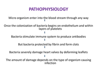
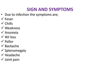
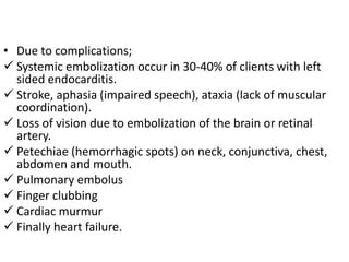
CARDIOVASCULAR
   DISORDERS
           BY;
 Mr. M.K LAMBA M.Sc. Nsg
The cardio vascular disorders includes the
  disorders related to heart and blood
  vessels like infections of heart layers and
  heart, valvular disorders, arithmetic
  disorders etc.
HEART DISORDERS
                 PERICARDITIS
It is an inflammation of the pericardium
   the outer most covering the heart. It may
   be primary illness or a variety of medical
   and surgical disorders. It is an acute and
   chronic illness.
CAUSES OF PERICARDITIS:
• Infections: viral, bacterial, parasitic, fungal
• Myocardial injuries: cardiac trauma, surgery
• Collagen      diseases:       Rheumatic       fever,
  scleroderma (connective tissue disease that
  involves changes in the skin, blood vessels,
  muscles, and internal organs), rheumatoid
  arthritis
• Drug reactions
• Radiation therapy
• Cobalt therapy
• Anemia
• Neoplasm
PAHOPHYSIOLOGY:
          Due to any of the above reason
                         ↓
 Either from infection or any other reason there
     will be increased WBC and chance of fluid
              accumulation in the cavity
                         ↓
    Leads to dyspnea and pain aggravated by
     breathing due to pericardial compression
                         ↓
 Results in decrease cardiac output and cardiac
                     temponade
SIGNS AND SYMPTOMS
• Pain: over precordium and may felt beneath of
  clavicle, in neck and pain left to scapular
  region.
• Pain aggravated by breathing, relieving by
  sitting
• Dyspnea may occur as a result of pericardial
  compression which leads to decreased cardiac
  output
• Fever may be present.
• ↑ WBC count
• Echocardiogram: accurately detects all
  pericardial effusions and provides clinically
  relevant information about their size and
  hemodynamic.
• Magnetic resonance imaging (MRI) and
  computed tomography (CT) in detecting
  pericardial thickening/ constriction and
  calcification as well as small loculated
  effusions is extremely useful in these
  conditions.
MEDICAL MANAGEMENT:
 Analgesics to relieve pain
 NSAID (Non steroidal anti- inflammatory agents):
  eg. Spirin, Indomethacin (Indocin) These
  drugs with analgesic and antipyretic effects and which have, in
  higher doses, anti-inflammatory effects.
 Corticosteroids: are two types;
  1. Gluco-corticoids such as cortisol control carbohydrate,
  fat and protein metabolism and are anti-inflammatory by
  preventing         phospho-           lipid       release,
  decreasing eosinophil action and a number of other
  mechanisms.
  2. Mineralo-corticoids such as aldosterone control
  electrolyte and water levels, mainly by promoting sodium
  retention.
MYOCARDITIS
It is an inflammatory process of myocardium It
   may cause heart dilation, thrombi on the
   heart wall, infiltration of blood cells around
   the coronary vessels and between the muscle
   fibers and degeneration of the muscle fibers
   themselves.
CAUSES

•   Infections
•   Rheumatic fever
•   Immunosuppressive therapy
•   Infective endocarditis
•   Dilated cardiomyopathy
SIGNS AND SYMPTOMS

Fatigue
Dyspnea
Palpitation
Occasionally discomfort in chest and abdomen
May have sudden congestive cardiac failure
 due to the damage of myocardial tissues.
DIAGNOSTIC FINDING

Auscultation; it shows faint heart sound and
 systolic murmur.
Pulsus alternans (a pulse in which there is
 regular alteration of weak and strong beats).
Fever
Tachycardia
Symptoms of CHF
Endomyocardial biopsy
MEDICAL MANAGEMENT
Bed rest the patient to decrease cardiac work load
 and to decrease myocardium damage.
Penicillin antibiotic to treat streptococci
If CHF Treatment;
     • ACE (angiotension converting enzyme) promote vasodilation
       which leads to diuresis.
     • Digitalis like digoxin, medication increase the force of
       myocardial contraction and slows down the conduction
       through AV needs
     • Dobutamin which increases cardiac contractility
ENDOCARDITIS
Endocarditis is an inflammation of endocardium,
   especially the valves. It is common in old age due
   to the decreased immunologic response.
The types;
i. Sub acute bacterial endocarditis: develop
    gradually in several weeks to months caused by
    streptoccus viridans.
ii. Acute bacterial endocarditis: develop in days to
    weeks caused by staphylococcus aurens.
iii. Native valve endocarditis: Caused to a damaged
     valve.
iv. Non bacterial thrombotic endocarditis: caused
     by thrombotic lesions.
ETIOLOGY
•    Infection by an organism; staphylococcus,
     streptococci and fungi etc.
•   Structural abnormalities of heart and great blood
    vessels.
•   History of infective endocarditis.
•   Rheumatic fever
•   Valvalar dysfunction
•   Cardiomyopathy
Some procedures may cause bacterial entry;
• Dental procedure
• Tonsillectomy
• Bronchoscopy
• Esophageal varices
• Esophageal dilation
• Gall bladder surgery
• Cystoscopy
• Urethral catheterization
• UTI surgery
PATHOPHYSIOLOGY
   Micro organism enter into the blood stream through any way

Once the colonization of bacteria begins on endothelium and within
                          layers of platelets

     Bacteria stimulate immune system to produce antibodies

          But bacteria protected by fibrin and form clots

   Bacteria severely damage heart valves by deforming leaflets

 The amount of damage depends on the type of organism causing
                          infection
SIGN AND SYMPTOMS
• Due to infection the symptoms are;
 Fever
 Chills
 Weakness
 Anorexia
 Wt loss
 Pallor
 Backache
 Splenomegaly
 Headache
 Joint pain
• Due to complications;
 Systemic embolization occur in 30-40% of clients with left
  sided endocarditis.
 Stroke, aphasia (impaired speech), ataxia (lack of muscular
  coordination).
 Loss of vision due to embolization of the brain or retinal
  artery.
 Petechiae (hemorrhagic spots) on neck, conjunctiva, chest,
  abdomen and mouth.
 Pulmonary embolus
 Finger clubbing
 Cardiac murmur
 Finally heart failure.
DIAGNOSTIC FINDING
Physical examination
Auscultation – cardiac murmur
Blood culture
ECG and echo
CBC
Chest x- ray
MEDICAL MANAGEMENT
Eradicating the infective organism- by antibiotics
Proper treating complication
Anti micro bacterial therapy
     - Penicillin
     - Gentamicin

Cardiovascular disorders

  • 1.
    CARDIOVASCULAR DISORDERS BY; Mr. M.K LAMBA M.Sc. Nsg
  • 2.
    The cardio vasculardisorders includes the disorders related to heart and blood vessels like infections of heart layers and heart, valvular disorders, arithmetic disorders etc.
  • 3.
    HEART DISORDERS PERICARDITIS It is an inflammation of the pericardium the outer most covering the heart. It may be primary illness or a variety of medical and surgical disorders. It is an acute and chronic illness.
  • 4.
    CAUSES OF PERICARDITIS: •Infections: viral, bacterial, parasitic, fungal • Myocardial injuries: cardiac trauma, surgery • Collagen diseases: Rheumatic fever, scleroderma (connective tissue disease that involves changes in the skin, blood vessels, muscles, and internal organs), rheumatoid arthritis • Drug reactions • Radiation therapy • Cobalt therapy • Anemia • Neoplasm
  • 5.
    PAHOPHYSIOLOGY: Due to any of the above reason ↓ Either from infection or any other reason there will be increased WBC and chance of fluid accumulation in the cavity ↓ Leads to dyspnea and pain aggravated by breathing due to pericardial compression ↓ Results in decrease cardiac output and cardiac temponade
  • 6.
    SIGNS AND SYMPTOMS •Pain: over precordium and may felt beneath of clavicle, in neck and pain left to scapular region. • Pain aggravated by breathing, relieving by sitting • Dyspnea may occur as a result of pericardial compression which leads to decreased cardiac output • Fever may be present. • ↑ WBC count
  • 7.
    • Echocardiogram: accuratelydetects all pericardial effusions and provides clinically relevant information about their size and hemodynamic. • Magnetic resonance imaging (MRI) and computed tomography (CT) in detecting pericardial thickening/ constriction and calcification as well as small loculated effusions is extremely useful in these conditions.
  • 8.
    MEDICAL MANAGEMENT:  Analgesicsto relieve pain  NSAID (Non steroidal anti- inflammatory agents): eg. Spirin, Indomethacin (Indocin) These drugs with analgesic and antipyretic effects and which have, in higher doses, anti-inflammatory effects.  Corticosteroids: are two types; 1. Gluco-corticoids such as cortisol control carbohydrate, fat and protein metabolism and are anti-inflammatory by preventing phospho- lipid release, decreasing eosinophil action and a number of other mechanisms. 2. Mineralo-corticoids such as aldosterone control electrolyte and water levels, mainly by promoting sodium retention.
  • 9.
    MYOCARDITIS It is aninflammatory process of myocardium It may cause heart dilation, thrombi on the heart wall, infiltration of blood cells around the coronary vessels and between the muscle fibers and degeneration of the muscle fibers themselves.
  • 10.
    CAUSES • Infections • Rheumatic fever • Immunosuppressive therapy • Infective endocarditis • Dilated cardiomyopathy
  • 11.
    SIGNS AND SYMPTOMS Fatigue Dyspnea Palpitation Occasionallydiscomfort in chest and abdomen May have sudden congestive cardiac failure due to the damage of myocardial tissues.
  • 12.
    DIAGNOSTIC FINDING Auscultation; itshows faint heart sound and systolic murmur. Pulsus alternans (a pulse in which there is regular alteration of weak and strong beats). Fever Tachycardia Symptoms of CHF Endomyocardial biopsy
  • 13.
    MEDICAL MANAGEMENT Bed restthe patient to decrease cardiac work load and to decrease myocardium damage. Penicillin antibiotic to treat streptococci If CHF Treatment; • ACE (angiotension converting enzyme) promote vasodilation which leads to diuresis. • Digitalis like digoxin, medication increase the force of myocardial contraction and slows down the conduction through AV needs • Dobutamin which increases cardiac contractility
  • 14.
    ENDOCARDITIS Endocarditis is aninflammation of endocardium, especially the valves. It is common in old age due to the decreased immunologic response. The types; i. Sub acute bacterial endocarditis: develop gradually in several weeks to months caused by streptoccus viridans. ii. Acute bacterial endocarditis: develop in days to weeks caused by staphylococcus aurens.
  • 15.
    iii. Native valveendocarditis: Caused to a damaged valve. iv. Non bacterial thrombotic endocarditis: caused by thrombotic lesions.
  • 16.
    ETIOLOGY • Infection by an organism; staphylococcus, streptococci and fungi etc. • Structural abnormalities of heart and great blood vessels. • History of infective endocarditis. • Rheumatic fever • Valvalar dysfunction • Cardiomyopathy
  • 17.
    Some procedures maycause bacterial entry; • Dental procedure • Tonsillectomy • Bronchoscopy • Esophageal varices • Esophageal dilation • Gall bladder surgery • Cystoscopy • Urethral catheterization • UTI surgery
  • 18.
    PATHOPHYSIOLOGY Micro organism enter into the blood stream through any way Once the colonization of bacteria begins on endothelium and within layers of platelets Bacteria stimulate immune system to produce antibodies But bacteria protected by fibrin and form clots Bacteria severely damage heart valves by deforming leaflets The amount of damage depends on the type of organism causing infection
  • 19.
    SIGN AND SYMPTOMS •Due to infection the symptoms are;  Fever  Chills  Weakness  Anorexia  Wt loss  Pallor  Backache  Splenomegaly  Headache  Joint pain
  • 20.
    • Due tocomplications;  Systemic embolization occur in 30-40% of clients with left sided endocarditis.  Stroke, aphasia (impaired speech), ataxia (lack of muscular coordination).  Loss of vision due to embolization of the brain or retinal artery.  Petechiae (hemorrhagic spots) on neck, conjunctiva, chest, abdomen and mouth.  Pulmonary embolus  Finger clubbing  Cardiac murmur  Finally heart failure.
  • 21.
    DIAGNOSTIC FINDING Physical examination Auscultation– cardiac murmur Blood culture ECG and echo CBC Chest x- ray
  • 22.
    MEDICAL MANAGEMENT Eradicating theinfective organism- by antibiotics Proper treating complication Anti micro bacterial therapy - Penicillin - Gentamicin