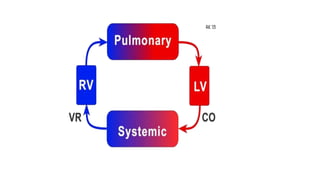
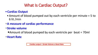
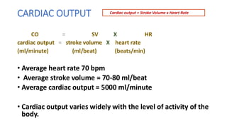
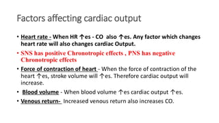
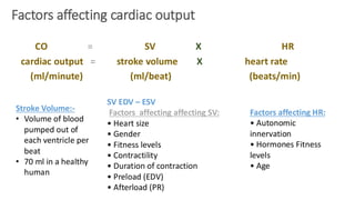
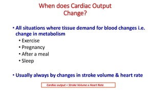
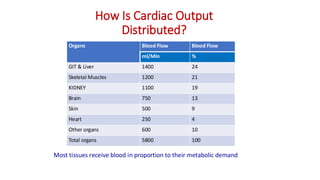
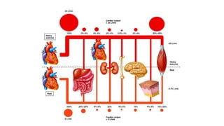
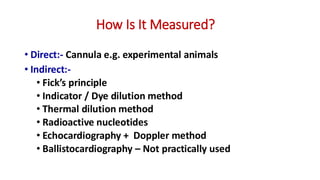
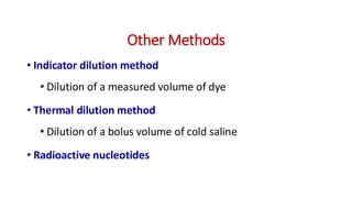
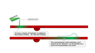
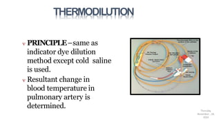
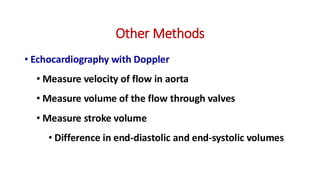
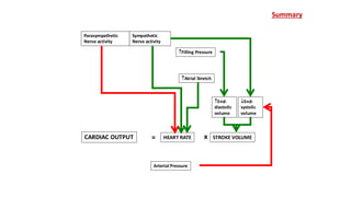
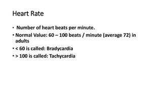
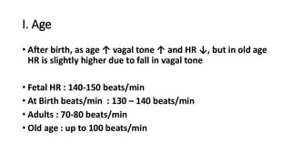
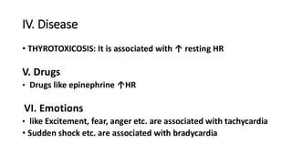
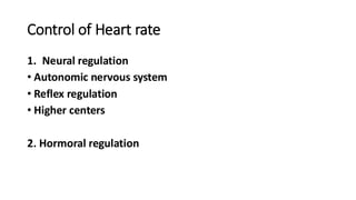
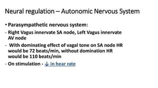
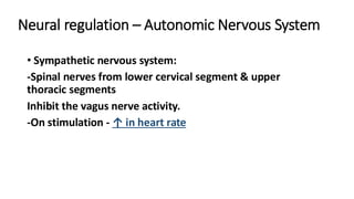
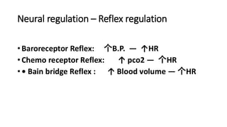
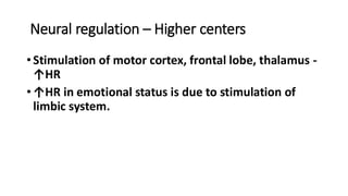
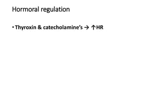
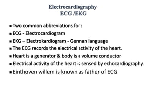
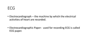
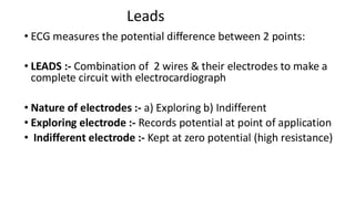
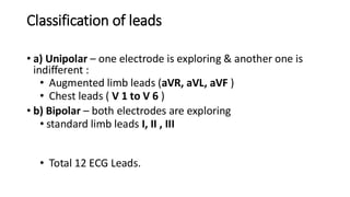
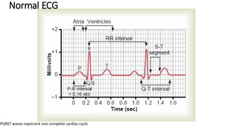
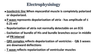
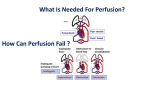
Cardiac Output, ECG & Shock
DEPARTMENT OF PHYSIOLOGY
Shweta kanwar
Learning Objectives
• What is cardiac output ?
• Measurement of cardiac output
• Heart rate & its regulating factors
• Normal ECG
• Shock
Cardiac Output
What makes the heart a good pump?
1
What Is Cardiac Output?
•Cardiac Output
•Amount of blood pumped out by each ventricle per minute = 5 to
6 lit /min
A measure of cardiac performance
Stroke volume
Amount of blood pumped by each ventricle per beat = 70ml
Heart Rate
Cardiac output = Stroke Volume x Heart Rate
CARDIAC OUTPUT
CO = SV X HR
cardiac output = stroke volume X heart rate
(ml/minute) (ml/beat) (beats/min)
• Average heart rate 70 bpm
• Average stroke volume = 70-80 ml/beat
• Average cardiac output = 5000 ml/minute
• Cardiac output varies widely with the level of activity of the
body.
Cardiac output = Stroke Volume x Heart Rate
Factors affecting cardiac output
• Heart rate - When HR ↑es - CO also ↑es. Any factor which changes
heart rate will also changes cardiac Output.
• SNS has positive Chronotropic effects , PNS has negative
Chronotropic effects
• Force of contraction of heart - When the force of contraction of the
heart ↑es, stroke volume will ↑es. Therefore cardiac output will
increase.
• Blood volume - When blood volume ↑es cardiac output ↑es.
• Venous return- Increased venous return also increases CO.
Factors affecting cardiac output
CO = SV X HR
cardiac output = stroke volume X heart rate
(ml/minute) (ml/beat) (beats/min)
Stroke Volume:-
• Volume of blood
pumped out of
each ventricle per
beat
• 70 ml in a healthy
human
SV EDV – ESV
Factors affecting affecting SV:
• Heart size
• Gender
• Fitness levels
• Contractility
• Duration of contraction
• Preload (EDV)
• Afterload (PR)
Factors affecting HR:
• Autonomic
innervation
• Hormones Fitness
levels
• Age
When does Cardiac Output
Change?
• All situations where tissue demand for blood changes i.e.
change in metabolism
• Exercise
• Pregnancy
• After a meal
• Sleep
• Usually always by changes in stroke volume & heart rate
Cardiac output = Stroke Volume x Heart Rate
Factors influencing CO
• Physiological:-
• Age - More in adults than in children because blood volume is more
• Gender - CO is more in male than females
• Surface area – Increase CO
• Emotions - CO increases during emotional expressions
• Pregnancy - CO increases during pregnancy.
• Muscular exercise - CO increases during exercise
• Sleep
• Pathological:-
• Hyperthyroidism, Anemia, Fever
• Hypovolemia
• Hemorrhage
• Myocardial infarction
How Is Cardiac Output
Distributed?
Organs Blood Flow Blood Flow
ml/Min %
GIT & Liver 1400 24
Skeletal Muscles 1200 21
KIDNEY 1100 19
Brain 750 13
Skin 500 9
Heart 250 4
Other organs 600 10
Total organs 5800 100
Most tissues receive blood in proportion to their metabolic demand
How Is It Measured?
• Direct:- Cannula e.g. experimental animals
• Indirect:-
• Fick’s principle
• Indicator / Dye dilution method
• Thermal dilution method
• Radioactive nucleotides
• Echocardiography + Doppler method
• Ballistocardiography – Not practically used
Measurement of Cardiac Output
Fick’s principle
Blood flow=Amt. of O2 absorption from lungs per min./A-V O2
CO = 200/40 = 5 lit/min
measured by collecting
expired air
Arterial blood
sample
Mixed venous
blood sample
Disadvantages & Advantages
• Disadvantages
• Invasive
• Slow
• Steady state required
• Advantages
• Quite accurate
Other Methods
• Indicator dilution method
• Dilution of a measured volume of dye
• Thermal dilution method
• Dilution of a bolus volume of cold saline
• Radioactive nucleotides
THERMODILUTION
 PRINCIPLE–same as
indicator dye dilution
method except cold saline
is used.
 Resultant change in
blood temperature in
pulmonary artery is
determined. Thursday,
November , 18,
2024
Other Methods
• Echocardiography with Doppler
• Measure velocity of flow in aorta
• Measure volume of the flow through valves
• Measure stroke volume
• Difference in end-diastolic and end-systolic volumes
Thursday,
November 18,
2024
PHYSICALMETHODS
Doppler
echocardiography–
.
CARDIAC OUTPUT STROKE VOLUME
Arterial Pressure
Filling Pressure
Atrial Stretch
Parasympathetic
Nerve activity
End-
diastolic
volume
HEART RATE
Sympathetic
Nerve activity
End-
systolic
volume
= x
Summary
Heart Rate
Heart Rate
• Number of heart beats per minute.
• Normal Value: 60 – 100 beats / minute (average 72) in
adults
• < 60 is called: Bradycardia
• > 100 is called: Tachycardia
Factors affecting Heart rate
• I. Age
• II. Gender
• III. Body temperature
• IV. Disease
• V. Drugs
• VI. Emotions
• VII. Exercise
I. Age
• After birth, as age ↑ vagal tone ↑ and HR ↓, but in old age
HR is slightly higher due to fall in vagal tone
• Fetal HR : 140-150 beats/min
• At Birth beats/min : 130 – 140 beats/min
• Adults : 70-80 beats/min
• Old age : up to 100 beats/min
II. Gender
• HR is slightly higher in females as compared to males.
•III. Body temperature
• HR is directly related to the body temp
• ↑ Temp - ↑ HR
• ↓ Temp. - ↓ HR
IV. Disease
• THYROTOXICOSIS: It is associated with ↑ resting HR
V. Drugs
• Drugs like epinephrine ↑HR
VI. Emotions
• like Excitement, fear, anger etc. are associated with tachycardia
• Sudden shock etc. are associated with bradycardia
VII. Exercise
• HR increases in linear pattern with the severity of the
exercise because :
• Increase in sympathetic activity
• Decrease in vagal tone
• Increase in Release of catecholamine's and thyroxine
• Change in blood chemistry (hypoxia and hypercapnia)
Control of Heart rate
1. Neural regulation
• Autonomic nervous system
• Reflex regulation
• Higher centers
2. Hormoral regulation
Neural regulation – Autonomic Nervous System
• Parasympathetic nervous system:
- Right Vagus innervate SA node, Left Vagus innervate
AV node
- With dominating effect of vagal tone on SA node HR
would be 72 beats/min, without domination HR
would be 110 beats/min
- On stimulation - ↓ in hear rate
Neural regulation – Autonomic Nervous System
• Sympathetic nervous system:
-Spinal nerves from lower cervical segment & upper
thoracic segments
Inhibit the vagus nerve activity.
-On stimulation - ↑ in heart rate
Neural regulation – Reflex regulation
•Baroreceptor Reflex: 个B.P. ― ↑HR
•Chemo receptor Reflex: ↑ pco2 ― 个HR
•• Bain bridge Reflex : ↑ Blood volume ― 个HR
Neural regulation – Higher centers
•Stimulation of motor cortex, frontal lobe, thalamus -
↑HR
•↑HR in emotional status is due to stimulation of
limbic system.
Hormoral regulation
•Thyroxin & catecholamine’s → ↑HR
ELECTROCARDIOGRAPHY
Electrocardiography
ECG /EKG
◼ Two common abbreviations for :
◼ ECG - Electrocardiogram
◼ EKG – Electrokardiogram - German language
◼ The ECG records the electrical activity of the heart.
◼ Heart is a generator & body is a volume conductor
◼ Electrical activity of the heart is sensed by echocardiography.
◼ Einthoven willem is known as father of ECG
ECG
• Electrocardiograph – the machine by which the electrical
activities of heart are recorded.
• Electrocardiographic Paper- used for recording ECG is called
ECG paper.
Clinical Importance
• Ionic changes
• Ischemia, infarction
• Arrhythmias
• Conduction defects
• Hypertrophy
• Damage to cardiac muscles
Electrophysiology
◼ If wave of depolarization
spreads toward the recording
electrode-positive deflection.
◼ If wave of depolarization
spreads away from recording
electrode, a negative deflection
Leads
• ECG measures the potential difference between 2 points:
• LEADS :- Combination of 2 wires & their electrodes to make a
complete circuit with electrocardiograph
• Nature of electrodes :- a) Exploring b) Indifferent
• Exploring electrode :- Records potential at point of application
• Indifferent electrode :- Kept at zero potential (high resistance)
Classification of leads
• a) Unipolar – one electrode is exploring & another one is
indifferent :
• Augmented limb leads (aVR, aVL, aVF )
• Chest leads ( V 1 to V 6 )
• b) Bipolar – both electrodes are exploring
• standard limb leads I, II , III
• Total 12 ECG Leads.
Normal ECG
PQRST waves represent one complete cardiac cycle
Electrophysiology
◼ Isoelectric line When myocardial muscle is completely polarized
or depolarized.
◼ P wave represents depolarization of atria - has amplitude of ≤
0.25 mV
• Repolarization of atria not normally detectable on an ECG
• Excitation of bundle of His and bundle branches occur in middle
of PR interval
• QRS complex reflects depolarization of ventricles - Q& S waves
are downward deflections
• T wave reflects repolarization of ventricular muscles
Circulatory
Shock
Generalized inadequacy of
blood flow
or
Inadequate tissue perfusion
Pump-Heart
Pipe -vessels
Fluid - blood
What Is Needed For Perfusion?
How Can Perfusion Fail ?
• Inadequate
fluid -
• Vascular
Vasodilatation
• Obstruction to
blood flow
Cardiogenic
Hypovolemic Obstructive Distributive
Inadequate
pumping of heart
Shock
Neurogenic
Anaphylactic
Septic
Hypovolemic
•In most types of shock CO & arterial pressure decreases
•Shock is a life-threatening condition of circulatory failure.
How Is Shock Classified?
Cardiac Output & Heart Rate introduction

Cardiac Output & Heart Rate introduction

  • 1.
    Cardiac Output, ECG& Shock DEPARTMENT OF PHYSIOLOGY Shweta kanwar
  • 2.
    Learning Objectives • Whatis cardiac output ? • Measurement of cardiac output • Heart rate & its regulating factors • Normal ECG • Shock
  • 3.
    Cardiac Output What makesthe heart a good pump?
  • 4.
  • 5.
    What Is CardiacOutput? •Cardiac Output •Amount of blood pumped out by each ventricle per minute = 5 to 6 lit /min A measure of cardiac performance Stroke volume Amount of blood pumped by each ventricle per beat = 70ml Heart Rate Cardiac output = Stroke Volume x Heart Rate
  • 6.
    CARDIAC OUTPUT CO =SV X HR cardiac output = stroke volume X heart rate (ml/minute) (ml/beat) (beats/min) • Average heart rate 70 bpm • Average stroke volume = 70-80 ml/beat • Average cardiac output = 5000 ml/minute • Cardiac output varies widely with the level of activity of the body. Cardiac output = Stroke Volume x Heart Rate
  • 7.
    Factors affecting cardiacoutput • Heart rate - When HR ↑es - CO also ↑es. Any factor which changes heart rate will also changes cardiac Output. • SNS has positive Chronotropic effects , PNS has negative Chronotropic effects • Force of contraction of heart - When the force of contraction of the heart ↑es, stroke volume will ↑es. Therefore cardiac output will increase. • Blood volume - When blood volume ↑es cardiac output ↑es. • Venous return- Increased venous return also increases CO.
  • 8.
    Factors affecting cardiacoutput CO = SV X HR cardiac output = stroke volume X heart rate (ml/minute) (ml/beat) (beats/min) Stroke Volume:- • Volume of blood pumped out of each ventricle per beat • 70 ml in a healthy human SV EDV – ESV Factors affecting affecting SV: • Heart size • Gender • Fitness levels • Contractility • Duration of contraction • Preload (EDV) • Afterload (PR) Factors affecting HR: • Autonomic innervation • Hormones Fitness levels • Age
  • 9.
    When does CardiacOutput Change? • All situations where tissue demand for blood changes i.e. change in metabolism • Exercise • Pregnancy • After a meal • Sleep • Usually always by changes in stroke volume & heart rate Cardiac output = Stroke Volume x Heart Rate
  • 10.
    Factors influencing CO •Physiological:- • Age - More in adults than in children because blood volume is more • Gender - CO is more in male than females • Surface area – Increase CO • Emotions - CO increases during emotional expressions • Pregnancy - CO increases during pregnancy. • Muscular exercise - CO increases during exercise • Sleep • Pathological:- • Hyperthyroidism, Anemia, Fever • Hypovolemia • Hemorrhage • Myocardial infarction
  • 11.
    How Is CardiacOutput Distributed? Organs Blood Flow Blood Flow ml/Min % GIT & Liver 1400 24 Skeletal Muscles 1200 21 KIDNEY 1100 19 Brain 750 13 Skin 500 9 Heart 250 4 Other organs 600 10 Total organs 5800 100 Most tissues receive blood in proportion to their metabolic demand
  • 13.
    How Is ItMeasured? • Direct:- Cannula e.g. experimental animals • Indirect:- • Fick’s principle • Indicator / Dye dilution method • Thermal dilution method • Radioactive nucleotides • Echocardiography + Doppler method • Ballistocardiography – Not practically used
  • 14.
    Measurement of CardiacOutput Fick’s principle Blood flow=Amt. of O2 absorption from lungs per min./A-V O2 CO = 200/40 = 5 lit/min measured by collecting expired air Arterial blood sample Mixed venous blood sample
  • 15.
    Disadvantages & Advantages •Disadvantages • Invasive • Slow • Steady state required • Advantages • Quite accurate
  • 16.
    Other Methods • Indicatordilution method • Dilution of a measured volume of dye • Thermal dilution method • Dilution of a bolus volume of cold saline • Radioactive nucleotides
  • 18.
    THERMODILUTION  PRINCIPLE–same as indicatordye dilution method except cold saline is used.  Resultant change in blood temperature in pulmonary artery is determined. Thursday, November , 18, 2024
  • 19.
    Other Methods • Echocardiographywith Doppler • Measure velocity of flow in aorta • Measure volume of the flow through valves • Measure stroke volume • Difference in end-diastolic and end-systolic volumes
  • 20.
  • 21.
    CARDIAC OUTPUT STROKEVOLUME Arterial Pressure Filling Pressure Atrial Stretch Parasympathetic Nerve activity End- diastolic volume HEART RATE Sympathetic Nerve activity End- systolic volume = x Summary
  • 22.
  • 23.
    Heart Rate • Numberof heart beats per minute. • Normal Value: 60 – 100 beats / minute (average 72) in adults • < 60 is called: Bradycardia • > 100 is called: Tachycardia
  • 24.
    Factors affecting Heartrate • I. Age • II. Gender • III. Body temperature • IV. Disease • V. Drugs • VI. Emotions • VII. Exercise
  • 25.
    I. Age • Afterbirth, as age ↑ vagal tone ↑ and HR ↓, but in old age HR is slightly higher due to fall in vagal tone • Fetal HR : 140-150 beats/min • At Birth beats/min : 130 – 140 beats/min • Adults : 70-80 beats/min • Old age : up to 100 beats/min
  • 26.
    II. Gender • HRis slightly higher in females as compared to males. •III. Body temperature • HR is directly related to the body temp • ↑ Temp - ↑ HR • ↓ Temp. - ↓ HR
  • 27.
    IV. Disease • THYROTOXICOSIS:It is associated with ↑ resting HR V. Drugs • Drugs like epinephrine ↑HR VI. Emotions • like Excitement, fear, anger etc. are associated with tachycardia • Sudden shock etc. are associated with bradycardia
  • 28.
    VII. Exercise • HRincreases in linear pattern with the severity of the exercise because : • Increase in sympathetic activity • Decrease in vagal tone • Increase in Release of catecholamine's and thyroxine • Change in blood chemistry (hypoxia and hypercapnia)
  • 29.
    Control of Heartrate 1. Neural regulation • Autonomic nervous system • Reflex regulation • Higher centers 2. Hormoral regulation
  • 30.
    Neural regulation –Autonomic Nervous System • Parasympathetic nervous system: - Right Vagus innervate SA node, Left Vagus innervate AV node - With dominating effect of vagal tone on SA node HR would be 72 beats/min, without domination HR would be 110 beats/min - On stimulation - ↓ in hear rate
  • 31.
    Neural regulation –Autonomic Nervous System • Sympathetic nervous system: -Spinal nerves from lower cervical segment & upper thoracic segments Inhibit the vagus nerve activity. -On stimulation - ↑ in heart rate
  • 32.
    Neural regulation –Reflex regulation •Baroreceptor Reflex: 个B.P. ― ↑HR •Chemo receptor Reflex: ↑ pco2 ― 个HR •• Bain bridge Reflex : ↑ Blood volume ― 个HR
  • 33.
    Neural regulation –Higher centers •Stimulation of motor cortex, frontal lobe, thalamus - ↑HR •↑HR in emotional status is due to stimulation of limbic system.
  • 34.
    Hormoral regulation •Thyroxin &catecholamine’s → ↑HR
  • 35.
  • 36.
    Electrocardiography ECG /EKG ◼ Twocommon abbreviations for : ◼ ECG - Electrocardiogram ◼ EKG – Electrokardiogram - German language ◼ The ECG records the electrical activity of the heart. ◼ Heart is a generator & body is a volume conductor ◼ Electrical activity of the heart is sensed by echocardiography. ◼ Einthoven willem is known as father of ECG
  • 37.
    ECG • Electrocardiograph –the machine by which the electrical activities of heart are recorded. • Electrocardiographic Paper- used for recording ECG is called ECG paper.
  • 38.
    Clinical Importance • Ionicchanges • Ischemia, infarction • Arrhythmias • Conduction defects • Hypertrophy • Damage to cardiac muscles
  • 39.
    Electrophysiology ◼ If waveof depolarization spreads toward the recording electrode-positive deflection. ◼ If wave of depolarization spreads away from recording electrode, a negative deflection
  • 40.
    Leads • ECG measuresthe potential difference between 2 points: • LEADS :- Combination of 2 wires & their electrodes to make a complete circuit with electrocardiograph • Nature of electrodes :- a) Exploring b) Indifferent • Exploring electrode :- Records potential at point of application • Indifferent electrode :- Kept at zero potential (high resistance)
  • 41.
    Classification of leads •a) Unipolar – one electrode is exploring & another one is indifferent : • Augmented limb leads (aVR, aVL, aVF ) • Chest leads ( V 1 to V 6 ) • b) Bipolar – both electrodes are exploring • standard limb leads I, II , III • Total 12 ECG Leads.
  • 42.
    Normal ECG PQRST wavesrepresent one complete cardiac cycle
  • 43.
    Electrophysiology ◼ Isoelectric lineWhen myocardial muscle is completely polarized or depolarized. ◼ P wave represents depolarization of atria - has amplitude of ≤ 0.25 mV • Repolarization of atria not normally detectable on an ECG • Excitation of bundle of His and bundle branches occur in middle of PR interval • QRS complex reflects depolarization of ventricles - Q& S waves are downward deflections • T wave reflects repolarization of ventricular muscles
  • 44.
    Circulatory Shock Generalized inadequacy of bloodflow or Inadequate tissue perfusion
  • 45.
    Pump-Heart Pipe -vessels Fluid -blood What Is Needed For Perfusion? How Can Perfusion Fail ? • Inadequate fluid - • Vascular Vasodilatation • Obstruction to blood flow Cardiogenic Hypovolemic Obstructive Distributive Inadequate pumping of heart
  • 46.
    Shock Neurogenic Anaphylactic Septic Hypovolemic •In most typesof shock CO & arterial pressure decreases •Shock is a life-threatening condition of circulatory failure. How Is Shock Classified?