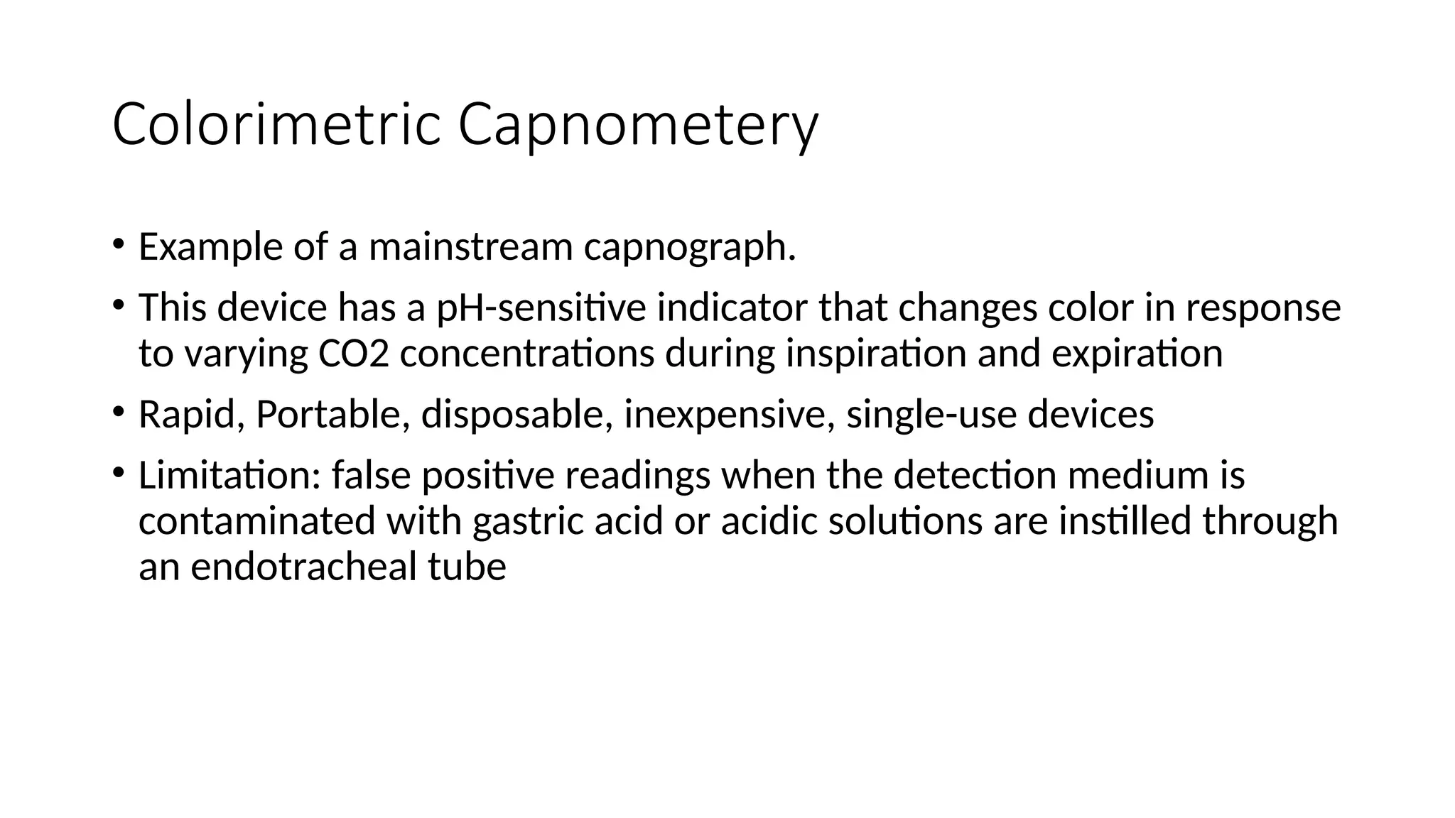
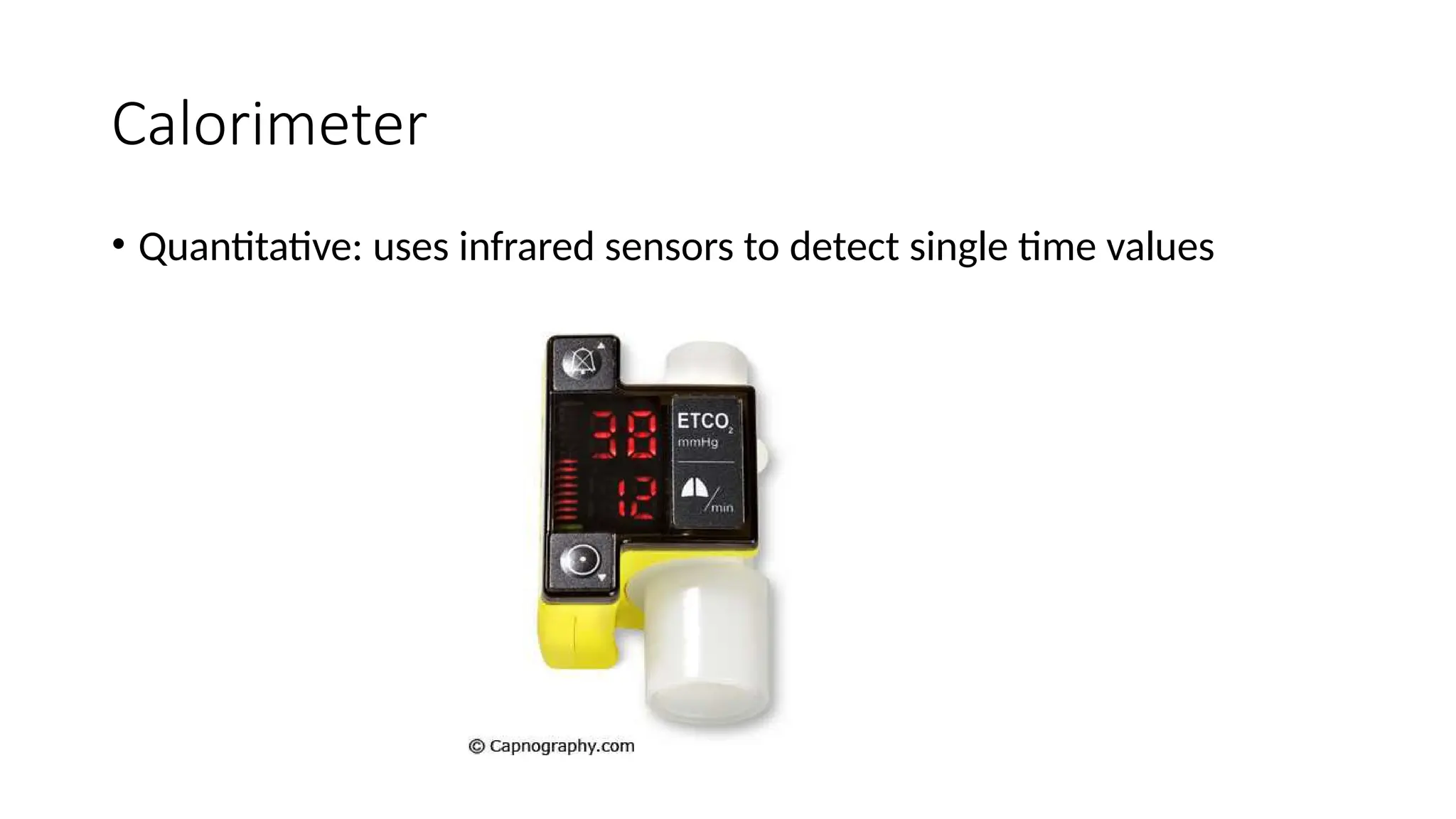
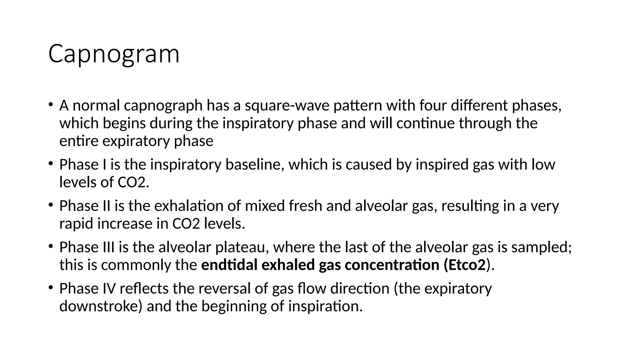
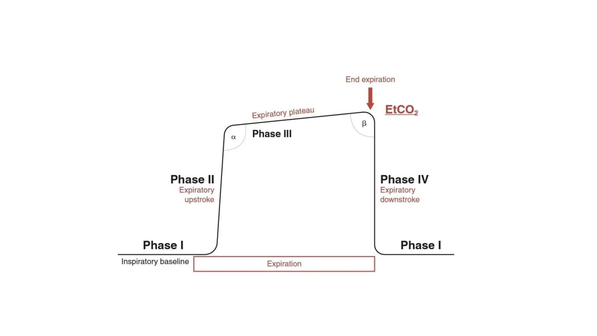
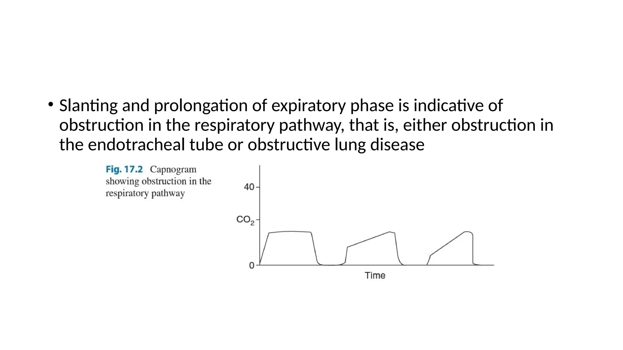
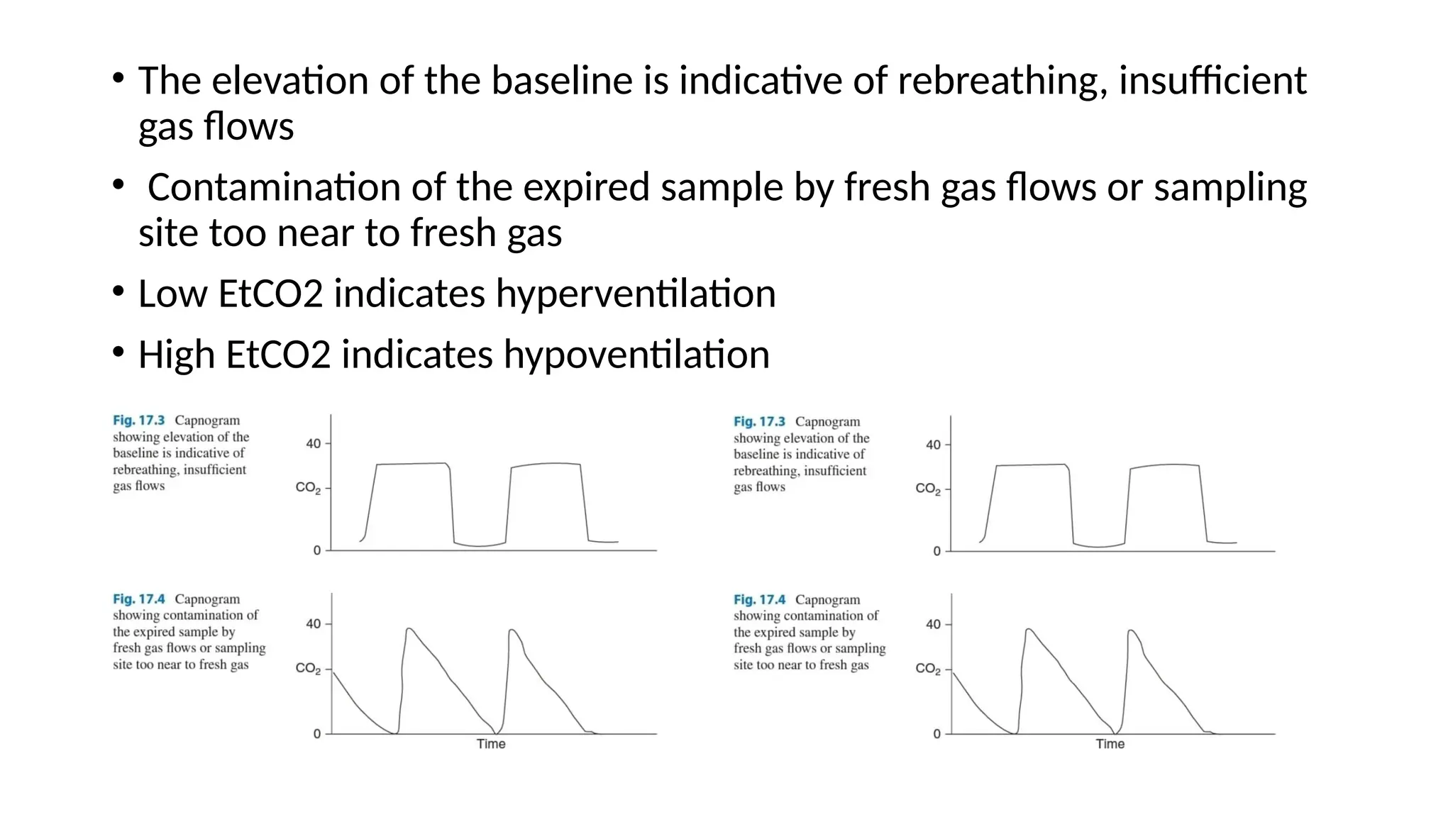
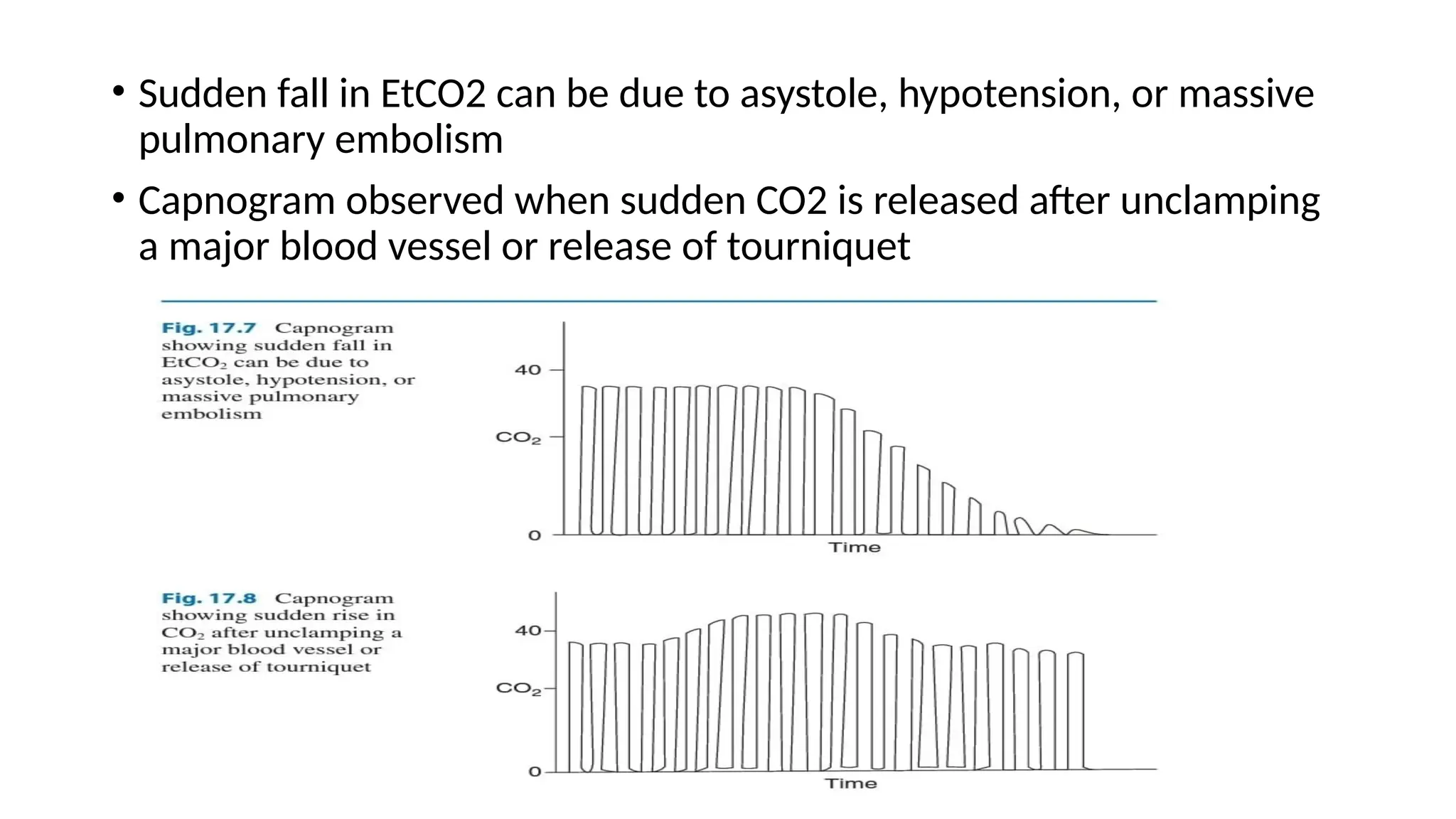
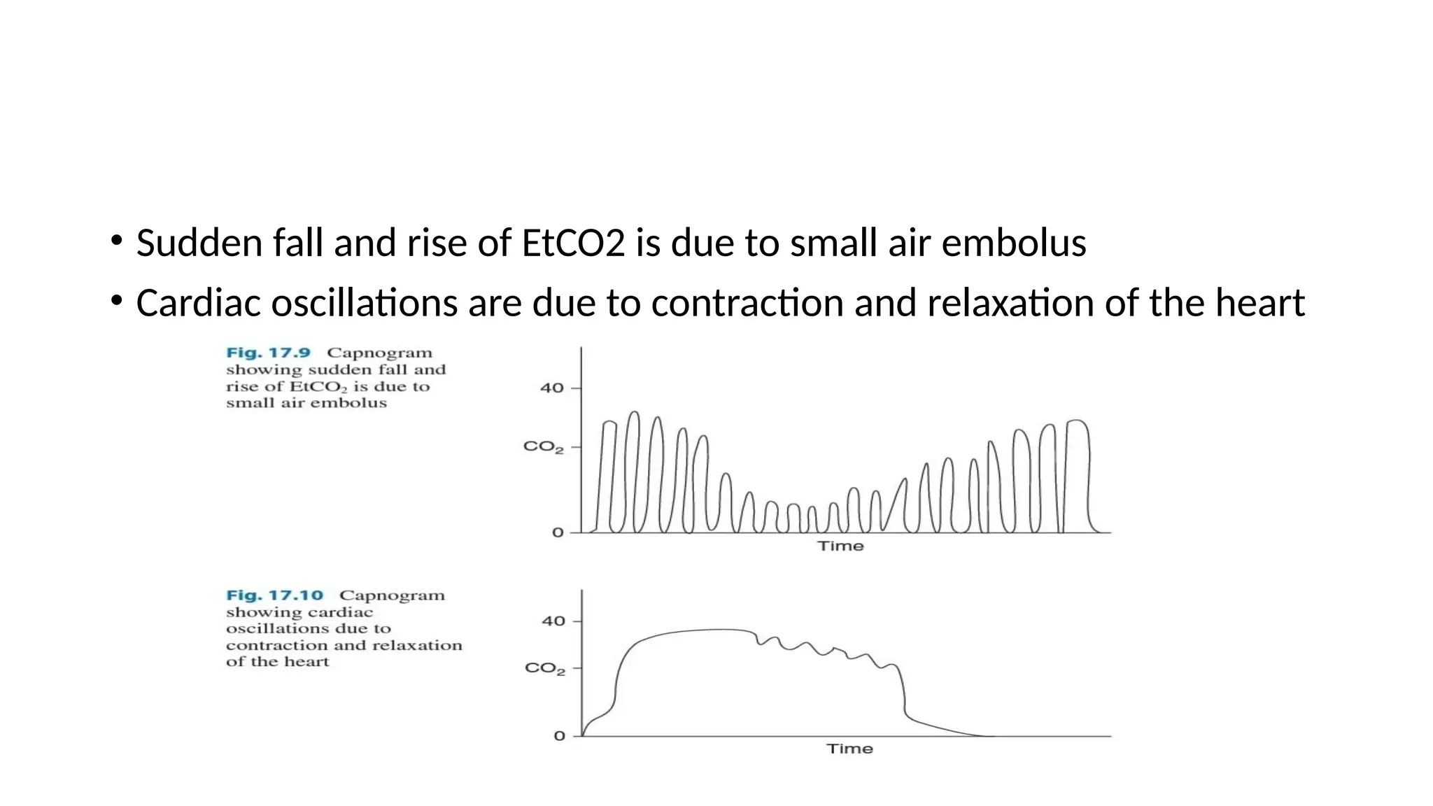
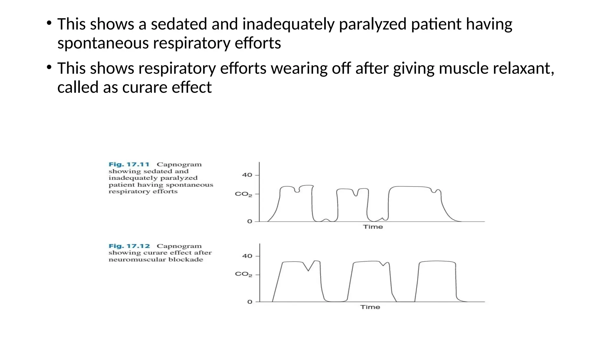
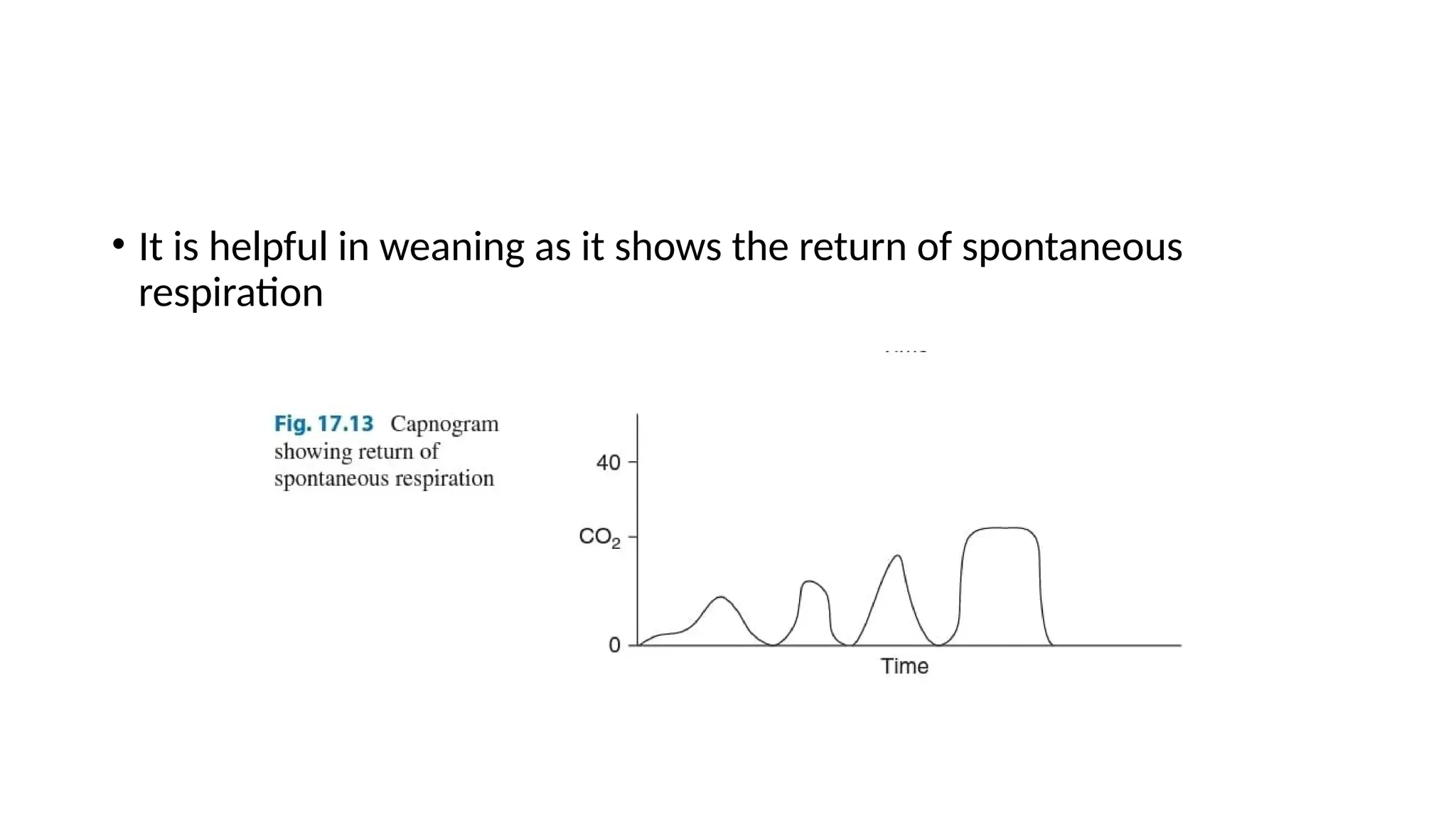
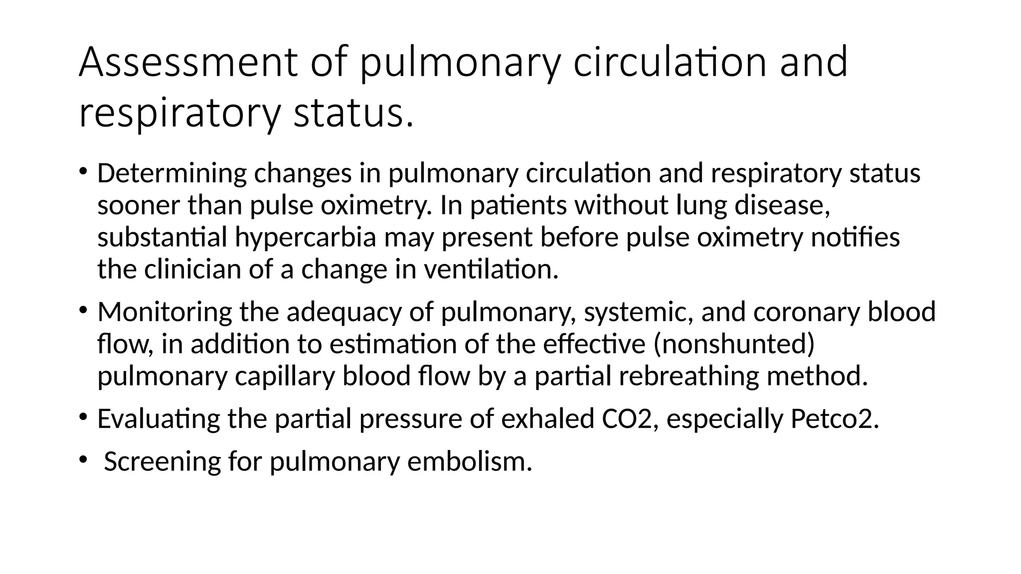
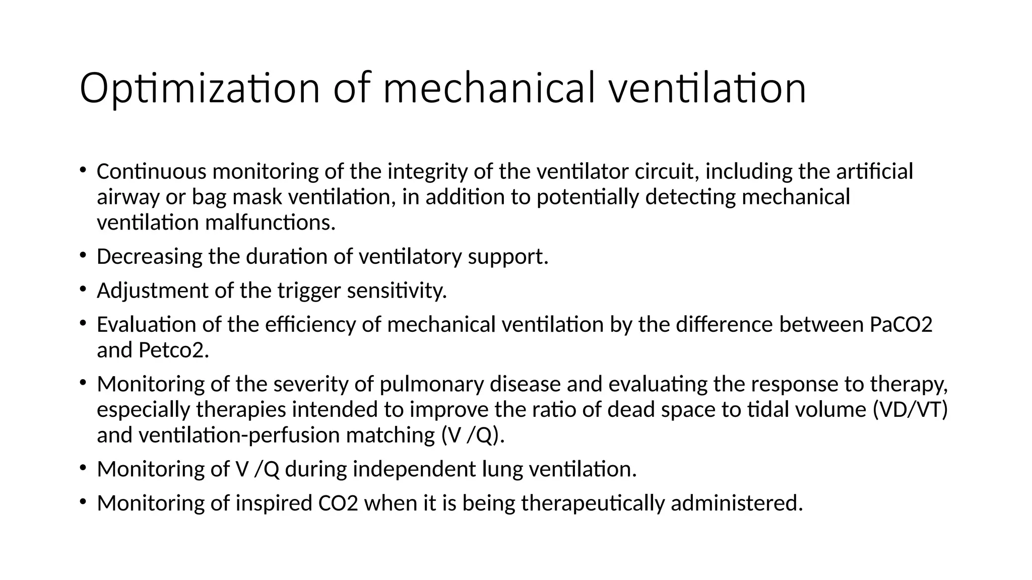
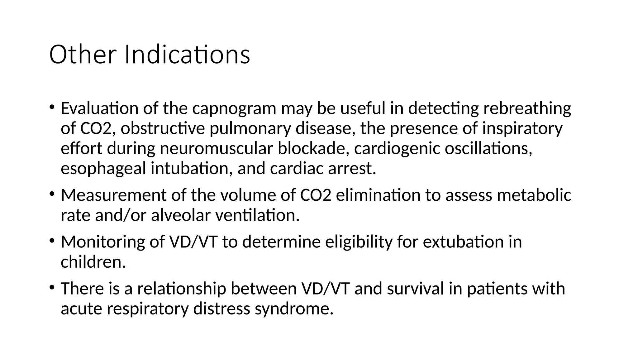
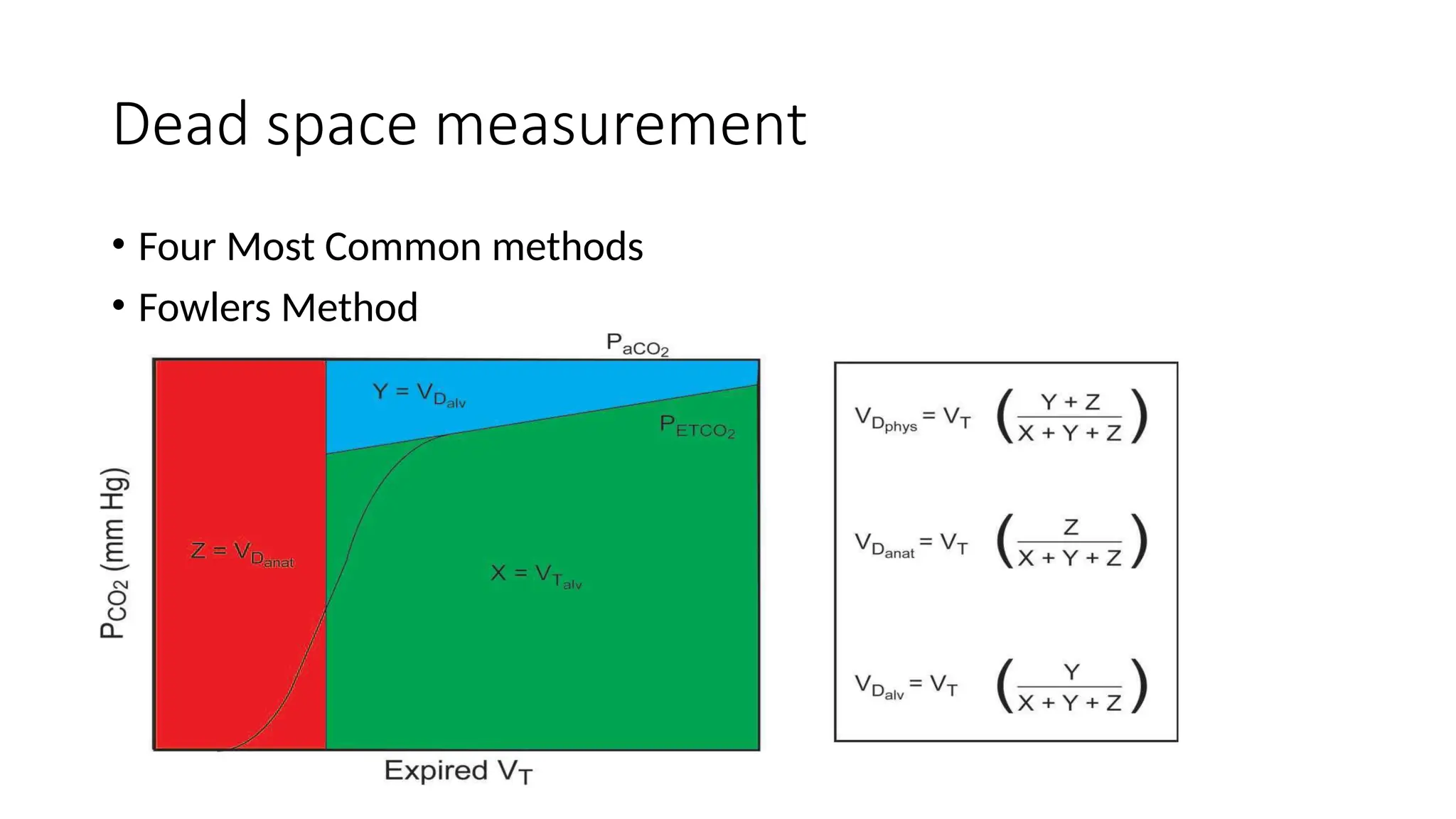
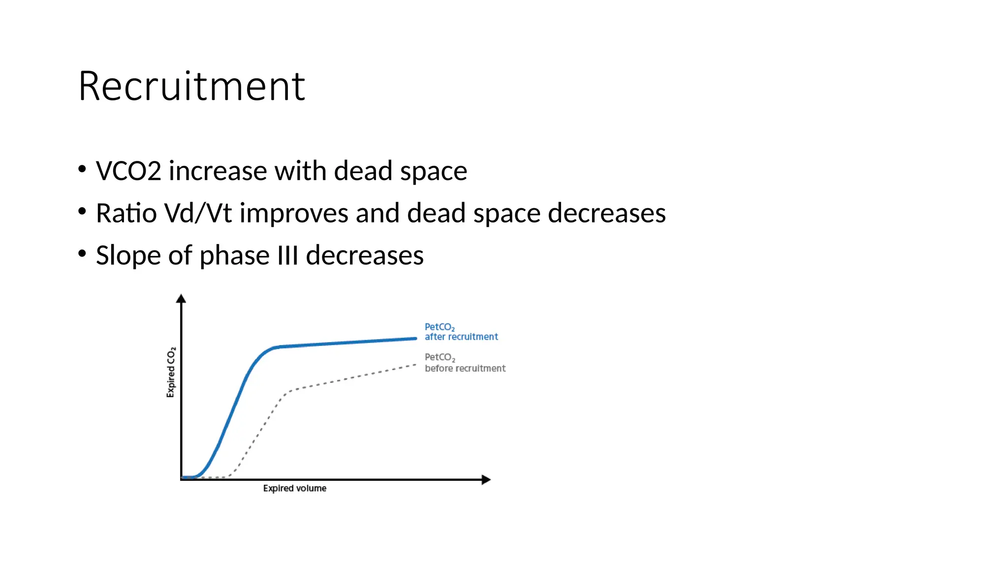
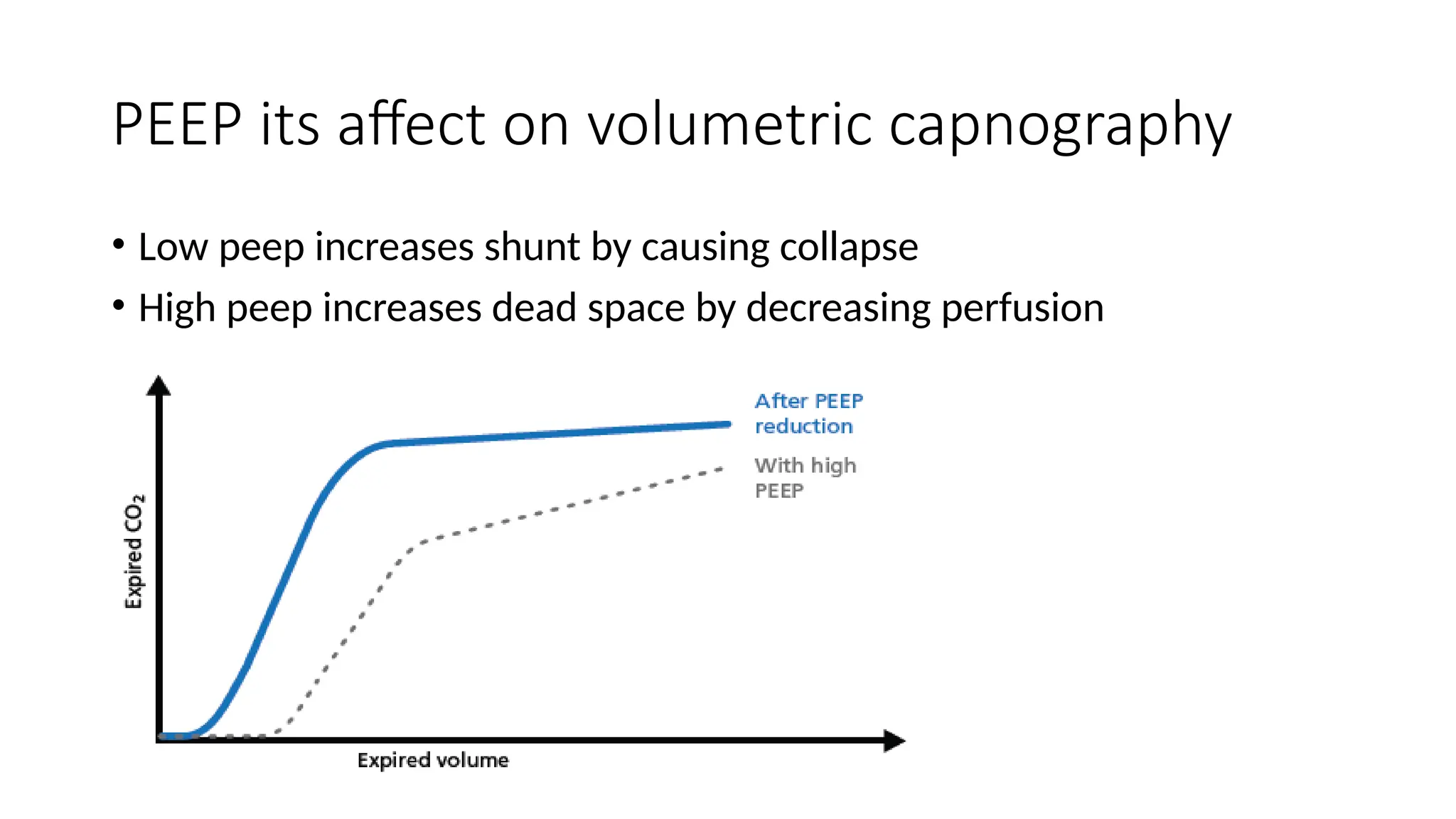
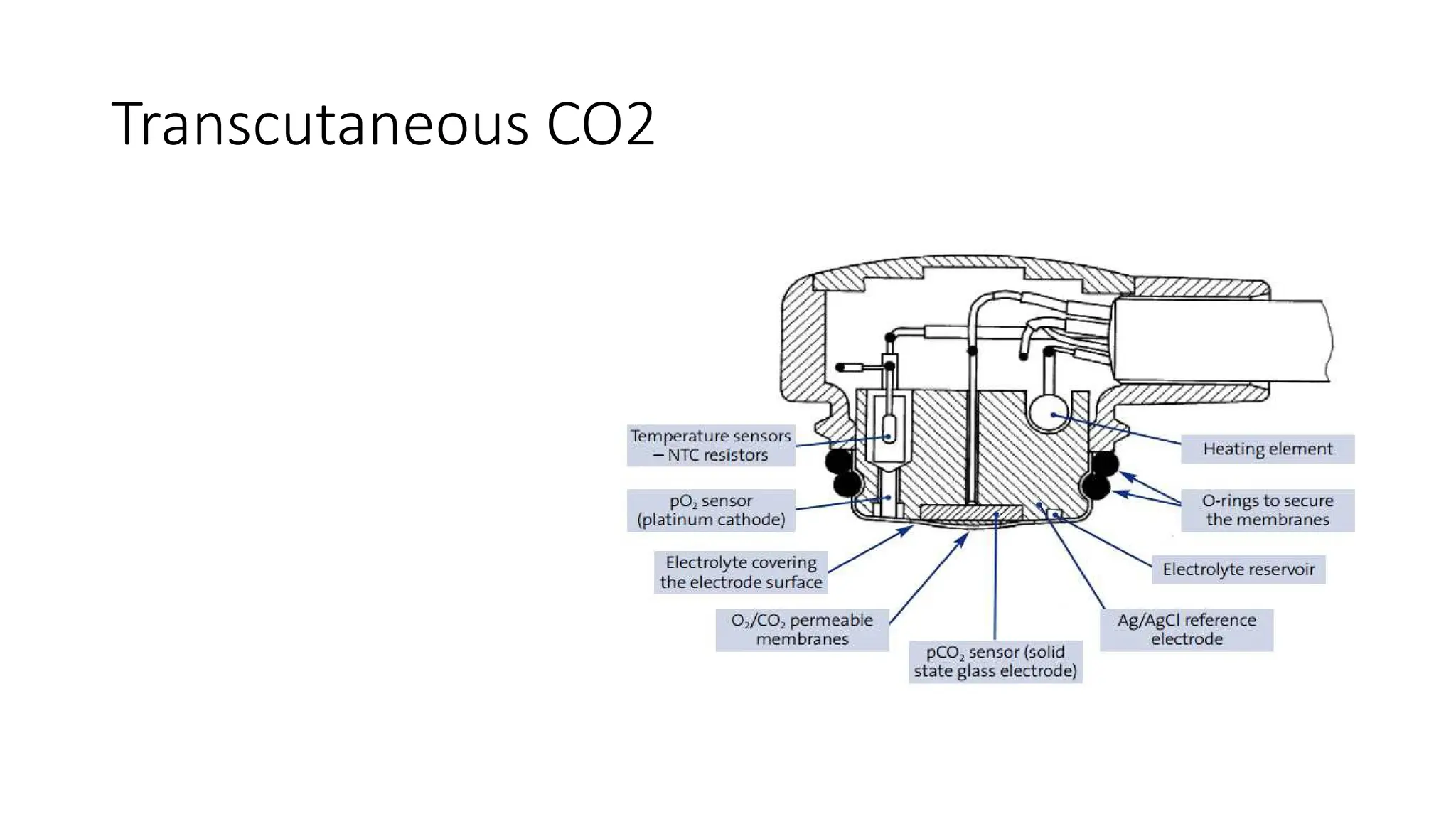
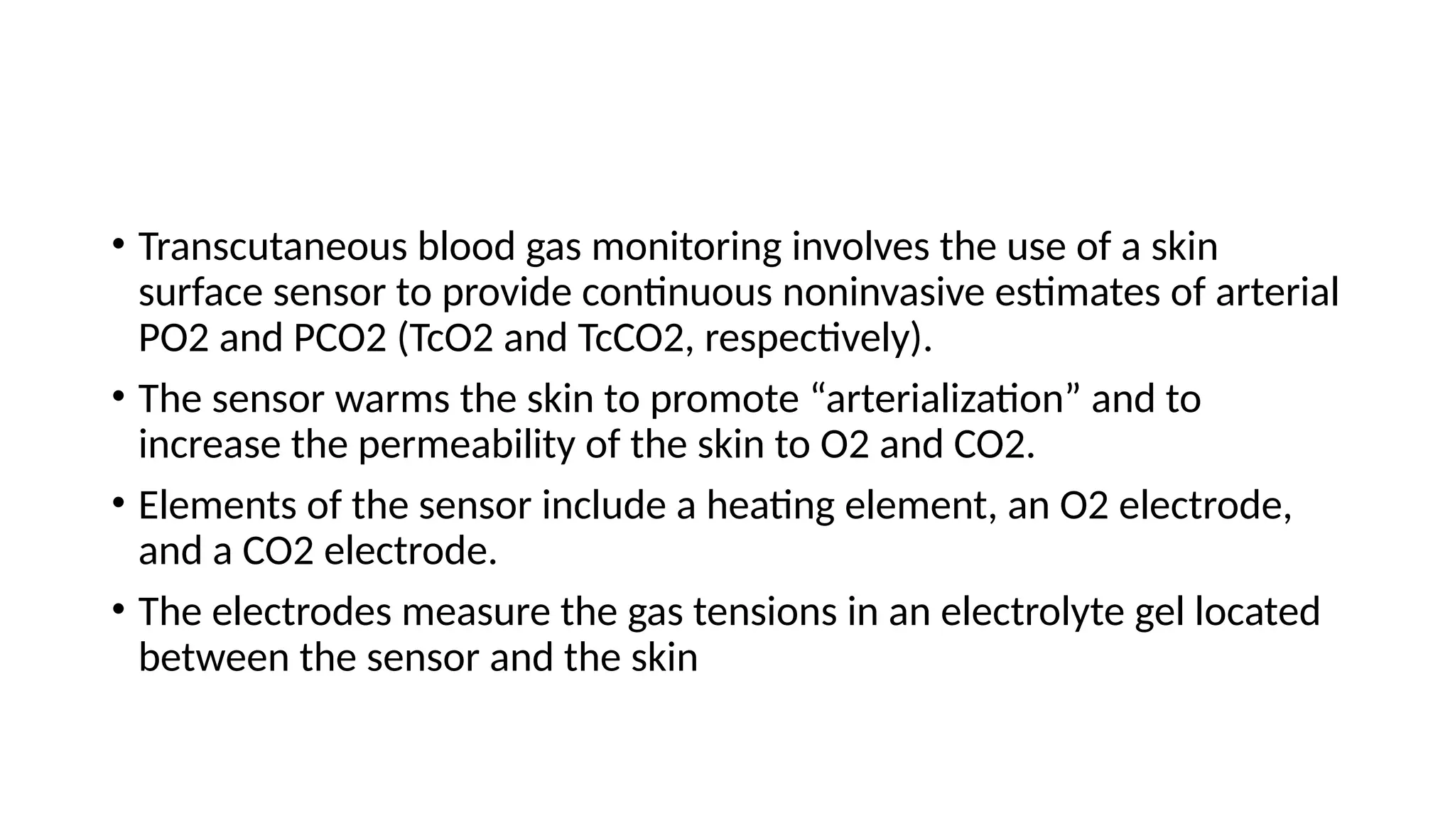
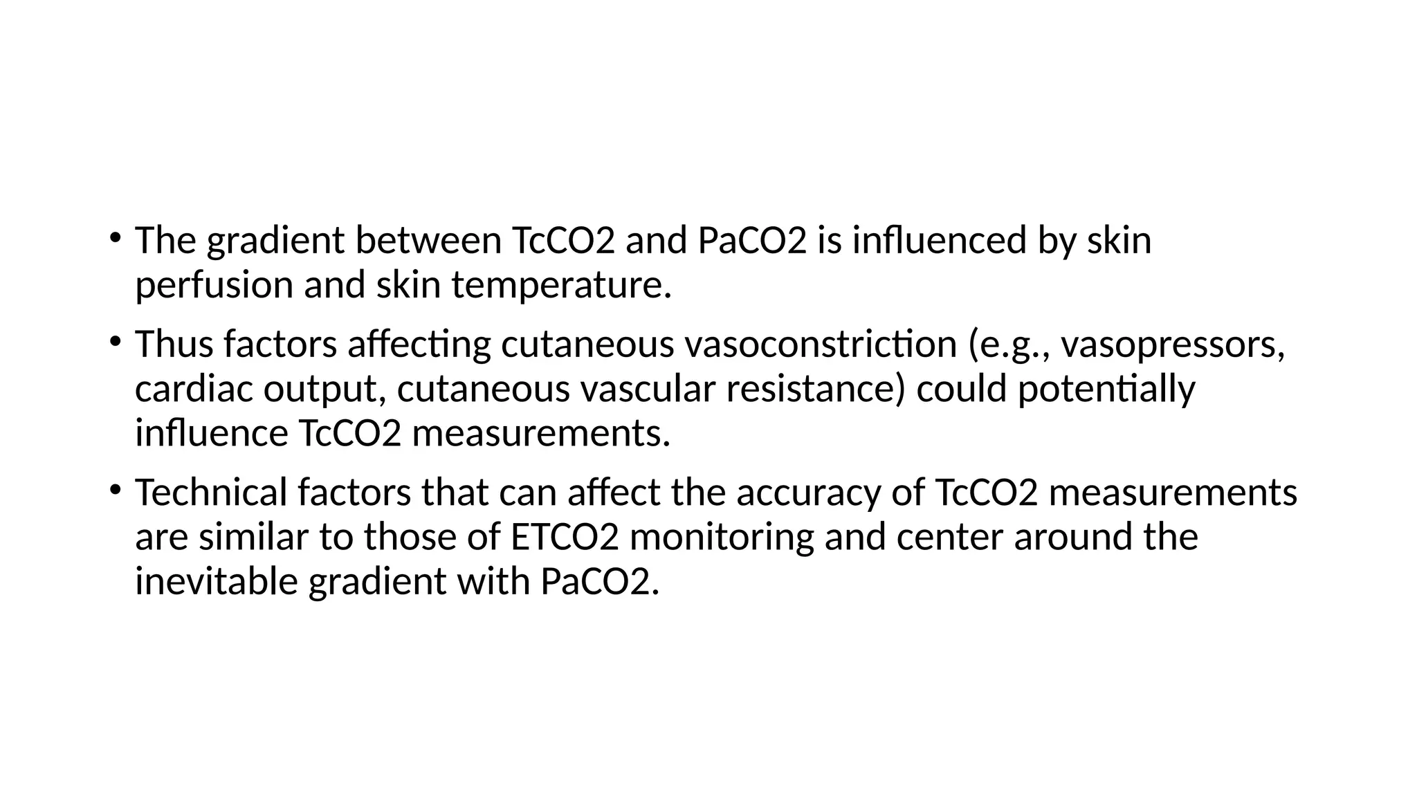
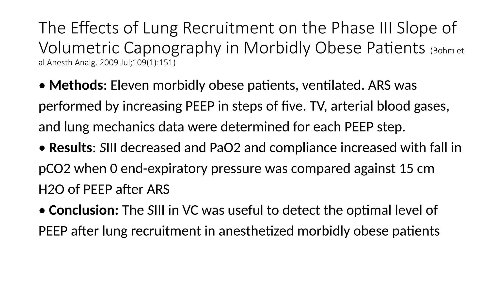
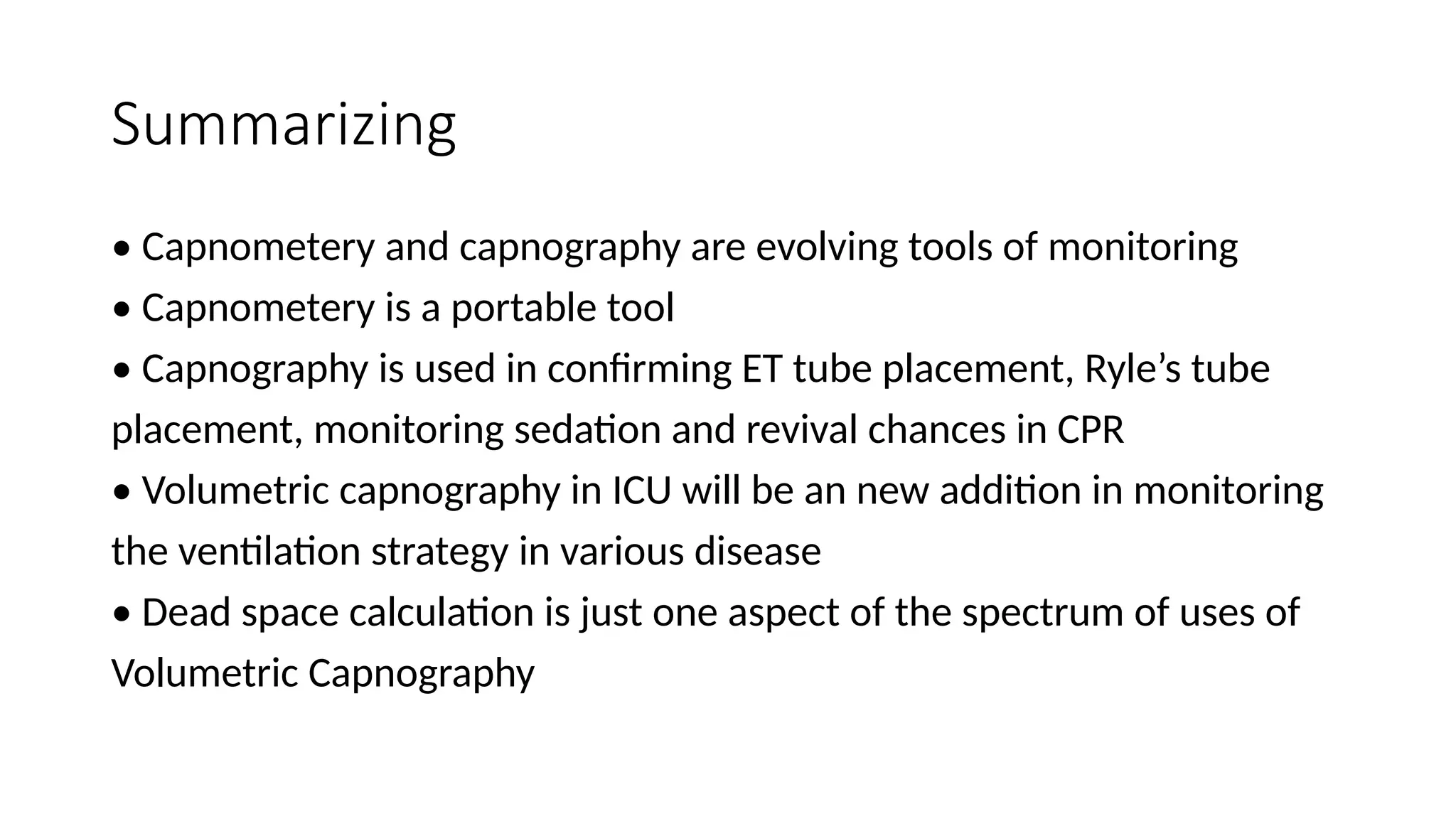
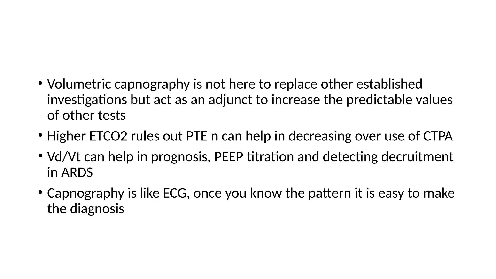
Capnometry
Dr Sanat Verenkar
Introduction
• “Capnos” is a Greek word meaning “Smoke”
• Measurement of CO2 in air was first used by Haldane and Tyndall for
the analysis of mine air.
• Aiken and Clark-Kennedy, in 1928, tried to analyze dead space by
using expired CO2 measurements
• Prof Bob Smalhout used the first CO2 analyzer on a patients in
Netherlands in 1962
Types of Monitoring
• Capnometery: Provides a single time quantitative value of CO2
fraction in one particular amount of air
• Capnography: Provides a real time graphical representation of CO2
that can be against time or tidal volume
• The waveform is refered to as Capnogram
Conditions that can effect CO2 in expired air
• Decreased production
• Decreased transport
• Decreased lung perfusion
• Decreased ventilation
• Ventilator malfunction
• Monitoring equipment malfunction
• Capnometry uses infrared light projected through a gas sampling
chamber to a detector on the opposite side.
• CO2 absorbs infrared light at a peak wavelength of approximately 4.27
mm.
• More infrared light passing through the sample chamber (i.e., less
CO2) causes a larger signal in the detector relative to the infrared light
passing through a reference cell.
Mainstream CO2 Monitoring
• An infrared optical bench placed within the gas flow pathway at the
airway.
• Real-time CO2 values within the airway and a real-time graphical
representation of the CO2 waveform
• Limitation: Cost, potential of damage to the sensor, increases dead
space, fouling with coughed up secretions and condensate, additional
weight of the adapter on the circuit
Colorimetric Capnometery
• Example of a mainstream capnograph.
• This device has a pH-sensitive indicator that changes color in response
to varying CO2 concentrations during inspiration and expiration
• Rapid, Portable, disposable, inexpensive, single-use devices
• Limitation: false positive readings when the detection medium is
contaminated with gastric acid or acidic solutions are instilled through
an endotracheal tube
Calorimeter
• Quantitative: uses infrared sensors to detect single time values
Side-stream CO2 Monitoring
• Devices aspirate a gas sample from a ventilator breathing circuit
• Utilize an infrared CO2 sensor in a monitor located away from the
patient
• Can be used in non intubated patients as well
• Side-stream sample method often requires use of a water trap to
prevent contamination.
• Limitation: Cost, time delay in analysis, can interfere with the trigger
system as air is actively aspirated.
Capnogram
• A normal capnograph has a square-wave pattern with four different phases,
which begins during the inspiratory phase and will continue through the
entire expiratory phase
• Phase I is the inspiratory baseline, which is caused by inspired gas with low
levels of CO2.
• Phase II is the exhalation of mixed fresh and alveolar gas, resulting in a very
rapid increase in CO2 levels.
• Phase III is the alveolar plateau, where the last of the alveolar gas is sampled;
this is commonly the endtidal exhaled gas concentration (Etco2).
• Phase IV reflects the reversal of gas flow direction (the expiratory
downstroke) and the beginning of inspiration.
Abnormal Capnographs
• The expired CO2 waveform can distinguish a variety of pulmonary and
airway pathologies.
• An esophageal intubation is discernible when the end-tidal waveform
becomes lower and lower with subsequent breaths and the patient
becomes more hypoxic.
• Other reasons for a flat tracing include capnograph disconnection,
near-complete airway obstruction, endotracheal tube perforation, or
cardiac arrest.
• Slanting and prolongation of expiratory phase is indicative of
obstruction in the respiratory pathway, that is, either obstruction in
the endotracheal tube or obstructive lung disease
• The elevation of the baseline is indicative of rebreathing, insufficient
gas flows
• Contamination of the expired sample by fresh gas flows or sampling
site too near to fresh gas
• Low EtCO2 indicates hyperventilation
• High EtCO2 indicates hypoventilation
• Sudden fall in EtCO2 can be due to asystole, hypotension, or massive
pulmonary embolism
• Capnogram observed when sudden CO2 is released after unclamping
a major blood vessel or release of tourniquet
• Sudden fall and rise of EtCO2 is due to small air embolus
• Cardiac oscillations are due to contraction and relaxation of the heart
• This shows a sedated and inadequately paralyzed patient having
spontaneous respiratory efforts
• This shows respiratory efforts wearing off after giving muscle relaxant,
called as curare effect
• It is helpful in weaning as it shows the return of spontaneous
respiration
American Association for Respiratory Care (AARC) Clinical
Practice Guideline:
Capnography/Capnometry During Mechanical Ventilation
There are three broad categories of indications for
capnography/capnometry:
• Verification of artificial airway placement
• Assessment of pulmonary circulation and respiratory status
• Optimization of mechanical ventilation.
Verification of Artificial Airway Placement
• Additional confirmation of airway placement with waveform
capnography or an exhaled CO2 or esophageal detector device
Assessment of pulmonary circulation and
respiratory status.
• Determining changes in pulmonary circulation and respiratory status
sooner than pulse oximetry. In patients without lung disease,
substantial hypercarbia may present before pulse oximetry notifies
the clinician of a change in ventilation.
• Monitoring the adequacy of pulmonary, systemic, and coronary blood
flow, in addition to estimation of the effective (nonshunted)
pulmonary capillary blood flow by a partial rebreathing method.
• Evaluating the partial pressure of exhaled CO2, especially Petco2.
• Screening for pulmonary embolism.
Optimization of mechanical ventilation
• Continuous monitoring of the integrity of the ventilator circuit, including the artificial
airway or bag mask ventilation, in addition to potentially detecting mechanical
ventilation malfunctions.
• Decreasing the duration of ventilatory support.
• Adjustment of the trigger sensitivity.
• Evaluation of the efficiency of mechanical ventilation by the difference between PaCO2
and Petco2.
• Monitoring of the severity of pulmonary disease and evaluating the response to therapy,
especially therapies intended to improve the ratio of dead space to tidal volume (VD/VT)
and ventilation-perfusion matching (V /Q).
• Monitoring of V /Q during independent lung ventilation.
• Monitoring of inspired CO2 when it is being therapeutically administered.
Other Indications
• Evaluation of the capnogram may be useful in detecting rebreathing
of CO2, obstructive pulmonary disease, the presence of inspiratory
effort during neuromuscular blockade, cardiogenic oscillations,
esophageal intubation, and cardiac arrest.
• Measurement of the volume of CO2 elimination to assess metabolic
rate and/or alveolar ventilation.
• Monitoring of VD/VT to determine eligibility for extubation in
children.
• There is a relationship between VD/VT and survival in patients with
acute respiratory distress syndrome.
Contraindications
• There are no absolute contraindications to capnography in
mechanically ventilated patients, provided the data obtained are
evaluated with consideration given to the patient’s clinical condition
Hazards/Complications
Mainstream
• Dead space. Adapters inserted into the airway between the airway and the ventilator
circuit should have a minimal amount of dead space. This effect is inversely
proportional to the size of the patient being monitored.
• The addition of the weight of a mainstream adapter can increase the risk of accidental
extubation in neonates and small children.
Sidestream:
• The gas sampling rate from some sidestream analyzers may be high enough to cause
auto-triggering when flow triggering of mechanical breaths is used. This effect is also
inversely proportional to the size of the patient.
• The gas sampling rate can diminish delivered VT in neonates and small patients while
using volume-targeted or volume-controlled ventilation modes.
Assessment of Need
• Capnography is considered a standard of care during general anesthesia.
• The American Society of Anesthesiologists has suggested that capnography be
available for patients with acute ventilatory failure on mechanical ventilator
support.
• The American College of Emergency Physicians recommends capnography as
an adjunctive method to ensure proper endotracheal tube position.
• The 2010 American Heart Association Guidelines for Cardiopulmonary
Resuscitation and Emergency Cardiovascular Care recommend capnography to
verify endotracheal tube placement in all age groups.
• Assessment of the need to use capnography with a specific patient should be
guided by the clinical situation.
Assessment of Outcome
• Results should reflect the patient’s condition and should validate the
basis for ordering the monitoring
• Documentation of results (along with all ventilator and hemodynamic
variables available), therapeutic interventions, and/or clinical
decisions made based on the capnogram should be included in the
patient’s chart
Monitoring
During capnography, the following should be considered and
monitored:
• Ventilatory variables: tidal volume, respiratory rate, positive end-
expiratory pressure, inspiratory-to-expiratory time ratio (I:E), peak
airway pressure, and concentrations of respiratory gas mixture.
• Hemodynamic variables: systemic and pulmonary blood pressures,
cardiac output, shunt, and ventilation-perfusion imbalances.
Drug-induced hypoventilation- 2 types
Type 1
• Bradypneic hypoventilation
• Characterized by a decreased
respiratory frequency
• Slightly decreased tidal volume,
• And an increased PETCO2 &
PaCO2
• Type 2
• Hypopneic hypoventilation
• Characterized by decrease in
tidal volume
• Slightly decreased respiratory
frequency
• And a decreased or normal
PETCO2 with elevated PaCO2
Monitoring During CPR
• The correct placement of an advanced airway
• The effectiveness of cardiac compressions
• The return of spontaneous circulation
• The prediction of outcome and survival during cardiac arrest
Role to Predict Survival
• Changes in PETCO2 during resuscitation attempts predicted outcomes
and return of spontaneous circulation
• PETCO2 less than 10 mm Hg during CPR was suggestive of poor outcome.
• This suggests that PETCO2 can be used in deciding when to terminate
resuscitation efforts in adult patients
• PETCO2 at 20 min after the initiation of CPR with a threshold of 14
mmHg can be used as a screening test to predict return of spontaneous
circulation
• Sensitivity, specificity, NPV, PPV of 100%.(Field et al Circulation
2010;122(18 Suppl 3): S640-S656)
Rescuer Fatigue
• PETCO2 monitoring during CPR can be used to detect the
effectiveness of external cardiac compression and the development of
rescuer fatigue
Volumetric Capnography
• Exhaled CO2 is plotted against the exhaled tidal volume.
• Can reveal important information in regarding efficiency of ventilation
and perfusion, the physiological dead space fraction, and the
metabolic rate of the patient
• These features are indications of use of Volumetric capnography in
ICU
Monitoring Adequacy of Ventilation
• PETCO2 closely approximates PaCO2 and is usually 2–5 mm Hg lower,
therefore can be an effective index of the adequacy of ventilation
• Diseased states, such as ARDS, COPD and asthma,ventilation/perfusion (V˙ /Q˙
) mismatch in the lungs can cause the PaCO2- PETCO2 difference to increase.
• PaCO2-PETCO2 difference can be an indication of the degree of lung injury
and correlates with increased VD/VT
• PaCO2-PETCO2 difference has been shown to predict mortality in trauma
patients.
• PETCO2 can be measured non invasively hence decreases frequency of
arterial sampling
Why bother if we have pulse ox?
• Reflects oxygenation not ventilation
• SpO2 changes lag behind the detection of hypoventilation
• Procedures are usually done with supplemental oxygen there by
decreasing sensitivity of picking up over-sedation
Few uses in critical care
• Measuring dead space and hence adequacy of ventilation
• Recruitment monitoring in ARDS
• Monitoring ventilation, tube positions, tube block, system leaks
• Diagnosing PTE
Dead space measurement
• Four Most Common methods
• Fowlers Method
• Bohr’s Equation
• Vd/Vt = PACo2 –PeCo2/ PACo2
• Enghoff modification of Bohr equation
CO2 is easily diffusible gas
So partial pressure of CO2 in alveoli and arterial blood should be same
PACO2 = PaCO2
• Vd/Vt(enghoff) = PaCo2 – PeCo2/PaCo2
• Frankenfield equation
• Vd/Vt = 0.32 + 0.0106 (PaCO2 - ETCO2) + 0.003 (RR) + 0.0015 (age)
Recruitment
• VCO2 increase with dead space
• Ratio Vd/Vt improves and dead space decreases
• Slope of phase III decreases
PEEP and its affect on Dead space
PEEP its affect on volumetric capnography
• Low peep increases shunt by causing collapse
• High peep increases dead space by decreasing perfusion
Capnography and PTE
• Various Capnographic parameters have been evaluated and corelated
• Have higher negative predictive value but not PPV
• Does not work alone, have to be used in combination with clinical
probability and D dimer
• etCO2, VCO2, Vd/Vt, are the most commonly used parameters.
End Tidal Carbon Dioxide as a Screening Tool for
Computed Tomography Angiogram (Ramne et al J Arthroplasty. 2016
Oct;31(10):2348-52)
• Method: All patients admitted for orthopedic surgery and who had a
CTA performed for PE were eligible
• Results: 121 patients enrolled, 84 had a negative CTA examination, 25
had a positive examination, and 12 had a non diagnostic examination.
• An ETCO2 cutoff value of 43 mm Hg was 100% sensitive with a
negative predictive value of 100% for absence of PE on CTA
• Conclusion: ETCO2 value >43 mm Hg is 100% sensitive for the
absence of PE.
D-Dimer and Exhaled CO2/O2 to Detect Segmental
Pulmonary Embolism in Moderate-Risk Patients (Kline et al Am J
Respir Crit Care Med Vol 182. pp 669–675, 2010)
• Methods: Patients with one sign, one symptom and CTPA done were enrolled.
Two groups on the basis of D Dimer cut off. The median etCO2/O2 less than 0.28
from seven or more breaths was group1 and etCO2/O2 greater than 0.45 was
group 2.
• Results: 495 patients, including 60 (12%) with segmental or larger, and 29 (6%)
with subsegmental. Positive D-dimer and etCO2/O2 less than 0.28 significantly
increases the probability of PE and etCO2/O2 greater than 0.45 predicts the
absence of PE.
• Conclusions: etCO2/O2 add to the specificity of D dimer in PPV for PTE
Volumetric Capnography as a Screening Test for
Pulmonary Embolism in the Emergency Department
(Verschuren et al CHEST 2004; 125:841–850)
Methods: 45 patients with D-dimer levels of > 500 ng/mL enrolled.
Several variables were calculated: Vd/Vt, Vdalv, Vdaw,, the slope of
phase 3; and the late dead space fraction (Fdlate)
• Results: Four variables of the VCap exhibited a statistical difference
between both groups, as follows: the VDalv/Vdaw, the slope of phase
3; the VDalv/VDphys; and Fdlate. PaCO2-EtCO2 gradient was also
significantly different in two groups
• Conclusion: Fdlate had a statistically better diagnostic performance in
suspected PE than the PaCO2-EtCO2 gradient. VCap is a promising
application of pulmonary pathophysiology.
Weaning and capnography
• During SBT successful weaning is represented by VCO2 staying stable
followed by slight increase suggestive of increasing work of breathing
• Unsuccessful weaning is represented with dramatic/ chaotic/
decrease in VCO2 or increase in dead space fraction
• Among several parameters ETCO2, Slope III, Expiratory time, VCO2
were significantly found to be different in two groups. (Rasera et al
Physiol Meas. 2015 Feb;36(2):231-42.(
Transcutaneous CO2
• Transcutaneous blood gas monitoring involves the use of a skin
surface sensor to provide continuous noninvasive estimates of arterial
PO2 and PCO2 (TcO2 and TcCO2, respectively).
• The sensor warms the skin to promote “arterialization” and to
increase the permeability of the skin to O2 and CO2.
• Elements of the sensor include a heating element, an O2 electrode,
and a CO2 electrode.
• The electrodes measure the gas tensions in an electrolyte gel located
between the sensor and the skin
Has the potential advantages over direct arterial blood gas sampling of
• Reducing the amount of blood drawn,
• Time spent for analysis,
• Patient discomfort,
• Associated costs.
• TcCO2 tends to be more reliable than ETCO2, most likely because of
the greater diffusion capacity of CO2 through the skin and the skin’s
own O2 consumption.
• The gradient between TcCO2 and PaCO2 is influenced by skin
perfusion and skin temperature.
• Thus factors affecting cutaneous vasoconstriction (e.g., vasopressors,
cardiac output, cutaneous vascular resistance) could potentially
influence TcCO2 measurements.
• Technical factors that can affect the accuracy of TcCO2 measurements
are similar to those of ETCO2 monitoring and center around the
inevitable gradient with PaCO2.
• Transcutaneous monitoring has been used in several clinical settings
to determine the presence of hypoventilation or respiratory
depression, including sleep studies, ventilator management,
bronchoscopies, and pulmonary function studies.
• However, some reports suggest that TcPO2 is not accurate enough to
be used clinically for ICU applications in adult populations or even in
preterm infants
Use of tCO2 in Chronic Hypercapenic
respiratory failures requiring NIV (Hajed et al Journal of Clinical and Diagnostic
Research. 2016 Sep, 10(9))
• Methods: Titration polysomnography studies were done on 12
patients with transcutaneous CO2 measurements in awake and asleep
state
• Results: Patients were followed up with Epworth sleepiness score
before and after transcutaneous CO2 guided titration. 6 patients
improved, 5 detiorated and one worsened
• Conclusion: Cutaneous capnography is feasible and permits the
optimization of non-invasive ventilation pressure settings in patients
with chronic hypercapenic respiratory failure
Monitoring dead space during recruitment
and PEEP titration in an experimental model (Tusman et al Intensive
Care Med (2006) 32:1863–1871)
• Methods: 8 lung lavaged pigs were ventilated at 6ml/kg and after
recruitment maneuver PEEP was decreased from 24cm of water at an
interval on 2 cm.
• VDalv, VDalv/VTalv, and Pa-etCO2 were monitored.
• Results: PEEP titration was achieved with compliance and Sat O2
monitoring. VDalv/VTalv and Pa-etCO2 showed good co-relation.
• Conclusion: Monitoring of dead space was useful for detecting lung
collapse and for establishing open-lung PEEP after a recruitment
maneuver.
Pulmonary dead-space fraction as a risk factor for death in the acute respiratory
distress syndrome Elevated values are
associated with an increased risk of death.
Nuckton et al N Engl J Med 2002; 346:1281-128
• • Methods: Dead-space fraction was prospectively measured in 179
intubated patients after the acute respiratory distress syndrome had
developed
• Results: Mean dead space was significantly increased in patients
who
died as compared to the patients who survived.
• Conclusions: Increased dead-space fraction is a feature of the early
phase of the acute respiratory distress syndrome.
The Effects of Lung Recruitment on the Phase III Slope of
Volumetric Capnography in Morbidly Obese Patients (Bohm et
al Anesth Analg. 2009 Jul;109(1):151)
• Methods: Eleven morbidly obese patients, ventilated. ARS was
performed by increasing PEEP in steps of five. TV, arterial blood gases,
and lung mechanics data were determined for each PEEP step.
• Results: SIII decreased and PaO2 and compliance increased with fall in
pCO2 when 0 end-expiratory pressure was compared against 15 cm
H2O of PEEP after ARS
• Conclusion: The SIII in VC was useful to detect the optimal level of
PEEP after lung recruitment in anesthetized morbidly obese patients
Summarizing
• Capnometery and capnography are evolving tools of monitoring
• Capnometery is a portable tool
• Capnography is used in confirming ET tube placement, Ryle’s tube
placement, monitoring sedation and revival chances in CPR
• Volumetric capnography in ICU will be an new addition in monitoring
the ventilation strategy in various disease
• Dead space calculation is just one aspect of the spectrum of uses of
Volumetric Capnography
• Volumetric capnography is not here to replace other established
investigations but act as an adjunct to increase the predictable values
of other tests
• Higher ETCO2 rules out PTE n can help in decreasing over use of CTPA
• Vd/Vt can help in prognosis, PEEP titration and detecting decruitment
in ARDS
• Capnography is like ECG, once you know the pattern it is easy to make
the diagnosis
Thank You

Capnometry:Introduction and uses and .pptx

  • 1.
  • 2.
    Introduction • “Capnos” isa Greek word meaning “Smoke” • Measurement of CO2 in air was first used by Haldane and Tyndall for the analysis of mine air. • Aiken and Clark-Kennedy, in 1928, tried to analyze dead space by using expired CO2 measurements • Prof Bob Smalhout used the first CO2 analyzer on a patients in Netherlands in 1962
  • 3.
    Types of Monitoring •Capnometery: Provides a single time quantitative value of CO2 fraction in one particular amount of air • Capnography: Provides a real time graphical representation of CO2 that can be against time or tidal volume • The waveform is refered to as Capnogram
  • 4.
    Conditions that caneffect CO2 in expired air • Decreased production • Decreased transport • Decreased lung perfusion • Decreased ventilation • Ventilator malfunction • Monitoring equipment malfunction
  • 5.
    • Capnometry usesinfrared light projected through a gas sampling chamber to a detector on the opposite side. • CO2 absorbs infrared light at a peak wavelength of approximately 4.27 mm. • More infrared light passing through the sample chamber (i.e., less CO2) causes a larger signal in the detector relative to the infrared light passing through a reference cell.
  • 6.
    Mainstream CO2 Monitoring •An infrared optical bench placed within the gas flow pathway at the airway. • Real-time CO2 values within the airway and a real-time graphical representation of the CO2 waveform • Limitation: Cost, potential of damage to the sensor, increases dead space, fouling with coughed up secretions and condensate, additional weight of the adapter on the circuit
  • 8.
    Colorimetric Capnometery • Exampleof a mainstream capnograph. • This device has a pH-sensitive indicator that changes color in response to varying CO2 concentrations during inspiration and expiration • Rapid, Portable, disposable, inexpensive, single-use devices • Limitation: false positive readings when the detection medium is contaminated with gastric acid or acidic solutions are instilled through an endotracheal tube
  • 10.
    Calorimeter • Quantitative: usesinfrared sensors to detect single time values
  • 11.
    Side-stream CO2 Monitoring •Devices aspirate a gas sample from a ventilator breathing circuit • Utilize an infrared CO2 sensor in a monitor located away from the patient • Can be used in non intubated patients as well • Side-stream sample method often requires use of a water trap to prevent contamination. • Limitation: Cost, time delay in analysis, can interfere with the trigger system as air is actively aspirated.
  • 13.
    Capnogram • A normalcapnograph has a square-wave pattern with four different phases, which begins during the inspiratory phase and will continue through the entire expiratory phase • Phase I is the inspiratory baseline, which is caused by inspired gas with low levels of CO2. • Phase II is the exhalation of mixed fresh and alveolar gas, resulting in a very rapid increase in CO2 levels. • Phase III is the alveolar plateau, where the last of the alveolar gas is sampled; this is commonly the endtidal exhaled gas concentration (Etco2). • Phase IV reflects the reversal of gas flow direction (the expiratory downstroke) and the beginning of inspiration.
  • 15.
    Abnormal Capnographs • Theexpired CO2 waveform can distinguish a variety of pulmonary and airway pathologies. • An esophageal intubation is discernible when the end-tidal waveform becomes lower and lower with subsequent breaths and the patient becomes more hypoxic. • Other reasons for a flat tracing include capnograph disconnection, near-complete airway obstruction, endotracheal tube perforation, or cardiac arrest.
  • 16.
    • Slanting andprolongation of expiratory phase is indicative of obstruction in the respiratory pathway, that is, either obstruction in the endotracheal tube or obstructive lung disease
  • 17.
    • The elevationof the baseline is indicative of rebreathing, insufficient gas flows • Contamination of the expired sample by fresh gas flows or sampling site too near to fresh gas • Low EtCO2 indicates hyperventilation • High EtCO2 indicates hypoventilation
  • 18.
    • Sudden fallin EtCO2 can be due to asystole, hypotension, or massive pulmonary embolism • Capnogram observed when sudden CO2 is released after unclamping a major blood vessel or release of tourniquet
  • 19.
    • Sudden falland rise of EtCO2 is due to small air embolus • Cardiac oscillations are due to contraction and relaxation of the heart
  • 20.
    • This showsa sedated and inadequately paralyzed patient having spontaneous respiratory efforts • This shows respiratory efforts wearing off after giving muscle relaxant, called as curare effect
  • 21.
    • It ishelpful in weaning as it shows the return of spontaneous respiration
  • 22.
    American Association forRespiratory Care (AARC) Clinical Practice Guideline: Capnography/Capnometry During Mechanical Ventilation There are three broad categories of indications for capnography/capnometry: • Verification of artificial airway placement • Assessment of pulmonary circulation and respiratory status • Optimization of mechanical ventilation.
  • 23.
    Verification of ArtificialAirway Placement • Additional confirmation of airway placement with waveform capnography or an exhaled CO2 or esophageal detector device
  • 24.
    Assessment of pulmonarycirculation and respiratory status. • Determining changes in pulmonary circulation and respiratory status sooner than pulse oximetry. In patients without lung disease, substantial hypercarbia may present before pulse oximetry notifies the clinician of a change in ventilation. • Monitoring the adequacy of pulmonary, systemic, and coronary blood flow, in addition to estimation of the effective (nonshunted) pulmonary capillary blood flow by a partial rebreathing method. • Evaluating the partial pressure of exhaled CO2, especially Petco2. • Screening for pulmonary embolism.
  • 25.
    Optimization of mechanicalventilation • Continuous monitoring of the integrity of the ventilator circuit, including the artificial airway or bag mask ventilation, in addition to potentially detecting mechanical ventilation malfunctions. • Decreasing the duration of ventilatory support. • Adjustment of the trigger sensitivity. • Evaluation of the efficiency of mechanical ventilation by the difference between PaCO2 and Petco2. • Monitoring of the severity of pulmonary disease and evaluating the response to therapy, especially therapies intended to improve the ratio of dead space to tidal volume (VD/VT) and ventilation-perfusion matching (V /Q). • Monitoring of V /Q during independent lung ventilation. • Monitoring of inspired CO2 when it is being therapeutically administered.
  • 26.
    Other Indications • Evaluationof the capnogram may be useful in detecting rebreathing of CO2, obstructive pulmonary disease, the presence of inspiratory effort during neuromuscular blockade, cardiogenic oscillations, esophageal intubation, and cardiac arrest. • Measurement of the volume of CO2 elimination to assess metabolic rate and/or alveolar ventilation. • Monitoring of VD/VT to determine eligibility for extubation in children. • There is a relationship between VD/VT and survival in patients with acute respiratory distress syndrome.
  • 27.
    Contraindications • There areno absolute contraindications to capnography in mechanically ventilated patients, provided the data obtained are evaluated with consideration given to the patient’s clinical condition
  • 28.
    Hazards/Complications Mainstream • Dead space.Adapters inserted into the airway between the airway and the ventilator circuit should have a minimal amount of dead space. This effect is inversely proportional to the size of the patient being monitored. • The addition of the weight of a mainstream adapter can increase the risk of accidental extubation in neonates and small children. Sidestream: • The gas sampling rate from some sidestream analyzers may be high enough to cause auto-triggering when flow triggering of mechanical breaths is used. This effect is also inversely proportional to the size of the patient. • The gas sampling rate can diminish delivered VT in neonates and small patients while using volume-targeted or volume-controlled ventilation modes.
  • 29.
    Assessment of Need •Capnography is considered a standard of care during general anesthesia. • The American Society of Anesthesiologists has suggested that capnography be available for patients with acute ventilatory failure on mechanical ventilator support. • The American College of Emergency Physicians recommends capnography as an adjunctive method to ensure proper endotracheal tube position. • The 2010 American Heart Association Guidelines for Cardiopulmonary Resuscitation and Emergency Cardiovascular Care recommend capnography to verify endotracheal tube placement in all age groups. • Assessment of the need to use capnography with a specific patient should be guided by the clinical situation.
  • 30.
    Assessment of Outcome •Results should reflect the patient’s condition and should validate the basis for ordering the monitoring • Documentation of results (along with all ventilator and hemodynamic variables available), therapeutic interventions, and/or clinical decisions made based on the capnogram should be included in the patient’s chart
  • 31.
    Monitoring During capnography, thefollowing should be considered and monitored: • Ventilatory variables: tidal volume, respiratory rate, positive end- expiratory pressure, inspiratory-to-expiratory time ratio (I:E), peak airway pressure, and concentrations of respiratory gas mixture. • Hemodynamic variables: systemic and pulmonary blood pressures, cardiac output, shunt, and ventilation-perfusion imbalances.
  • 32.
    Drug-induced hypoventilation- 2types Type 1 • Bradypneic hypoventilation • Characterized by a decreased respiratory frequency • Slightly decreased tidal volume, • And an increased PETCO2 & PaCO2 • Type 2 • Hypopneic hypoventilation • Characterized by decrease in tidal volume • Slightly decreased respiratory frequency • And a decreased or normal PETCO2 with elevated PaCO2
  • 33.
    Monitoring During CPR •The correct placement of an advanced airway • The effectiveness of cardiac compressions • The return of spontaneous circulation • The prediction of outcome and survival during cardiac arrest
  • 34.
    Role to PredictSurvival • Changes in PETCO2 during resuscitation attempts predicted outcomes and return of spontaneous circulation • PETCO2 less than 10 mm Hg during CPR was suggestive of poor outcome. • This suggests that PETCO2 can be used in deciding when to terminate resuscitation efforts in adult patients • PETCO2 at 20 min after the initiation of CPR with a threshold of 14 mmHg can be used as a screening test to predict return of spontaneous circulation • Sensitivity, specificity, NPV, PPV of 100%.(Field et al Circulation 2010;122(18 Suppl 3): S640-S656)
  • 35.
    Rescuer Fatigue • PETCO2monitoring during CPR can be used to detect the effectiveness of external cardiac compression and the development of rescuer fatigue
  • 36.
    Volumetric Capnography • ExhaledCO2 is plotted against the exhaled tidal volume. • Can reveal important information in regarding efficiency of ventilation and perfusion, the physiological dead space fraction, and the metabolic rate of the patient • These features are indications of use of Volumetric capnography in ICU
  • 37.
    Monitoring Adequacy ofVentilation • PETCO2 closely approximates PaCO2 and is usually 2–5 mm Hg lower, therefore can be an effective index of the adequacy of ventilation • Diseased states, such as ARDS, COPD and asthma,ventilation/perfusion (V˙ /Q˙ ) mismatch in the lungs can cause the PaCO2- PETCO2 difference to increase. • PaCO2-PETCO2 difference can be an indication of the degree of lung injury and correlates with increased VD/VT • PaCO2-PETCO2 difference has been shown to predict mortality in trauma patients. • PETCO2 can be measured non invasively hence decreases frequency of arterial sampling
  • 38.
    Why bother ifwe have pulse ox? • Reflects oxygenation not ventilation • SpO2 changes lag behind the detection of hypoventilation • Procedures are usually done with supplemental oxygen there by decreasing sensitivity of picking up over-sedation
  • 39.
    Few uses incritical care • Measuring dead space and hence adequacy of ventilation • Recruitment monitoring in ARDS • Monitoring ventilation, tube positions, tube block, system leaks • Diagnosing PTE
  • 40.
    Dead space measurement •Four Most Common methods • Fowlers Method
  • 41.
    • Bohr’s Equation •Vd/Vt = PACo2 –PeCo2/ PACo2 • Enghoff modification of Bohr equation CO2 is easily diffusible gas So partial pressure of CO2 in alveoli and arterial blood should be same PACO2 = PaCO2 • Vd/Vt(enghoff) = PaCo2 – PeCo2/PaCo2 • Frankenfield equation • Vd/Vt = 0.32 + 0.0106 (PaCO2 - ETCO2) + 0.003 (RR) + 0.0015 (age)
  • 42.
    Recruitment • VCO2 increasewith dead space • Ratio Vd/Vt improves and dead space decreases • Slope of phase III decreases
  • 43.
    PEEP and itsaffect on Dead space
  • 44.
    PEEP its affecton volumetric capnography • Low peep increases shunt by causing collapse • High peep increases dead space by decreasing perfusion
  • 45.
    Capnography and PTE •Various Capnographic parameters have been evaluated and corelated • Have higher negative predictive value but not PPV • Does not work alone, have to be used in combination with clinical probability and D dimer • etCO2, VCO2, Vd/Vt, are the most commonly used parameters.
  • 46.
    End Tidal CarbonDioxide as a Screening Tool for Computed Tomography Angiogram (Ramne et al J Arthroplasty. 2016 Oct;31(10):2348-52) • Method: All patients admitted for orthopedic surgery and who had a CTA performed for PE were eligible • Results: 121 patients enrolled, 84 had a negative CTA examination, 25 had a positive examination, and 12 had a non diagnostic examination. • An ETCO2 cutoff value of 43 mm Hg was 100% sensitive with a negative predictive value of 100% for absence of PE on CTA • Conclusion: ETCO2 value >43 mm Hg is 100% sensitive for the absence of PE.
  • 47.
    D-Dimer and ExhaledCO2/O2 to Detect Segmental Pulmonary Embolism in Moderate-Risk Patients (Kline et al Am J Respir Crit Care Med Vol 182. pp 669–675, 2010) • Methods: Patients with one sign, one symptom and CTPA done were enrolled. Two groups on the basis of D Dimer cut off. The median etCO2/O2 less than 0.28 from seven or more breaths was group1 and etCO2/O2 greater than 0.45 was group 2. • Results: 495 patients, including 60 (12%) with segmental or larger, and 29 (6%) with subsegmental. Positive D-dimer and etCO2/O2 less than 0.28 significantly increases the probability of PE and etCO2/O2 greater than 0.45 predicts the absence of PE. • Conclusions: etCO2/O2 add to the specificity of D dimer in PPV for PTE
  • 48.
    Volumetric Capnography asa Screening Test for Pulmonary Embolism in the Emergency Department (Verschuren et al CHEST 2004; 125:841–850) Methods: 45 patients with D-dimer levels of > 500 ng/mL enrolled. Several variables were calculated: Vd/Vt, Vdalv, Vdaw,, the slope of phase 3; and the late dead space fraction (Fdlate) • Results: Four variables of the VCap exhibited a statistical difference between both groups, as follows: the VDalv/Vdaw, the slope of phase 3; the VDalv/VDphys; and Fdlate. PaCO2-EtCO2 gradient was also significantly different in two groups • Conclusion: Fdlate had a statistically better diagnostic performance in suspected PE than the PaCO2-EtCO2 gradient. VCap is a promising application of pulmonary pathophysiology.
  • 49.
    Weaning and capnography •During SBT successful weaning is represented by VCO2 staying stable followed by slight increase suggestive of increasing work of breathing • Unsuccessful weaning is represented with dramatic/ chaotic/ decrease in VCO2 or increase in dead space fraction • Among several parameters ETCO2, Slope III, Expiratory time, VCO2 were significantly found to be different in two groups. (Rasera et al Physiol Meas. 2015 Feb;36(2):231-42.(
  • 50.
  • 51.
    • Transcutaneous bloodgas monitoring involves the use of a skin surface sensor to provide continuous noninvasive estimates of arterial PO2 and PCO2 (TcO2 and TcCO2, respectively). • The sensor warms the skin to promote “arterialization” and to increase the permeability of the skin to O2 and CO2. • Elements of the sensor include a heating element, an O2 electrode, and a CO2 electrode. • The electrodes measure the gas tensions in an electrolyte gel located between the sensor and the skin
  • 52.
    Has the potentialadvantages over direct arterial blood gas sampling of • Reducing the amount of blood drawn, • Time spent for analysis, • Patient discomfort, • Associated costs. • TcCO2 tends to be more reliable than ETCO2, most likely because of the greater diffusion capacity of CO2 through the skin and the skin’s own O2 consumption.
  • 53.
    • The gradientbetween TcCO2 and PaCO2 is influenced by skin perfusion and skin temperature. • Thus factors affecting cutaneous vasoconstriction (e.g., vasopressors, cardiac output, cutaneous vascular resistance) could potentially influence TcCO2 measurements. • Technical factors that can affect the accuracy of TcCO2 measurements are similar to those of ETCO2 monitoring and center around the inevitable gradient with PaCO2.
  • 54.
    • Transcutaneous monitoringhas been used in several clinical settings to determine the presence of hypoventilation or respiratory depression, including sleep studies, ventilator management, bronchoscopies, and pulmonary function studies. • However, some reports suggest that TcPO2 is not accurate enough to be used clinically for ICU applications in adult populations or even in preterm infants
  • 55.
    Use of tCO2in Chronic Hypercapenic respiratory failures requiring NIV (Hajed et al Journal of Clinical and Diagnostic Research. 2016 Sep, 10(9)) • Methods: Titration polysomnography studies were done on 12 patients with transcutaneous CO2 measurements in awake and asleep state • Results: Patients were followed up with Epworth sleepiness score before and after transcutaneous CO2 guided titration. 6 patients improved, 5 detiorated and one worsened • Conclusion: Cutaneous capnography is feasible and permits the optimization of non-invasive ventilation pressure settings in patients with chronic hypercapenic respiratory failure
  • 56.
    Monitoring dead spaceduring recruitment and PEEP titration in an experimental model (Tusman et al Intensive Care Med (2006) 32:1863–1871) • Methods: 8 lung lavaged pigs were ventilated at 6ml/kg and after recruitment maneuver PEEP was decreased from 24cm of water at an interval on 2 cm. • VDalv, VDalv/VTalv, and Pa-etCO2 were monitored. • Results: PEEP titration was achieved with compliance and Sat O2 monitoring. VDalv/VTalv and Pa-etCO2 showed good co-relation. • Conclusion: Monitoring of dead space was useful for detecting lung collapse and for establishing open-lung PEEP after a recruitment maneuver.
  • 57.
    Pulmonary dead-space fractionas a risk factor for death in the acute respiratory distress syndrome Elevated values are associated with an increased risk of death. Nuckton et al N Engl J Med 2002; 346:1281-128 • • Methods: Dead-space fraction was prospectively measured in 179 intubated patients after the acute respiratory distress syndrome had developed • Results: Mean dead space was significantly increased in patients who died as compared to the patients who survived. • Conclusions: Increased dead-space fraction is a feature of the early phase of the acute respiratory distress syndrome.
  • 58.
    The Effects ofLung Recruitment on the Phase III Slope of Volumetric Capnography in Morbidly Obese Patients (Bohm et al Anesth Analg. 2009 Jul;109(1):151) • Methods: Eleven morbidly obese patients, ventilated. ARS was performed by increasing PEEP in steps of five. TV, arterial blood gases, and lung mechanics data were determined for each PEEP step. • Results: SIII decreased and PaO2 and compliance increased with fall in pCO2 when 0 end-expiratory pressure was compared against 15 cm H2O of PEEP after ARS • Conclusion: The SIII in VC was useful to detect the optimal level of PEEP after lung recruitment in anesthetized morbidly obese patients
  • 59.
    Summarizing • Capnometery andcapnography are evolving tools of monitoring • Capnometery is a portable tool • Capnography is used in confirming ET tube placement, Ryle’s tube placement, monitoring sedation and revival chances in CPR • Volumetric capnography in ICU will be an new addition in monitoring the ventilation strategy in various disease • Dead space calculation is just one aspect of the spectrum of uses of Volumetric Capnography
  • 60.
    • Volumetric capnographyis not here to replace other established investigations but act as an adjunct to increase the predictable values of other tests • Higher ETCO2 rules out PTE n can help in decreasing over use of CTPA • Vd/Vt can help in prognosis, PEEP titration and detecting decruitment in ARDS • Capnography is like ECG, once you know the pattern it is easy to make the diagnosis
  • 61.