


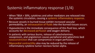
![Formula Fluid in First 24 Hours Crystalloid in Second 24-Hours Colloid in Second 24-Hours
Parkland RL at 4 mL/kg per percentage burn 20-60% estimated plasma volume Titrated to urinary output of 30 mL/h
Evans[2]
NS at 1 mL/kg per percentage burn, 2000 mL
D5W*, and colloid at 1 mL/kg per percentage
burn
50% of first 24-hour volume plus 2000 mL D5W 50% of first 24-hour volume
Slater[2] RL at 2 L/24 h plus fresh frozen plasma at 75
mL/kg/24 h
Brooke[2]
RL at 1.5 mL/kg per percentage burn, colloid at
0.5 mL/kg per percentage burn, and 2000 mL
D5W
50% of first 24-hour volume plus 2000 mL D5W 50% of first 24-hour volume
Modified Brooke RL at 2 mL/kg per percentage burn
MetroHealth
(Cleveland)
RL solution with 50 mEq sodium bicarbonate per
liter at 4 mL/kg per percentage burn
Half NS titrated to urine output
1 U fresh frozen plasma for each liter of half NS
used plus D5W as needed for hypoglycemia
Monafo hypertonic
Demling[22, 23]
250 mEq/L saline titrated to urine output at 30
mL/h, dextran 40 in NS at 2 mL/kg/h for 8 hours,
RL titrated to urine output at 30 mL/h, and fresh
frozen plasma 0.5 mL/h for 18 hours beginning 8
hours postburn
One-third NS titrated to urine output
*D5W is dextrose 5% in water solution
Table 2. Resuscitation Formulas](https://image.slidesharecdn.com/burns-generic-160906154859/85/Burns-26-320.jpg)
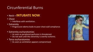


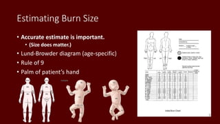
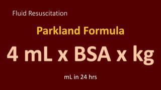
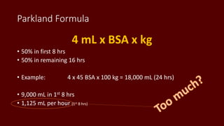
This document provides guidance on transport considerations for burned patients. It discusses burn classification, initial resuscitation, and fluid management. Burns are classified based on depth and percentage of total body surface area affected. The Parkland formula is commonly used to calculate initial fluid resuscitation, with 4 ml of lactated Ringer's solution per kg of body weight per percentage of burn over 24 hours. Accurate assessment of burn size and depth is important for determining fluid needs. Complications like edema formation, systemic inflammatory response, and hypothermia are also addressed.


