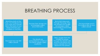
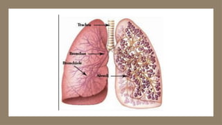
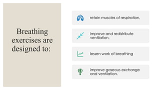
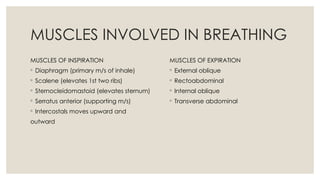
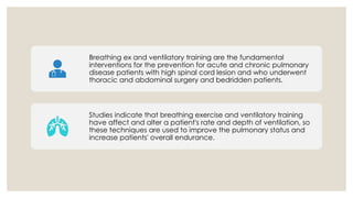
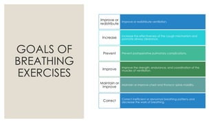
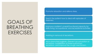
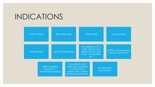
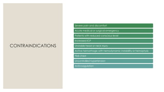
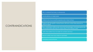
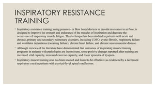
BREATHING
EXERCISES
Submitted by: RITIKA RIA
MPT 2ND
SEM
ORTHOPEDICS
Submitted to: Dr. Suraj Kumar
Professor, CPRS
BREATHING PROCESS
Breathing starts at the
nose. You inhale air into
your nose and it travels
down the back of your
throat and into your
windpipe or trachea.
Trachea then divides into
air passages called
bronchial tubes.
Bronchial tubes pass
through the lungs, they
are divided into smaller
air passages called
bronchioles or bronchial
tree.
The bronchioles end in
tiny balloon-like air sacs
called alveoli.
The body has over 300
million alveoli.
The alveoli are
surrounded by a mesh of
tiny blood vessels called
capillaries.
Here, oxygen from the
inhaled air passes
through the alveoli walls
and into the blood and
carbon dioxide from the
blood into the alveoli.
BREATHING EXERCISES
◦ Breathing is a function controlled by the autonomic nervous system and is also under
volitional control.
◦ Breathing exercises are a form of exercise which can improve the overall efficiency at
which the lungs function.
◦ They can be helpful in individual with both healthy lungs as well as those with impaired
lung function.
◦ In the presence of disease our breathing becomes altered with stress and when left
unchecked over time can result in disordered breathing patterns (breathing pattern
disorder)
◦ Disordered breathing can upset the oxygen and carbon dioxide exchange and contribute
to anxiety, panic attacks, fatigue, and other physical and emotional disturbances.
Breathing
exercises are
designed to:
retain muscles of respiration,
improve and redistribute
ventilation,
lessen work of breathing
improve gaseous exchange
and ventilation.
MUSCLES INVOLVED IN BREATHING
MUSCLES OF INSPIRATION
◦ Diaphragm (primary m/s of inhale)
◦ Scalene (elevates 1st two ribs)
◦ Sternocleidomastoid (elevates sternum)
◦ Serratus anterior (supporting m/s)
◦ Intercostals moves upward and
outward
MUSCLES OF EXPIRATION
◦ External oblique
◦ Rectoabdominal
◦ Internal oblique
◦ Transverse abdominal
Breathing ex and ventilatory training are the fundamental
interventions for the prevention for acute and chronic pulmonary
disease patients with high spinal cord lesion and who underwent
thoracic and abdominal surgery and bedridden patients.
Studies indicate that breathing exercise and ventilatory training
have affect and alter a patient's rate and depth of ventilation, so
these techniques are used to improve the pulmonary status and
increase patients' overall endurance.
GOALS OF
BREATHING
EXERCISES
Improve or redistribute ventilation.
Improve or
redistribute
Increase the effectiveness of the cough mechanism and
promote airway clearance.
Increase
Prevent postoperative pulmonary complications.
Prevent
Improve the strength, endurance, and coordination of the
muscles of ventilation.
Improve
Maintain or improve chest and thoracic spine mobility.
Maintain or
improve
Correct inefficient or abnormal breathing patterns and
decrease the work of breathing.
Correct
GOALS OF
BREATHING
EXERCISES
Promote relaxation and relieve stress.
Teach the patient how to deal with episodes of
dyspnea.
Improve a patient’s overall functional capacity for
daily living, occupational, and recreational activities.
Assisting in removal of secretions.
Aid in bronchial hygiene---Prevent accumulation of
secretions, mobilization of these pulmonary
secretions, and improve the cough mechanism.
INDICATIONS
Cystic fibrosis Bronchiectasis Atelectasis Lung abscess
Pneumonias Acute lung disease
For patients with a
high spinal cord
lesion/ spinal cord
injury, myopathies
etc.
COPD –emphysema,
chronic bronchitis
After surgeries
(thoracic or
abdominal surgery)
For patients with
prolonged bedrerst
must remain in
(obstruction due to
retained secretions).
As relaxation
procedure.
CONTRAINDICATIONS
Severe pain and discomfort
Acute medical or surgical emergency
Patients with reduced conscious level
Increased ICP
Unstable head or neck injury
Active hemorrhage with hemodynamic instability or hemoptysis
Flail chest
Uncontrolled hypertension
Anticoagulation
CONTRAINDICATIONS
Rib or vertebral fractures or osteoporosis
Acute asthma or tuberculosis
Patients who have recently experienced a heart attack.
Patients with skin grafts or spinal fusions will have undue stress placed on
areas of repair.
Bony metastases, brittle bones, bronchial hemorrhage, and emphysema are
contraindications because of undue stress to the thoracic area.
Verify that patient has not eaten for at least one hour.
Recent (within one hour) meal or tube feed
Untreated pneumothorax
PRINCIPLES
Area of exercises
Explanation &Instructions to the patient
Patients position
Evaluate the patient
Demonstration of exercise
Patient practice
Guidelines for Teaching
Breathing Exercises
◦ If possible, choose a quiet area for instruction in which you can interact with
the patient with minimal distractions.
◦ Explain to the patient the aims and rationale of breathing exercises or
ventilatory training specific to his or her particular impairments and functional
limitations.
◦ Have the patient assume a comfortable, relaxed position and loosen restrictive
clothing. Initially, a semi-Fowler’s position with the head and trunk elevated
approximately 45, is desirable. By supporting the head and trunk, flexing the
hips and knees, and supporting the legs with a pillow, the abdominal muscles
remain relaxed. Other positions, such as supine, sitting, or standing, may be
used initially or as the patient progresses during treatment.
Guidelines for Teaching
Breathing Exercises
◦ Observe and assess the patient’s spontaneous breathing pattern while at rest and later with activity.
◦ Determine whether ventilatory training is indicated.
◦ Establish a baseline for assessing changes, progress, and outcomes of intervention.
◦ If necessary, teach the patient relaxation techniques. This relaxes the muscles of the upper thorax, neck,
and shoulders to minimize the use of the accessory muscles of ventilation.
◦ Pay particular attention to relaxation of the sternocleidomastoids, upper trapezius, and levator scapulae
muscles.
◦ Depending on the patient’s underlying pathology and impairments, determine whether to emphasize
the inspiratory or expiratory phase of ventilation.
◦ Demonstrate the desired breathing pattern to the patient.
◦ Have the patient practice the correct breathing pattern in a variety of positions at rest and with activity.
PRECAUTIONS
Never allow a patient to force expiration. Expiration should be relaxed or lightly controlled. Forced
expiration only increases turbulence in the airways, leading to bronchospasm and increased airway
restriction.
Do not allow a patient to take a highly prolonged expiration. This causes the patient to gasp with the
next inspiration. The patient’s breathing pattern then becomes irregular and inefficient.
Do not allow the patient to initiate inspiration with the accessory muscles and the upper chest. Advise
the patient that the upper chest should be relatively quiet during breathing.
Allow the patient to perform deep breathing for only three or four inspirations and expirations at a time
to avoid hyperventilation.
TYPES OF
BREATHING
EXERCISES
DIAPHRAGMATIC
• Diaphragmatic training using weight
• Inspiratory resistance training
• Incentive Respiratory spirometry
VENTILATORY MUSCLE TRAINING
• Lateral costal expansion
• Posterior basal expansion
• Right lobe lingula expansion
• Apical expansion
SEGMENTAL
GLOSSOPHARYNGEAL
PURSED LIP
DIAPHRAGMATIC BREATHING
◦ When the diaphragm is functioning effectively in its role as the primary muscle of inspiration, ventilation
is efficient and the oxygen consumption of the muscles of ventilation is low during relaxed (tidal)
breathing.
◦ When a patient relies substantially on the accessory muscles of inspiration, the mechanical work of
breathing (oxygen consumption) increases and the efficiency of ventilation decreases.
◦ Although the diaphragm controls breathing at an involuntary level, a patient with primary or secondary
pulmonary dysfunction can be taught how to control breathing by optimal use of the diaphragm and
decreased use of accessory muscles.
◦ Controlled breathing techniques, which emphasize diaphragmatic breathing, are designed to improve the
efficiency of ventilation, decrease the work of breathing, increase the excursion (descent or ascent) of the
diaphragm, and improve gas exchange and oxygenation.
◦ Diaphragmatic breathing exercises also are used during postural drainage to mobilize lung secretions.
SEGMENTAL BREATHING
◦ It is questionable whether a patient can be taught to expand localized areas of
the lungs while keeping other areas quiet.
◦ It is known, however, that hypoventilation does occur in certain areas of the
lungs because of chest wall fibrosis, pain, and muscle guarding after surgery,
atelectasis, and pneumonia.
◦ Therefore, there are certain instances such as during postural drainage or
following thoracic surgery when it is important to emphasize expansion of
problem areas of the lungs and chest wall.
PURSED LIP BREATHING
◦ Pursed-lip breathing is a strategy that involves lightly pursing the lips together during controlled
exhalation. This breathing pattern often is adopted spontaneously by patients with COPD to deal with
episodes of dyspnea. Patients with COPD using pursed-lip breathing report a decrease in their perceived
level of exertion during activity.
◦ However, whether it is beneficial to teach a patient pursed-lip breathing often is debated. Many therapists
believe that gentle pursed-lip breathing is a useful procedure, particularly to relieve dyspnea if it is
performed appropriately. It is thought to keep airways open by creating back-pressure in the airways.
◦ Studies suggest that pursed-lip breathing decreases the respiratory rate and the work of breathing (oxygen
consumption), increases the tidal volume, and improves exercise tolerance.
GLOSSOPHARYNGEAL BREATHING
◦ Glossopharyngeal breathing is a technique that became known to therapists during the 1950s through
patients with severe ventilatory impairment as the result of poliomyelitis. It is a means of increasing the
inspiratory capacity when there is severe weakness of the muscles of inspiration.
◦ Today, it is used primarily by patients who are ventilator-dependent because of absent or incomplete
innervation of the diaphragm as the result of a high cervical-level spinal cord lesion or other
neuromuscular disorders.
◦ Glossopharyngeal breathing combined with the inspiratory action of the neck musculature can reduce
ventilator dependence or can be used as an emergency procedure should a malfunction of a patient’s
ventilator occur.
◦ It also can be used to improve the force (and therefore the effectiveness) of a cough or increase the
volume of the voice.
RESPIRATORY RESISTANCE
TRAINING
◦ The process of improving the strength or endurance of the muscles of ventilation is known as respiratory
resistance training (RRT). Other descriptions used to denote this form of breathing exercises are
ventilatory muscle training, inspiratory (or expiratory) muscle training, inspiratory resistance training,
and flow-controlled endurance training.
◦ These techniques typically focus on training the muscles of inspiration, although expiratory muscle
training also has been described.
◦ RRT is advocated to improve ventilation in patients with pulmonary dysfunction associated with
weakness, atrophy, or inefficiency of the muscles of inspiration or to improve the effectiveness of the
cough mechanism in patients with weakness of the abdominal muscles or other expiratory muscles.
◦ With support from animal studies, it has been suggested that the principles of overload and specificity of
training apply to skeletal muscles throughout the body, including the muscles of ventilation. In humans,
it is not feasible to use invasive procedures to evaluate morphological or histochemical changes in the
diaphragm that may occur as the result of strength or endurance training.
◦ Instead, strength or endurance changes must be assessed indirectly. Increases in respiratory muscle
strength and endurance are determined by ultrasonographic measurements of the thickness of the
diaphragm, maximal voluntary ventilation, and decreased reliance on accessory muscles of inspiration.
Respiratory muscle strength (either inspiratory or expiratory) also is evaluated indirectly with
measurements of inspiratory capacity, forced expiratory volume, inspiratory mouth pressure using a
spirometer, vital capacity, and increased cough effectiveness.
INSPIRATORY RESISTANCE
TRAINING
◦ Inspiratory resistance training, using pressure- or flow based devices to provide resistance to airflow, is
designed to improve the strength and endurance of the muscles of inspiration and decrease the
occurrence of inspiratory muscle fatigue. This technique has been studied in patients with acute and
chronic, primary and secondary pulmonary disorders, including COPD, cystic fibrosis, respiratory failure
and ventilator dependence (weaning failure), chronic heart failure, and chronic neuromuscular disease.
◦ Although reviews of the literature have demonstrated that outcomes of inspiratory muscle training
programs in patients with pathologies are inconsistent, some positive changes reported after training are
increased vital capacity, increased exercise capacity, and fewer episodes of dyspnea.
◦ Inspiratory muscle training also has been studied and found to be effective (as evidenced by a decreased
respiratory rate) in patients with cervical-level spinal cord lesions.
INCENTIVE RESPIRATORY
SPIROMETRY
◦ Incentive spirometry is a form of ventilatory training that emphasizes sustained maximum inspirations.
The patient inhales as deeply as possible through a small, handheld that provides visual or auditory
feedback about whether a target maximum inspiration was reached.
◦ Typically, this breathing technique is performed while using a spirometer, but it also may be performed
without the equipment.
◦ The purpose of incentive spirometry is to increase the volume of air inspired. It is used primarily to
prevent alveolar collapse and atelectasis in postoperative patients.
◦ Despite the widespread use of incentive spirometry for patients after surgery, the effectiveness of this
technique alone or in addition to general deep breathing and coughing for the prevention of postoperative
pulmonary complications is not clear
THANKYOU

Breathing Exercises with recent evidences

  • 1.
    BREATHING EXERCISES Submitted by: RITIKARIA MPT 2ND SEM ORTHOPEDICS Submitted to: Dr. Suraj Kumar Professor, CPRS
  • 2.
    BREATHING PROCESS Breathing startsat the nose. You inhale air into your nose and it travels down the back of your throat and into your windpipe or trachea. Trachea then divides into air passages called bronchial tubes. Bronchial tubes pass through the lungs, they are divided into smaller air passages called bronchioles or bronchial tree. The bronchioles end in tiny balloon-like air sacs called alveoli. The body has over 300 million alveoli. The alveoli are surrounded by a mesh of tiny blood vessels called capillaries. Here, oxygen from the inhaled air passes through the alveoli walls and into the blood and carbon dioxide from the blood into the alveoli.
  • 4.
    BREATHING EXERCISES ◦ Breathingis a function controlled by the autonomic nervous system and is also under volitional control. ◦ Breathing exercises are a form of exercise which can improve the overall efficiency at which the lungs function. ◦ They can be helpful in individual with both healthy lungs as well as those with impaired lung function. ◦ In the presence of disease our breathing becomes altered with stress and when left unchecked over time can result in disordered breathing patterns (breathing pattern disorder) ◦ Disordered breathing can upset the oxygen and carbon dioxide exchange and contribute to anxiety, panic attacks, fatigue, and other physical and emotional disturbances.
  • 5.
    Breathing exercises are designed to: retainmuscles of respiration, improve and redistribute ventilation, lessen work of breathing improve gaseous exchange and ventilation.
  • 6.
    MUSCLES INVOLVED INBREATHING MUSCLES OF INSPIRATION ◦ Diaphragm (primary m/s of inhale) ◦ Scalene (elevates 1st two ribs) ◦ Sternocleidomastoid (elevates sternum) ◦ Serratus anterior (supporting m/s) ◦ Intercostals moves upward and outward MUSCLES OF EXPIRATION ◦ External oblique ◦ Rectoabdominal ◦ Internal oblique ◦ Transverse abdominal
  • 7.
    Breathing ex andventilatory training are the fundamental interventions for the prevention for acute and chronic pulmonary disease patients with high spinal cord lesion and who underwent thoracic and abdominal surgery and bedridden patients. Studies indicate that breathing exercise and ventilatory training have affect and alter a patient's rate and depth of ventilation, so these techniques are used to improve the pulmonary status and increase patients' overall endurance.
  • 8.
    GOALS OF BREATHING EXERCISES Improve orredistribute ventilation. Improve or redistribute Increase the effectiveness of the cough mechanism and promote airway clearance. Increase Prevent postoperative pulmonary complications. Prevent Improve the strength, endurance, and coordination of the muscles of ventilation. Improve Maintain or improve chest and thoracic spine mobility. Maintain or improve Correct inefficient or abnormal breathing patterns and decrease the work of breathing. Correct
  • 9.
    GOALS OF BREATHING EXERCISES Promote relaxationand relieve stress. Teach the patient how to deal with episodes of dyspnea. Improve a patient’s overall functional capacity for daily living, occupational, and recreational activities. Assisting in removal of secretions. Aid in bronchial hygiene---Prevent accumulation of secretions, mobilization of these pulmonary secretions, and improve the cough mechanism.
  • 10.
    INDICATIONS Cystic fibrosis BronchiectasisAtelectasis Lung abscess Pneumonias Acute lung disease For patients with a high spinal cord lesion/ spinal cord injury, myopathies etc. COPD –emphysema, chronic bronchitis After surgeries (thoracic or abdominal surgery) For patients with prolonged bedrerst must remain in (obstruction due to retained secretions). As relaxation procedure.
  • 11.
    CONTRAINDICATIONS Severe pain anddiscomfort Acute medical or surgical emergency Patients with reduced conscious level Increased ICP Unstable head or neck injury Active hemorrhage with hemodynamic instability or hemoptysis Flail chest Uncontrolled hypertension Anticoagulation
  • 12.
    CONTRAINDICATIONS Rib or vertebralfractures or osteoporosis Acute asthma or tuberculosis Patients who have recently experienced a heart attack. Patients with skin grafts or spinal fusions will have undue stress placed on areas of repair. Bony metastases, brittle bones, bronchial hemorrhage, and emphysema are contraindications because of undue stress to the thoracic area. Verify that patient has not eaten for at least one hour. Recent (within one hour) meal or tube feed Untreated pneumothorax
  • 13.
    PRINCIPLES Area of exercises Explanation&Instructions to the patient Patients position Evaluate the patient Demonstration of exercise Patient practice
  • 14.
    Guidelines for Teaching BreathingExercises ◦ If possible, choose a quiet area for instruction in which you can interact with the patient with minimal distractions. ◦ Explain to the patient the aims and rationale of breathing exercises or ventilatory training specific to his or her particular impairments and functional limitations. ◦ Have the patient assume a comfortable, relaxed position and loosen restrictive clothing. Initially, a semi-Fowler’s position with the head and trunk elevated approximately 45, is desirable. By supporting the head and trunk, flexing the hips and knees, and supporting the legs with a pillow, the abdominal muscles remain relaxed. Other positions, such as supine, sitting, or standing, may be used initially or as the patient progresses during treatment.
  • 15.
    Guidelines for Teaching BreathingExercises ◦ Observe and assess the patient’s spontaneous breathing pattern while at rest and later with activity. ◦ Determine whether ventilatory training is indicated. ◦ Establish a baseline for assessing changes, progress, and outcomes of intervention. ◦ If necessary, teach the patient relaxation techniques. This relaxes the muscles of the upper thorax, neck, and shoulders to minimize the use of the accessory muscles of ventilation. ◦ Pay particular attention to relaxation of the sternocleidomastoids, upper trapezius, and levator scapulae muscles. ◦ Depending on the patient’s underlying pathology and impairments, determine whether to emphasize the inspiratory or expiratory phase of ventilation. ◦ Demonstrate the desired breathing pattern to the patient. ◦ Have the patient practice the correct breathing pattern in a variety of positions at rest and with activity.
  • 16.
    PRECAUTIONS Never allow apatient to force expiration. Expiration should be relaxed or lightly controlled. Forced expiration only increases turbulence in the airways, leading to bronchospasm and increased airway restriction. Do not allow a patient to take a highly prolonged expiration. This causes the patient to gasp with the next inspiration. The patient’s breathing pattern then becomes irregular and inefficient. Do not allow the patient to initiate inspiration with the accessory muscles and the upper chest. Advise the patient that the upper chest should be relatively quiet during breathing. Allow the patient to perform deep breathing for only three or four inspirations and expirations at a time to avoid hyperventilation.
  • 17.
    TYPES OF BREATHING EXERCISES DIAPHRAGMATIC • Diaphragmatictraining using weight • Inspiratory resistance training • Incentive Respiratory spirometry VENTILATORY MUSCLE TRAINING • Lateral costal expansion • Posterior basal expansion • Right lobe lingula expansion • Apical expansion SEGMENTAL GLOSSOPHARYNGEAL PURSED LIP
  • 18.
    DIAPHRAGMATIC BREATHING ◦ Whenthe diaphragm is functioning effectively in its role as the primary muscle of inspiration, ventilation is efficient and the oxygen consumption of the muscles of ventilation is low during relaxed (tidal) breathing. ◦ When a patient relies substantially on the accessory muscles of inspiration, the mechanical work of breathing (oxygen consumption) increases and the efficiency of ventilation decreases. ◦ Although the diaphragm controls breathing at an involuntary level, a patient with primary or secondary pulmonary dysfunction can be taught how to control breathing by optimal use of the diaphragm and decreased use of accessory muscles. ◦ Controlled breathing techniques, which emphasize diaphragmatic breathing, are designed to improve the efficiency of ventilation, decrease the work of breathing, increase the excursion (descent or ascent) of the diaphragm, and improve gas exchange and oxygenation. ◦ Diaphragmatic breathing exercises also are used during postural drainage to mobilize lung secretions.
  • 20.
    SEGMENTAL BREATHING ◦ Itis questionable whether a patient can be taught to expand localized areas of the lungs while keeping other areas quiet. ◦ It is known, however, that hypoventilation does occur in certain areas of the lungs because of chest wall fibrosis, pain, and muscle guarding after surgery, atelectasis, and pneumonia. ◦ Therefore, there are certain instances such as during postural drainage or following thoracic surgery when it is important to emphasize expansion of problem areas of the lungs and chest wall.
  • 21.
    PURSED LIP BREATHING ◦Pursed-lip breathing is a strategy that involves lightly pursing the lips together during controlled exhalation. This breathing pattern often is adopted spontaneously by patients with COPD to deal with episodes of dyspnea. Patients with COPD using pursed-lip breathing report a decrease in their perceived level of exertion during activity. ◦ However, whether it is beneficial to teach a patient pursed-lip breathing often is debated. Many therapists believe that gentle pursed-lip breathing is a useful procedure, particularly to relieve dyspnea if it is performed appropriately. It is thought to keep airways open by creating back-pressure in the airways. ◦ Studies suggest that pursed-lip breathing decreases the respiratory rate and the work of breathing (oxygen consumption), increases the tidal volume, and improves exercise tolerance.
  • 22.
    GLOSSOPHARYNGEAL BREATHING ◦ Glossopharyngealbreathing is a technique that became known to therapists during the 1950s through patients with severe ventilatory impairment as the result of poliomyelitis. It is a means of increasing the inspiratory capacity when there is severe weakness of the muscles of inspiration. ◦ Today, it is used primarily by patients who are ventilator-dependent because of absent or incomplete innervation of the diaphragm as the result of a high cervical-level spinal cord lesion or other neuromuscular disorders. ◦ Glossopharyngeal breathing combined with the inspiratory action of the neck musculature can reduce ventilator dependence or can be used as an emergency procedure should a malfunction of a patient’s ventilator occur. ◦ It also can be used to improve the force (and therefore the effectiveness) of a cough or increase the volume of the voice.
  • 23.
    RESPIRATORY RESISTANCE TRAINING ◦ Theprocess of improving the strength or endurance of the muscles of ventilation is known as respiratory resistance training (RRT). Other descriptions used to denote this form of breathing exercises are ventilatory muscle training, inspiratory (or expiratory) muscle training, inspiratory resistance training, and flow-controlled endurance training. ◦ These techniques typically focus on training the muscles of inspiration, although expiratory muscle training also has been described. ◦ RRT is advocated to improve ventilation in patients with pulmonary dysfunction associated with weakness, atrophy, or inefficiency of the muscles of inspiration or to improve the effectiveness of the cough mechanism in patients with weakness of the abdominal muscles or other expiratory muscles.
  • 24.
    ◦ With supportfrom animal studies, it has been suggested that the principles of overload and specificity of training apply to skeletal muscles throughout the body, including the muscles of ventilation. In humans, it is not feasible to use invasive procedures to evaluate morphological or histochemical changes in the diaphragm that may occur as the result of strength or endurance training. ◦ Instead, strength or endurance changes must be assessed indirectly. Increases in respiratory muscle strength and endurance are determined by ultrasonographic measurements of the thickness of the diaphragm, maximal voluntary ventilation, and decreased reliance on accessory muscles of inspiration. Respiratory muscle strength (either inspiratory or expiratory) also is evaluated indirectly with measurements of inspiratory capacity, forced expiratory volume, inspiratory mouth pressure using a spirometer, vital capacity, and increased cough effectiveness.
  • 25.
    INSPIRATORY RESISTANCE TRAINING ◦ Inspiratoryresistance training, using pressure- or flow based devices to provide resistance to airflow, is designed to improve the strength and endurance of the muscles of inspiration and decrease the occurrence of inspiratory muscle fatigue. This technique has been studied in patients with acute and chronic, primary and secondary pulmonary disorders, including COPD, cystic fibrosis, respiratory failure and ventilator dependence (weaning failure), chronic heart failure, and chronic neuromuscular disease. ◦ Although reviews of the literature have demonstrated that outcomes of inspiratory muscle training programs in patients with pathologies are inconsistent, some positive changes reported after training are increased vital capacity, increased exercise capacity, and fewer episodes of dyspnea. ◦ Inspiratory muscle training also has been studied and found to be effective (as evidenced by a decreased respiratory rate) in patients with cervical-level spinal cord lesions.
  • 26.
    INCENTIVE RESPIRATORY SPIROMETRY ◦ Incentivespirometry is a form of ventilatory training that emphasizes sustained maximum inspirations. The patient inhales as deeply as possible through a small, handheld that provides visual or auditory feedback about whether a target maximum inspiration was reached. ◦ Typically, this breathing technique is performed while using a spirometer, but it also may be performed without the equipment. ◦ The purpose of incentive spirometry is to increase the volume of air inspired. It is used primarily to prevent alveolar collapse and atelectasis in postoperative patients. ◦ Despite the widespread use of incentive spirometry for patients after surgery, the effectiveness of this technique alone or in addition to general deep breathing and coughing for the prevention of postoperative pulmonary complications is not clear
  • 31.