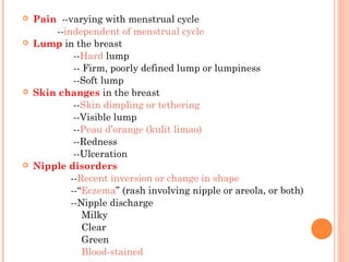
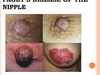
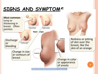
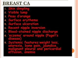
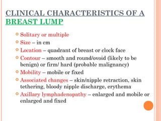
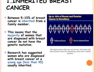
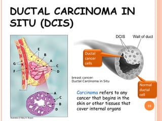
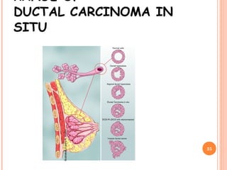
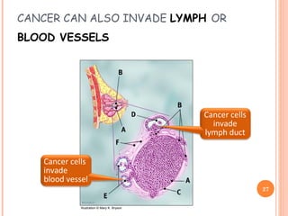
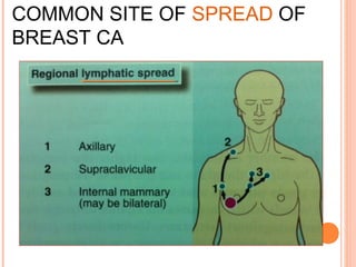
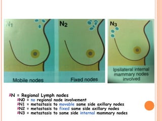
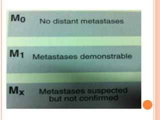
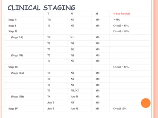
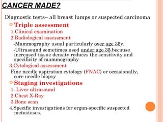
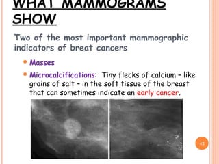
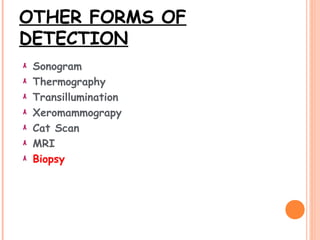
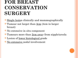
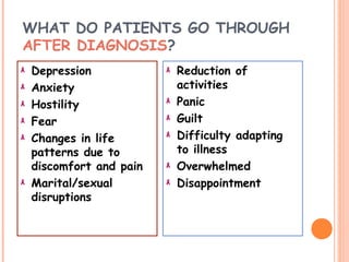
By Dr. Celine Tey
TECHNIQUE OF BREAST EXAMINATION
 Pain --varying with menstrual cycle
--independent of menstrual cycle
 Lump in the breast
--Hard lump
-- Firm, poorly defined lump or lumpiness
--Soft lump
 Skin changes in the breast
--Skin dimpling or tethering
--Visible lump
--Peau d’orange (kulit limao)
--Redness
--Ulceration
 Nipple disorders
--Recent inversion or change in shape
--“Eczema” (rash involving nipple or areola, or both)
--Nipple discharge
Milky
Clear
Green
Blood-stained
NIPPLE DISORDERS
 --Recent inversion or change in shape
suggests a fibrosing underlying lesion such as a carcinoma or
mammary duct ectasia but can be malignancy (refer urgently)
 --“Eczema” (rash involving nipple or areola, or both)
if unilateral and persistent, this is the classic sign of Paget’s
desease of the nipple, a presentation of breast ca
(refer urgently if not responding to treatment)
 --Nipple discharge
1. Milky—pregnancy of hyperprolactinaemia
2. Clear – physiological
3. Green –perimenopausal, duct ectasia, fibroadenotic cyst
4. Blood-stained –possible carcinoma or intraduct papilloma (refer
urgently)
PAGET’S DISEASE OF THE
NIPPLE
SIGNS AND SYMPTOMS
9
Most common:
lump or
thickening in
breast. Often
painless
Change in color
or appearance
of areola
Redness or pitting
of skin over the
breast, like the
skin of an orange
Discharge
or
bleeding
Change in size
or contours of
breast
rg
BREAST CA
1. Skin dimpling
2. Visible lump
3. Peau d’orange
4. Surface erythema
5. Surface ulceration
6. Recent nipple inversion
7. Blood-stained nipple discharge
8. ‘eczema’ around nipple (Paget’s
disease)
9. Systemic features:weight loss,
anorexia, bone pain, jaundice,
malignant pleural and pericardial
effusion, anemia
CHARACTERISTIC SIGNS OF
BREAST CA
BREAST CA
CLINICAL CHARACTERISTICS OF A
BREAST LUMP
 Solitary or multiple
 Size – in cm
 Location – quadrant of breast or clock face
 Contour – smooth and round/ovoid (likely to be
benign) or firm/ hard (probable malignancy)
 Mobility – mobile or fixed
 Associated changes – skin/nipple retraction, skin
tethering, bloody nipple discharge, erythema
 Axillary lymphadenopathy – enlarged and mobile or
enlarged and fixed
CAUSES
1. Inherited
2. Risk Factors
3. Environmental Factors
1.INHERITED BREAST
CANCER
Between 5-10% of breast
cancer is inherited from a
family member.
This means that the
majority of women that
are diagnosed with breast
cancer do not have the
genetic mutation.
Research has suggested
women who are diagnosed
with breast cancer at a
young age (less than 45)
usually inherited.
This figure shows that one out of every 10 women will
obtain breast cancer by inheriting a gene from a family
member.
INHERITED
GENES
BRCA1 (Breast Cancer 1)
BRCA2 (Breast Cancer 2)
TP53 gene
ATM gene
BRCA 1 AND BRCA 2
Both of these genes code for DNA repair.
If a woman has a mutation on either one of these
genes, the risk of her getting breast cancer
increases from 10% to 80% in her lifetime.
Mutations in BRCA1 or BRCA2 account for 40-50%
of all cases of inherited breast cancer.
These genes are also associated with ovarian cancer
in women and prostate cancer in men.
These genes can be inherited either from the
mother or the father.
OTHER INHERITED
GENES THAT CAUSE
CANCER
TP53 gene
This gene codes for the
tumor suppressor protein
p53.
Mutations of this gene
cause Li-Fraumeni
syndrome, which is a
condition that is
associated with early
onset breast cancer.
ATM gene
Females with one
defective copy of the
ATM gene and one
normal copy of the
gene are at
increased risk for
breast cancer.
2.RISK FACTORS CAUSE BREAST CANCER
Factors that Cannot
be Prevented
Gender
Aging (40-55 y-o)
Genetic Risk Factors
(inherited)
Family History
Personal History
Menstrual Cycle
Estrogen
Lifestyle Risks
Oral Contraceptive Use
Nulliparity
Hormone Replacement
Therapy
Not Breast Feeding
Alcohol Use
Obesity
High Fat Diets
Physical Inactivity
Smoking
3.ENVIRONMENTAL
FACTORS
Exposure to irradiation
Electromagnetic Fields
Xenoestrogens
Exposure to Chemicals
NORMAL BREAST
Breast profile
A ducts
B lobules
C dilated section of duct to hold milk
D nipple
E fat
F pectoralis major muscle
G chest wall/rib cage
20
Enlargement
A normal duct cells
B basement membrane (duct wall)
C lumen (center of duct)
Illustration © Mary K. Bryson
DIAGRAM OF THE
BREAST
The breast is a glandular
organ.
It is made up of a network of
mammary ducts.
Each breast has about 15-20
mammary ducts that lead to
lobes that are made up of
lobules.
The lobules contain cells that
secrete milk that are
stimulated by estrogen and
progesterone which are
ovarian hormones.
IN SITU BREAST
CANCER
In Situ Breast Cancer remains within the ducts or
lobules of the breasts.
This type of cancer is only detected by mammograms
– not by a physical examination.
If the cancer is in the duct it is called Ductal
Carcinoma in situ.
If the cancer is in the lobule of the breast, it is
called Lobular Carcinoma in situ.
This type of cancer is most common among pre-menopausal
women.
There is also a slight chance that if a woman has this type
of cancer she is at risk that it would occur in the other.
DUCTAL CARCINOMA IN
SITU (DCIS)
Carcinoma refers to any
cancer that begins in the
skin or other tissues that
cover internal organs
23
Illustration © Mary K. Bryson
Ductal
cancer
cells
Normal
ductal
cell
INVASIVE DUCTAL CARCINOMA
(IDC – 80% OF BREAST CANCER)
 The cancer has spread to the
surrounding tissues
24
Illustration © Mary K. Bryson
Ductal cancer cells
breaking through
the wall
RANGE OF
DUCTAL CARCINOMA IN
SITU
25
Illustration©MaryK.Bryson
INVASIVE LOBULAR CARCINOMA
(ILC)
26
Illustration © Mary K. Bryson
Lobular cancer
cells breaking
through the wall
CANCER CAN ALSO INVADE LYMPH OR
BLOOD VESSELS
27
Illustration © Mary K. Bryson
Cancer cells
invade
lymph duct
Cancer cells
invade
blood vessel
INFILTRATING BREAST CANCER
Breast cancer is
considered infiltrating
or invasive if the
cancer cells have
penetrated the
membrane that
surrounds a duct or
lobule.
This type of cancer
forms a lump that can
eventually be felt by a
physical examination.
Breast cancer cells cross the lining of
the milk duct or lobule, and begin to
invade adjacent tissues. This type of
cancer is called "infiltrating cancer."
In this picture, you can see the breast
cancer cells invading the milk duct.
MORE ON INFILTRATING BREAST
CANCER
Infiltrating cancer of
the duct
Called “Infiltrating
Ductal Carcinoma”
It is the most common
type of breast cancer.
Cancer cells that are
invading the fatty
tissue around the duct,
they stimulate the
growth of non-
cancerous scar like
tissue that surrounds
the cancer making it
easier to spot.
Infiltrating cancer of
the lobules
Called “Infiltrating
Lobular Carcinoma”
Occurs when cells stream
out in a single file into
the surrounding breast
tissue.
This type of cancer is
harder to detect on a
mammogram because
there is no fibrous
growth.
OTHER TYPES OF BREAST CANCER
Cystosarcoma Phyllodes
Inflammatory Cancer
Accounts for less than one percent of all breast
cancers and looks as though the breast is
infected.
Breast Cancer During Pregnancy
Paget’s Disease
COMMON SITE OF SPREAD OF
BREAST CA
TNM STAGING
IN BREAST CA
T = Primary Tumor
Tis (T0) = carcinoma in situ
T1 = less than 2 cm in diameter
T2 = between 2 and 5 cm in
diameter
T3 = more than 5 cm in diameter
T4 = any size, but extends to the
skin or chest wall
N = Regional Lymph nodes
N0 = no regional node involvement
N1 = metastasis to movable same side axillary nodes
N2 = metastasis to fixed same side axillary nodes
N3 = metastasis to same side internal mammary nodes
CLINICAL STAGING
  T N M 5-Year Survival
Stage 0 Tis N0 M0 > 95%
Stage I T1 N0 M0 Overall = 85%
Stage II       Overall = 66%
    (Stage IIA) T0 N1 M0  
  T1 N1 M0  
  T2 N0 M0  
    (Stage IIB) T2 N1 M0  
  T3 N0 M0  
Stage III       Overall = 41%
    (Stage IIIA) T0 N2 M0  
  T1 N2 M0  
  T2 N2 M0  
  T3 N1, N2 M0  
    (Stage IIIB) T4 Any N M0  
  Any T N3 M0  
Stage IV Any T Any N M1 Overall 10%
THE EFFECT OF TUMOR SIZE ON
SURVIVAL
Survival
Tumor Size
As tumor size
increases, the chance
of survival
decreases.
HOW DO
YOU
DETECT
BREAST
CANCER?
 Triple assessment
1.Clinical examination
2.Radiological assessment
-Mammography usual particularly over age 35y.
-Ultrasound sometimes used under age 35 because
increased tissue density reduces the sensitivity and
specificity of mammography
3.Cytological assessment
Fine needle aspiration cytology (FNAC) or occasionally,
core needle biopsy
 Staging investigations
1. Liver ultrasound
2.Chest X-Ray
3.Bone scan
4.Specific investigations for organ-specific suspected
metastases.
Diagnostic tests– all breast lumps or suspected carcinoma
CANCER MADE?
MAMMOGRAM
A Mammogram is a X-ray of
the breast that takes
pictures of the fat, fibrous
tissues, ducts, lobes, and
blood vessels.
When should a mammogram
be performed?
If a lump has been found
during self-examination or
by a physician
Younger women who have a
strong history of breast
cancer in their family
All women over forty
Women who have had
previous diagnosis of breast
cancer.
WHAT MAMMOGRAMS
SHOW
Two of the most important mammographic
indicators of breat cancers
Masses
Microcalcifications: Tiny flecks of calcium – like
grains of salt – in the soft tissue of the breast
that can sometimes indicate an early cancer.
42
DETECTION OF MALIGNANT
MASSES
Malignant masses have a more spiculated appearance
43
malignant
benign
BREAST SELF
EXAMINATIO
N
OTHER FORMS OF
DETECTION
Sonogram
Thermography
Transillumination
Xeromammograpy
Cat Scan
MRI
Biopsy
TREATMENTS OF
BREAST CANCER
MEDICAL TREATMENT
NON- METASTASIS DISEASE
 Adjuvant to reduce the risk of systemic relapse usually after
primary surgery.
 Occasionally used as treatment of choise in elderly or those
unfit/inappropriate for surgery
Endocrine Therapy
1. Anti-estrogens (e.g tamoxifen, LHRH antagonists,
aromatase inhibitors)
2. Most effective in ER +ve tumours
Chemotherapy
1. Anthracyclines, cyclophosphamide,5-FU, methotrexate
2. Offered to patients with high risk features (+ve nodes,
poor grade)
MEDICAL TREATMENT
METASTASIS DISEASE
 Palliative to increase survival time
Endocrine Therapy
As above
Chemotherapy
Anthracyclines, tanaxest
Radiotherapy
To reduce pain of bony metastases or
symptoms from cerebral or liver disease
SURGERYMAINSTAY FOR NON METASTASIS DISEASE
Mastectomy
(radical,modified radical
simple)
A mastectomy is the
surgical removal of the
breast, non-protruding
breast tissue, the lymph
nodes in the armpits and
some pectoral muscle.
Breast reconstruction
surgery may be
conducted after the
removal of the breast.
Breast conservation
(lumpectomy, wide local
excision, quadrantectomy)
In this surgical procedure,
the breast is conserved and
the tumor is removed.
Radiation commonly follows a
lumpectomy to try to rid the
body of any other cancerous
cells.
BREAST CA
 Wide local excision-commenest procedure
-breast conserving provided breast is adequate size and
tumour location appropriate (not central/retro-areolar)
-usually combined with local radiotherapy
 Simple mastectomy-best treatment and cosmetic result
 Surgical management of regional lymph nodes
-Axillary node sampling
-Axillary node clearance
-Sentinel node biopsy
Usually the first axillary node to receive lymphatic drainage
from the tumour. Before operation, a blue dye and a radiotracer
are injected into subareolar areas and at operation the sentinel
node is identified visually and by using a device to detect
radioactivity.
 Surgery for metastastic disease: limited to procedures for
symptomatic control of local disease (e.g mastectomy to
remove fungating tumour)
FOR BREAST
CONSERVATION
SURGERY
 Single lesion clinically and mammographically
 Tumour not larget than 3cm (4cm in larger
breast)
 No extensive in situ component
 Tumours more than 2cm away from nipple/areola
 Lesion of lower histological grade
 No extensive nodal involvement
PSYCHOLOGICA
L IMPACTS OF
BREAST
CANCER
WHAT DO PATIENTS GO THROUGH
AFTER DIAGNOSIS?
Depression
Anxiety
Hostility
Fear
Changes in life
patterns due to
discomfort and pain
Marital/sexual
disruptions
Reduction of
activities
Panic
Guilt
Difficulty adapting
to illness
Overwhelmed
Disappointment
REOCCURRENCES OF BREAST
CANCER
Reoccurrences
Personal
Responsibility
Loss of Hope
Denial
Grief
Therapies
Group Therapies
Single session groups
Time limited groups
Long Term groups
Traditional
Single session with
psychologists
PREVENTIO
N
FAT
Research shows that dietary fat should
be 20% or less in order to gain
meaningful protection against cancer.
Fat cells make estrogen, which promotes
breast cancer.
Diets high in fat are associated with the
increasing breast density in mammograms,
which makes interpretation more
difficult.
FIBER
Fiber provides protection against breast
cancer because it has a mechanism that
decreases the amount of estrogen in the
body.
The amount of fiber in the diet affects the
activities of intestinal bacteria, which affects
the amount of reabsorbed estrogens.
ANTIOXIDANT
NUTRIENTS
Antioxidants are important in fighting
breast cancer because they can disarm
cancer-causing substances called free
radicals.
Vitamin C
Vitamin E
Beta-carotene
Vitamin A
Selenium
OTHER PREVENTATIVE MEASURES
Early Detection!!!!
Exercise
No Smoking!!
Good Diet
REFERANCE
 Essential surgery 4th
edition
 Oxford handbook of clinical surgery 3rd
edition
 TheNational Cancer Institutewedsite

breastca-121013104443-phpapp01

  • 1.
  • 2.
  • 6.
     Pain --varyingwith menstrual cycle --independent of menstrual cycle  Lump in the breast --Hard lump -- Firm, poorly defined lump or lumpiness --Soft lump  Skin changes in the breast --Skin dimpling or tethering --Visible lump --Peau d’orange (kulit limao) --Redness --Ulceration  Nipple disorders --Recent inversion or change in shape --“Eczema” (rash involving nipple or areola, or both) --Nipple discharge Milky Clear Green Blood-stained
  • 7.
    NIPPLE DISORDERS  --Recentinversion or change in shape suggests a fibrosing underlying lesion such as a carcinoma or mammary duct ectasia but can be malignancy (refer urgently)  --“Eczema” (rash involving nipple or areola, or both) if unilateral and persistent, this is the classic sign of Paget’s desease of the nipple, a presentation of breast ca (refer urgently if not responding to treatment)  --Nipple discharge 1. Milky—pregnancy of hyperprolactinaemia 2. Clear – physiological 3. Green –perimenopausal, duct ectasia, fibroadenotic cyst 4. Blood-stained –possible carcinoma or intraduct papilloma (refer urgently)
  • 8.
  • 9.
    SIGNS AND SYMPTOMS 9 Mostcommon: lump or thickening in breast. Often painless Change in color or appearance of areola Redness or pitting of skin over the breast, like the skin of an orange Discharge or bleeding Change in size or contours of breast rg
  • 10.
    BREAST CA 1. Skindimpling 2. Visible lump 3. Peau d’orange 4. Surface erythema 5. Surface ulceration 6. Recent nipple inversion 7. Blood-stained nipple discharge 8. ‘eczema’ around nipple (Paget’s disease) 9. Systemic features:weight loss, anorexia, bone pain, jaundice, malignant pleural and pericardial effusion, anemia
  • 11.
  • 12.
    CLINICAL CHARACTERISTICS OFA BREAST LUMP  Solitary or multiple  Size – in cm  Location – quadrant of breast or clock face  Contour – smooth and round/ovoid (likely to be benign) or firm/ hard (probable malignancy)  Mobility – mobile or fixed  Associated changes – skin/nipple retraction, skin tethering, bloody nipple discharge, erythema  Axillary lymphadenopathy – enlarged and mobile or enlarged and fixed
  • 13.
    CAUSES 1. Inherited 2. RiskFactors 3. Environmental Factors
  • 14.
    1.INHERITED BREAST CANCER Between 5-10%of breast cancer is inherited from a family member. This means that the majority of women that are diagnosed with breast cancer do not have the genetic mutation. Research has suggested women who are diagnosed with breast cancer at a young age (less than 45) usually inherited. This figure shows that one out of every 10 women will obtain breast cancer by inheriting a gene from a family member.
  • 15.
    INHERITED GENES BRCA1 (Breast Cancer1) BRCA2 (Breast Cancer 2) TP53 gene ATM gene
  • 16.
    BRCA 1 ANDBRCA 2 Both of these genes code for DNA repair. If a woman has a mutation on either one of these genes, the risk of her getting breast cancer increases from 10% to 80% in her lifetime. Mutations in BRCA1 or BRCA2 account for 40-50% of all cases of inherited breast cancer. These genes are also associated with ovarian cancer in women and prostate cancer in men. These genes can be inherited either from the mother or the father.
  • 17.
    OTHER INHERITED GENES THATCAUSE CANCER TP53 gene This gene codes for the tumor suppressor protein p53. Mutations of this gene cause Li-Fraumeni syndrome, which is a condition that is associated with early onset breast cancer. ATM gene Females with one defective copy of the ATM gene and one normal copy of the gene are at increased risk for breast cancer.
  • 18.
    2.RISK FACTORS CAUSEBREAST CANCER Factors that Cannot be Prevented Gender Aging (40-55 y-o) Genetic Risk Factors (inherited) Family History Personal History Menstrual Cycle Estrogen Lifestyle Risks Oral Contraceptive Use Nulliparity Hormone Replacement Therapy Not Breast Feeding Alcohol Use Obesity High Fat Diets Physical Inactivity Smoking
  • 19.
    3.ENVIRONMENTAL FACTORS Exposure to irradiation ElectromagneticFields Xenoestrogens Exposure to Chemicals
  • 20.
    NORMAL BREAST Breast profile Aducts B lobules C dilated section of duct to hold milk D nipple E fat F pectoralis major muscle G chest wall/rib cage 20 Enlargement A normal duct cells B basement membrane (duct wall) C lumen (center of duct) Illustration © Mary K. Bryson
  • 21.
    DIAGRAM OF THE BREAST Thebreast is a glandular organ. It is made up of a network of mammary ducts. Each breast has about 15-20 mammary ducts that lead to lobes that are made up of lobules. The lobules contain cells that secrete milk that are stimulated by estrogen and progesterone which are ovarian hormones.
  • 22.
    IN SITU BREAST CANCER InSitu Breast Cancer remains within the ducts or lobules of the breasts. This type of cancer is only detected by mammograms – not by a physical examination. If the cancer is in the duct it is called Ductal Carcinoma in situ. If the cancer is in the lobule of the breast, it is called Lobular Carcinoma in situ. This type of cancer is most common among pre-menopausal women. There is also a slight chance that if a woman has this type of cancer she is at risk that it would occur in the other.
  • 23.
    DUCTAL CARCINOMA IN SITU(DCIS) Carcinoma refers to any cancer that begins in the skin or other tissues that cover internal organs 23 Illustration © Mary K. Bryson Ductal cancer cells Normal ductal cell
  • 24.
    INVASIVE DUCTAL CARCINOMA (IDC– 80% OF BREAST CANCER)  The cancer has spread to the surrounding tissues 24 Illustration © Mary K. Bryson Ductal cancer cells breaking through the wall
  • 25.
    RANGE OF DUCTAL CARCINOMAIN SITU 25 Illustration©MaryK.Bryson
  • 26.
    INVASIVE LOBULAR CARCINOMA (ILC) 26 Illustration© Mary K. Bryson Lobular cancer cells breaking through the wall
  • 27.
    CANCER CAN ALSOINVADE LYMPH OR BLOOD VESSELS 27 Illustration © Mary K. Bryson Cancer cells invade lymph duct Cancer cells invade blood vessel
  • 28.
    INFILTRATING BREAST CANCER Breastcancer is considered infiltrating or invasive if the cancer cells have penetrated the membrane that surrounds a duct or lobule. This type of cancer forms a lump that can eventually be felt by a physical examination. Breast cancer cells cross the lining of the milk duct or lobule, and begin to invade adjacent tissues. This type of cancer is called "infiltrating cancer." In this picture, you can see the breast cancer cells invading the milk duct.
  • 29.
    MORE ON INFILTRATINGBREAST CANCER Infiltrating cancer of the duct Called “Infiltrating Ductal Carcinoma” It is the most common type of breast cancer. Cancer cells that are invading the fatty tissue around the duct, they stimulate the growth of non- cancerous scar like tissue that surrounds the cancer making it easier to spot. Infiltrating cancer of the lobules Called “Infiltrating Lobular Carcinoma” Occurs when cells stream out in a single file into the surrounding breast tissue. This type of cancer is harder to detect on a mammogram because there is no fibrous growth.
  • 30.
    OTHER TYPES OFBREAST CANCER Cystosarcoma Phyllodes Inflammatory Cancer Accounts for less than one percent of all breast cancers and looks as though the breast is infected. Breast Cancer During Pregnancy Paget’s Disease
  • 31.
    COMMON SITE OFSPREAD OF BREAST CA
  • 33.
  • 34.
    T = PrimaryTumor Tis (T0) = carcinoma in situ T1 = less than 2 cm in diameter T2 = between 2 and 5 cm in diameter T3 = more than 5 cm in diameter T4 = any size, but extends to the skin or chest wall
  • 35.
    N = RegionalLymph nodes N0 = no regional node involvement N1 = metastasis to movable same side axillary nodes N2 = metastasis to fixed same side axillary nodes N3 = metastasis to same side internal mammary nodes
  • 37.
    CLINICAL STAGING   TN M 5-Year Survival Stage 0 Tis N0 M0 > 95% Stage I T1 N0 M0 Overall = 85% Stage II       Overall = 66%     (Stage IIA) T0 N1 M0     T1 N1 M0     T2 N0 M0       (Stage IIB) T2 N1 M0     T3 N0 M0   Stage III       Overall = 41%     (Stage IIIA) T0 N2 M0     T1 N2 M0     T2 N2 M0     T3 N1, N2 M0       (Stage IIIB) T4 Any N M0     Any T N3 M0   Stage IV Any T Any N M1 Overall 10%
  • 38.
    THE EFFECT OFTUMOR SIZE ON SURVIVAL Survival Tumor Size As tumor size increases, the chance of survival decreases.
  • 39.
  • 40.
     Triple assessment 1.Clinicalexamination 2.Radiological assessment -Mammography usual particularly over age 35y. -Ultrasound sometimes used under age 35 because increased tissue density reduces the sensitivity and specificity of mammography 3.Cytological assessment Fine needle aspiration cytology (FNAC) or occasionally, core needle biopsy  Staging investigations 1. Liver ultrasound 2.Chest X-Ray 3.Bone scan 4.Specific investigations for organ-specific suspected metastases. Diagnostic tests– all breast lumps or suspected carcinoma CANCER MADE?
  • 41.
    MAMMOGRAM A Mammogram isa X-ray of the breast that takes pictures of the fat, fibrous tissues, ducts, lobes, and blood vessels. When should a mammogram be performed? If a lump has been found during self-examination or by a physician Younger women who have a strong history of breast cancer in their family All women over forty Women who have had previous diagnosis of breast cancer.
  • 42.
    WHAT MAMMOGRAMS SHOW Two ofthe most important mammographic indicators of breat cancers Masses Microcalcifications: Tiny flecks of calcium – like grains of salt – in the soft tissue of the breast that can sometimes indicate an early cancer. 42
  • 43.
    DETECTION OF MALIGNANT MASSES Malignantmasses have a more spiculated appearance 43 malignant benign
  • 44.
  • 45.
  • 46.
  • 47.
    MEDICAL TREATMENT NON- METASTASISDISEASE  Adjuvant to reduce the risk of systemic relapse usually after primary surgery.  Occasionally used as treatment of choise in elderly or those unfit/inappropriate for surgery Endocrine Therapy 1. Anti-estrogens (e.g tamoxifen, LHRH antagonists, aromatase inhibitors) 2. Most effective in ER +ve tumours Chemotherapy 1. Anthracyclines, cyclophosphamide,5-FU, methotrexate 2. Offered to patients with high risk features (+ve nodes, poor grade)
  • 48.
    MEDICAL TREATMENT METASTASIS DISEASE Palliative to increase survival time Endocrine Therapy As above Chemotherapy Anthracyclines, tanaxest Radiotherapy To reduce pain of bony metastases or symptoms from cerebral or liver disease
  • 49.
    SURGERYMAINSTAY FOR NONMETASTASIS DISEASE Mastectomy (radical,modified radical simple) A mastectomy is the surgical removal of the breast, non-protruding breast tissue, the lymph nodes in the armpits and some pectoral muscle. Breast reconstruction surgery may be conducted after the removal of the breast. Breast conservation (lumpectomy, wide local excision, quadrantectomy) In this surgical procedure, the breast is conserved and the tumor is removed. Radiation commonly follows a lumpectomy to try to rid the body of any other cancerous cells.
  • 50.
    BREAST CA  Widelocal excision-commenest procedure -breast conserving provided breast is adequate size and tumour location appropriate (not central/retro-areolar) -usually combined with local radiotherapy  Simple mastectomy-best treatment and cosmetic result  Surgical management of regional lymph nodes -Axillary node sampling -Axillary node clearance -Sentinel node biopsy Usually the first axillary node to receive lymphatic drainage from the tumour. Before operation, a blue dye and a radiotracer are injected into subareolar areas and at operation the sentinel node is identified visually and by using a device to detect radioactivity.  Surgery for metastastic disease: limited to procedures for symptomatic control of local disease (e.g mastectomy to remove fungating tumour)
  • 51.
    FOR BREAST CONSERVATION SURGERY  Singlelesion clinically and mammographically  Tumour not larget than 3cm (4cm in larger breast)  No extensive in situ component  Tumours more than 2cm away from nipple/areola  Lesion of lower histological grade  No extensive nodal involvement
  • 52.
  • 53.
    WHAT DO PATIENTSGO THROUGH AFTER DIAGNOSIS? Depression Anxiety Hostility Fear Changes in life patterns due to discomfort and pain Marital/sexual disruptions Reduction of activities Panic Guilt Difficulty adapting to illness Overwhelmed Disappointment
  • 54.
    REOCCURRENCES OF BREAST CANCER Reoccurrences Personal Responsibility Lossof Hope Denial Grief Therapies Group Therapies Single session groups Time limited groups Long Term groups Traditional Single session with psychologists
  • 55.
  • 56.
    FAT Research shows thatdietary fat should be 20% or less in order to gain meaningful protection against cancer. Fat cells make estrogen, which promotes breast cancer. Diets high in fat are associated with the increasing breast density in mammograms, which makes interpretation more difficult.
  • 57.
    FIBER Fiber provides protectionagainst breast cancer because it has a mechanism that decreases the amount of estrogen in the body. The amount of fiber in the diet affects the activities of intestinal bacteria, which affects the amount of reabsorbed estrogens.
  • 58.
    ANTIOXIDANT NUTRIENTS Antioxidants are importantin fighting breast cancer because they can disarm cancer-causing substances called free radicals. Vitamin C Vitamin E Beta-carotene Vitamin A Selenium
  • 59.
    OTHER PREVENTATIVE MEASURES EarlyDetection!!!! Exercise No Smoking!! Good Diet
  • 60.
    REFERANCE  Essential surgery4th edition  Oxford handbook of clinical surgery 3rd edition  TheNational Cancer Institutewedsite